Rimklb mutation causes male infertility in mice - Nature
←
→
Page content transcription
If your browser does not render page correctly, please read the page content below
www.nature.com/scientificreports
OPEN Rimklb mutation causes male
infertility in mice
Koji Maekura1, Satoshi Tsukamoto2, Michiko Hamada‑Kanazawa1 & Masaoki Takano1*
Rimklb is a mammalian homologue of the E. coli enzyme RimK, which catalyzes addition of glutamic
acid to the ribosomal protein S6. To date, no previous studies have shown any physiological role for
Rimklb in mammals. In this study, using Western blotting, we found that Rimklb is distributed and
expressed in mouse testis and heart. Rimklb was subsequently localized to the testicular Leydig cells
using immunohistochemistry with an anti-Rimklb antibody. We generated a Rimklb mutant mouse in
which a three-base deletion results in deletion of Ala 29 and substitution of Leu 30 with Val, which we
named the RimklbA29del, L30V mutant mouse. RimklbA29del, L30V mutant mice show a decrease in testicular
size and weight, and in vitro fertilization demonstrates complete male infertility. Furthermore, we
found that a key factor in the mammalian target of the rapamycin/ribosomal protein S6 transcriptional
pathway is hyperphosphorylated in the seminiferous tubules of the mutant testis. We conclude that
Rimklb has important roles that include spermatogenesis in seminiferous tubules. In summary, male
RimklbA29del, L30V mice are infertile.
RimK is a unique protein, which in Escherichia coli acts as an enzyme that post-translationally modulates ribo-
somal protein S6 (S6)1. Bacterial S6 is a target for oligo-glutamylation by the ATP-dependent glutamate ligase
RimK2. Post-translational modification of S6 involves addition of a glutamic acid residue to the C-terminus to
regulate ribosomal f unction3. In Pseudomonas aeruginosa, deficiency of RimK affects its survival, toxicity, and
plant infectivity, due to the functional effects of RimK on ribosomal properties4.
Rimklb is a mammalian homologue of RimK, and cDNA had been cloned in mammals5 resulting in a
β-citrylglutamate (β-CG) or N-Acetylaspartylglutamate synthase that adds glutamic acid to s ubstrates6. However,
the physiological functions of Rimklb in mammals are still unknown, and this has not been studied previously.
The mammalian target of rapamycin (mTOR) plays a critical role in spermatogenesis7, and treatment with
rapamycin suppresses mTOR, reducing sperm count and proliferation of spermatogonia8. Binding to Raptor,
mTOR forms the mTORC1 complex that activates mammalian S6 protein kinase (S6K) and the S6 protein, using
phosphorylation to regulate blood-testis barrier (BTB) d ynamics9,10. Studies of animal models have shown that
overexpression and phosphorylation of S6 disrupts the BTB and results in defective spermatogenesis, indicating
that S6 plays an important role in BTB dynamics, regulating spermatogenesis11. These lines of study have shown
that the mTORC/p70s6k/S6 pathway plays a crucial role in spermatogenesis.
To elucidate the physiological function of Rimklb in mammals, we generated Rimklb mutant mice that show
a three-base deletion in exon 2 of the Rimklb gene, described as R imklbA29del, L30V. We observed that R
imklbA29del,
L30V
mice clearly show male infertility: testicular size and weight are reduced, resulting in a reduction in the suc-
cess rate of in vitro fertilization. Our results suggested that Rimklb plays an essential role in spermatogenesis
in mice.
Results
Expression of Rimklb in organs and tissues. The expression of Rimklb in various organs of adult mice
was analyzed by Western blotting. As shown in Fig. 1A, Rimklb was robustly expressed in heart and testis, and
slightly expressed in brain and liver, which is consistent with a previous study where Rimklb was extracted from
testes5. Moreover, we analyzed the expression pattern of Rimklb in various reproductive organs, and Rimklb was
only expressed in the testis, with no expression in organs such as the ovary, uterus, epididymis (caput, corpus or
cauda), prostate or seminal vesicle (Fig. 1B). Furthermore, immunohistochemical analysis revealed that Rimklb
was mainly expressed in the Leydig cells of the testis (Fig. 1C).
1
Laboratory of Molecular Cellular Biology, School of Pharmaceutical Sciences, Kobe Gakuin University, 1‑1‑3
Minatojima, Chuo‑ku, Kobe 650‑8586, Japan. 2Laboratory Animal and Genome Sciences Section, National Institute
for Quantum and Radiological Science and Technology, 4‑9‑1 Anagawa, Inage‑ku, Chiba 263‑8555, Japan. *email:
takano@pharm.kobegakuin.ac.jp
Scientific Reports | (2021) 11:4604 | https://doi.org/10.1038/s41598-021-84105-z 1
Vol.:(0123456789)
www.nature.com/scientificreports/
Figure 1. Expression profile of Rimklb in mouse tissues. (A) Western blot analysis of Rimklb. Br brain,
He heart, Lu lung, Li liver, Ki kidney, Sp spleen, Th thymus, Te testis, (B) Western blot analysis of Rimklb
in reproductive organs. Ov ovary, Ut uterus, Cap caput epididymidis, Cor corpus epididymidis, Cau cauda
epididymidis, Pr prostate gland, SV seminal vesicle. GAPDH was used as a loading control. (C) Rimklb
expression in mouse testis sections as determined by immunostaining, using normal rabbit IgG as a negative
control. Enlarged images of the boxed area are shown (Right). Note that Rimklb is mainly expressed in the
Leydig cells of the testis.
Generation of RimklbA29del, L30V mutant mice. To examine the physiological roles of Rimklb in vivo, we
generated Rimklb mutant mice using a CRISPR/Cas9-mediated genome-editing approach. Using this approach,
we obtained three lines of homozygous mutant mice, including two male mice and one female mouse. The
DNA sequence obtained from the mutant mouse is shown as an electrophoretogram indicating a three-base
deletion mutation (Fig. 2A). The deletion of three bases results in deletion of Ala 29 and substitution of Leu 30
with Val 30 (Fig. 2B); we call these RimklbA29del, L30V mutant mice. Potential off-target sites were identified using
Off-spotter (https://cm.jefferson.edu) and CHOPCHOP (https://chopchop.cbu.uib.no). There were no genomic
DNA sequences that differed from the Rimklb target site in one or two locations. Three sites with high similarity
were selected and the nucleotide sequence was analyzed by direct sequencing; there were no deletions or inser-
tions at these sites (Supplementary Fig. S1).
Genotyping was performed by PCR with associated use of the restriction enzyme Mwo I (Fig. 2C): PCR
products from wild mice were cut by Mwo I, but the amplicon from mutant mice was not digested (Fig. 2C). On
analyzing the expression of Rimklb protein, in which the signals were not different between mutant and wild-type
mice testes (Fig. 2D, Supplementary Fig. S2A), the mutated Rimklb protein was assumed to be the same size as
the wild type protein due to the single amino acid deletion of Ala 29 and the substitution of Leu 30 with Val 30.
RimklbA29del, L30V mutation causes male infertility in mice. We tested the fertility of RimklbA29del, L30V
male mice by mating them with wild C57BL/6 females for a two-month period (from eight weeks to 16 weeks
of age). As shown in Fig. 2E,F, female mice showed plugs after mating with R imklbA29del, L30V male mice, but did
not become pregnant and did not have pups, compared with the 60% litter rate per plug after mating with wild
C57BL/6 male mice. These results indicate that R imklbA29del, L30V male mice were able to mate but were com-
pletely infertile. In addition, the weights of testes from R imklbA29del, L30V male mice were significantly reduced
compared with wild C57BL/6 mice at age 13 or 20 weeks (Fig. 3A,B). Sperm counts were obviously decreased
(Fig. 3C), and attenuation of sperm motility was observed in R imklbA29del, L30V male mice (Supplementary Video
1). There was no significant change in testosterone levels between wild type (WT) and RimklbA29del, L30V male
mice (Supplementary Fig. S2B).
Histological analysis revealed large vacuoles in seminiferous tubules in the testes of eight-week-old
RimklbA29del, L30V mice, which became prominent at 13 weeks (Fig. 3D), and the incidence of seminiferous tubules
with large vacuoles was markedly increased in seminiferous tubules of RimklbA29del, L30V male mice at both 8 and
13 weeks (Fig. 3E). These results suggest that incomplete spermatogenesis occurs in testes of R imklbA29del, L30V
male mice. Morphological evaluation of RimklbA29del, L30V sperm shows an abnormal head and a marked increase
in the percentage of sperm head abnormalities, compared with the wild type (Fig. 3F,G). To evaluate sperm fertil-
ity, we performed in vitro fertilization (IVF) using the spermatozoa of three-month-old male mice, and further
analysis revealed that R imklbA29del, L30V spermatozoa showed no fertility with intact oocytes; 53.8 ± 2.6% fertilized
eggs were observed when oocytes were treated with wild type spermatozoa, whereas 1.7 ± 1.7% fertilized eggs
were observed when oocytes were treated with RimklbA29del, L30V spermatozoa (Fig. 3H). We were able to observe
RimklbA29del, L30V spermatozoa binding to the zona pellucida (ZP), and some eggs showed two pronuclei, 6 h
after insemination (Supplementary Fig. S2C). These results suggest that the attenuation in the fertilization rate
of oocytes is probably caused by multiple factors such as decreased motility and abnormal morphology of the
Scientific Reports | (2021) 11:4604 | https://doi.org/10.1038/s41598-021-84105-z 2
Vol:.(1234567890)
www.nature.com/scientificreports/
Figure 2. RimklbA29del, L30V mutant mice are infertile. (A) Diagram illustrating the RimklbA29del, L30V gene.
RimklbA29del, L30V mice had three bases deleted in exon 2 (c.351_353del). (B) The deletion of 3 bp altered the Rimklb
ORF. The amino acid sequence corresponding to the codons (DNA sequence) is shown in smaller letters below.
(C) Genotyping of WT, RimklbA29del, L30V. The electrophoretic image of the PCR product after Mwo I digestion. (D)
Western blot analysis of Rimklb in WT and RimklbA29del, L30V mouse testis at age 12 weeks. β-Actin was used as a
sample processing control. (E) The average number of pups per litter. Data are presented as mean ± standard error
of the mean (SEM); Student’s t-test; ***p < 0.001. (F) Number of deliveries after vaginal plug formation.
Scientific Reports | (2021) 11:4604 | https://doi.org/10.1038/s41598-021-84105-z 3
Vol.:(0123456789)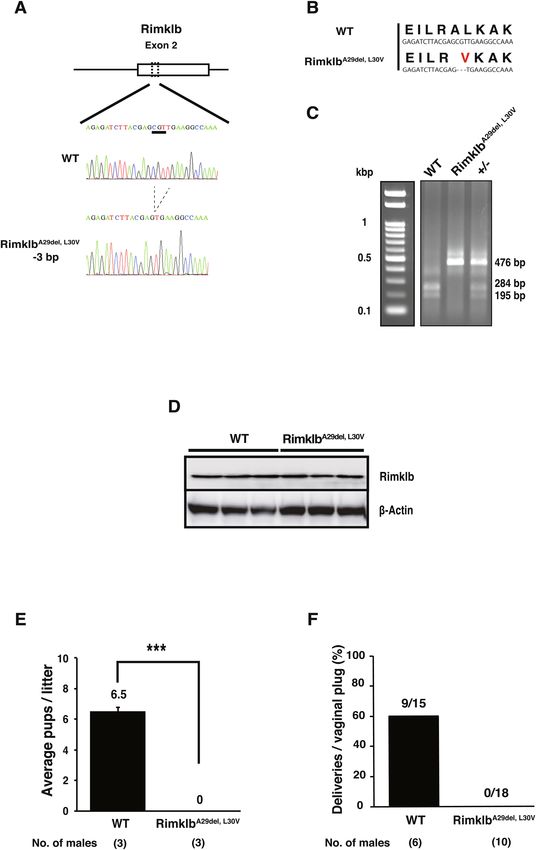
www.nature.com/scientificreports/

          Scientific Reports |   (2021) 11:4604 |   https://doi.org/10.1038/s41598-021-84105-z   4

Vol:.(1234567890)
www.nature.com/scientificreports/
◂Figure 3. RimklbA29del, L30V mice show abnormal spermatogenesis. (A) Average testis/body weights of WT and
imklbA29del, L30V at 8, 13 and 20 weeks of age. Data are presented as mean ± SEM; Student’s t-test; **p < 0.01,
R
*p < 0.05, NS not significant. (n = 3) (B) Representative images of WT and R imklbA29del, L30V testis at 20 weeks
of age. Scale bar = 1 mm. (C) Average sperm counts of WT and RimklbA29del, L30V at 13 weeks of age. Data are
presented as mean ± SEM; Student’s t-test; **p < 0.01. (D) Hematoxylin and eosin staining of tissue from WT
(upper) and RimklbA29del, L30V (bottom) mice at 8 and 13 weeks of age. Vacuolated tubules in the testis are
indicated by black arrows. Scale bar = 250 μm. (E) Percentage of tubules with large vacuoles. We scored 59–212
tubules from each animal. Data are presented as mean ± SEM; Student’s t-test; **p < 0.01. (n = 3) (F) Morphology
of spermatozoa from WT (upper, left) and R imklbA29del, L30V (upper, right). The black arrows indicate sperm
with abnormal head. Enlarged image of WT and RimklbA29del, L30V spermatozoal heads (bottom). Scale
bar = 10 μm. (G) Cauda epididymidis sperm head abnormality ratio for WT and R imklbA29del, L30V mice. Data
are presented as mean ± SEM; Student’s t-test; **p < 0.01 (H) In vitro fertilization rate with WT and RimklbA29del,
L30V
spermatozoa. The percentage of two-cell embryos 24 h after in vitro fertilization. Data are presented as
mean ± SEM; Student’s t-test; **p < 0.01 (I) Western blot analysis of VASA, MIWI, GAPDH-S and IZUMO1 in
WT and RimklbA29del, L30V mouse testes at 12 weeks of age. β-Actin was used as a sample processing control.
sperm head. During spermatogenesis, some sperm-specific proteins are expressed. In the mutant Rimklb mouse
testis we found reduced IZUMO1, a protein that is well known to play a role in sperm-egg fusion. Conversely,
the sperm- and spermatocyte-specific proteins, V ASA12, MIWI13 and GAPDH-S14 were not significantly changed
in the mutation vs wild mouse testis (Fig. 3I).
Rimklb has a critical role in the process of spermatogenesis in seminiferous tubules; the mutation of
RimklbA29del, L30V results in incomplete spermatozoa, which have been shown to be completely infertile.
Rimklb mutation enhanced S6 phosphorylation. Rimklb is a member of the rimK family, which
modifies the ribosomal protein S6 in prokaryotes15. It has been reported that during spermatogenesis the mam-
malian S6 protein is downstream to the mTOR pathway, regulating the BTB and spermatogenesis9. To examine
the relationship between Rimklb, mTOR and S6, we analyzed the effect of the Rimklb mutation on expression
and phosphorylation of mTOR and S6 in the testis, comparing wild vs R imklbA29del, L30V. We found that phospho-
rylated-S6 (p-S6) was obviously increased in the testes of RimklbA29del, L30V mutant mice. However, with p-AKT,
p-mTOR, p-4E-BP1, p-p70S6K and S6, no significant changes could be observed (Fig. 4A,B). S6 is known to be
the target protein of mTOR, which for spermatogenesis to occur is activated by phosphorylation via p70S6K16.
To analyze the cell-specific expression of p-S6 in testes, we performed immunohistochemistry (IHC) using p-S6
antibody with hematoxylin staining. We found a weak p-S6 signal on the basement membrane side of the semi-
niferous tubules (Fig. 5A–D) for spermatocytes in stage VII-VIII or IX-XI germinal epithelia of WT testes. In
addition, focal adhesion kinase (FAK) is known as a regulator of BTB dynamics in the testis17, the signals of
which were detected near the basement membrane in the seminiferous tubule; p-S6 signals are also expressed
in the seminiferous epithelium (Supplementary Fig. S2D). In the RimklbA29del, L30V mutant testis, stronger p-S6
protein expression was observed in the vacuoles of seminiferous tubules (Fig. 5E–H), and p-S6 positive tubules
were obviously increased in the R imklbA29del, L30V mutant testis. Moreover, p-S6 positive tubules with vacuoles
were distinctly increased in the RimklbA29del, L30V mutant testis (Table. 1).
Discussion
In this study, we have shown that R imklbA29del, L30V males were completely infertile: both the ratios of average
pups/litter and deliveries/plugs were zero on mating with RimklbA29del, L30V male mice. In addition, the Inter-
national Mouse Phenotyping Consortium (https://www.mousephenotype.org) indicates that knockout (KO) of
the Rimklb gene causes male infertility18. In RimklbA29del, L30V mutant mice, the testis weight was lower; sperm
morphology analysis showed small, abnormally shaped heads; sperm counts were decreased; and when we tested
imklbA29del, L30V male mice by mating them with wild C57BL/6 female for a two-month period
the fertility of R
(from eight weeks to 16 weeks of age) pregnancy failed to occur. IZUMO1, which plays an important role in
sperm-egg fusion, was obviously reduced in the testes of R imklbA29del, L30V mutant male mice compared with
wild mice. Furthermore, to determine how Rimklb is involved in spermatogenesis, we examined the expression
of mTOR/S6 and the sperm-specific proteins that play crucial roles in spermatogenesis. We found that p-S6 was
up-regulated around the vacuoles in seminiferous tubules within R imklbA29del, L30V testes.
RimklbA29del, L30V mutant mice have a deletion at amino acid 29 and the leucine at position 30 has been
replaced with valine. Rimk has two ATP-binding sites: the lysine at position 158 and arginine at position 219
(UniProtKB-Q80WS1 (RIMKB_MOUSE)). One possibility is that A29del and L30V mutations in Rimklb affect
the overall conformation and activity of Rimklb. We still need to conduct further experiments and analyses in
order to elucidate the function of the Rimklb in spermatogenesis by Rimklb KO mice.
A previous study that found ß-CG in the adult rat t estis19 is consistent with our finding that Rimklb is
expressed in the mouse testis (Fig. 1). RimklbA29del, L30V male mice showed severe infertility including failure of
spermatogenesis. One possible explanation is that Rimklb has a crucial role in the process of spermatogenesis
through synthesis of ß-CG; however, we still have no direct evidence of the relationship between ß-CG and
spermatogenesis.
Conversely, mammalian S6 is a key regulator in spermatogenesis16, especially in the mTOR/S6 pathway that
is a critical signal transduction process in the Sertoli c ell11. In this study, we have shown that the expression of
mTOR, p-mTOR and S6 were unchanged in mutant vs wild mouse testis, however, p-S6 was obviously enhanced
in the R imklbA29del, L30V mouse testis. Boyer et al. used conditional knockout mice ( Mtorflox/flox; Amhr2cre/+ mice)
Scientific Reports | (2021) 11:4604 | https://doi.org/10.1038/s41598-021-84105-z 5
Vol.:(0123456789)www.nature.com/scientificreports/
Figure 4. Hyperphosphorylation of ribosomal protein S6 in RimklbA29del, L30V mouse testis. (A) Western blot
analysis of p-AKT, p-mTOR, p-4E-BP1, p-p70S6K, p-S6 and S6 in WT and RimklbA29del, L30V mouse testis at
12 weeks of age. β-Actin was used as a loading control except S6. Loading control of S6 was β-Actin as shown in
Fig. 2D. (B) Graphic presentations show the expressions of p-AKT, p-mTOR, p-4E-BP1, p-p70S6K, p-S6 and S6.
Data are presented as mean ± SEM; Student’s t-test; **p < 0.01, NS not significant. (n = 3).
Figure 5. Hyperphosphorylation of ribosomal protein S6 in seminiferous tubules. (A,B)
Immunohistochemistry of WT testis for p-S6. Seminiferous tubules stages are shown. (C,D) Enlarged
images: immunohistochemistry of WT testis for p-S6. The black arrows indicate p-S6-positive cells. (E,F)
Immunohistochemistry of R imklbA29del, L30V mouse testis for p-S6. (G,H) Enlarged images of the boxed area are
shown. Scale bar = 100 μm.
Total tubules Vacuoles p-S6-positive p-S6-positive with vacuoles
Number of seminiferous tubules in testis
WT 149.0 ± 11.4 4.3 ± 0.5 30.7 ± 13.1 1.7 ± 1.3
RimklbA29del, L30V 81.3 ± 18.9 34.0 ± 6.5 48.0 ± 11.0 23.0 ± 11.0
Table 1. The number of p-S6-positive seminiferous tubules with increased vacuoles in the R imklbA29del, L30V
mouse testis. The total number of tubules, tubules with vacuoles, p-S6-positive tubules, p-S6-positive tubules
with vacuoles in WT and RimklbA29del, L30V mouse testis at 12 weeks of age. Data are presented as mean ± SD.
Scientific Reports | (2021) 11:4604 | https://doi.org/10.1038/s41598-021-84105-z 6
Vol:.(1234567890)www.nature.com/scientificreports/
to target mTOR in Sertoli cells, revealing the presence of large vacuoles in seminiferous tubules as well as severe
male infertility. In addition, phosphorylation of RPS6 at S235/236 was upregulated in the testes of these m ice18.
This data indicates that down-regulation of mTOR in Sertoli cells inhibits spermatogenesis and leads to male
infertility, resulting in enhanced phosphorylation of rps6. Their data are consistent with our observation for
hyperphosphorylation of rpS6 and male infertility.
Interestingly, the p-S6 signal was observed in the vacuoles of seminiferous tubules, suggesting that the induc-
tion of p-S6 is possibly associated with seminiferous tubules and Sertoli cell function. Li et al. carried out
experiments showing that the over-expressed and phosphorylated ribosomal protein S6 regulates the BTB,
thereby negatively affecting s permatogenesis9, and rapamycin promotes autophagy and leads to suppression of
spermatogenesis in the rat testis by inhibiting mTOR and p70S6 k inase16. Evidence has thus accumulated that
p-S6 plays an important role in spermatogenesis.
Rimklb is expressed in Leydig cells, which are known to be involved in spermatogenesis by producing hor-
mones such as testosterone. R imklbA29del, L30V mutant mice showed no difference in testosterone levels on compar-
ing RimklbA29del, L30V mutants and wild male mice, suggesting that the mutation of Rimklb may not directly affect
testosterone levels. A few studies have been conducted on S6 in Leydig cells: luteinizing hormone stimulated the
phosphorylation of a 33,000 kDa protein in Leydig tumor cells20, and human chorionic gonadotropin (hCG)
hormone enhanced p-S6 in primary cultures of porcine Leydig cells21. In this study, p-S6 expression was difficult
to identify in Leydig cells, so the function of p-S6 in Leydig cells remains unclear. Further studies will be needed.
We have also shown that the expression of IZUMO1 was downregulated in R imklbA29del, L30V mutant testes.
IZUMO1 is present in the acrosomal membrane and is known to play an important role during fertilization.
Although it is not clear why IZUMO1 is decreased in the testes of R imklbA29del, L30V mutant mice, the functional
changes putatively caused by the RimklbA29del, L30V mutation may suppress IZUMO1 expression.
Taken together, Rimklb is essential for spermatogenesis, and Rimklb is thought to be involved in all processes:
spermatogenesis, spermatocyte-to-sperm differentiation, proliferation, and sperm fertilization. However, detailed
mechanisms have not been elucidated, and further research must be conducted. Understanding the fine details
of Rimklb may lead to elucidation of unknown mechanisms of male infertility.
Methods
All experiments were performed in accordance with the relevant guidelines and regulations.
Animal subjects. All mouse experiments were approved by the Kobe Gakuin Animal Experiment Commit-
tee (protocol No. A17-50) and the Animal Care and Use Committee of the National Institute of Quantum and
Radiological Science and Technology (protocol No. 1610111 and 1610121). Mice were sacrificed using cervi-
cal dislocation performed by trained experimenters, or perfused and dissected under the three types of mixed
anesthetic agents (0.3 mg/kg of medetomidine, 4.0 mg/kg of midazolam, and 5.0 mg/kg of butorphanol). Animal
studies were conducted following the ARRIVE guidelines.
Immunohistochemistry. The tissues were perfused and additionally fixed using Bouin fixation22 for 48 h.
After fixation, the tissues were embedded in paraffin wax. Paraffin-embedded tissues were sliced to a thickness of
six microns, attached to polylysine-coated slides, and dried at 40 °C overnight. The sliced tissues were deparaffi-
nized using xylene, and immersed in ethanol and PBS. Antigens were retrieved in HistoVT One (Nacalai Tesque,
Kyoto, Japan) by boiling for 20 min. In this study, tissue antigen signals were detected using the VECTASTAIN
Elite ABC Kit (Vector Laboratories, Burlingame, CA, USA). In brief, for blocking, tissues were incubated in
PBS containing normal goat serum for 20 min; the primary antibodies then used were anti-Rimklb (ab15783,
Abcam, Cambridge, UK, Anti-RIMKB antibody N-terminal 1:100) and anti-Phospho-S6 Ribosomal Protein
(#2211, Cell Signaling Technology, Danvers, MA, USA, Phospho-S6 Ribosomal Protein (Ser235/236) Antibody
1:400), applied overnight. Endogenous peroxidase was inactivated by 3% hydrogen peroxide for 15 min. The
secondary antibody used was biotinyl-labeled anti rabbit antibody for 20 min; signal detection was performed by
avidin-labeled peroxidase and DAB using the VECTASTAIN Elite ABC Kit. The sections (Phospho-S6 Riboso-
mal Protein) were counterstained with Mayer’s hematoxylin solution (FUJIFILM Wako Pure Chemical, Osaka,
Japan). At least 50 effectively round seminiferous tubules were used for measurement of p-S6-positive tubules
or p-S6-positive tubules with vacuoles. “P-S6-positive tubules” were counted if seminiferous tubules contained
p-S6-positive cells, and “p-S6-positive tubules with vacuoles” were counted if seminiferous tubules contained
positive cells and vacuoles.
Generation of Rimklb mutant mouse. Rimklb mutant mice were generated using the CRISPR/Cas9
system and cytoplasmic microinjection of mouse embryos. Guide gRNAs (gRNAs) were designed to delete exon
2 of the Rimklb gene, and synthesized from 130 bp of chemically synthesized double-stranded DNA (gBlocks
Gene Fragments, Integrated DNA Technologies, Coralville, IA, USA) that included the T7 promoter, the gRNA
target sequence (AGAGATCTTACGAGCGTTGA) and the gRNA-scaffold sequence as a template using the
MEGAshortscript T7 Transcription Kit (Life Technologies, Carlsbad, CA, USA) followed by RNA purifica-
tion using a MEGAclear kit (Life Technologies). Embryo manipulation and microinjection were performed
escribed23,24. Briefly, MII-oocytes were collected from superovulated C57BL/6J females (aged
as previously d
8–12 weeks, Japan SLC, Shizuoka, Japan), fertilized in vitro, and cultured in KSOM medium until use. Fertilized
one-cell embryos underwent cytoplasmic microinjection with a mixture of recombinant Cas9 protein (50 ng/μl,
NIPPON GENE, Tokyo, Japan) and two gRNAs (25 ng/μl). After the microinjection, the embryos were cultured
in KSOM medium until the two-cell stage, and transferred to the oviduct of pseudopregnant ICR females (CLEA
Japan, Tokyo, Japan) on the day of the vaginal plug (Day 0.5). Genomic DNA of offspring (F0 founders) was
Scientific Reports | (2021) 11:4604 | https://doi.org/10.1038/s41598-021-84105-z 7
Vol.:(0123456789)www.nature.com/scientificreports/
extracted from tail samples and used for genotyping. F0 founders harboring potential mutant alleles were bred
with wild-type C57BL/6J mice, and mutations in the F1 generation were analyzed using the Guide-it Mutation
Detection Kit (Takara Bio, Shiga, Japan). The mutant F2 females were crossed with wild BL/6 male mice; after
mating mutant F3 mice with each other, TA cloning was used to obtain litter DNA for sequencing. One mouse
line with deletion of three bases was chosen and used for this study.
Genotyping. Mouse tails were lysed at 55 °C overnight, using lysis buffer containing Proteinase K (Sigma-
Aldrich, St. Louis, MO, USA), and the lysate was directly used as a template for PCR. Genotyping of Rimklb
mutant mice was performed using Ex Taq polymerase (Takara Bio) with a specific primer (Rimk1bCheckF:
5ʹ-CCTCATCCTCCTGTGCCTAA-3ʹ and Rimk1bCheckR: 5ʹ-GCACTCAGCTCTCCAGCTCT-3ʹ). PCR prod-
ucts were digested by the restriction enzyme Mwo I; the amplicon from the mutant allele was insensitive to Mwo I.
Western blotting. The Western blotting shown in Fig. 1 was performed as previously described25. For the
Western blotting of Fig. 4, mouse tissues were collected and lysed using a bead homogenizer (Mini bead-beater,
WakenBtech, Kyoto, Japan) with 2.0 mm Zirconia beads (BioSpec Products, Bartlesville, OK, USA) in 1 mL cell-
lysis buffer (#9803, Cell Signaling Technology) containing a proteinase inhibitor cocktail (#5871, Cell Signaling
Technology). The lysates were centrifuged at 12,000 rpm for 20 min at 4 °C, and the supernatants were boiled
in 4 × sample buffer (FUJIFILM Wako Pure Chemical) for 5 min. Protein samples were separated on a 10–20%
gel (SuperSep Ace, FUJIFILM Wako Pure Chemical) and subsequently transferred to a PVDF membrane using
the Turbo Transfer System (Bio-Rad, Hercules, CA, USA) with a seven-minute protocol. The membrane was
blocked with Blocking One (Nacalai Tesque) for 30 min or 3% BSA (Rockland Immunochemicals, Inc., Potts-
town, PA, USA) in T-BST for 1 h at room temperature, and incubated overnight at 4 °C with the following
primary antibodies diluted 1:1000 in Blocking ONE: anti-phospho-S6 ribosomal protein (#4858, Cell Signaling
Technology), anti-S6 ribosomal protein (#2317, Cell Signaling Technology), anti-phospho-mTOR (#5536, Cell
Signaling Technology), anti-phospho-AKT (#4060, Cell Signaling Technology), anti-phospho-p70S6K (#9234
Cell Signaling Technology), anti-phospho-4E-BP1 (#2855, Cell Signaling Technology), horseradish peroxidase-
conjugated anti-actin (A00730, GenScript, Piscataway, NJ, USA), anti-Miwi (#2079, Cell Signaling Technology),
anti-GAPDH-S (13937-1-AP, Proteintech, Rosemont, IL, USA), and anti-Mouse Vasa Homologue (MVH/Vasa,
ab13840, Abcam). Anti-IZUMO1 antibody was kindly provided by Dr. Masaru Okabe (Osaka University). Each
membrane was incubated with an anti-mouse or rabbit IgG HRP-linked antibody (Cell Signaling Technology) as
a secondary antibody diluted 1:10,000 in Blocking One at room temperature for 1 h. Chemiluminescence reac-
tions were performed with Lightning Ultra (PerkinElmer, Waltham, MA, USA). The signals were detected using
a ChemiDoc-It imaging system (Bio-Rad) with a BioChemi camera (UVP, Upland, CA, USA). Signal intensities
were analyzed with NIH ImageJ (https://imagej.nih.gov/ij/). For some figures, unrelated lanes were cropped out.
Full size images are provided in the Supplementary Figs. S3, S4.
Histological staining. Testis sections were stained with hematoxylin and eosin after deparaffination. Slides
were mounted and observed by microscopy (Model IX71, Olympus Corporation, Tokyo, Japan). At least 50
effectively round seminiferous tubules were used for measurement of vacuoles greater than ~ 30 μm in greatest
diameter and located on or near the seminiferous tubule basement membrane, similar to previously reported
methods26. Vacuoles values are all represented as the percentage of total tubules affected per total tubules counted.
Testosterone assay. Serum testosterone analyses were performed using ELISA kits (Testosterone ELISA
Kit, ADI-900-065, Enzo Life Sciences, Inc., Farmingdale, NY, USA). Serum was separated from all blood sam-
ples after centrifugation at 16,099×g for 15 min and frozen at − 90 °C for later hormonal analysis.
Sperm counts and morphology. Sperm counts were performed as described by Wang27. The caudae
A29del, L30V
epididymides of 12–13 week-old R imklb and WT mice were collected in PBS, and minced using scal-
pel blades. After incubating for 15 min at 37 °C, sperm were diluted 1:4 in PBS and sperm counts determined
on duplicate samples using a hemocytometer. Sperm were collected in the same manner and observed with a
microscope. At least 250 sperm were observed for each experimental condition. Spermatozoa with round, thin,
or bent heads were determined to be abnormal.
Fertility test and IVF. Two C57BL/6J female mice and one male were kept in the same cage for two months
until pregnancy resulted. Copulation was checked by examining for vaginal plugs every morning. IVF was per-
formed as follows. The C57BL/6J female mice were injected intraperitoneally with pregnant mare serum gon-
adotropin (PMSG) (7.5 units, ASKA Pharmaceutical, Tokyo, Japan) and injected with human chorionic gon-
adotropin (hCG) (7.5 units, ASKA Pharmaceutical) 48 h later. MII-oocytes were collected from the ampulla of
each oviduct of superovulated female mice 15 h after the injection of hCG. Spermatozoa were collected from the
cauda epididymidis of three-month-old male mice and incubated in TYH medium for two hours. Capacitated
spermatozoa were incubated in a drop with MII-oocytes, at a final concentration of 2 × 10⁵ sperm/mL. After
incubation for 24 h, two-cell embryos were counted under a microscope.
Received: 6 May 2020; Accepted: 11 February 2021
Scientific Reports | (2021) 11:4604 | https://doi.org/10.1038/s41598-021-84105-z 8
Vol:.(1234567890)www.nature.com/scientificreports/
References
1. Kino, K., Arai, T. & Arimura, Y. Poly-alpha-glutamic acid synthesis using a novel catalytic activity of RimK from Escherichia coli
K-12. Appl. Environ. Microbiol. 77, 2019–2025. https://doi.org/10.1128/AEM.02043-10 (2011).
2. Pletnev, P. I. et al. Oligoglutamylation of E. coli ribosomal protein S6 is under growth phase control. Biochimie 167, 61–67. https
://doi.org/10.1016/j.biochi.2019.09.008 (2019).
3. Kang, W. K., Icho, T., Isono, S., Kitakawa, M. & Isono, K. Characterization of the gene rimK responsible for the addition of glu-
tamic acid residues to the C-terminus of ribosomal protein S6 in Escherichia coli K12. Mol. Gen. Genet. 217, 281–288. https://doi.
org/10.1007/bf02464894 (1989).
4. Grenga, L., Little, R. H. & Malone, J. G. Quick change: Post-transcriptional regulation in Pseudomonas. FEMS Microbiol Lett https
://doi.org/10.1093/femsle/fnx125 (2017).
5. Collard, F., Vertommen, D., Constantinescu, S., Buts, L. & Van Schaftingen, E. Molecular identification of beta-citrylglutamate
hydrolase as glutamate carboxypeptidase 3. J. Biol. Chem. 286, 38220–38230. https://doi.org/10.1074/jbc.M111.287318 (2011).
6. Collard, F. et al. Molecular identification of N-acetylaspartylglutamate synthase and beta-citrylglutamate synthase. J. Biol. Chem.
285, 29826–29833. https://doi.org/10.1074/jbc.M110.152629 (2010).
7. Jesus, T. T., Oliveira, P. F., Sousa, M., Cheng, C. Y. & Alves, M. G. Mammalian target of rapamycin (mTOR): A central regulator of
male fertility?. Crit. Rev. Biochem. Mol. Biol. 52, 235–253. https://doi.org/10.1080/10409238.2017.1279120 (2017).
8. Oliveira, P. F., Cheng, C. Y. & Alves, M. G. Emerging role for mammalian target of rapamycin in male fertility. Trends Endocrinol.
Metab. 28, 165–167. https://doi.org/10.1016/j.tem.2016.12.004 (2017).
9. Li, S. Y. T. et al. mTORC1/rpS6 regulates blood-testis barrier dynamics and spermatogenetic function in the testis in vivo. Am. J.
Physiol. Endocrinol. Metab. 314, E174–E190. https://doi.org/10.1152/ajpendo.00263.2017 (2018).
10. Wen, Q. et al. Signaling pathways regulating blood-tissue barriers: Lesson from the testis. Biochim. Biophys. Acta https://doi.
org/10.1016/j.bbamem.2017.04.020 (2018).
11. Xu, H. et al. mTOR/P70S6K promotes spermatogonia proliferation and spermatogenesis in Sprague Dawley rats. Reprod. Biomed.
Online 32, 207–217. https://doi.org/10.1016/j.rbmo.2015.11.007 (2016).
12. Kim, J. Y., Jung, H. J. & Yoon, M. J. VASA (DDX4) is a putative marker for spermatogonia, spermatocytes and round spermatids
in stallions. Reprod. Domest. Anim. 50, 1032–1038. https://doi.org/10.1111/rda.12632 (2015).
13. Grivna, S. T., Pyhtila, B. & Lin, H. MIWI associates with translational machinery and PIWI-interacting RNAs (piRNAs) in regulat-
ing spermatogenesis. Proc. Natl. Acad. Sci. USA 103, 13415–13420. https://doi.org/10.1073/pnas.0605506103 (2006).
14. Feiden, S., Wolfrum, U., Wegener, G. & Kamp, G. Expression and compartmentalisation of the glycolytic enzymes GAPDH and
pyruvate kinase in boar spermatogenesis. Reprod. Fertil. Dev. 20, 713–723. https://doi.org/10.1071/rd08004 (2008).
15. Zhao, G. et al. Structure and function of Escherichia coli RimK, an ATP-grasp fold L-glutamyl ligase enzyme. Proteins 81, 1847–
1854. https://doi.org/10.1002/prot.24311 (2013).
16. Liu, S. et al. Rapamycin inhibits spermatogenesis by changing the autophagy status through suppressing mechanistic target of
rapamycin-p70S6 kinase in male rats. Mol. Med. Rep. 16, 4029–4037. https://doi.org/10.3892/mmr.2017.7120 (2017).
17. Siu, E. R., Wong, E. W., Mruk, D. D., Porto, C. S. & Cheng, C. Y. Focal adhesion kinase is a blood-testis barrier regulator. Proc.
Natl. Acad. Sci. USA 106, 9298–9303. https://doi.org/10.1073/pnas.0813113106 (2009).
18. Boyer, A. et al. mTOR regulates gap junction alpha-1 protein trafficking in sertoli cells and is required for the maintenance of
spermatogenesis in mice. Biol. Reprod. 95, 13. https://doi.org/10.1095/biolreprod.115.138016 (2016).
19. Miyake, M., Kakimoto, Y. & Sorimachi, M. A gas chromatographic method for the determination of N-acetyl-L-aspartic acid,
N-acetyl-alpha-aspartylglutamic acid and beta-citryl-L-glutamic acid and their distributions in the brain and other organs of
various species of animals. J. Neurochem. 36, 804–810. https://doi.org/10.1111/j.1471-4159.1981.tb01665.x (1981).
20. Bakker, G. H., Hoogerbrugge, J. W., Rommerts, F. F. & van der Molen, H. J. Lutropin increases phosphorylation of a 33000-dalton
ribosomal protein in rat tumour Leydig cells. Biochem. J. 204, 809–815. https://doi.org/10.1042/bj2040809 (1982).
21. Dazord, A. et al. hCG-Increased phosphorylation of proteins in primary culture of Leydig cells: Further characterization. Biochem.
Biophys. Res. Commun. 118, 8–13. https://doi.org/10.1016/0006-291x(84)91059-3 (1984).
22. Ellenburg, J. L. et al. Formalin versus bouin solution for testis biopsies: Which is the better fixative?. Clin Pathol https://doi.
org/10.1177/2632010X19897262 (2020).
23. Tatsumi, T. et al. Forced lipophagy reveals that lipid droplets are required for early embryonic development in mouse. Development
https://doi.org/10.1242/dev.161893 (2018).
24. Aizawa, R. et al. Synthesis and maintenance of lipid droplets are essential for mouse preimplantation embryonic development.
Development https://doi.org/10.1242/dev.181925 (2019).
25. Takano, M. et al. Proteomic analysis of the brain tissues from a transgenic mouse model of amyloid beta oligomers. Neurochem.
Int. 61, 347–355. https://doi.org/10.1016/j.neuint.2012.05.018 (2012).
26. Moffit, J. S., Boekelheide, K., Sedivy, J. M. & Klysik, J. Mice lacking Raf kinase inhibitor protein-1 (RKIP-1) have altered sperm
capacitation and reduced reproduction rates with a normal response to testicular injury. J. Androl. 28, 883–890. https://doi.
org/10.2164/jandrol.107.002964 (2007).
27. Wang, Y. Epididymal sperm count. Curr. Protoc. Toxicol. https://doi.org/10.1002/0471140856.tx1606s14 (2003).
Acknowledgements
This research is supported by the Ministry of Education, Culture, Sports, Science and Technology Basic Research
C, Young Researcher’s Research B, Kobe Gakuin University Research Grant A and Health Science Grant. This
work was also supported by the Japan Society for the Promotion of Science (KAKENHI program; 20K18153 to
K.M., and 20K09633 to M.T.), the Kobe Gakuin University Research Grant A (to M.T.), and the Takeda Science
Foundation (to S.T.). We are very grateful for helpful advice from and discussions with Dr. Tomoki Yamano
(Shionogi & Co., Ltd.) and Professor Masaru Okabe (Osaka University). We also thank ASCA Corporation for
editing a draft of this manuscript.
Author contributions
M. T. conceived the experiments and wrote the main manuscript text, K. M. and S. T. conducted the experi-
ments, K. M., S. T. and M. H. analyzed the results, and K. M. and S. T. prepared Figs. 1–5. All authors reviewed
the manuscript.
Competing interests
The authors declare no competing interests.
Scientific Reports | (2021) 11:4604 | https://doi.org/10.1038/s41598-021-84105-z 9
Vol.:(0123456789)www.nature.com/scientificreports/
Additional information
Supplementary Information The online version contains supplementary material available at https://doi.
org/10.1038/s41598-021-84105-z.
Correspondence and requests for materials should be addressed to M.T.
Reprints and permissions information is available at www.nature.com/reprints.
Publisher’s note Springer Nature remains neutral with regard to jurisdictional claims in published maps and
institutional affiliations.
Open Access This article is licensed under a Creative Commons Attribution 4.0 International
License, which permits use, sharing, adaptation, distribution and reproduction in any medium or
format, as long as you give appropriate credit to the original author(s) and the source, provide a link to the
Creative Commons licence, and indicate if changes were made. The images or other third party material in this
article are included in the article’s Creative Commons licence, unless indicated otherwise in a credit line to the
material. If material is not included in the article’s Creative Commons licence and your intended use is not
permitted by statutory regulation or exceeds the permitted use, you will need to obtain permission directly from
the copyright holder. To view a copy of this licence, visit http://creativecommons.org/licenses/by/4.0/.
© The Author(s) 2021
Scientific Reports | (2021) 11:4604 | https://doi.org/10.1038/s41598-021-84105-z 10
Vol:.(1234567890)You can also read