BREAST TOMOSYNTHESIS: A BETTER MAMMOGRAPHY - Mastology
←
→
Page content transcription
If your browser does not render page correctly, please read the page content below
REVIEW ARTICLE
                                           DOI: 10.29289/Z2594539420180000254

                              BREAST TOMOSYNTHESIS:
                              A BETTER MAMMOGRAPHY
                          Tomossíntese mamária: uma mamografia melhor
                             Vera Lucia Nunes Aguillar1*, Selma di Pace Bauab2, Aron Belfer3

    ABSTRACT
Digital mammography is an excellent method for detecting breast cancer at an early stage, but overlap of breast structures may lead
to both false-positive and false-negative results. The digital breast tomosynthesis (DBT) is addressed to overcome this limitation of
conventional 2D mammography. The purpose of this study was to discuss the multiple aspects related to this new tool, including its
role in breast cancer screening, through a literature review. DBT, or 3D mammography, provides a three-dimensional representation
of the breast, with the ability to scroll through breast tissue in the reconstructed images, thereby reducing the effect of tissue
superimposition. This leads to improved sensitivity and specificity in breast cancer screening. In diagnostic cases, tomosynthesis
increases the accuracy with better characterization and localization of the lesions. Prospective and retrospective studies confirm
that DBT is superior to digital mammography in breast cancer screening, with 27 to 53% increase in cancer detection and 17 to 37%
reduction in false-positive recalls. A total of 40 to 49% of the cancers detected by DBT were invasive: 40 to 48% of histological
grade 2 or 3 and more than 75% were node negative. DBT is the most promising new modality for breast cancer screening. Further
studies are needed to evaluate the reduction of interval cancers with this modality.
KEYWORDS: mammography; mass screening; breast neoplasms.

    RESUMO
Mamografia digital é um método excelente para detecção precoce do câncer de mama. Porém, a sobreposição das estruturas
mamárias pode levar a resultados falso-positivos e falso-negativos. A tomossíntese mamária é dirigida para superar essa limitação
da mamografia 2D convencional. O objetivo deste estudo é discutir os múltiplos aspectos relacionados a essa nova ferramenta,
incluindo, através de uma revisão da literatura, seu papel no rastreamento do câncer de mama. A tomossíntese mamária, ou
mamografia 3D, proporciona uma representação tridimensional da mama, com a habilidade de podermos visualizar as imagens
reconstruídas em diversos planos, reduzindo o efeito da sobreposição. Isso conduz a uma melhora da sensibilidade e da
especificidade no rastreamento mamográfico. Nos casos diagnósticos, aumenta a acurácia com melhor caracterização e localização
das lesões. Estudos prospectivos e retrospectivos confirmam que, no rastreamento do câncer de mama, a tomossíntese mamária
é superior à mamografia digital, com aumento da detecção de 27 a 53%, e na redução das reconvocações falso-positivas entre 17
e 30%. De 40 a 49% dos cânceres detectados pela tomossíntese foram invasivos: de 40 a 48% de grau histológico 2 ou 3 e mais de
75% foram linfonodo negativos. Tomossíntese mamária é a nova modalidade mais promissora para o rastreamento do câncer de
mama. São necessários estudos adicionais com essa nova modalidade para a avaliação da redução dos cânceres de intervalo.
PALAVRAS-CHAVE: mamografia; programas de rastreamento; neoplasias da mama.

1
 Departamento de Imagem da Mulher do Laboratório Fleury – São Paulo (SP), Brazil.
2
  Clínica Mama Imagem – São José do Rio Preto (SP), Brazil.
3
  Centro de Diagnósticos Brasil (Premium) – São Paulo (SP), Brazil.
*Corresponding author: veraaguillar@uol.com.br
Conflict of interests: nothing to declare.
Received on: 07/31/2017. Accepted on: 02/13/2018.

                                                  Mastology, 2018;28(1):51-66                                                   51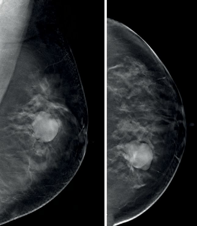
Aguillar VLN, Bauab SP, Belfer A

INTRODUCTION                                                            thickness : thus, in a 5-cm breast, 50 slices per mammographic
Breast tomosynthesis or 3D mammography is a technological               incidence are obtained. All images are analyzed in high resolu-
advancement in digital mammography, which allows three-                 tion monitors, either individually or in cine mode (Figure 2)4-6.
dimensional representation of breast tissue from two-dimen-                 The are different commercially systems available with varia-
tional projections. It was developed to overcome an impor-              tions in scan angle, number of projections and scan time.
tant limitation of conventional digital mammography (2D),
which is normal overlapping tissue during the acquisition of            Exam technique
the radiographic image. This limitation of 2D mammography               Patient positioning and breast compression at tomosynthe-
may lead to low sensitivity in detecting some cancers and               sis are similar to that at DM, typically using standard cranio-
high false-positive recall rates (due to summation images).             caudal and mediolateral oblique projections. The approval of
Tomosynthesis reduces the impact of overlapping breast tis-             tomosynthesis by the Food and Drug Administration (FDA),
sue by depicting tissue on a dynamic sequence of thin cross-            in the United States, in 2011, was based on the “combo mode”:
sectional images which results in a considerable increase in            3D images are obtained along with 2D DM images during the
diagnostic accuracy (Figure 1A)1-4.                                     same compression. Although tomosynthesis is superior to
                                                                        detect non-calcified lesions (nodules and architectural distor-
                                                                        tions), the 2D images gives us a panoramic view of the breasts,
HOW IS TOMOSYNTHESIS OBTAINED?                                          which allows better analysis of asymmetries and comparison
Tomosynthesis uses a digital mammograph, in which the X-ray             with previous examinations3,4.
source moves in an arc above the compressed breast and a series             Similar to conventional mammography, two views of each
of low-dose x-ray projections are acquired at different angles          breast are recommended with tomosynthesis, since some lesions
(Figure 1B). From these two-dimensional projections, 3D images          may be seen only in one projection. Clinical studies have shown
(tomosynthesis slices) are reconstructed, with 1 mm thickness,          that about 9% of the tumors may be missed if only one incidence
parallel to the detector. The number of slices depends on breast        of tomosynthesis is used.

 A                                                   B

                                                                                              tube displacement

      compressor

                                                                                        compressor

                    digital detector

                                                                   digital detector
                                                                                                        3D slice

          2D
                                                                      projections

          Suspicious lesion

          Normal fibroglandular tissue
          X-ray tube

          Overlapped suspicious lesion

Figure 1. Schematic acquisition of mammographic images during breast compression: (A) conventional digital mammography (2D)
shows overlap of normal structures and suspicious lesion; (B) tomosynthesis (3D) reduces tissue overlap by detecting the suspicious
lesion in one of the slices.

     52                                                    Mastology, 2018;28(1):51-66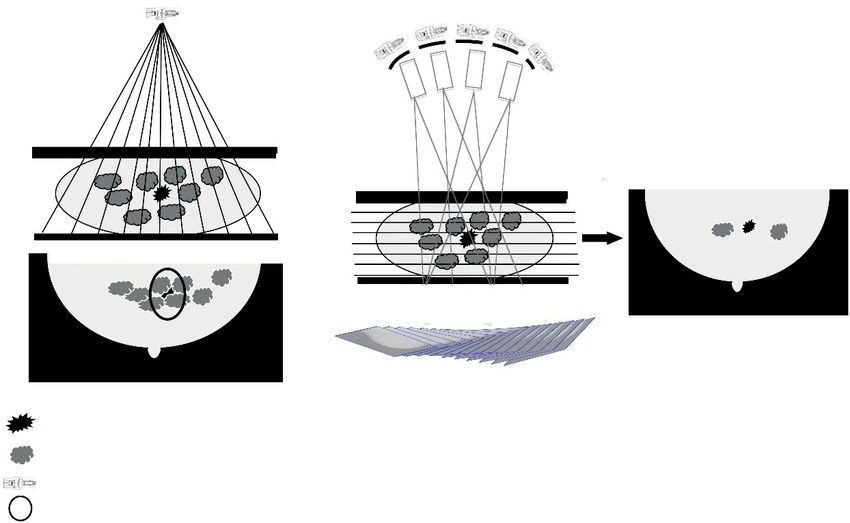
Breast tomosynthesis: a better mammography

Radiation dose                                                            Breast anatomy in tomosynthesis
The disadvantage of combination of DM and tomosynthesis is                Although normal anatomy is similar in 2 D and 3Ds images,
an approximate twofold increase in radiation dose, although               the reduction of overlapping tissue in the tomosynthesis slices
the total dose still falls within the safety limits determined by         allows better individualization of breast structures. In slices
American and European regulatory agencies7. Concerns regard-              closest to the detector and the compressor, the dermis and
ing increased radiation dose has led to other alternatives, such as:      cutaneous pores (round radiolucent images) and eventual cuta-
1. Replace 2D mammography by SM (synthesized mammography),                neous lesions, such as nevi, or calcifications are better seen.
    in combination with tomosynthesis. In this case, the                  Cooper ligaments and linear structures in pre-parenchymal fat
    performance of the combined test is similar, without increasing       are seen brighter in tomosynthesis slices than in conventional
    the total radiation dose8-10;                                         mammography. The fibroglandular tissue is gray, with a lower
2. Tomosynthesis with only one view, combined to 2D                       density in relation to the 2D image, due to the smaller amount
    mammography 4,11. However, this option can reduce sensitivity         of breast tissue evidenced in each slice. High density objects,
    and especificity of the exam, when compared to tomosynthesis          such as markers and metal clips and larger calcifications, may
    performed in two projections11-13.                                    produce artifacts that, however, do not interfere with image
                                                                          interpretation. Tomosynthesis increase lesion conspicuity with
2D SYNTHESIZED MAMMOGRAPHY                                                better definition of margins and spicules, due to reduction in
Synthesized mammography (SM) is a technique that generates                tissue overlapping. 3,4.
two-dimensional images from the DBT dataset, eliminating the                  The characteristics of the parenchyma texture in the tomo-
need for a separately acquired FFDM examination and thereby               synthesis correlate better with breast density than digital mam-
decreasing the radiation dose to the patient. 8,10. Although SM’s         mography, and may become a method of measuring breast den-
initial version was not well accepted due to its limited techni-          sity, resulting in a quantitative biomarker to estimate the risk
cal quality, recent studies found that its updated version pre-           of cancer.
sented the same performance of the conventional 2D image.                     SM presents its own characteristics, different from the con-
Although the mathematical algorithms to obtain synthesized                ventional image: brighter mammary parenchyma, blurring of skin
images are different among the manufacturers, the objective is            and subcutaneous tissue, loss of resolution of axillary area, pseu-
similar and consists in obtaining the information provided by             docalcifications and artifacts generated by clips or other high-
2D images – panoramic view of the breasts, comparison with                density images. SM enhances linear structures - such as Cooper’s
previous mammograms and evaluation of microcalcifications                 ligaments or spicules associated with masses or architectural
- without extra dose of radiation. It is important to emphasize           distortions - and increases brightness and contrast of calcifica-
that the SM must be analyzed along with the tomosynthesis                 tions. The extreme dense breasts (ACR D) appear brighter in the
slices, never as an isolated study10.                                     SM due to the summation of the tomosynthesis slices (Figure 3).

  A                                                                                        B

Figure 2. (A) Low-dose radiation projections; (B) from which 1 mm thick tomosynthesis slices are reconstructed.

                                                     Mastology, 2018;28(1):51-66                                                        53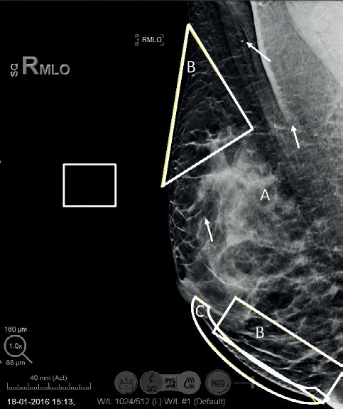
Aguillar VLN, Bauab SP, Belfer A
Exam interpretation alternatives proposed to reduce interpretation time is to
Due to the large number of images obtained with tomosynthe- abolish the double reading of tomosynthesis examination,
sis, the analysis of the exam must be performed in a systematic which, according to Houssami et al.17, does not change the
way, with implementation of a hanging protocol in the high- benefits of tomosynthesis (increase in detection of invasive
resolution monitor, to optimize reading time. Interpretation cancer and reduction of false-positive recalls), compared
of the exam starts with the analysis of the 2D images (conven- to DM alone.
tional or synthesized) for detection of calcifications, asym- The learning curve and the optimization of reading time
metries, masses or architectural distortions. Next, a com- with the implementation of a flowchart in the monitor (report
parative analysis is performed with previous mammograms f low) are fundamental to reduce the time of interpretation
to characterize developing asymmetries or other new lesions. in tracing 14 . The availability of computer-aided detection
Tomosynthesis images are interpreted with two-dimensional (CAD) systems for synthesized image and tomosynthesis
mammography, using the latter as a “road map” for each mam- slices and the use of thicker tomosynthesis images (slabs),
mographic incidence1,4,14. instead of 1 mm slices, are alternatives for the reduction of
It is necessary to obtain conventional (2D) digital mammog- interpretation time.
raphy along with tomosynthesis, since each method provides
different types of information in the analysis of mammographic
findings (Chart 1). IMPACT OF TOMOSYNTHESIS
Tomosynthesis slices must be seen sequentially (cine mode), IN COMPUTER DEPARTMENTS
breast by breast and incidence by incidence. It is important to In tomosynthesis examination, the images of each slice gen-
mentally divide the breast into 2 or 3 segments and look at each of erate the same volume of data as a 2D. The number of slices
them separately, during at least one cine mode cycle, to increase is proportional to breast thickness and usually 30 to 70 slices
detection of small lesions1,14. are obtained, with the total volume of data generated by the
tomosynthesis, by incidence, significantly higher than in 2D.
Exam interpretation time This fact has several implications for the storage of images.
Tomosynthesis is an easy-to-implement technology because The size of the tomosynthesis file also implies the time
the examination is performed in the same way as conventional and speed of transmission and retrieval of the examination.
or digital mammography. However, interpretation time is lon- Compaction softwares are used for better storage and transmis-
ger due to a large number of images to be analyzed, which sion performance of tomosynthesis.
requires more concentration and focus from the radiologist,
resulting in greater mental and visual fatigue15,16 . One of the
Chart 1. Degree of information provided by tomosynthesis and
conventional digital mammography on different aspects in the
analysis of the images.
2D Tomosynthesis
Visibility of lesions ++ +++
Margin analysis + +++
2D SM Location of the lesions +++
Extent of the lesions +++
Multifocality + +++
Density associated with
+++
calcifications
Reduction of summation + +++
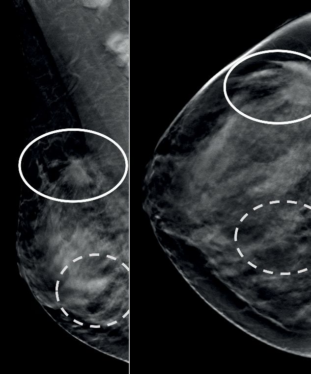
Figure 3. Comparison of 2D with synthesized mammography: Recall reduction + +++
in synthesized mammography (SM), there was an increase of Asymmetries +++ +
the contrast with the parenchyma (A) with greater intensity
Interval changes +++ +
(whiter), greater enhancement of the ligaments (B) and of
Comparison with previous ones +++ +
the linear structures in pre-parenchymatous fat, greater
prominence of calcifications (arrows). The increased intensity in Calcifications (detection) +++ ++
the peripheral area of the image (C) dos not represent greater Calcifications (distribution) +++ +
skin thickness and is associated to the reconstruction program, +: low grade; ++: medium grade; +++: high grade.
noticing lesser prominence in more recent versions. Source: Rafferty and Belfer15.
54 Mastology, 2018;28(1):51-66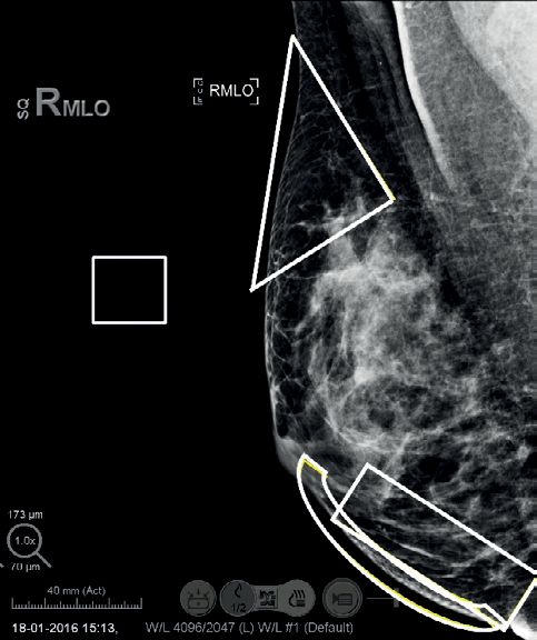
Breast tomosynthesis: a better mammography

INDICATIONS OF TOMOSYNTHESIS                                                A                      B            C             D
The indications for tomosynthesis are the same as for conven-
tional digital mammography: it can be used as a diagnostic or
screening test for early cancer detection. Due to the reduction
of tissue overlap, tomosynthesis presents several advantages
over the 2D, such as:
1. Increase in cancer detection rate (Figures 4, 5 and 6);
2. Reduction of fa lse-positive reca lls and additiona l
    mammographic projections, especially spot compression,
    which means reduction of the total dose of radiation (Figure 7);
3. Increase in the conspicuity of noncalcified lesions (masses,           Figure 6. Architectural distortion (arrows and details)
    asymmetries and architectural distortions) (Figures 8, 9              evidenced in tomosynthesis slices (B and D) and not expressed
    and 10);                                                              in 2D (A and C). Histology: invasive lobular carcinoma.
4. Reduction in the percentage of category 3 lesions (ACR
    BI-RADS), especially asymmetries;
5. Detection and location of lesions seen in only one incidence
    (Figures 11 and 12);
                                                                            A                                 B
6. Confirm cutaneous lesions (Figure 13);

 A                                  B

                                                                                   2D                  3D           2D            3D
                                                                          Figure 7. The density (arrows) observed in conventional
                                                                          digital mammography (2D) does not correspond to
                                                                          tomosynthesis slices (3D), representing overlap of normal
     2D                3D               2D                3D
                                                                          tissues (pseudolesion): (A) craniocaudal incidence; (B)
Figure 4. Conventional digital mammography (2D) and
                                                                          mediolateral incidence.
tomosynthesis representative slice (3D), 1 mm thick,
evidenced spiculated nodule (circle): (A) mediolateral incidence;
(B) craniocaudal incidence. Histology: invasive carcinoma.

                                                                            A                  B            C                D

 A                                  B

                                                                          Figure 8. Patient was recalled for evaluation of irregular
          2D             3D              2D                 3D            asymmetry in the upper/posterior third of her left breast (arrow),
                                                                          in the oblique mediolateral incidence in 2D (A). Tomosynthesis
Figure 5. Conventional digital mammography (2D) and                       slice (B) was shown to be tissue overlap. In tomosynthesis slice,
representative tomosynthesis slice (3D), 1 mm thick, evidenced            in craniocaudal incidence (D), an architectural distortion area
spiculated nodule (arrows) identified only in tomosynthesis: (A)          (larger circle) was observed, not evidenced in 2D in craniocaudal
mediolateral incidence; (B) craniocaudal incidence. Histology:            incidence (C) and an irregular nodule (smaller circle) was better
invasive carcinoma.                                                       characterized in the same slice (D).

                                                     Mastology, 2018;28(1):51-66                                                       55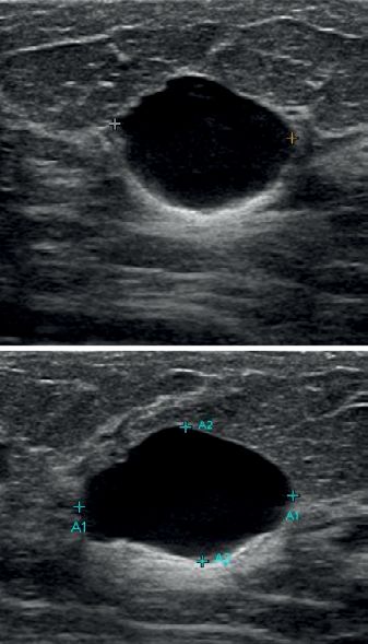
Aguillar VLN, Bauab SP, Belfer A

TOMOSYNTHESIS IN SCREENING                                                  (15 to 37%) as its primary advantage18,19. The multicenter study,
Performance of tomosynthesis in screening was investigated                  published by Rafferty et al. in 2013, was crucial in demonstrat-
in a series of prospective, retrospective, single-institution or            ing that tomosynthesis, in addition to reducing the number of
multi-center studies, all using 2D mammography along with                   recalls, significantly increased the diagnostic accuracy in rela-
tomosynthesis versus the 2D one alone. The first publications               tion to 2D mammography 20.
on tomosynthesis emphasized the reduction of the recall rate
                                                                            Prospective studies
                                                                            Prospective clinical studies are all European, although they have
 A                                     C                                    different study designs. The largest one, carried out in Oslo (Oslo
                                                                            Tomosynthesis Screening Trial – OTST), used independent dou-
                                                                            ble reading and analyzed the first results in 12,631 women in the
                                                                            screening program at the University of Oslo, aged between 50 and
                                                                            69, who performed 2D together with tomosynthesis, two views
                                                                            per breast and per modality. The combination of tomosynthesis
                                                                            and 2D increased the cancer detection rate by 27% compared

 B
                                                                              A                           B              C

Figure 9. Correlation between tomosynthesis (A),
ultrasonography (B) and magnetic resonance imaging (B)
                                                                                                                         D
showing the nodule (smaller circle) and area of architectural
distortion (larger circle).

 A                B                C                 D
                                                                            Figure 12. Correlation of the finding observed only in
                                                                            craniocaudal incidence in (A) and in tomosynthesis (B) with
                                                                            ultrasound (C) and with magnetic resonance imaging (D).
                                                                            Histology: Invasive Lobular carcinoma grade I.

Figure 10. Nodule evidenced in craniocaudal incidence, in 2D
(A) shows radiolucent center in tomosynthesis slice (B) and
represents intramammary lymph node.

 A                B                C                D

Figure 11. Finding observed only in craniocaudal incidence                  H: head; F: feet; M: medial; L: lateral.
in 2D (A). Lesion’s infiltrative margins are characterized in               Figure 13. Location of the lesion in tomosynthesis. The lateral
tomosynthesis slices in craniocaudal incidence (B). The lesion              bar demonstrates the lesions are in the lower (F) and medial
is not observed in 2D nor in tomosynthesis in mediolateral                  (M) quadrant, at the extremity of the bar, demonstrating the
incidence (C and D). Histology: Invasive Lobular carcinoma grade I.         quadrant and its cutaneous location.

  56                                                           Mastology, 2018;28(1):51-66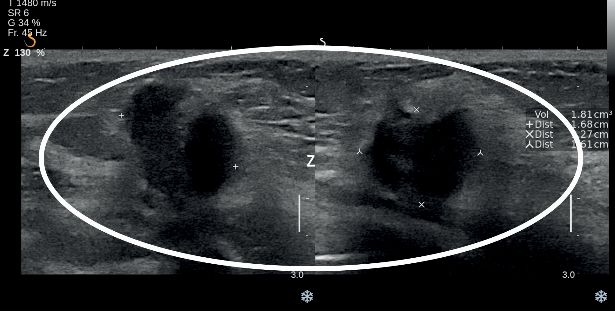
Breast tomosynthesis: a better mammography
to 2D alone (absolute increase of 1.9 cancers/thousand women It is important to remember that these results reflect prevalent
screened by tomosynthesis) and, at the same time, reduced the exams, that is, the first tomosynthesis examinations in these
recall rate17. populations (Table 1)24.
The Italian study (Screening with Tomosynthesis OR stan-
dard Mammography – STORM1), carried out in two cities Trento Retrospective studies
and Verona, included 7,292 women who did both - 2D and tomo- American studies on tomosynthesis are retrospective, compar-
synthesis – in two views per breast per modality, with double ing isolated 2D versus digital mammography associated with
sequential reading. Their final results were promising: a rela- tomosynthesis. Some of them should be highlighted as pioneers:
tive increase of 53% in cancer detection with tomosynthesis Rose et al.19, with a 25% increase in cancer detection, in a pri-
(an increase of 2.8 cancers/thousand) and 17% fewer recalls21. vate clinic; Haas et al.18, with an increase of 10% in an academic
STORM 2 is another population-based prospective study, center and reduction of recall rate from 37 to 30%. The work of
based on STORM 1, which included 9,672 women. In this study, all McCarthy et al.25 has, in fact, demonstrated the impact of tomo-
patients were screened with 2D and tomosynthesis, and SM was synthesis on screening after its implementation in a population
also used: the combined examination, either with a 2D (COMBO in a single academic center in the United States. The screening
mode) or with SM (TOMOHD mode), detected more cancers was performed in all women, from October 2011 to February 2013
than the digital mammography alone. There was an increase (n = 15,571 women) with a significant reduction in the number
of 35 and 40% (COMBO and TOMOHD, respectively) in cancer of recalls in the group with tomosynthesis. Overall, the increase
detection rate, with an increase of 2.2 and 2.5 c ancers/thousand). in cancer detection was small (0.9 additional cancers/thousand
However, there was a slight increase in false-positive recall with women), but analysis of subgroup showed a significant increase
the combined examination, compared to 2D alone, especially in cancer detection among women younger than 50 years25 .
with the use of SM22. In 2014 , a large US retrospective multicenter study was published
The study performed in Malmö, Sweden (Malmö Breast (13 academic or private institutions), including 281.187 women
Tomosynthesis Screening Trial – MBTST), differs from the pre- undergoing FFDM only and 173.663 women having 2D+ 3D .
vious ones because it was designed to evaluate the efficacy of The results showed a 29% increase in cancer detection rate and
only one view of tomosynthesis (mediolateral oblique) versus two 15% decrease of recalls, after implementation of tomosnthesis,.
views of 2D mammography (craniocaudal and MLO). The first In 2015, data from the TOMMY TRIAL (Comparison of
results included 7,500 women, aged 40 to 74 years old, invited TOMosynthesis with digital Mammography in the UK NHS
to perform both modalities. Tomosynthesis ( in just one view- Breast Screening Programme), a retrospective, multicenter
MLO) increased cancer detection rate by 43% (2.6 more can- study with the participation of several radiologists was pub-
cers/1000 women). Although radiation dose was lower, there was lished, comparing the performance of tomosynthesis and 2D
an increase in the recall rate, probably due to the use of only one versus isolated 2D in the mammography screening program
view of tomosynthesis23. of the United Kingdom. Women aged between 29 and 85 years
In summary, prospective studies showed a 26 to 43% increase (mean 56 years) were recruited from July 2011 to February 2013
in cancer detection rate with the addition of tomosynthesis to 2D and the final analysis consisted of 7,060 cases. All participants
mammography, basically at the expense of invasive carcinomas. performed 2D (on two incidences) and tomosynthesis (on two
Table 1. Resultas obtained with prospective and retrospective studies in the screening of breast cancer.
Detection Detection Relative
Design of Type of Age range
Population (n) Recall rate rate /1000 rate/1000 carcinoma
the study Reading (years)
(2D) (tomosynthesis) increase
Prospective Sequential
7,292 48 to 71 -17% 5.3 8.1 53%
paired pair
Prospective Independent
12,631 50 to 69 -13% 6.1 8.0 27%
paired pair
Prospective Sequential
7,500 40 to 74 43% 6.3 8.9 43%
paired pair
Prospective Sequential 16% 6.3 8.8 35%
9,672 53 to 63
4 arms pair 30% 6.3 8.8 40%
Retrospective 2D: 281,187
Single reading -16% 4.2 5.4 29%
unpaired 3D: 173,663
Mastology, 2018;28(1):51-66 57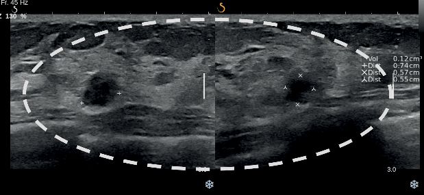
Aguillar VLN, Bauab SP, Belfer A

incidences), with SM replacing 2D, as of 2011. The results showed         Malmö23 and 45% in the American multicenter retrospective
increased specificity with tomosynthesis in all subgroups: age            study 26. Forty to forty-eigth had histological grade 2 or 3 and 76
range, breast density and mammographic findings. Regarding                to 90% presented with negative sentinel lymph node14,23. There
sensitivity, tomosynthesis, compared with isolated 2D, was                was no significant increase in carcinoma in situ.
superior in dense breasts, invasive carcinomas of 11 to 20 mm                 Another important finding were the preliminary results from
and lesions presented as nodules, with no significant difference          Malmo Trial (MBTST), presented at the European Congress of
between 2D and SM 27.                                                     Radiology (Vienna, 2017), including all participants of the study
    In Brazil, we have few published data regarding the perfor-           (15.000 twomen). Among the additional invasive carcinomas
mance of tomosynthesis. In the private clinic data obtained by            detected only by tomosynthesis, 58% were ductal and 26% were
one of the authors (Bauab SP) from July 2, 2012 to August 31, 2012,       lobular, whereas in the group detected by 2D, only 17% were lob-
which included 1,220 women aged 40-83 years, submitted to tomo-           ular. The authors suggest that tomosynthesis may have a higher
synthesis (COMBO mode or 2D complementary tomosynthesis),                 sensitivity for detection of lobular carcinomas compared to 2D
12 cases of invasive cancer were found in asymptomatic patients.          mammography (Table 2, Figures 14 and 15)28.
Tomosynthesis showed the lesion better in 9 cases, and in 3 cases
the lesion was detected only by tomosynthesis, resulting in a             Diagnostic tomosynthesis:
33% increase in the diagnosis of cancer in asymptomatic women.            In diagnostic cases, tomosynthesis increases accuracy, reduces the
    Data from another private clinic (Aguillar VLN), included             number of additional mammographic incidences29,30 and reduces
4,314 women - 82% between 40 and 69 years and 58% with                    the probability of category 3 lesions of ACR BI-RADS, mainly focal
dense breasts (ACR C or D) - from September 2011 to August                asymmetries31-33. Tomosynthesis plays an important role in the
2014, who were submitted to COMBO mode tomosynthesis, two                 confirmation of cutaneous lesions, in the evaluation of findings in
views for each modality. Twenty-one carcinomas (10 in situ and            a single incidence (mainly asymmetries) and in palpable lesions.
11 invasive) were detected by 2D and 27 carcinomas (10 in situ            It reduces need for additional mammographic incidences, with a
and 17 invasive) were detected by 3D. The six invasive carcino-           faster and cheaper workflow and lower radiation dose34,35.
mas detected by tomosynthesis-only, were all seen as archi-                    Cutaneous lesions such as warts, sebaceous cysts or calcifica-
tectural distortions and stage I, positive hormone receptors              tions are observed in the more superficial slices of tomosynthesis,
and only one SL positive, with micromatastases. The cancer                in which dermis and cutaneous pores are also visualized, con-
detection rate with 2D was 4.87 carcinomas/thousand exams;                firming the superficial location of these lesions. Tomosynthesis
and with 3D, 6.26 carcinomas/thousand exams, representing                 does not require tangential incidences34.
an absolute increase of 1.4 carcinomas/thousand exams and a                    For asymmetries, tomosynthesis proves that most of the
relative increase of 28.6%. Invasive detection rate with 2D was           findings observed in only one incidence represent normal breast
2.54 invasive carcinomas/thousand exams, whereas with 3D it               overlapping with no need for additional projections33.
was 3.94 invasive carcinomas/thousand exams, representing a                    For palpable lesions, tomosynthesis, together with ultra-
relative increase of 54%.                                                 sonography, in general, are diagnostic, with no need for extra
    Regarding the characteristics of the carcinomas detected              views, such as spot compression or roll angle incidences
only by tomosynthesis, similar results were demonstrated in               (Figure 16)34,35.
the prospective and retrospective studies. There was a signifi-                In the presence of a nodule of circumscribed margins evi-
cant increase in the detection of invasive carcinomas with tomo-          denced in the tomosynthesis, an ultrasonography should be
synthesis: 40% in the study by Oslo13, 49% in STORM 121, 41% in           used to characterize its cystic or solid nature (Figures 17 and 18).

Table 2. Carcinomas detected only by tomosynthesis.
                                  Detection          Detection            Relative        Relative invasive                     Negative
                                                                                                              Histological
 Study                          rate/thousand      rate/thousand         carcinoma           carcinoma                         sentinela
                                                                                                              grade 2 or 3
                                     (2D)         (tomosynthesis)         increase            increase                        lymph node
 STORM121                             5.3                8.1                53%                 49%                               60%
 OTST  17
                                      6.1                8.0                27%                 45%              40%              76%
 MBTST 23                             6.3                8.9                43%                 42%              48%              90%
                                      6.3                8.8                35%
 STORM222                                                                                                        69%              86%
                                      6.3                8.8                40%
 Multicenter United State26           4.2                5.4                29%                 41%

  58                                                           Mastology, 2018;28(1):51-66
Breast tomosynthesis: a better mammography
A B C Breast density and tomosynthesis
A multicentric study specifically developed to correlate the effi-
ciency of tomosynthesis in different mammographic patterns of
ACR BI-RADS found that tomosynthesis is better than 2D DM
alone in dense or non-dense breasts, justifying its use in any
mammographic pattern. However, the subgroup analysis showed
that the sensitivity of tomosynthesis is lower in extremely dense
breasts (ACR D) than in the other groups due to lack of adipose
tissue in the breast, necessary for lesions detection. (Table 3)36.
Preliminary results with 15,000 participants from the Swedish
prospective study by Malmö23 show that tomosynthesis increases
cancer detection in all mammographic patterns. Of the tumors
detected only by tomosynthesis, 46% were in women with non-dense
breasts (ACR A and B) and 54% in dense breasts (ACR C and D).
Figure 14. Focal architecture distortion is not evidenced in 2D
In daily use, the greatest gain in tomosynthesis screening is
(A) nor in synthesized 2D mammography (B). It is characterized
observed in women with scattered fibroglandular densities (ACR B)
in tomosynthesis (C).
A B C
Figure 15. Focal architecture distortion only in oblique mediolateral (A) and craniocaudal (B) incidences in tomosynthesis (B). It is
characterized in ultrasound (C). Histology: invasive lobular carcinoma grade I.
A B A B
Craniocaudal Médio-lateral
Ultrasonography
Figure 16. Palpable nodule to the right in heterogeneous dense breast, not observed in 2D (A) is presented in tomosynthesis (B) with
spiculated margins. Ultrasonography shows a 1.4 cm nodule . Histopathology: invasive carcinoma g2.
Mastology, 2018;28(1):51-66 59Aguillar VLN, Bauab SP, Belfer A
Tomosynthesis
Ultrasonography
and heterogeneously dense breasts (ACR C), which represent the
A B
majority of breast density patterns. In women with extremely
dense breasts (ACR D), complementary ultrasonography will
continue to play an important role due to the lower sensitivity
of tomosynthesis in this group (Figure 19)33.
Detection of calcifications in tomosynthesis
Although there is no doubt that tomosynthesis is superior to 2D in
the detection and characterization of noncalcified lesions (nodules,
asymmetries and distortions), there are still questions related to
grouped calcifications. Some studies showed that tomosynthesis is
at least equal to 2D in detecting microcalcifications37,38. However, the
Left breast
detection of clusters of small and faint calcifications can be a chal-
SMQ MQJ lenge to be perceived in tomosynthesis for several reasons:
SMQ: superomedial quadrant; MQJ: medial quadrant junction. 1. The low radiation dose in the raw images can reduce
Figure 17. Heterogeneously dense breast. Tomosynthesis shows reconstructed images’ spatial resolution;
circumscribed margins of the lesion: (A) mediolateral incidence; 2. Movement of the patient during the acquisition of the
(B) craniocaudal incidence. Ultrasonography shows cyst. tomosynthesis or the 2D image;
3. Tomosynthesis slices have parallel reconstruction to the
detector, while suspicious microcalcifications have radial
Tomosynthesis
distribution in the breast. Therefore, grouped calcifications
A B Ultrasonography
with linear or segmental distribution will be visualized in
more than one slice of tomosynthesis, which makes it difficult
to analyze its morphology and distribution. The addition of
several 1-mm slices in a single image (slab) facilitates the
visualization of the whole group although with loss of spatial
resolution and bigger impact in small particles.
SM increases the brightness and contrast of calcifications
and can improve their perception and characterization when
Right breast
combined with tomosynthesis. However, the detection of clus-
MQJ
tered calcifications in tomosynthesis or SM does not exclude the
MQJ: medial quadrante junction.
Figure 18. Heterogeneously dense breast. Tomosynthesis shows need for extra views with magnification spot compression ampli-
circumscribed margins of the lesion: (A) mediolateral incidence; fied in both incidences (craniocaudal and 90 degrees mediolat-
(B) craniocaudal incidence. Ultrasonography characterizes the eral), essential for the characterization of their morphology and
solid nature of the lesion. Histology: fibroadenoma. distribution and recommendation of management (Figure 20).
Table 3. Breast density and tomosynthesis.
Sparse Heterogeneous Extremely
Adipose Not dense Dense
densities density dense
2D 3D 2D 3D 2D 3D 2D 3D 2D 3D 2D 3D
Recall/thousand
57 55 97 84 128 110 114 98 90 79 127 109
screenings
Cancer/thousand
3.2 4.2 4.4 5.3 4.5 6.1 3.8 3.9 4.2 5.1 4.5 5.8
screenings
Invasive câncer/
2.3 3.5 3.2 4.1 3.0 4.5 1.9 2.6 3.0 4.0 2.9 4.2
thousand screenings
Positive predictive
6.2 8.4 4.9 6.9 3.8 5.9 3.7 4.3 5.1 7.1 6.2 8.4
value by recall
Source: adapted36.
60 Mastology, 2018;28(1):51-66Breast tomosynthesis: a better mammography
In some cases, tomosynthesis may add to the final mammo-
graphic diagnosis by detecting radiographic changes associated
with clustered calcifications, such as focal architectural distor-
tion, asymmetries or nodules (Figure 21).
Management of lesions
detected only in tomosynthesis 2D SM
Some lesions are detected only in tomosynthesis (hidden in the
2D), mainly spiculated nodes and architectural distortions, due
to better resolution of spicules in tomosynthesis. In cases where
the change is only suspected, selective compression can be per-
formed to confirm the finding33.
Tomosynthesis facilitates the localization of a lesion in the
breast, through the lateral bar shown in its slices: for example,
in the craniocaudal incidence, the bar demonstrates whether
the lesion is in the lower or upper quadrant and, in the medio- 3D MAG
lateral incidence, it demonstrates whether the lesion is lateral
MAG: magnified radiography.
or medial. The sidebar also guides the location of the lesion on Figure 20. Calcifications in 2D synthesized mammography (SM)
the second look examination and helps when the image is only presents better conspicuity than in 2D. Although calcifications
seen in one incidence39. can be detected in tomosynthesis (3D), the magnified spot
Most of the changes observed only in tomosynthesis are compression (MAG) better characterizes their shape and
also characterized by second look ultrasound which allows for distribution. Histopathology: ductal carcinoma in situ.
Tomosynthesis
Ultrasonography
Right breast
LQJ PALP COND
Right breast
LQJ PALP COND
SMQ: superomedial quadrant; PALP COND: palpable density; LQJ: lower quadrant junction.
Figure 19. In breasts with extremely dense pattern (D pattern), even in tomosynthesis the lesion may be obscured by dense tissue
(dashed line). Ultrasonography is able to detect these lesions. The largest and most peripheral lesion (continuous line) was detected
in both methods. Histopathology: multicentric invasive ductal carcinoma.
Mastology, 2018;28(1):51-66 61Aguillar VLN, Bauab SP, Belfer A
a percutaneous biopsy by this method. In cases where the lesion associated to several pathologies (Figure 24). However, it has
is not evident on ultrasonography, magnetic resonance imaging a high positive predictive value (PPV) for malignancy. Recent
(MRI) is an option, if it enhances. Percutaneous vacuum biopsy studies, with retrospective analysis of findings detected only
may be guided by this modality. In cases where the lesion is by tomosynthesis, show PPV from 21 to 53%. The work of
not characterized on ultrasonography or does not show MRI Partyka et al.41, a retrospective analysis of 9,982 tomosynthesis
enhancement or MRI is not available, a tomosynthesis-guided
percutaneous biopsy may be performed, when available, on the
biopsy prone table or in the biopsy equipment coupled to the A B Slice C Slice
tomosynthesis equipment. If the tomosynthesis biopsy equip-
ment is not available, the tomosynthesis-guided preoperative
location can be performed through tomosynthesis equipment :
the procedure is performed similarly to the biplanar preopera-
tive location (Figures 22 and 23)40.
Among the findings seen only in tomosynthesis, architec-
tural focal distortion is the most frequent finding and may be
Figure 23. Preoperative location in mediolateral incidence,
assisted by tomosynthesis, of the focal architecture distortion
not seen in ultrasound and MRI (Figure 23). The positioning of the
A needle on axes x and y (dashed line) is provided by synthesized
A B mammography (A) and depth (Z axis) by tomosynthesis (B).
Positioning of the needle confirmed by tomosynthesis slice (C).
Invasive carcinoma Invasive lobular carcinoma
B
Figure 21. Conventional digital mammography (A) shows
suspicious calcifications (arrows) and tomosynthesis
(B) shows architectural distortion as an additional finding
to microcalcifications, which may represent an invasive Radial scar Complex
component of the lesion. sclerosing lesion
A B C
Surgical scar Fat necrosis
Figure 22. Focal architecture distortion is not evidenced
in 2D (A). In mediolateral incidence, it is characterized in
SM (B) and in tomosynthesis (C). Ultrasound and MRI not Figure 24. Architectural distortion may be associated with
showing this finding. several diseases, requiring a histological study
62 Mastology, 2018;28(1):51-66Breast tomosynthesis: a better mammography
examinations, found 26 cases of architectural distortion, of as well as tomosynthesis, detect small invasive cancers, not seen
which 19 (73%) were detected only in tomosynthesis, with a on mammography, even retrospectively, in women with dense
PPV of 21% (4/19). Ray et al.42 retrospectively analyzed 19 lesions breasts. Tomosynthesis has the advantage of being only one
detected by tomosynthesis only (14 cases of architectural dis- exam (an improved mammography), with high PPV, but with a
tortion and 5 of spiculated masses), of which 10 were malignant higher implementation and maintenance cost. Ultrasonography
(5 invasive ductal carcinomas and 3 invasive lobular carcino- is an additional exam, with low cost and widely available, with-
mas), with PPV of 53%. out radiation addition, though it has low predictive value and
Risk factors for breast cancer should be taken into account, needs an experienced radiologist to be performed. There is little
and biopsy may be indicated instead of follow-up for high-risk information on which exam to choose, as a complement to 2D,
patients. A modified algorithm by Durand et al. 39 is proposed, in women with dense breasts: only ultrasonography, only tomo-
considering the risk of breast cancer and the availability or not synthesis or both?
of MRI (Figure 25). Tagliafico et al. 43 published a multicenter study with
Future studies are needed to determine the likelihood of 3,231 women, mean age of 51 years, in which tomosynthesis
malignancy in architectural distortions detected only in tomo- and ultrasonography were performed, with independent inter-
synthesis, without characterization in ultrasound or in MRI, to pretation, in women with dense breasts and negative mam-
establish new protocols related to these lesions. mography. The study observed the detection of 24 additional
cancers in relation to digital mammography, with 23 invasive
Could tomosynthesis replace ultrasound? ones. The detection rate was 4/1000 with tomosynthesis and
Dense breasts reduce mammography’s sensitivity, being rec- 7.1/1000 with ultrasonography, with a similar recall rate in
ommended, in those cases, complementary tracing with other both methods. The study concludes that ultrasound detects
methods. Numerous studies have found that ultrasonography, more cancers than tomosynthesis in women with dense breasts.
Architectural distortion in tomosynthesis
guided ultrasound
NO match WITH match
BX by tomo if available Bx by tomo NOT available US guided Bx
MRI NOT available MRI available
High risk NO enhancement WITH enhancement
Bx or LOC by tomo Average risk High risk Bx or LOC by MRI
Short term f-up or LOC by tomo Bx or LOC by tomo
Bx: biopsy; tomo: tomosynthesis; US: ultrasonography; MRI: magnetic resonance imaging; LOC: preoperative location; F-up: follow-up.
Figure 25. Algorithm for management in architectural distortion evidenced in tomosynthesis.
Mastology, 2018;28(1):51-66 63Aguillar VLN, Bauab SP, Belfer A
Therefore, tomosynthesis does not exempt complementary ultra- Interval carcinomas and
sonography in dense breast breasts. tomosynthesis screening
An important finding was the detection, through tomosyn- The rate of interval cancers (ICR) after introduction of tomosyn-
thesis, of more than 50% of the additional cancers found, showing thesis into screening still needs to be investigated. Evidence that
that potentially, tomosynthesis could replace 2D as the primary tomosynthesis (combined with 2D) reduces the rate of interval
cancer screening method in dense breasts. Unlike ultrasonogra- carcinoma is still limited .Skaane et al.44 didn’t show reduction
phy, tomosynthesis increases cancer detection without increas- in the ICR after the first year of introduction of tomosynthesis:
ing the rate of false positives, which is the most critical point in 2,0 IC/1000 with 2D and 2,1 IC/1000 with 2D+3D. On the other
relation to ultrasound screening. hand, a study from Pensylvannia 45 which analyzed the results of
We must remember that the results published are prelimi- three years follow-up with tomosynthesis, demonstrated a small
nary and that this study needs to be reproduced in other centers. reduction in ICR with tomosynthesis (0,7/1000 with 2D versus
In addition, it was the first shift with tomosynthesis in this group 0,5/1000 with tomo). However, these are individual studies with
(little experience of the authors with tomosynthesis), while most small number of patients and more data is needed.
of the ultrasound examinations were incident and performed with
professionals experienced in the method (Table 4) 43.
In clinical practice, it can be observed that tomosynthesis CONCLUSION
does not completely replace ultrasound. In very dense and het- Tomosynthesis (3D mammography) is a new mammographic
erogeneously dense breasts, complementary ultrasonography technique that increases sensitivity and specificity when com-
should still be recommended, since it is possible to detect small bined to 2D mammography (conventional or synthesized)
lesions on ultrasound, not characterized in tomosynthesis, espe- In screening, tomosynthesis has a positive impact because
cially in breasts with extremely dense pattern, in which there is increases detection of small , low grade, RH positive and LN neg-
insufficient adipose tissue to make contrast in tomosynthesis ative invasive cancers and reduces unnecessary recalls or need
slices (Figure 19). for additional mammographic incidences.
In diagnostic cases, the technique increases diagnostic accu-
racy by allowing for a better characterization and location of the
Table 4. Carcinomas detected only by tomosynthesis. lesions, making the conventional diagnostic approach unneces-
Positive Negative Total
sary, with reduction of patients’ anxiety and lower financial cost.
ultrasound ultrasound n (%) With the development of SM, reconstructed from tomo-
Positive synthesis to replace conventional 2D imaging, the problem of
12 1 13 (54.2)
tomography increased total radiation dose — considered an obstacle to this
Negative method in screening — has been solved.
11 0 11 (45.8)
tomography Future studies of interval cancer incidence and cost-benefit
Total n (%) 23 (95.8) 8.9 (4.2) analysis of this technology should provide new data for implemen-
Source: adapted .
43
tation of tomosynthesis in large-scale breast cancer screening.
REFERENCES
1. Skaane P, Gullien R, Bjorndal H, Eben EB, Eksebh UH, Jahr G, Radiology. 2015;272(3):663-84. https://doi.org/10.1148/
et al. Digital breast tomosynthesis (DBT): initial experience radiol.2015141303
in a clinical setting. Acta Radiol. 2012;53(5):524-9. https://doi. 5. Sechopoulos I. A review of breast tomosynthesis. Part I. The
org/10.1258/ar.2012.120062 image acquisition process. Med Phys. 2013;40:014301. https://
2. Hardesty LA. Issues to Consider Before Implementing Digital doi.org/10.1118/1.4770279
Breast Tomosynthesis Into a Breast Imaging Practice. 6. Sechopoulos I. A review of breast tomosynthesis. Part
Am J Roentgenol. 2015;204:681-4. https://doi.org/10.2214/ II. Image reconstruction, processing and analysis, and
AJR.14.13094 advanced applications. Med Phys. 2013;40:14302. https://doi.
3. Kopans DB. Digital breast tomosynthesis from concept to org/10.1118/1.4770281
clinical care. Am J Roentgenol. 2014;202:299-308. https://doi. 7. Svahn TM, Houssami N, Sechopoulos I, Mattsson S. Review of
org/10.2214/AJR.13.11520 radiation dose estimates in digital breast tomosynthesis relative
4. Vedantham S, Karellas A, Vizayaraghavan GP, Kopans to those in two-view full-field digital mammography. Breast.
DB. Digital Breast Tomosynthesis: state of the art. 2015;24(2):93-9. https://doi.org/10.1016/j.breast.2014.12.002
64 Mastology, 2018;28(1):51-66Breast tomosynthesis: a better mammography
8. Skaane P, Bandos AI, Eben EB, Jebsen IN, Krager M, screening practice: an observational study. Am J Roentgenol.
Haakenaasen U, et al. Two-view digital breast tomosynthesis 2013;200(6):1401-8. https://doi.org/10.2214/AJR.12.9672
screening with synthetically reconstructed projection images: 20. Rafferty EA, Park JM, Philpotts LE, Poplack SP, Sumkin JH,
comparison with digital breast tomosynthesis with full-field Halpern EF, et al. Assessing radiologist performance using
digital mammographic images. Radiology. 2014;271(3):655-63. combined digital mammography and breast tomosynthesis
https://doi.org/10.1148/radiol.13131391 compared with digital mammography alone: results of a
9. Zuley ML, Guo B, Catullo VJ, Chough DM, Kelly AE, Lu multicenter, multireader trial. Radiology. 2013;266(1):104-13.
AH, et al. Comparison of two-dimensional synthesized https://doi.org/10.1148/radiol.12120674
mammograms versus original digital mammograms alone 21. Ciatto S, Houssami N, Bernardi D, Caumo F, Pellegrini M,
and in combination with tomosynthesis images. Radiology. Brunelli S, et al. Integration of 3D digital mammography
2014;271(3):664-71. https://doi.org/10.1148/radiol.13131530 with tomosynthesis for population breast-cancer screening
10. Zuckerman SP, Conant EF, Keller BM, Maidment ADA, (STORM): a prospective comparison study. Lancet Oncol.
Barufaldi B, Weinstein SP, et al. Implementation of synthesized 2013:14(7):583-9. https://doi.org/10.1016/S1470-2045(13)70134‑7
two-dimensional mammography in a population based 22. Bernardi D, Macaskill P, Pellegrini M, Valentini M, Fantò C,
digital breat tomosynthesis screening program. Radiology. Ostillio L, et al. Breast cancer screening with tomosynthesis
2016;281(3):730-6. https://doi.org/10.1148/radiol.2016160366 (3D mammography) with acquired or synthetic 2D
11. Wallis MG, Moa E, Zanca F, Leifland K, Danielsson M. Two- mammography compared with 2D mammography alone
view and single-view tomosynthesis versus full-field digital (STORM 2): a population-based prospective study. Lancet Oncol.
mammography: high resolution X-ray imaging observer 2016;17(8):1105-13. https://doi.org/10.1016/S1470-2045(16)30101-2
study. Radiology. 2012;262(3):788-96. https://doi.org/10.1148/ 23. Lang K, Andersson I, Rosso A, Tingberg A, Timberg P,
radiol.11103514 Zackrisson S. Performance of one-view breast tomosynthesis
12. Rafferty EA, Park JM, Philpotts LE, Poplack SP, Sumkin JH, as a stand-alone breast cancer screening modality: results
Halpern EF, et al. Diagnostic accuracy and recall rates for from the Malmo Breast Tomosynthesis Screening Trial, a
digital mammography and digital mammography combined population-based study. Eur Radiol. 2016;26:184-90. https://
with one-view and two-view tomosynthesis: results of an doi.org/10.1007/s00330-015-3803-3
enriched reader study. Am J Roentgenol. 2014;202(2):273-81. 24. Houssami N, Skaane P. Overview of the evidence on digital
https://doi.org/10.2214/AJR.13.11240 breast tomosynthesis in breast cancer detection. Breast.
13. Beck N, Butler R, Durand M, Andrejeva, Hooley R, Horvath 2013;22(2):101-8. https://doi.org/10.1016/j.breast.2013.01.017
L, et al. One-View Versus Two-View Tomosynthesis: 25. McCarthy AM, Kontos D, Synnestvedt M, Tan KS, Heitjan
A Comparison of Breast Cancer Visibility in the Mediolateral DF, Schnall M, at al. Screening Outcomes Following
Oblique and Craniocaudal Views. In: ARRS; 2013. Scientific Implementation of Digital Breast Tomosynthesis in a
Session 27, Breast Imaging. 2013. General Population Screening Program. J Natl Cancer Inst.
2014;106(11):1-7. https://doi.org/10.1093/jnci/dju316
14. Skaane P, Bandos A, Gullien R, Eben EB, Ekseth U, Haakenaasen
U, et al. Comparison of digital mammography alone and digital 26. Friedewald SM, Rafferty EA, Rose SL, Durand M, Plecha DM,
mammography plus tomosynthesis in a population-based Greenberg JS, et al. Breast cancer screening using tomosynthesis
screening program. Radiology. 2013;267(1):47-56. https://doi. in combination with digital mammography. JAMA.
org/10.1148/radiol.12121373 2014;311(24):2499-507. https://doi.org/10.1001/jama.2014.6095
15. Rafferty EA., Belfer AJ. Tomosynthesis & synthesized 2-D 27. Gilbert FJ, Tucker L, Gillan MG, Willsher P, Cooke J, Duncan
imaging part II: the evolution of mammography. Applied KA, et al. Accuracy of Digital Breast Tomosynthesis for
Radiology Experts Forum Webinars. October, 2013. depicting breast cancer subgroups in a UK retrospective
reading study (TOMMY Trial). Radiology. 2015;277(3):697-706.
16. Dang PA, Free PE, Humphrey KL, Halpern EF, Rafferty https://doi.org/10.1148/radiol.2015142566
EA. Addition of tomosynthesis to conventional diagnostic
mammography: effect on image interpretation time of 28. Lang K, Experiences from the Malmö breast tomosynthesis
screening examinations. Radiology. 2014;270(1):49-56. https:// screening trial Presented at the ECR Annual Meeting, Vienna
doi.org/10.1148/radiol.13130765 2017, Scientific Session Breast tomosynthesis symposium:
Is digital breast tomosynthesis ready for mammo screening?
17. Houssami N, Bernardi D, Pellegrini M, Valentini M, Fanto C, In: ECR Annual Meeting, Vienna, 2017. 2017.
Ostilliò L, et al. Breast cancer detection using single-reading
29. Noroozian M, Hadjiiski L, Rahnama-Moghadam S, Klein KA,
for breast tomosynthesis (3 D mammography) compared
Jeffries DO, Pinsky RW, et al. Digital Breast Tomosynthesis
to double reading of 2 D mammography: evidence from
Is Comparable to Mammographic Spot Views for Mass
a population-based trial. Cancer Epidemiol. 2017;47:94-9.
Characterization. Radiology. 2012;262(1):61-8. https://doi.
https://doi.org/10.1016/j.canep.2017.01.008
org/10.1148/radiol.11101763
18. Haas BM, Kalra V, Geisel J, Raghu M, Durand M, Philpotts LE.
30. Brandt KR, Craig DA, Hoskins TL, Henrichsen TL, Bendel EC,
Comparison of tomosynthesis plus digital mammography and
Brandt SR, et al. Can digital breast tomosynthesis replace
digital mammography alone for breast cancer screening. Radiology.
conventional diagnostic mammography views for screening
2013;269(3):694-700. https://doi.org/10.1148/radiol.13130307
recalls without calcifications? A comparison study in a
19. Rose SL, Tidwell AL, Bujnoch LJ, Kushwaha AC, Nordmann AS, simulated clinical setting. Am J Roentgenol. 2013;200(2):291-8.
Sexton R Jr. Implementation of breast tomosynthesis in a routine https://doi.org/10.2214/AJR.12.8881
Mastology, 2018;28(1):51-66 65Aguillar VLN, Bauab SP, Belfer A
31. Durand MA, Haas BM, Yao X, Geisel JL, Raghu M, Hooley 39. Durand MA, Wang S, Hooley RJ, Raghu M, Philpotts LE.
RJ, et al. Early clinical experience with digital breast Tomosynthesis-detected Architectural Distortion: Management
tomosynthesis for screening mammography. Radiology. Algorithm with Radiologic-Pathologic Correlation, RadioGraphics.
2015;274(1):85-92. https://doi.org/10.1148/radiol.14131319 2016;36:311-21. https://doi.org/10.1148/rg.2016150093
32. Lourenço AP, Barry-Brooks M, Baird GL, Tuttle A, Mainiero 40. Freer PE, Niell B, Rafferty EA. Preoperative Tomosynthesis-
MB. Changes in recall type and patient treatment following guided needle localization of Mammographically
implementation of screening digital breast tomosynthesis. and sonographically Occult Breast lesions, Radiology.
Radiology. 2015;274:337-42. https://doi.org/10.1148/radiol.14140317 2015;275(2):377-83. https://doi.org/10.1148/radiol.14140515
33. Hooley RJ, Durand MA, Philpotts LE. Advances in Digital 41. Partyka L, Lourenco AP, Mainiero MB. Detection of
Breast Tomosynthesis. Am J Roentgenol. 2017;208:256-66. mammographically occult architectural distortion on digital
https://doi.org/10.2214/AJR.16.17127 breast tomosynthesis screening: initial clinical experience.
Am J Roentgenol. 2014;203(1):216-22. https://doi.org/10.2214/
34. Chong A, McDonald ES, Weinstein S, Roth S, Tobey J, AJR.13.11047
Conant EF. Digital breast tomosynthesis: challenging the
traditional workflow dogma: poster. In: Radiological Society 42. Ray KM, Turner E, Sickles E, Joe BN. Suspicious findings at
of North America, 2016. Scientific Assembly and Annual digital breast tomosynthesis occult to conventional digital
Meeting [Internet]. 2016. Disponível em: archive.rsna. mammography: imaging features and pathological findings.
org/2016/16004083.html Breast J. 2015;21(5):538-42. https://doi.org/10.1111/tbj.12446
35. Conant EF, Beaber EF, Sprague BL, Herschorn SD, Weaver 43. Tagliafico AS, Calabrese M, Mariscotti G, Durando M, Tosto
DL, Onega T, et al. Breast cancer screening using S, Monetti F, et al. Adjunct Screening With Tomosynthesis or
tomosynthesis. Breast Cancer Res Treat. 2016;156:109-16. Ultrasound in Women With Mammography-Negative Dense
https://doi.org/10.1007/s10549-016-3695-1 Breasts: Interim Report of a Prospective Comparative Trial.
J Clin Oncol. 2016;34(16):1882-88. https://doi.org/10.1200/
36. Rafferty EA, Durand MA, Conant EF, Copit DS, Friedewald JCO.2015.63.4147
SM, Plecha DM, et al. Breast cancer screening with
44. Skaane P, Sebuodegard S, Gur D, Gullien R, Hofvind SS.
tomosynthesis and Digital Mammography in Dense and
Screen-detected and interval cancers before, during, and
Non-dense Breasts. JAMA. 2016;315(16):1784-6. https://doi.
after implementation of digital breast tomosynthesis in
org/10.1001/jama.2016.1708
a population-based mammography screening program
37. Spangler ML, Zuley ML, Sumkin JH, Abrams G, Ganott MA, [Internet]. In: Radiological Society of North America, 2015.
Hakim C, et al. Detection and classification of calcifications on Scientific Assembly and Annual Meeting. 2015. Disponível em:
digital breast tomosynthesis and 2D digital mammography: a archive.rsna.org/2015/15017637.html
comparison. Am J Roentgenol. 2011:196(2):320-4. https://doi.
45. McDonald ES, Oustimov A, Weinstein SP, Synnestvedt MB, Schnall
org/10.2214/AJR.10.4656
M, Conant EF. Effectiveness of Digital Breast Tomosynthesis
38. Kopans D, Gavenonis S, Halpern E, Moore R. Calcifications Compared With Digital Mammography Outcomes Analysis from
in the breast and digital breast tomosynthesis. Breast J. 3 Years of Breast Cancer Screening JAMA Oncol. 2016;2(6):737-43.
2011;17:638‑44. https://doi.org/10.1111/j.1524-4741.2011.01152.x https://doi.org/10.1001/jamaoncol.2015.5536
66 Mastology, 2018;28(1):51-66You can also read