STAINLESS STEEL AND NITI TORQUE ARCHWIRES AND APICAL ROOT RESORPTION
←
→
Page content transcription
If your browser does not render page correctly, please read the page content below
J Orofac Orthop (2021) 82:1–12
https://doi.org/10.1007/s00056-020-00244-4

 ORIGINAL ARTICLE

Stainless steel and NiTi torque archwires and apical root resorption
Andrea Wichelhaus1 · Marc Dulla1 · Hisham Sabbagh1 · Uwe Baumert1 · Thomas Stocker1

Received: 17 December 2018 / Accepted: 24 April 2020 / Published online: 1 September 2020
© The Author(s) 2020

Abstract
Objective The amount of apical root resorption when using the torque-segmented archwire (TSA) was investigated as
well as the extent and direction of the therapeutically indicated apical movement and the treatment duration.
Materials and methods The degree of apical root resorption in 18 randomly chosen Class II and Class I patients treated
with the TSA, as well as in 18 conventionally treated patients were evaluated using pre- and posttreatment panoramic
radiographs. The sagittal and vertical apical movements and inclination changes were determined based on pre- and
posttreatment lateral cephalograms. Nonparametric tests were applied to test between treatment groups and steps. The
Mann–Whitney U test, Kruskal–Wallis, Pearson correlation and Wilcoxon signed-rank test were applied for statistical
analysis (p < 0.05).
Results The incidence of root resorptions was 89–94.4% in low or moderate level. The relative root–crown ratio (rRCR)
was not statistically different between the TSA and control groups except tooth 12. The axis of the incisors in the TSA
group was significantly improved. The main direction of movement of the apices of the central incisors was retrusion
and extrusion. No interdependence between the amount of resorption and the parameters of treatment duration, extent and
direction of apical movement were found.
Conclusion The results of the study showed that the amount of apical root resorption with the TSA is slight to moderate
and can be compared to conventional orthodontic treatment. The TSA is hence a suitable method for applying targeted
torques to the incisors.

Keywords Incisors · Orthodontic therapy · Tooth movement · Root-crown ratio · Orthodontic appliances

Edelstahl- und NiTi-Torquesegmentbogen und apikale Wurzelresorption

Zusammenfassung
Ziel Der Umfang der apikalen Wurzelresorption ebenso wie das Ausmaß und die Richtung der therapeutisch angenomme-
nen apikalen Bewegung sowie die Behandlungsdauer wurden bei Anwendung des Torquesegmentbogens (TSB) untersucht.
Methoden Das Ausmaß der apikalen Wurzelresorption wurde an 18 willkürlich ausgewählten Klasse-II- und -I-Patienten,
die mit dem TSB behandelt wurden, und an 18 konventionell behandelten Patienten anhand von prä- und posttherapeu-
tisch erstellten Panoramaschichtaufnahmen beurteilt. Die sagittalen und vertikalen apikalen Bewegungen sowie die Än-
derungen der Inklination wurden anhand der FRS(Fernröntgenseitenbilder)-Aufnahmen bestimmt. Unterschiede zwischen
Behandlungsgruppen und -schritten wurden mit nichtparametrischen Tests bestimmt. Mann-Whitney-U-, Kruskal-Wallis-,
Pearson-Korrelation- und Wilcoxon-Rangsummen-Tests wurden zur statischen Analyse verwendet (p < 0.05).

 Prof. Dr. Andrea Wichelhaus
     kfo.sekretariat@med.uni-muenchen.de

1
     Department of Orthodontics and Dentofacial
     Orthopedics, University Hospital, LMU Munich,
     Goethestraße 70, 80336 Munich, Germany

                                                                                                             K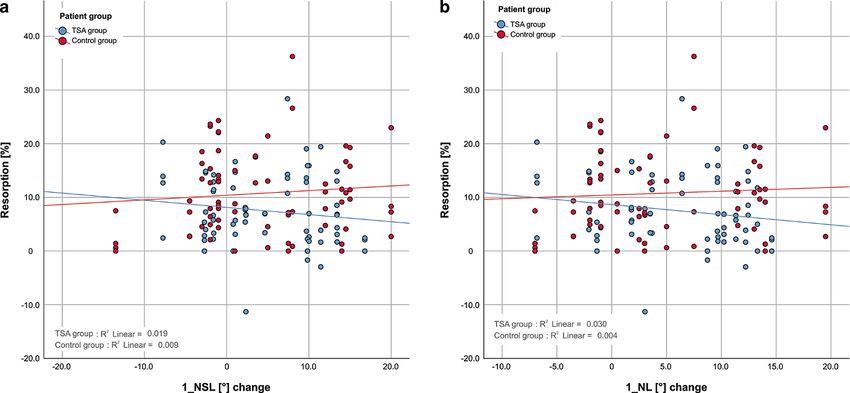
2 A. Wichelhaus et al.
Ergebnisse Die Inzidenz der Wurzelresorption betrug 89–94,4 % für geringes bis moderates Ausmaß. Es gab keinen
statistisch signifikanten rRCR(„relative root–crown ratio“)-Unterschied zwischen der TSB- und der Kontrollgruppe bis auf
Zahn 12. Die Inzisivusachse in der TSB-Gruppe wurde signifikant verbessert. Die Hauptbewegungsrichtung der Apices
der zentralen Schneidezähne waren Retrusion und Extrusion. Es fand sich keine Interdependenz zwischen dem Umfang
der Resorption und den Parametern Behandlungsdauer, Ausmaß und Richtung der apikalen Bewegung.
Schlussfolgerung Die Studienergebnisse zeigen, dass der Umfang der apikalen Wurzelresorption mit TSB leicht bis
moderat ist und mit der konventionellen orthodontischen Behandlung vergleichbar. Der TSB ist daher geeignet, Torque auf
die Schneidezähne zu übertragen.
Schlüsselwörter Frontzähne · Kieferorthopädische Therapie · Zahnbewegung · Wurzel-Kronen-Verhältnis ·
Orthodontische Apparaturen
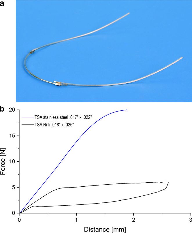
Introduction adequately stretched or loaded. As a result, defined forces
and torques can be orthodontically applied as well as small
The root torque of the anterior teeth is an important tool forces and torques. To utilize the superelasticity of NiTi
in orthodontic treatment. The buccopalatal angulation of alloy in order to change incisors’ root torque a segment
the root known as palatal root torque in orthodontics is an of a stainless steel archwire was replaced with NiTi alloy.
important step in creating functionally correct static and dy- This combination provides the rigidness and stability of
namic occlusal relationships and guaranteeing good support stainless steel on the “side-parts” on the one hand, as well
to the anterior teeth. Bodily retraction of the anterior teeth as the small forces and torques produced by the superelastic
and simultaneous application of torque are often necessary, NiTi alloy on the other hand. Torque segmented archwires
especially in the course of premolar extraction treatment. (TSA), which were developed specifically for this purpose
For correct axial adjustment of the roots of the incisors, [47, 55], are available in prefabricated form (Forestadent,
torque must be transferred to the teeth via orthodontic ap- Pforzheim, Germany). The TSA consists of a pretorqued su-
pliances. The torque angle in the bracket when using the perelastic archwire component (NiTi) for the incisors and
straight wire technique does not transfer suitable torques in two steel segments attached via a clamp connection for the
many cases because of slot geometry [53]. In these cases, posterior teeth (Fig. 1a). If the mechanical load-deflection
a defined, biomechanically effective torque therefore needs behavior of the entire TSA compound is measured as pub-
to be produced via torsion of wires. The torques dependent lished by Wichelhaus and Sander [55], the resulting curve
on applied force and range between 15 and 20 Nmm as rec- shows an incomplete superelasticity even though the frontal
ommended in the literature [5] for all four upper incisors segment is made from superelastic material. This can be at-
[55]. However, in some instances torques of around 5 Nmm tributed to the fact that it is not possible to separate the NiTi
are also specified [39]. These values ascertained from the hysteretic behavior from the linear elasticity of the stain-
literature are a guide and are dependent on the root ge- less steel component. However, it has to be considered that
ometry of the teeth or the biological circumstances in the the mechanical performance of the entire TSA compound
individual patient. If forces and torques are biomechanically is clinically almost irrelevant because the main stresses and
uncontrolled or too high, the risk of apical root resorptions moments are generated in the interbracket segments which
will increase [10, 13, 34, 45]. In addition to the force and are made of either stainless steel or NiTi. To prove the
torque magnitude, the duration of force/torque application, superelastic character of the TSA’s NiTi part, three-point
force direction, treatment mechanics and treatment period bending measurements were conducted (Fig. 1b).
should be noted as influencing variables in relation to re- As apical root resorptions are a very commonly occur-
sorption [2, 17, 23, 28, 31, 33, 34]. Torque movement of the rence with the upper incisors [2, 46], the aim of the present
apex causes a local concentration of pressure at the root tip study in using the TSA was to investigate the amount of api-
and can reach a fourfold level compared with pure transla- cal root resorption compared with conventional orthodon-
tory tooth movements [47]. However, root apex resorptions tic appliances. Furthermore, the extent and direction of the
are dependent on genetic and biological factors as well as therapeutically indicated apical movement and the treat-
the effects of the orthodontic treatment [51]. ment duration were also evaluated.
Nickel–titanium alloys (NiTi) are particularly well suited
to transfer small forces and torques to the teeth because
of their low Young’s modulus [6, 54]. In addition, the al-
loy is characterized by its superelastic material behavior.
However, this behavior only arises when the material is
K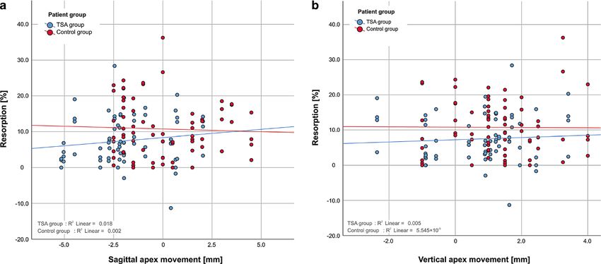
Stainless steel and NiTi torque archwires and apical root resorption                                                               3

Materials and methods                                                  (project number 19-815). On average, torque treatment of
                                                                       the patients in the study group started at 18.6 ± 7.5 years of
Patient groups                                                         age (range 11.4–37.3 years) and lasted for 105 ± 44 days.
                                                                       Within the control group patient torque treatment started
In a randomized retrospective follow-up study, we inves-               at 14.8 ± 1.8 years of age (range 12.6–18.9 years). These
tigated the effect of TSA versus conventional orthodontic              patients were treated for 146 ± 52 days.
appliance application on the amount of apical root resorp-                The torque application by means of the TSA took place
tion. As such, the study group comprised of 18 randomly                using a standardized archwire sequence: 0.01400 NiTi,
chosen successfully treated Class I and Class II patients              0.01600 NiTi, 0.01600 × 0.02200 NiTi, 0.01600 × 0.02200 stain-
(11 female, 7 male) from the Department of Orthodontics                less steel, TSA (0.01800 × 0.02500 NiTi with 45° pretorque)
and Pediatric Medicine of Basel University and included                and 0.01800 × 0.0250 stainless steel in the 0.02200 slot tech-
7 extraction cases. The complementary control group con-               nique with MBT prescription. The effective torque in the
sisted of 18 randomly chosen Class I and Class II patients             0.01800 × 0.02500 wire dimension is about 2° [11]. If sweep
(11 female, 7 male) from the Department of Orthodontics                is necessary, an additional torque of 15° occurs. If no
and Dentofacial Orthopedics of the University of Munich                sweep is necessary, a torque of 30° was bent into the wire
and included 10 extraction cases. In all cases, there was              applying a moment of 20 Nmm [8].
a therapeutic necessity to apply torque on the maxillary in-              The tooth axis in relation to the occlusal plane was clini-
cisors. In our treatment concept, torque with the stainless            cally controlled with a special torque key [55]. The archwire
steel wire is not applied in patients with critical anchorage.         sequence applied in the control group was identical to the
In this case, we prefer the TSA because the sagittal force is          one previously described, but without TSA.
lower in comparison to the stainless steel arch wire. Both
methods apply torque changes to the maxillary incisors.
The local ethics committee approved the study protocol

Hier steht eine Anzeige.

K

                                                                                                                          K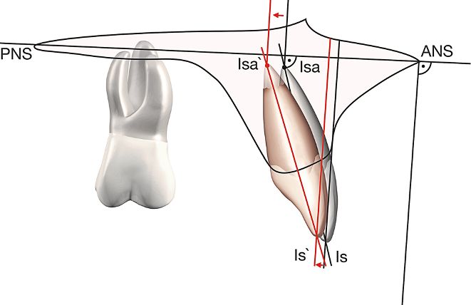
4                                                                                                                          A. Wichelhaus et al.

                                                                         angles or nonstandardized conditions [15, 26]. Using these
                                                                         measures, the root-to-crown-ratio (RCR) before and after
                                                                         treatment for each of the examined teeth was calculated
                                                                         (Fig. 2). The relative root-to-crown ratio (rRCR) (Fig. 2,
                                                                         formula) is the quotient of the RCR values of a given tooth
                                                                         before and after treatment and, thus, reflects the amount
                                                                         of root resorption [15, 16, 26]. As such, a rRCR ≥100%
                                                                         means “no resorption” and rRCR values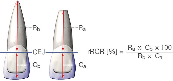
Stainless steel and NiTi torque archwires and apical root resorption                                                                            5

                                                                           Results

                                                                           Based on a pretreatment tooth length of 100%, the mean
                                                                           relative root–crown ratio (rRCR) after completion of the
                                                                           orthodontic treatment with the TSA was 93% and in the
                                                                           orthodontic-treated control group 89%. The difference be-
                                                                           tween the groups was significant (U = 3208, p = 0.014; Ta-
                                                                           ble 1). The average rRCR for the individual incisors in
                                                                           the TSA group ranged from 89 to 95% and in the control
                                                                           group from 87 to 90% (Table 1, Fig. 5). The individual teeth
                                                                           (tooth 12, 11, 21, and 22) did not differ significantly within
                                                                           both groups (Kruskal–Wallis test; TSA group: H = 7.512,
                                                                           df = 3, p = 0.057; control group: H = 1.373, df = 3, p = 0.712).
Fig. 3 Analysis of the amount of movement of the apex based on pre-        But a significant difference was found for tooth 12 between
and posttreatment lateral cephalograms. Sagittal and vertical move-        TSA and the control group (92 vs. 96; U = 236.0, p = 0.019;
ments (mm) were determined. ANS anterior nasal spine, PNS posterior
                                                                           Table 1, Fig. 5). The median rRCR in the control group was
nasal spine, Isa incision superius apicalis, Is incision superius
Abb. 3 Analyse des Bewegungsausmaßes des Apex anhand prä- und              smaller than in the TSA group (Fig. 5).
posttherapeutischer Fernröntgenseitenbilder. Es wurden sagittale und          Concerning the incidence of root resorption, 89% (TSA
vertikale Bewegungen bestimmt (mm). ANS „anterior nasal spine“,            group) and 94.4% (control group) of the teeth examined
PNS „posterior nasal spine”, Isa „incision superius apicalis”, Is „in-
                                                                           showed resorptions (Table 2). In the TSA group 8/72
cision superius”
                                                                           (11.1%) teeth were not affected with posttreatment root
                                                                           resorption, in the control group only 5.6%. The severity of
separately. The Pearson correlation was applied to test the                the resorptions was predominantly in the light to moderate
relationship between root resorption and apical movements                  range (55.6% and 30.6% within the TSA group and 48.6%
and between torque changes of the maxillary incisors and                   and 33.3% in the control group). Severe resorption was
root resorption. The level of significance was set at p < 0.05.

                                                                           Fig. 5 Relative change in root–crown ratio (rRCR) for tooth 11, 12, 21,
                                                                           22, for torque-segmented archwire (TSA) treated and conventionally
                                                                           orthodontic treated patients. Pairwise comparisons were done using the
Fig. 4 Analysis of torque change for the maxillary central incisors        Mann–Whitney U test (asterisk p < 0.05). For the comparison within
between pretreatment (black line) and posttreatment (red line) lateral     each patient group the Kruskal–Wallis test was applied (p < 0.05)
cephalogram relative to the anterior base of the skull and the maxillary   Abb. 5 Relative Änderung des Wurzel-Kronen-Verhältnisses (rRCR)
base plane                                                                 für die Zähne 11, 12, 21 und 22 bei mit TSA (Torquesegmentbogen)
Abb. 4 Analyse der Torqueveränderung der zentralen Oberkieferinzi-         sowie konventionell orthodontisch behandelten Patienten. Paarweise
siven zwischen prä- (schwarze Linie) und posttherapeutischem (rote         Vergleiche wurden mit dem Mann-Whitney-U-Test durchgeführt (As-
Linie) Fernröntgenseitenbild zur vorderen Schädelbasis und der Ober-       terisk p < 0,05), für den Vergleich innerhalb der Gruppen wurde der
kiefergrundebene                                                           Kruskal-Wallis-Test angewendet (p < 0,05)

                                                                                                                                      K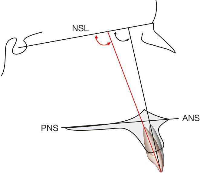
6                                                                                                                           A. Wichelhaus et al.

Table 1 Comparison between the torque-segmented archwire (TSA) and control group concerning the amount of movement of the apices of
maxillary incisors in the sagittal and vertical direction relative to the ANS-PNS plane and the relative root–crown ratio (rRCR). Number of
patients (N), mean, standard deviation (SD), range (minimum and maximum), median and interquartile range (IQR) were reported. U statistics and
significance (p) were given for pairwise comparisons using the Mann–Whitney U test
Tab. 1 Vergleich zwischen TSA(Torquesegmentbogen)- und Kontrollgruppe bezüglich der sagittalen und vertikalen Apexbewegung der Oberkiefer-
frontzähne und des relativen Wurzel-Kronen-Verhältnisses (rRCR). Anzahl der Patienten (N), Mittelwert, Standardabweichung (SD), Bereich
(Minimum, Maximum), Median und das Interquartilsabstand (IQR) wurden dokumentiert. Für die paarweisen Vergleiche mit dem Mann-Whit-
ney-U-Test wurden U-Statistik und p-Wert gezeigt
                           Patient group                                                                                      Mann–
                           TSA group (N = 18)                                 Control group (N = 18)                          Whitney
                                                                                                                              U test
                          N     Mean SD       Range      Median IQR          N     Mean SD      Range      Median IQR            U       P
Retrusion/   Retrusion    14    –2.8   1.3    –5.2 to    –2.4    [–3.2;      9     –1.7   0.7   –2.5 to    –2.0    [–2.0;        33.0    0.062
protrusion                                    –0.9               –2.0]                          –0.5               –1.5]
(sagittal    No           0     –      –      –          –       –           2     –      –     –          –       –             –       –
apex         move-
move-        ment
ment,
mm)          Protrusion   4     1.0    0.7    0.4 to     0.7     [0.5;       7     2.4    1.4   0.5 to     2.0     [1.5;         6.0     0.164
                                              2.0                1.4]                           4.5                3.5]
             Total        18    –1.9   2.0    –5.2 to    –2.2    [–2.7;      18    0.1    2.2   –2.5 to    –0.2    [–2.0;        83.0    0.012*
                                              2.0                –0.9]                          4.5                1.5]
Extrusion/   Intrusion    5     –1.1   0.7    –2.4 to    –0.9    [–0.9;      2     –1.0   0.0   –1.0 to    –1.0    [–1.0;        8.0     0.381
intrusion                                     –0.6               –0.9]                          –1.0               –1.0]
(vertical    No           0     –      –      –          –       –           2     –      –     –          –       –             –       –
apex         move-
move-        ment
ment,
mm)          Extrusion    13    1.5    0.8    0.4 to     1.2     [0.9;       14    1.8    1.0   0.5 to     1.5     [1.0;         74.0    0.430
                                              3.4                1.7]                           4.0                2.3]
             Total        18    0.7    1.4    –2.4 to    1.0     [–0.6;      18    1.3    1.3   –1.0 to    1.3     [0.5;         134.0   0.389
                                              3.4                1.6]                           4.0                2.0]
rRCR, %      Tooth 11     18    92     6      80 to      93      [87;        18    87     8     64 to      91      [82;          218.0   0.079
                                              100                97]                            96                 93]
             Tooth 12     18    95     6      81 to      96      [93;        18    90     7     77 to      92      [85;          236.0   0.019*
                                              111                98]                            100                93]
             Tooth 21     18    94     6      85 to      93      [89;        18    90     7     73 to      91      [86;          213.0   0.111
                                              103                100]                           99                 95]
             Tooth 22     18    89     7      72 to      88      [84;        18    90     9     76 to      90      [83;          136.5   0.424
                                              100                95]                            100                99]
             Central      36    93     6      80 to      93      [88;        36    88     8     64 to      91      [83;          441.5   0.020*
             incisors                         103                97]                            99                 94]
             Lateral      36    92     7      72 to      94      [86;        36    90     8     76 to      91      [84;          573.5   0.401
             incisors                         111                97]                            100                98]
             Total        72    93     6      72 to      93      [87;        72    89     8     64 to      91      [84;          3208.0 0.014*
                                              111                97]                            100                95]

present in 2.8% of the TSA group and 12.5% of the control                 in the TSA group from 98.2 to 102.5° (Z = –2.286, p = 0.022;
group.                                                                    Table 3). In the control group, no significant change in
   Evaluation of the lateral cephalograms showed that the                 the axial position (Z = –1.613, p = 0.107) was observed (Ta-
main directions of movement of the apices of the upper                    ble 3).
central incisors were retrusion and extrusion (Table 1). The                  Pearson’s correlation coefficients for resorptions at max-
median sagittal apex movement of the total TSA group                      illary incisors and horizontal direction of apical movement
(–2.2 mm) was statistically significant larger than that of               were r = 0.136 (p = 0.255) for the TSA group and r = 0.042
the control group (median: –0.2 mm; U = 83.0, p = 0.012),                 (p = 0.727) for the control group and were not statistically
whereas there was no statistical difference in vertical apex              significant (Fig. 6a). For resorptions at maxillary incisors
movement (U = 134.0, p = 0.389; Table 1).                                 and vertical direction of apical movements the Pearson’s
   Regarding the axial position of the front teeth, the me-               correlation coefficients were r = 0.067 (p = 0.575) for the
dian of the front tooth axis (1_NSL) significantly increased              TSA group and r = –0.007 (p = 0.950) for the control group

K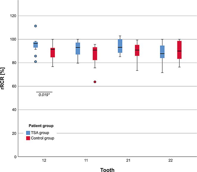
Stainless steel and NiTi torque archwires and apical root resorption                                                                                7

Table 2 Severity of root resorption (RR). Crosstabulation of tooth and patient group with levels of severity of root resorption classified according
to relative root–crown ratio (rRCR)
Tab. 2 Schweregrad der Wurzelresorption (RR). Kreuztabelle aus Zahn und Patientengruppe mit dem Niveau des Schweregrades der Wurzelre-
sorption entsprechend des relativen Wurzel-Kronen-Verhältnisses (rRCR)
Severity                   12                     11                        21                       22                        Total
of RR                      TSA       Control      TSA         Control       TSA       Control        TSA         Control       TSA         Control
                               group      group     group      group        group        group          group       group      group      group
No RR         No. of           1          1         1          0            5            0              1           3          8          4
(rRCR         teeth
≥100%)        %                5.6        5.6       5.6        0.0          27.8         0.0            5.6         16.7       11.1       5.6
Slight RR No. of               15         9         10         10           8            10             7           6          40         35
(90 ≤ x <     teeth
 100)         %                83.3       50.0      55.6       55.6         44.4         55.6           38.9        33.3       55.6       48.6
Moderate      No. of           2          6         6          6            5            6              9           6          22         24
RR            teeth
(80 ≤ x < 90) %                11.1       33.3      33.3       33.3         27.8         33.3           50.0        33.3       30.6       33.3
Severe        No. of           0          2         1          2            0            2              1           3          2          9
RR            teeth
(
8 A. Wichelhaus et al.
Fig. 6 a Relationship between resorption of the central and lateral incisors and sagittal apex movement for both patient groups. No significant
correlation between sagittal apex movement and resorption was found in either group. b Relationship between resorption of the central and lateral
incisors and vertical apex movement for both patient groups. No significant correlation between vertical apex movement and resorption was found
in either group. TSA torque-segmented archwire
Abb. 6 a Beziehung zwischen Wurzelresorption der zentralen und lateralen Schneidezähne und der sagittalen Apexbewegung in beiden Pati-
entengruppen. Eine signifikante Korrelation zwischen sagittaler Apexbewegung und Resorption ließ sich in beiden Gruppen nicht feststellen.
b Beziehung zwischen Wurzelresorption der zentralen und lateralen Schneidezähne und der vertikalen Apexbewegung in beiden Gruppen. Eine
signifikante Korrelation zwischen vertikaler Apexbewegung und Resorption konnte in beiden Gruppen nicht festgestellt werden
Discussion torque range is discussed in order to minimize resorptions
at the apical root tip [10, 12, 13, 18, 19, 21, 24, 42, 44, 48].
The accurate and controlled biomechanics of the TSA [47, The methodology to assess the resorption rate during or-
55] makes it possible to exert defined moments on the thodontic treatment applied in our study is known from the
teeth and thereby achieve clinically very efficient results literature and well established [15, 16, 26]. It allows use
[3, 47]. The results also show that within a 1_NSL range of of X-rays, which are recorded in the diagnostic routine of
85.3–109.8° torque adjustment with defined moments even an orthodontic treatment. Therefore, it avoids additional ra-
with preactivated torque wires remains difficult. In addi- diation exposure of the patient. As panoramic radiographs
tion, biomechanical and biological factors can play a role. are accompanied by perspective distortions, the use of the
Torque can be applied with stainless steel arch wires as well relative root-to-crown ratio (rRCR) allows one to calculate
as the TSA. The inconsistency of data between the two a decresase in root length [15]. With the assumption of
methods shows that clinically the adjustment of the M/F a constant crown length, these distortions are compensated.
ratio depends on other factors. The application of the TSA A much more detailed analysis of root resorptions is only
simplifies clinical application of torque. However prospec- possible using three-dimensional radiographic techniques
tively, for better M/F ratio control different moments should like cone-beam computed tomography [4, 43]. Unfortu-
be used. For bodily retraction, moments in the range of nately, these X-ray techniques are accompanied by high
15–20 Nmm are reported [5, 9, 14], whereas for controlled additional radiation exposure and have a strong indication.
tipping lower moments can be applied. Further clinical stud- The average loss of apical root length (rRCR) of 7% in
ies must show whether this will lead to a better adjustment the TSA group and 11% in the control group recorded in
of the M/F ratio. the present study can be regarded as not clinically relevant.
In most of the studies on continuous forces and moments, The median rRCR of the maxillary incisors was significant
there was nevertheless a loss of force or moment during the larger in the TSA group as compared to the control group.
progress of the treatment [1, 30, 36–38], although the study Therefore, root resorption was less expressed in the TSA
design in these studies is generally a matter of discussion. group although the difference was only 2%. In our study,
The torque we used on the four maxillary incisors with an no significant differences were found between the maxillary
overall magnitude of 15–20 Nmm [55] is within the physi- incisors within one group and between the groups except
ological range of the ideal level of torque [5, 9, 14]. This tooth 12. Our results for the maxillary central incisor coin-
KStainless steel and NiTi torque archwires and apical root resorption 9
Fig. 7 a Relationship between resorption of the central and lateral incisors and 1_NSL change for both patient groups. No significant correlation
between 1_NSL change and resorption was found in either group. b Relationship between resorption of the central and lateral incisors and 1_NL
change for both patient groups. No significant correlation between change of 1_NL and resorption was found in either group. TSA torque-segmented
archwire
Abb. 7 a Beziehung zwischen Wurzelresorption der zentralen und lateralen Schneidezähne und 1_NSL-Änderung in beiden Patientengruppen.
Eine signifikante Korrelation zwischen 1_NSL-Änderung und Resorption konnte in beiden Gruppen nicht festgestellt werden. b Beziehung zwi-
schen Wurzelresorption der zentralen und lateralen Schneidezähne und 1_NL-Änderung in beiden Gruppen. Eine signifikante Korrelation zwischen
Änderung des 1_NL und Resorption ließ sich in beiden Gruppen nicht feststellen
cide with the amount of resorption recorded in most other No correlation between root resorption and incisor axis
studies that analyzed the extent of root resorption found change was found in our study. A review of the literature
on upper central incisors after orthodontic treatment with shows that there are contradictory findings concerning this
fixed appliances [7, 12, 15, 22, 29, 32, 40, 43, 50]. In the correlation [12, 32, 40, 41]. Multifactorial influences on
present study, however, no statistically significant correla- the amount of root resorption are discussed. Due to the
tion was noted for resorptions and treatment-related api- variety of study designs, no consistent statement is found
cal movement in the horizontal or vertical direction. On in the literature. Most of the authors have not found any
a cautionary note, however, it should be mentioned that the correlation between the difference of incisor inclination and
apical movement distances were calculated on the lateral root resorption [12, 32, 40, 41].
cephalograms, for which the interval between pre- and post- Although we found no significant correlation (p ≥ 0.101)
treatment X-rays does not always coincide precisely with between duration of torque application and root resorption,
the period when the TSA is in place. Hence, a treatment- it seems reasonable to assume that the amount and the dura-
related unwanted jiggling effect in other treatment phases tion of the torque application have an influence on the grade
cannot be ruled out as an important contributory factor to of root resorption [10, 45]. A longer treatment duration is
root resorption. The major advantage of utilizing the su- associated with significantly higher root resorption rates for
perelasticity of these materials for the practitioner is that, maxillary central incisors [20, 25, 49]. Only a few studies
even if the wire is excessively activated, the torque acting deal with the time dependence of treatment-related root re-
on the tooth remains within physiological limits. Hence, sorption when continuous forces are applied by means of
rectangular wires can be used even at an early stage; these NiTi components [10, 35]. Although the correlation was
allow better control of tooth movement, especially during not significant in our study, there was a trend towards an
torque application [52]. An evaluative comparison of the increased risk of root resorption with a longer treatment
different types of moment application is only possible if duration.
a controlled comparative test is done in the same individual, The results of this study show no correlation between
e.g., by applying a split-mouth design, but this is clinically vertical and sagittal apex movement and root resorption.
not realistic. This correlation is controversially discussed in the litera-
ture [7, 12, 15, 22, 29, 32, 43, 50]. Factors like study de-
K10 A. Wichelhaus et al.
sign, patient number and methodology should be discussed. Open Access This article is licensed under a Creative Commons At-
tribution 4.0 International License, which permits use, sharing, adapta-
In 4 patients from the TSA group and 7 patients from the tion, distribution and reproduction in any medium or format, as long as
control group, a sagittal apex movement of 1 or 2 mm was you give appropriate credit to the original author(s) and the source, pro-
observed, respectively. This indicates an uncontrolled tip- vide a link to the Creative Commons licence, and indicate if changes
ping of the incisors. This effect arises due to differences in were made. The images or other third party material in this article are
included in the article’s Creative Commons licence, unless indicated
root geometry and nonideal force–moment application. otherwise in a credit line to the material. If material is not included
The results of this retrospective study should be validated in the article’s Creative Commons licence and your intended use is not
with further prospective studies with a larger patient group permitted by statutory regulation or exceeds the permitted use, you will
and measurements of the actual torque moment acting on need to obtain permission directly from the copyright holder. To view
a copy of this licence, visit http://creativecommons.org/licenses/by/4.
the teeth, to gain more knowledge about the ideal level of 0/.
moment for continuous torque application, and to obtain
more clear-cut results.
References
Conclusion 1. Acar A, Canyurek U, Kocaaga M, Erverdi N (1999) Continuous vs.
discontinuous force application and root resorption. Angle Orthod
Use of the torque-segmented archwire (TSA) for contin- 69:159–163. https://doi.org/10.1043/0003-3219(1999)0692.3.co;2 (discussion 163–154)
uous torque application on the maxillary anterior teeth 2. Apajalahti S, Peltola JS (2007) Apical root resorption after or-
leads to slight, clinically nonrelevant apical root resorp- thodontic treatment—a retrospective study. Eur J Orthod 29:408–
tion. 412. https://doi.org/10.1093/ejo/cjm016
The incisors axis in the TSA group was on average sig- 3. Aras B, Cheng LL, Turk T, Elekdag-Turk S, Jones AS, Daren-
deliler MA (2012) Physical properties of root cementum: part 23.
nificantly improved. Independently, though defined mo- Effects of 2 or 3 weekly reactivated continuous or intermittent
ments were applied, the adjustment of the M/F ratio was orthodontic forces on root resorption and tooth movement: a mi-
still problematic. In order to achieve a better adjustment crocomputed tomography study. Am J Orthod Dentofacial Orthop
of the M/F ratio, the application of different moments is 141:e29–e37. https://doi.org/10.1016/j.ajodo.2011.07.018
4. Aras I, Unal I, Huniler G, Aras A (2018) Root resorption due to
advised. orthodontic treatment using self-ligating and conventional brack-
No differences were found between the amount of apical ets : A cone-beam computed tomography study. J Orofac Orthop
root resorption of maxillary lateral and central incisors in 79:181–190. https://doi.org/10.1007/s00056-018-0133-5
the TSA and control groups. 5. Bantleon HP, Droschl H (1988) Front torque using a partial arch
technic. Fortschr Kieferorthop 49:203–212. https://doi.org/10.
Between the TSA and control group significant differ- 1007/bf02163378
ences in the relative root–crown ratio (rRCR) were found 6. Bartzela TN, Senn C, Wichelhaus A (2007) Load-deflection char-
for tooth 12, the central incisors, and for all maxillary in- acteristics of superelastic nickel-titanium wires. Angle Orthod
cisors. Although root resorption in the TSA group was 77:991–998. https://doi.org/10.2319/101206-423.1
7. Baumrind S, Korn EL, Boyd RL (1996) Apical root resorption in
less pronounced, the difference between the groups was orthodontically treated adults. Am J Orthod Dentofacial Orthop
only 2%. 110:311–320
There is no correlation between root resorption and treat- 8. Brauchli LM, Steineck M, Wichelhaus A (2012) Active and pas-
ment duration, the distance of vertical and sagittal move- sive self-ligation: a myth? Part 1: torque control. Angle Orthod
82:663–669. https://doi.org/10.2319/062011-673.1
ment of the apex and the change in inclination of the in- 9. Burstone CJ (1966) The mechanics of the segmented arch tech-
cisal axis relative to the anterior base of the skull. niques. Angle Orthod 36:99–120. https://doi.org/10.1043/0003-
3219(1966)036h0099:TMOTSAi2.0.CO;2
Funding Open Access funding provided by Projekt DEAL. 10. Casa MA, Faltin RM, Faltin K, Sander FG, Arana-Chavez VE
(2001) Root resorptions in upper first premolars after application of
continuous torque moment. Intra-individual study. J Orofac Orthop
Compliance with ethical guidelines 62:285–295
11. Creekmore TD (1979) Dr. Thomas D. Creekmore on torque. J Clin
Conflict of interest A. Wichelhaus, M. Dulla, H. Sabbagh, U. Baumert Orthod 13:305–310
and T. Stocker declare that they have no competing interests. 12. DeShields RW (1969) A study of root resorption in treated
Class II, Division I malocclusions. Angle Orthod 39:231–245.
Ethical standards All procedures performed in studies involving hu- https://doi.org/10.1043/0003-3219(1969)039h0231:asorrii2.0.co;2
man participants or on human tissue were in accordance with the eth- 13. Faltin RM, Arana-Chavez VE, Faltin K, Sander FG, Wichelhaus A
ical standards of the institutional and/or national research committee (1998) Root resorptions in upper first premolars after application of
and with the 1975 Helsinki declaration and its later amendments or continuous intrusive forces. Intra-individual study. J Orofac Orthop
comparable ethical standards. The local ethics committee approved 59:208–219
the study protocol (project number 19-815). Informed consent was ob- 14. Feldner JC, Sarkar NK, Sheridan JJ, Lancaster DM (1994) In vitro
tained from all individual participants included in the study. torque-deformation characteristics of orthodontic polycarbonate
KStainless steel and NiTi torque archwires and apical root resorption 11
brackets. Am J Orthod Dentofacial Orthop 106:265–272. https:// 33. Motokawa M, Sasamoto T, Kaku M, Kawata T, Matsuda Y,
doi.org/10.1016/S0889-5406(94)70046-X Terao A, Tanne K (2012) Association between root resorption
15. Fritz U, Diedrich P, Wiechmann D (2003) Apical root resorption incident to orthodontic treatment and treatment factors. Eur J Or-
after lingual orthodontic therapy. J Orofac Orthop 64:434–442. thod 34:350–356. https://doi.org/10.1093/ejo/cjr018
https://doi.org/10.1007/s00056-003-0243-5 34. Nakano T, Hotokezaka H, Hashimoto M, Sirisoontorn I, Arita K,
16. Gay G, Ravera S, Castroflorio T, Garino F, Rossini G, Parrini S, Kurohama T, Darendeliler MA, Yoshida N (2014) Effects of differ-
Cugliari G, Deregibus A (2017) Root resorption during orthodon- ent types of tooth movement and force magnitudes on the amount
tic treatment with Invisalign(R): a radiometric study. Prog Orthod of tooth movement and root resorption in rats. Angle Orthod
18:12. https://doi.org/10.1186/s40510-017-0166-0 84:1079–1085. https://doi.org/10.2319/121913-929.1
17. Goel P, Tandon R, Agrawal KK (2014) A comparative study of 35. Noda K, Arai C, Nakamura Y (2010) Root resorption after exper-
different intrusion methods and their effect on maxillary incisors. imental tooth movement using superelastic forces in the rat. Eur J
J Oral Biol Craniofac Res 4:186–191. https://doi.org/10.1016/j. Orthod 32:681–687. https://doi.org/10.1093/ejo/cjq016
jobcr.2014.11.007 36. Owman-Moll P, Kurol J, Lundgren D (1995) Continuous ver-
18. Goldin B (1989) Labial root torque: effect on the maxilla and in- sus interrupted continuous orthodontic force related to early
cisor root apex. Am J Orthod Dentofacial Orthop 95:208–219 tooth movement and root resorption. Angle Orthod 65:395–401.
19. Goldson L, Henrikson CO (1975) Root resorption during Begg https://doi.org/10.1043/0003-3219(1995)065h0395:cvicofi2.0.co;2
treatment; a longitudinal roentgenologic study. Am J Orthod (discussion 401–392)
68:55–66 37. Owman-Moll P, Kurol J, Lundgren D (1996) Effects of a dou-
20. Harris EF, Baker WC (1990) Loss of root length and crestal bone bled orthodontic force magnitude on tooth movement and root
height before and during treatment in adolescent and adult or- resorptions. An inter-individual study in adolescents. Eur J Orthod
thodontic patients. Am J Orthod Dentofac Orthop 98:463–469 18:141–150
21. Harris EF, Butler ML (1992) Patterns of incisor root resorption be- 38. Owman-Moll P, Kurol J, Lundgren D (1996) The effects of a four-
fore and after orthodontic correction in cases with anterior open fold increased orthodontic force magnitude on tooth movement and
bites. Am J Orthod Dentofacial Orthop 101:112–119. https://doi. root resorptions. An intra-individual study in adolescents. Eur J Or-
org/10.1016/0889-5406(92)70002-r thod 18:287–294
22. Ho KH, Liao YF (2012) Pre-treatment radiographic features pre- 39. Papageorgiou SN, Sifakakis I, Doulis I, Eliades T, Bourauel C
dict root resorption of treated impacted maxillary central incisors. (2016) Torque efficiency of square and rectangular archwires into
Orthod Craniofac Res 15:198–205. https://doi.org/10.1111/j.1601- 0.018 and 0.022 in. conventional brackets. Prog Orthod 17:5.
6343.2012.01545.x https://doi.org/10.1186/s40510-016-0118-0
23. Janson GR, De Luca Canto G, Martins DR, Henriques JF, De Fre- 40. Parker RJ, Harris EF (1998) Directions of orthodontic tooth move-
itas MR (2000) A radiographic comparison of apical root resorption ments associated with external apical root resorption of the maxil-
after orthodontic treatment with 3 different fixed appliance tech- lary central incisor. Am J Orthod Dentofacial Orthop 114:677–683
niques. Am J Orthod Dentofac Orthop 118:262–273. https://doi. 41. Phillips JR (1955) Apical root resorption under orthodontic therapy.
org/10.1067/mod.2000.99136 Angle Orthod 25:1–22. https://doi.org/10.1043/0003-3219(1955)
24. Kaley J, Phillips C (1991) Factors related to root resorption in edge- 0252.0.co;2
wise practice. Angle Orthod 61:125–132. https://doi.org/10.1043/ 42. Plets JH, Isaacson RJ, Speidel TM, Worms FW (1974) Maxillary
0003-3219(1991)0612.0.co;2 central incisor root length in orthodontically treated and untreated
25. Kalha AS, Kachiwala VA, Govardhan SN, McLaughlin RP, Khur- patients. Angle Orthod 44:43–47. https://doi.org/10.1043/0003-
shaid SZ (2010) Redefining orthodontic space closure: sequential 3219(1974)044h0043:mcirlii2.0.co;2
repetitive loading of the periodontal ligament—a clinical study. 43. Puttaravuttiporn P, Wongsuwanlert M, Charoemratrote C, Leethanakul C
World J Orthod 11:221–229 (2018) Volumetric evaluation of root resorption on the upper in-
26. Krieger E, Drechsler T, Schmidtmann I, Jacobs C, Haag S, cisors using cone beam computed tomography after 1 year of
Wehrbein H (2013) Apical root resorption during orthodontic orthodontic treatment in adult patients with marginal bone loss.
treatment with aligners? A retrospective radiometric study. Head Angle Orthod 88:710–718. https://doi.org/10.2319/121717-868.1
Face Med 9:21. https://doi.org/10.1186/1746-160X-9-21 44. Remington DN, Joondeph DR, Artun J, Riedel RA, Chapko MK
27. Linge BO, Linge L (1983) Apical root resorption in upper anterior (1989) Long-term evaluation of root resorption occurring during
teeth. Eur J Orthod 5:173–183. https://doi.org/10.1093/ejo/5.3.173 orthodontic treatment. Am J Orthod Dentofacial Orthop 96:43–46
28. Linge L, Linge BO (1991) Patient characteristics and treatment 45. Roscoe MG, Meira JB, Cattaneo PM (2015) Association of or-
variables associated with apical root resorption during orthodontic thodontic force system and root resorption: a systematic review. Am
treatment. Am J Orthod Dentofacial Orthop 99:35–43. https://doi. J Orthod Dentofacial Orthop 147:610–626. https://doi.org/10.1016/
org/10.1016/s0889-5406(05)81678-6 j.ajodo.2014.12.026
29. Liou EJ, Chang PM (2010) Apical root resorption in orthodontic pa- 46. Sameshima GT, Sinclair PM (2001) Predicting and preventing root
tients with en-masse maxillary anterior retraction and intrusion with resorption: Part I. Diagnostic factors. Am J Orthod Dentofac Orthop
miniscrews. Am J Orthod Dentofac Orthop 137:207–212. https:// 119:505–510. https://doi.org/10.1067/mod.2001.113409
doi.org/10.1016/j.ajodo.2008.02.027 47. Sander FG, Wichelhaus A (1995) Clinical experiences with the
30. Lundgren D, Owman-Moll P, Kurol J, Martensson B (1996) Accu- torque-segmented arch (TSA). Fortschr Kieferorthop 56:194–201.
racy of orthodontic force and tooth movement measurements. Br J https://doi.org/10.1007/bf02168132
Orthod 23:241–248 48. Spurrier SW, Hall SH, Joondeph DR, Shapiro PA, Riedel RA
31. Martins DR, Tibola D, Janson G, Maria FR (2012) Effects of in- (1990) A comparison of apical root resorption during orthodontic
trusion combined with anterior retraction on apical root resorption. treatment in endodontically treated and vital teeth. Am J Orthod
Eur J Orthod 34:170–175. https://doi.org/10.1093/ejo/cjq178 Dentofacial Orthop 97:130–134. https://doi.org/10.1016/0889-
32. Mirabella AD, Artun J (1995) Risk factors for apical root resorption 5406(90)70086-r
of maxillary anterior teeth in adult orthodontic patients. Am J Or- 49. Taithongchai R, Sookkorn K, Killiany DM (1996) Facial and den-
thod Dentofac Orthop 108:48–55. https://doi.org/10.1016/s0889- toalveolar structure and the prediction of apical root shortening. Am
5406(95)70065-x J Orthod Dentofacial Orthop 110:296–302
K12 A. Wichelhaus et al.
50. Tieu LD, Saltaji H, Normando D, Flores-Mir C (2014) Radiolog- 53. Wichelhaus A (2017) A new elastic slot system and V-wire
ically determined orthodontically induced external apical root re- mechanics. Angle Orthod 87:774–781. https://doi.org/10.2319/
sorption in incisors after non-surgical orthodontic treatment of class 121516-899
II division 1 malocclusion: a systematic review. Prog Orthod 15:48. 54. Wichelhaus A, Brauchli L, Ball J, Mertmann M (2010) Mechani-
https://doi.org/10.1186/s40510-014-0048-7 cal behavior and clinical application of nickel-titanium closed-coil
51. Topkara A, Karaman AI, Kau CH (2012) Apical root resorption springs under different stress levels and mechanical loading cycles.
caused by orthodontic forces: A brief review and a long-term ob- Am J Orthod Dentofacial Orthop 137:671–678. https://doi.org/10.
servation. Eur J Dent 6:445–453 1016/j.ajodo.2008.06.029
52. Wichelhaus A (1999) Die Entwicklung und klinische Anwendung 55. Wichelhaus A, Sander FG (1995) Biomechanical testing of the new
superelastischer Bögen und Teilbögen in der Kieferorthopädie. torque-segmented arch (TSA). Fortschr Kieferorthop 56:224–235.
Quintessenz, Berlin https://doi.org/10.1007/bf02168135
KYou can also read