Vital root submergence of immature permanent incisors after complicated crown-root fracture followed by orthodontic space maintenance: A ...
←
→
Page content transcription
If your browser does not render page correctly, please read the page content below
Clinical cases

Vital root submergence of immature permanent incisors
after complicated crown-root fracture
followed by orthodontic space maintenance: A presentation of two cases
Pozostawienie „zatopionego” korzenia stałych siekaczy
z niezakończonym rozwojem po ciężkim złamaniu koronowo-korzeniowym
w celu ortodontycznego zachowania miejsca w łuku – opis dwóch przypadków
Adrian Strzecki1,A,D, Agnieszka Bruzda-Zwiech2,B-C, Renata Filipińska2,B,E
1
    Department of Orthodontics, Medical University of Lodz, Poland
2
    Department of Pediatric Dentistry, Medical University of Lodz, Poland

A – research concept and design; B – collection and/or assembly of data; C – data analysis and interpretation;
D – writing the article; E – critical revision of the article; F – final approval of the article

    Dental and Medical Problems, ISSN 1644-387X (print), ISSN 2300-9020 (online)                                                            Dent Med Probl. 2018;55(1):91–98

Address for correspondence
Adrian Strzecki
                                                                 Abstract
E-mail: adrian.strzecki@gmail.com                                Complicated crown-root fractures are considered rare occurrences in young permanent dentition; however,
                                                                 they pose a particularly difficult clinical challenge, especially when the traumatized tooth is immature.
Funding sources
None declared                                                    Two cases of complicated crown-root fractures of immature incisors are presented. In both cases, vital
                                                                 root submergence with no treatment to the exposed pulp was introduced as a stage in a complex
Conflict of interest                                             treatment plan with the primary goal of preserving the shape of the alveolar ridge in the traumatized
None declared
                                                                 area until the root was completely formed or facial growth was finished. No inflammatory symptoms
                                                                 were detected either radiographically or clinically during the 15- and 16-month follow-up periods.
                                                                 In the chosen clinical cases, vital root submergence followed by orthodontic space maintenance can be
Received on October 29, 2017
Reviewed on November 9, 2017                                     beneficial to young patients if other treatment options are limited by the depth of the crown-root fracture
Accepted on January 24, 2018                                     or if the patient suffers from high dental fear and presents a particularly low level of compliance.
                                                                 Key words: dental trauma, crown-root fracture, vital root submergence
                                                                 Słowa kluczowe: uraz zęba, złamanie koronowo-korzeniowe, pozostawienie zatopionego korzenia

DOI
10.17219/dmp/83744

Copyright
© 2018 by Wroclaw Medical University
and Polish Dental Society
This is an article distributed under the terms of the
Creative Commons Attribution Non-Commercial License
(http://creativecommons.org/licenses/by-nc-nd/4.0/)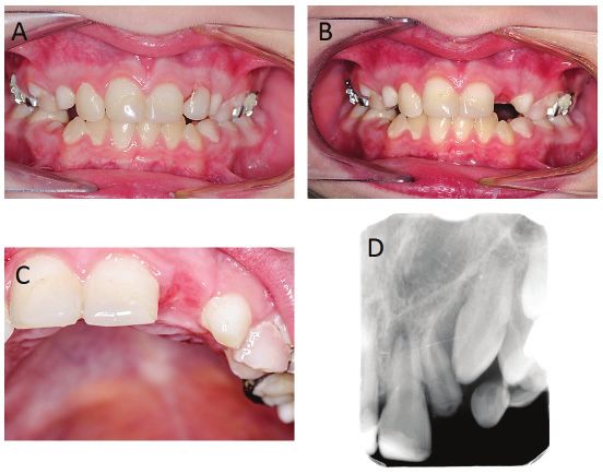
92                                                                  A. Strzecki, A. Bruzda-Zwiech, R. Filipińska. Post-traumatic vital root submergence

Introduction                                                     relations between the alveolar margin and remaining
                                                                 tooth portion. Orthodontic extrusion and submerging
   Traumatic dental injuries (TDI) in patients in devel-         vital root portion in situ are procedures burdened with
opmental age are considered a particularly difficult chal-       limited invasiveness.
lenge, as the prognosis regarding tooth vitality is usually         Most case reports describe crown-root fractures oc-
poor and the results of dental management difficult to           curring in teeth with closed apex or in adult patients.
predict. Clinicians need to make the correct clinical deci-      However, most traumatic dental injuries to the perma-
sion as quickly as possible while considering its life-long      nent dentition occur in schoolchildren.15 When the cli-
consequences. The early loss of a tooth prior to the pu-         nician needs to manage crown-root fracture in an im-
bertal growth spurt can have profound social, aesthetic,         mature tooth, treatment modalities are limited by the
occlusal and functional consequences,1–3 as the alveolar         stage of root development. As presented above, although
ridge in the traumatized, post-extraction areas ceases to        a plethora of treatment options can be mentioned, almost
develop and bone atrophy begins. Although the bone loss          every one of them requires non-delayed, permanent obtu-
of the alveolar ridge is most significant in the horizontal      ration of the root canal system at one of the clinical stages.
aspect, vertical bone loss also occurs, with the buccal side     For this reason, traumatized developing teeth with open
of the alveolar ridge being most prone to resorption.4,5         apices are particularly challenging to clinicians, especially
As a result, the alveolus is reduced, becomes more nar-          when the fracture line is located subgingivally. According
row and more lingually proclined.6 What is of major con-         to current guidelines regarding traumatic dental injuries
cern in early adolescence is permanent anterior maxillary        (TDI) management,15 “every effort should be made to
tooth loss due to alveolar bone arrest and aesthetic ridge       preserve pulpal vitality in the immature permanent tooth
deformation, which preclude the insertion of osseointe-          to ensure continuous root development”. Young age re-
grated implant with a good emergence profile that is im-         stricts the range of available treatment modalities, both
possible to achieve without ridge augmentation.7,8               due to the stage of root development and the limited level
   The central maxillary incisors are the teeth most likely to   of patient compliance. Hence, in young children with im-
be injured, and account for 80% of all traumatized teeth.1       mature permanent teeth demonstrating crown-root frac-
Maxillary lateral incisors and mandibular incisors are vul-      ture, the main focus should be on choosing methods with
nerable to injury to a lesser extent.1 Crown-root fracture       minimal invasiveness to the surrounding bone and peri-
defined as a fracture involving enamel, dentin and cemen-        odontium, retaining the ability of the root to continue its
tum accounts for 5% of all dental traumata to permanent          development and preventing the early loss of the tooth,
dentition,1 with transverse fractures being observed less        thus preserving the height and width of the alveolus in
frequently,9 especially in children10,11 and when the root       the traumatized site, negating the possible consequences
development is incomplete.10 The fracture line can also          of the injury to the occlusion and providing a temporary
expose the pulpal tissue, thus dividing the crown-root           aesthetic solution to minimize the social problems en-
fracture into 2 groups: complicated and uncomplicated.           countered by the child. The aim of this article is to present
Treatment options in cases of complicated crown-root             2 cases of subgingival crown-root fracture of immature
fractures are complex, and require a multidisciplinary ap-       teeth treated by a minimally invasive multidisciplinary
proach,12 the choice of which largely depends on the po-         treatment approach which can be also introduced in pa-
sition and direction of fracture line, pulpal involvement,       tients with a high level of dental fear.
tooth maturity, and the length of root remaining in the al-
veolus.2,3,13 Even when the fracture is positioned subgingi-
vally, bacterial invasion should be expected to the fracture     Case presentation
region, leading in most cases to subsequent inflamma-
tion.1,14 For this reason, most treatment protocols of deep
crown-root fracture require removal of the coronal tooth         Case 1
fragment; however, it can be used for further prosthetic
tooth reconstruction.                                              A 10-year-old male patient was referred to the De-
   The treatment modalities described in previous stud-          partment of Pediatric Dentistry of our institution due to
ies can be divided into 2 groups: surgical and conserva-         a dental injury suffered the previous evening after falling
tive. Surgical methods other than extraction focus on the        from a chair. His medical history was not relevant. Clini-
immediate reposition of the intra-osseus tooth portion           cal examination revealed a fractured restoration of the
followed by endodontic treatment. It can be either in-           medial corner of the left upper central incisor, as well
tentionally replanted or surgically extruded and fixed in        as movable composite build-up of the left lateral incisor
a more favorable, supragingival position. Surgical extru-        with marginal gum swelling and bleeding. Tooth #22 was
sion can be achieved with or without raising the muco-           very tender to percussion and the lower lip was swollen.
periosteal flap. The semi-conservative approach involves         The prior dental history revealed that he had suffered
gingivectomy and osteotomy in order to alter the spatial         trauma to the same maxillary region 6 months before,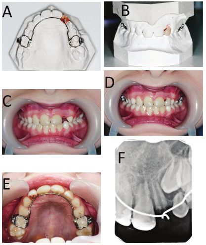
Dent Med Probl. 2018;55(1):91–98                                                                                                                        93

diagnosed as uncomplicated crown fracture of teeth #11
and #21, and uncomplicated crown-root fracture of tooth
#22, with an oblique fracture line penetrating subgingi-
vally. The teeth underwent immediate conservative resto-
ration with no further complications. The second trauma
led to a crown-root fracture of the lateral left incisor as
the mobile composite reconstruction was attached to the
fractured crown portion. The fracture line was horizon-
tal and reached below the alveolar ridge margin. A radio-
graphic examination confirmed the diagnosis of a crown
root fracture with pulp exposure and additionally showed
incomplete root development of tooth #22 (Fig. 1a). After
a holistic assessment of clinical and radiographic findings,
the decision was made to perform in situ submergence
of the vital root fragment. The coronal part of tooth #22
was surgically removed under local anesthesia, and the
fracture margin was exposed. The root fragment with the
bleeding exposed pulp was gently rinsed with 0.9% saline,
and the gingival tissue was soundly sutured. Antibiotic
therapy with 150 mg clindamicine administered 4 times
a day was prescribed.
  This clinical decision had the following rationale: 1. as
the crown root fracture occurred at the level of the al-
veolar ridge, the remaining apical portion of injured                         Fig. 2. A, B – upper lingual arch with acrylic tooth #22 on the dental
tooth #22 did not reach the criteria of 1:1 crown-root                        cast. Orthodontic bands fitted on the upper second deciduous molars.
                                                                              C – upfront view of patient’s anterior dentition without the prepared
ratio and so could barely support a future permanent
                                                                              appliance. D, E – patient’s dentition with the cemented lingual arch:
prosthetic build-up; gingivectomy/osteotomy with sub-                         upfront (D) and palatal view (E). F – dental radiograph taken after the
sequent apexification or mineral trioxide aggregate                           placement of orthodontic appliance – 4 weeks after the trauma
(MTA) pacement in apical region and root canal obtu-
ration, followed by orthodontic extrusion would not al-                       bands were fitted to the deciduous upper second molars
leviate this problem; 2. due to the patient’s young age,                      and dental impressions were taken to restore the missing
extraction of the remaining root fragment would lead to                       crown of tooth #22 by the use of an upper lingual arch
severe alveolar atrophy in anaesthetically-sensitive re-                      with an attached acrylic tooth (Fig. 2a, b). The patient
gion. The root was submerged with the intention of re-                        did not have any orthodontic treatment needs besides
taining the shape of the alveolar process for as long as                      the insertion of a space retainer after the dental trauma,
possible, preferably until the facial growth was finished                     and his first permanent molars were in I Angle Class in-
and a dental implant could be placed in the traumatized                       tercuspidation on both sides of the dental arch. The lin-
area. After 2 weeks of uneventful healing, the sutures                        gual arch was cemented on the next dental visit with
were removed (Fig. 1b, c). Subsequently, the orthodontic                      satisfactory aesthetic and functional effect (Fig. 2c–f ).
                                                                              As the oral hygiene of the patient was sub-optimal, the de-
                                                                              ciduous second molars were chosen as a support for the
                                                                              lingual arch instead of the permanent first molars to pro-
                                                                              tect the latter from the increased risk of caries associated
                                                                              with the presence of orthodontic bands. Therefore, the
                                                                              mentioned appliance should be perceived as a long-term
                                                                              solution, but not a permanent one: after root resorption
                                                                              occurs in the deciduous second molars, the lingual arch
                                                                              will be modified and fitted to the permanent first molars.
                                                                              No inflammatory symptoms could be detected either clin-
                                                                              ically or radiographically at any stage of treatment or dur-
                                                                              ing a 13-month follow-up (Fig. 3a). The submerged root
                                                                              manifested continued development. Although the shape
                                                                              of the alveolar ridge slightly altered vertically, the bone loss
                                                                              was not significantly pronounced in the horizontal aspect
Fig. 1. A – radiograph taken at the day of trauma – crown root fracture
of the upper left lateral incisor (#22). B, C – upfront view of traumatized   (Fig. 3b–d). The orthodontic appliance was well tolerated
area after 2 weeks of healing and suture removal                              by the patient, who underwent check-ups every 6 weeks.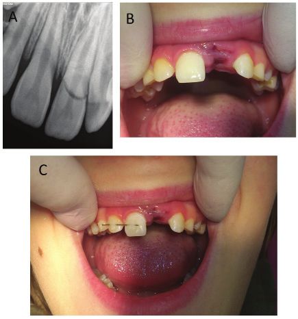
94 A. Strzecki, A. Bruzda-Zwiech, R. Filipińska. Post-traumatic vital root submergence
were unsatisfactory. Certain clarification of the observed
situation was given, with the parents mentioning that our
patient was recently diagnosed with Asperger’s syndrome.
The treatment focusing on retaining the space after the
lost tooth #22 was continued by means of removable
orthodontic appliance (modified Schwarz-type expander)
with the attached acrylic crown.
Case 2
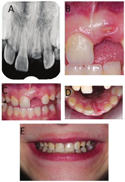
An 8-year-old female patient was referred to the Depart-
ment of Pediatric Dentistry of our institution for traumatic
dental injury suffered the previous day at school. The med-
ical history was non-contributory. The maxillary anterior
region was most severely injured, with the right central
Fig. 3. A – dental radiograph taken after 13-month follow-up period. No incisor subluxated and left central incisor crown horizon-
inflammatory symptoms could be detected. B–D – bone loss in the injured tally fractured at the level of the alveolar ridge (classified
area appears to be only slightly pronounced in vertical aspect and the as crown-root fracture with pulp exposure). The clinical
overall esthetic outcome is satisfactory
examination also revealed poor oral hygiene and many
carious lesions requiring immediate treatment. Tooth
After 15 months from trauma, the upper second decidu- #21 was not visible in the mouth, and the dental socket in
ous molars became mobile due to the physiological root the site of the tooth was filled with a blood clot (Fig. 5b).
resorption and additional occlusal load connected with The fractured coronal tooth fragment, brought in by the
supporting the artificial crown of the tooth #22. The ap- parents, was rinsed with 0.9% saline and stored in saline
pliance was removed and the patient was referred to the at 4°C for possible future adhesive build-up. The parents
surgical clinic to have teeth #55 and #65 extracted. Clini- also provided clinicians with a dental radiograph taken on
cal examination revealed the retained shape of alveolar the day of the trauma; however, it did not show the whole
process with slightly pronounced vertical bone loss (Fig. apical region of tooth #21 (Fig. 5a). Tooth #11 was sub-
4a–b). No inflammatory symptoms could be observed luxated and manifested slight pathological mobility, along
(Fig. 4a–c). Closed root apex with no signs of chronic
periapical inflammation can be observed on the dental
radiograph taken during the appointment (Fig. 4d). The
patient was highly anxious towards the planned surgical
procedures and both his compliance and oral hygiene
Fig. 4. A, B – patient after 15-month follow-up period with and without the
attached acrylic tooth #22. Slight inflammation of the mucosa observed Fig. 5. A – dental radiograph taken at the day of trauma. Tooth #21 is
in the injured region can be attributed to the poor oral hygiene combined horizontally fractured at the cervical area. Periapical region of the tooth
with the contact with the acrylic resin. C – the bone loss in the horizontal #21 cannot be assessed. B – missing clinical crown and the dental socket
aspect can be hardly observed. D – dental radiograph taken after of the tooth #21 filled with a blood clot – clinical situation during the first
15-month follow-up period: apex of the root of the tooth #22 is closed; appointment. C – semi-rigid splint connecting the teeth #11–#13 applied
previously exposed pulp area is covered with radiopaque tissue due to the subluxation of the tooth #11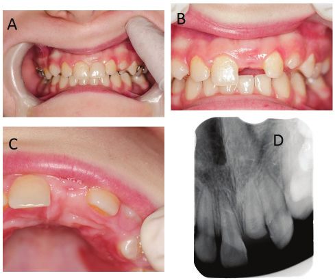
Dent Med Probl. 2018;55(1):91–98                                                                                                                          95

                                                                               were removed. The patient failed to attend a scheduled
                                                                               check-up visit 4 weeks following the trauma, but did so
                                                                               after 7 weeks, when it was possible to assess the course
                                                                               of the treatment both clinically and radiographically
                                                                               (Fig. 6a–e). No inflammatory symptoms were present at
                                                                               the time of examination and the traumatized region had
                                                                               healed properly. A dental radiograph showed an incom-
                                                                               pletely closed apex root of tooth #21, the root portion ap-
                                                                               peared to be of sufficient length for future core build-up.
                                                                               The area of the previously-exposed pulp appeared to be
                                                                               covered with hard tissue resembling radiographically re-
                                                                               parative dentine.
                                                                                  After the apex closure process is completed and patient
                                                                               compliance will be sufficient, it is planned to extirpate the
                                                                               pulp and extrude the root orthodontically above the gin-
                                                                               gival level, thus enabling a long-term temporary prosthet-
                                                                               ic build-up. It was also noted that the margin of the frac-
                                                                               tured root was visible above the mucosa in a very limited
                                                                               area. It is possible that the root either began to spontane-
                                                                               ously erupt or the root exposure occurred due to alveolar
                                                                               ridge recontouring. Due to the patient’s young age, poor
                                                                               oral hygiene and narrow maxillary arch, it was decided to
                                                                               combine a temporary replacement for the crown of tooth
                                                                               #21 with treatment using an orthodontic removable ap-
                                                                               pliance. An active palatal plate with an acrylic tooth #21
                                                                               and expansion screw positioned in the midline was de-
Fig. 6. A – dental radiograph of the injured area taken after 7-week period.
                                                                               signed (Fig. 7a–d). After a 10-month follow-up, no in-
No inflammatory symptoms could be detected and the process of the
closure of the root apex continued. Note the mineralized tissue covering       flammatory symptoms could be observed either clinically
the area of previously exposed pulp. B–E – view of the patient’s dentition     or radiographically (Fig. 8a–d). The apexification of the
after 7-weeks from the dental injury. No inflammatory symptoms could be        submerged root continued, thus indicating that the pulp
detected clinically
                                                                               retains its vitality. The patient showed improved compli-
                                                                               ance and was pleased with the esthetics of the proposed
with tenderness to vertical percussion. The sensitivity                        clinical solution (Fig. 8e).
of the pulp was positive when tested with a cold stimulus.                        Certain difficulties occurred in further monitoring
The subluxated tooth was splinted with semi-rigid wire                         of treatment outcome, as the patient failed to show up
and composite resin for 2 weeks (Fig. 5c). The anatomic re-                    for a pre-scheduled appointment at the 12-month stage
lationship between the fracture line and the alveolar ridge                    of follow-up. It was not until 16 months after the trauma
margin could be assessed, despite the parents not giving                       when we could get in touch with the patient’s parents
consent for another dental X-ray to be taken on the day
of admission. Although the stage of root formation could
not be evaluated, it was assumed on the basis of age of the
patient that root development was not completed.
   As the length of the root of tooth #11 seemed to be ad-
equate for supporting post-retained crown reconstruction,
immediate gingivectomy or even osteotomy followed by
the subsequent MTA placement in apical region of the root
to form a hard barrier prior to gutta-percha root obtura-
tion were considered. However, this treatment option was
abandoned due to dental fear and low level of compliance
by the patient. Vital root submergence in situ was discussed
with the parents as a treatment option, which might allow
for root apex closure of tooth #22 and the use of an apical
fragment for later post-retained crown reconstruction.
   The traumatized area with its remaining root segment
                                                                               Fig. 7. A, B – removable appliance fitted on the dental cast. C, D – upfront
was rinsed with 0.9% saline and soundly sutured. Af-                           view of patient’s anterior dentition without the appliance and with the
ter 2 weeks of uneventful healing, the sutures and splint                      appliance inserted into the oral cavity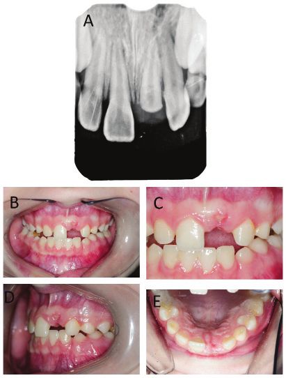
96                                                                            A. Strzecki, A. Bruzda-Zwiech, R. Filipińska. Post-traumatic vital root submergence

and the control clinical and radiological examination
could be performed. No inflammatory symptoms were
either mentioned by the patient or discovered during the
check-up (Fig. 9a, b). The crest of the alveolar process
in the injured area was on the level of cervical gingival
margin surrounding the tooth #11, which could be de-
scribed as a major improvement (Fig. 9c). A radiograph-
ic examination showed closed root apex of the tooth
#21 with no periapical lesions and the area of previously
exposed pulp covered with radiopaque tissue (Fig. 9d).
The patient and her parents were reluctant to the pro-
posed plan of the elective endodontic treatment and
a subsequent forced orthodontic eruption of the root
and wished to continue the treatment with the current
appliance. It was established that the treatment options
will be re-discussed after the eruption of all premolar
teeth, which will necessitate the change of the remov-
                                                                           Fig. 9. A–C – clinical examination after 16-months. Both vertical and
able appliance. Nevertheless, sustaining the alveolar                      horizontal dimension of the alveolar ridge remained unchanged. No
bone level and completed closure of the root apex made                     inflammatory state could be detected. D – dental radiograph taken after
all of the permanent treatment options ranging from the                    16-month follow-up period: apex of the root of the tooth #22 is closed;
                                                                           previously exposed pulp area is covered with radiopaque tissue
combined orthodontic and prosthetic treatment to the
implant placement possible.
                                                                           Discussion
                                                                              Although intentional submerging in situ of vital roots af-
                                                                           ter crown-root fracture is mentioned in up-to-date guide-
                                                                           lines regarding the management of traumatic dental inju-
                                                                           ries,15 reports of cases presenting it are rarely encountered
                                                                           in scientific literature. A review of existing literature reveals
                                                                           only one such study by Mackie et al.16 Although the main
                                                                           advantage of this approach is the possibility of retaining the
                                                                           shape and size of the alveolar ridge in the traumatized re-
                                                                           gion until the facial growth is finished, it also offers limited
                                                                           invasiveness, which requires little compliance, continued
                                                                           root development and no additional iatrogenic trauma to
                                                                           the surrounding tissues. Furthermore, it offers other bene-
                                                                           fits for the patient in cases when the remaining root cannot
                                                                           support any post-retained crown due to inadequate length
                                                                           and needs to be extracted.
                                                                              The fact that the untreated, exposed pulp showed no im-
                                                                           mediate or delayed inflammatory symptoms, either clini-
                                                                           cally or radiographically, can be expected to a certain ex-
                                                                           tent. This can be probably attributed to the stage of tooth
                                                                           development and relatively good perfusion of pulpal tis-
                                                                           sue in immature teeth. Furthermore, radiographs taken
                                                                           at various treatment stages indicated that the pulp tissue
                                                                           remained vital. The rationale behind root submergence is
                                                                           derived from studies concerning fractured and retained
                                                                           roots following unsuccessful extractions of vital teeth.
                                                                           The vast majority of studies show that such retained roots
                                                                           remain either vital or uneventful.17,18 Some authors sug-
                                                                           gest that the submerged root can be covered with new
Fig. 8. A – dental radiograph taken after the 10-month follow-up period.   bone.19 Attempts to preserve the volume of the alveolar
The apex of the root of the tooth #21 is closed and no sign of chronic     ridge by means of vital root submergence in older pros-
inflammation could be detected. B–E – 10 months from the injury. No
inflammatory symptoms could also be detected during the clinical           thetic patients, when the crown-root ratio is unfavorable,
examination                                                                have also been suggested.20 As it was mentioned above,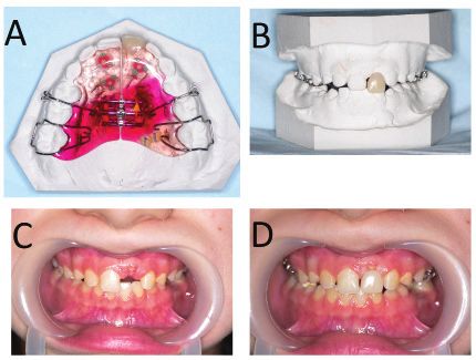
Dent Med Probl. 2018;55(1):91–98                                                                                                           97

the decoronation of deeply impacted lower third molars           and requiring very low level of compliance. The retained
bears a certain resemblance to the intentional submer-           root may remain within the alveolus until the implant
gence of traumatized roots. Despite obvious differences,         placement is possible or serve as a prosthetic post follow-
both procedures treat the exposed pulp with 0.9% saline,         ing delayed pulp extirpation. The possibility of delaying
which is then covered with a tightly sutured mucoperios-         pulp extirpation may prove beneficial in certain clinical
teal flap. The most important similarity is, however, the        situations with the emphasis of the immature trauma-
lack of subsequent symptoms of pulp inflammation de-             tized teeth. Orthodontic appliances with attached arti-
spite the lack of any capping. Interestingly, in some cases,     ficial dental crowns prove to be aesthetically acceptable,
the passive eruption of retained teeth occurs.21                 multi-purpose clinical solutions in cases of early clinical
  What should, and could, be done with the remaining             crown loss in growing patients. The root submergence
apical tooth segment is clearly the main problem encoun-         procedure also allows for dental fragments that would
tered in crown-root fracture management in the devel-            otherwise be extracted to remain within the alveolus and
opmental age. The 4 key issues that need to be addressed         at least partially contribute to retaining the shape of the
are as follows: 1. whether the length of the remaining por-      alveolar ridge.
tion of the root is adequate and whether it may be used             As the pulpal tissue in the submerged roots showed no
after surgical procedures/orthodontic extrusion for fixed        inflammatory symptoms, despite being left untreated and
prosthesis purposes; 2. what can be done and achieved at         only tightly covered with mucosa, its immune defensive
the current development stage of traumatized tooth root          capabilities in teeth with an open apex may be higher than
and patient growth stage; 3. the level of patient motiva-        commonly perceived. For this reason, root submergence
tion and compliance and whether the clinician is likely to       is worth considering in young patients experiencing deep
be limited by technical constraints or adolescent patient        crown-root fracture, especially when they suffer from
cooperation, and finally; 4. what can be done to protect         high dental fear and a low level of compliance.
the height and width of the alveolar process.
  When the submergence of the vital root is performed for        References
the preservation of the alveolar ridge prior to permanent         1. Andreasen JO, Andreasen FM, Andersson L. Textbook and Colour
prosthetic reconstruction with a dental implant, spontane-           Atlas of Traumatic Injuries to the Teeth. 4th ed. Oxford, England: Black-
                                                                     well Munksgaard; 2007:314–336.
ous eruption of the root may be regarded as an undesirable        2. Andreasen JO. Etiology and pathogenesis of traumatic dental inju-
complication.22 However, in our case of the submergence              ries. A clinical study of 1298 cases. Scand J Dent Res. 1970;78:329–342.
of a vital root with a length adequate to support further         3. Naudi AB, Fung DE. Tooth fragment reattachment in multiple com-
                                                                     plicated permanent incisor crown-root fractures: A report of two
post-retained crown reconstruction, the fact that the root           cases. Dent Traumatol. 2008;24:248–252.
began to erupt can be beneficial, as described in a case          4. Araujo MG, Lindhe J. Dimensional ridge alterations following tooth
presented by Johnson and Jensen,22 in which spontaneous              extraction: An experimental study in the dog. J Clin Periodontol.
                                                                     2005;32:212–218.
eruption eliminated the need for surgical exposure and            5. Araujo MG, Lindhe J. Ridge alterations following tooth extraction
orthodontic extrusion of the root fragment and allowed for           with and without flap elevation: An experimental study in the dog.
successful root canal obturation and restoration with a post         Clin Oral Implants Res. 2009;20:545–549.
                                                                  6. Pinho MN, Roriz VL, Novaes AB, et al. Titanium membranes in pre-
and porcelain fused to metal crown.                                  vention of alveolar collapse after tooth extraction. Implant Dent.
  In the first of our cases, the possibility of using the root       2006;15:53–61.
fragment for the prosthetic purposes, even after orth-            7. Darby I, Chen ST, Buser D. Ridge preservation techniques for implant
                                                                     therapy. Int J Oral Maxillofac Implants. 2009;24(Suppl):260–271.
odontic extrusion, would be very limited as the crown to          8. Jung RE, Philipp A, Annen BM. Radiographic evaluation of differ-
root ratio would be significantly higher than 1:1. In the            ent techniques for ridge preservation after tooth extraction: A ran-
second case, the remaining fragment of the central inci-             domized controlled clinical trial. J Clin Periodontol. 2013;40:90–98.
                                                                  9. Fariniuk LF, Ferreira EL, Soresini GC, Cavali AE, Baratto Filho F. Inten-
sor is sufficient for prosthetic build up and forced erup-           tional replantation with 180 degree rotation of a crown-root frac-
tion is planned. However, in our opinion, prosthetic                 ture: A case report. Dent Traumatol. 2003;19:321–325.
reconstruction should be delayed, as the root shows no           10. Díaz SC, Ferreyra S, Gutmann JL. Healing of a horizontal root frac-
                                                                     ture with lateral luxation of the coronal fragment: A 3-year follow-
inflammatory symptoms, root development persists and
                                                                     up ENDO (Lond Engl). 2013;7:291–297.
mildly pronounced passive eruption can be observed. Af-          11. Orhan K, Orhan AI, Tulga F. Management of untreated traumatized
ter apexification is completed, a forced eruption should             permanent incisors with crown and root fractures: A case report.
be performed, thus enabling at least temporary prosthetic            Quintessence Int. 2009;40:647–654.
                                                                 12. Robertson A. A retrospective evaluation of patients with uncompli-
restoration of the fractured crown.                                  cated crown fractures and luxation injuries. Endod Dent Traumatol.
                                                                     1998;14:245–256.
                                                                 13. Wang Z, Heffernan M, Vann WF Jr. Management of a complicated
                                                                     crown-root fracture in a young permanent incisor using intention-
Conclusions                                                          al replantation. Dent Traumatol. 2008;24:100–103.
                                                                 14. Bevelander G. Tissue reactions in experimental tooth fracture.
   Vital roots submergence could be considered as the                J Dent Res. 1942;21:481–487.
                                                                 15. DiAngelis AJ, Andreasen JO, Ebeleseder KA, et al. Guidelines for
first stage of a more complex treatment scheme in juve-              the management of traumatic dental injuries: 1. Fractures and lux-
nile patients burdened with few possible complications               ations of permanent teeth. Dent Traumatol. 2012;28:2–12.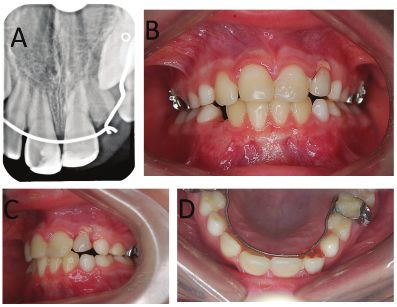
98                                                                           A. Strzecki, A. Bruzda-Zwiech, R. Filipińska. Post-traumatic vital root submergence

16. Mackie IC, Quayle AA. Alternative management of a crown root
    fractured tooth in a child. Br Dent J. 1992;173:60–62.
17. Simpson HE. Histological changes on retained roots. J Can Dent
    Assoc. 1959;25:287.
18. Glickman I, Pruzansky S, Ostrach M. The healing of extraction
    wounds in the presence of retained root remnants and bone frag-
    ments. Am J Orthod. 1974;33:263.
19. Von Wowern N, Winther S. Submergence of roots for alveolar ridge
    preservation. Int J Oral Surg. 1981;10:247–250.
20. Sharma A, Oberoi SS, Saxena S. Submergence of vital roots for the
    preservation of residual ridge: A clinical study. Oral Health Prev
    Dent. 2012;10:259–265.
21. Renton T, Hankins M, Sproate C, McGurk M. A randomised con-
    trolled clinical trial to compare the incidence of injury to the infe-
    rior alveolar nerve as a result of coronectomy and removal of man-
    dibular third molars. Br J Oral Maxillofac Surg. 2005;43:7–12.
22. Johnson BR, Jensen MR. Treatment of a horizontal root fracture by
    vital root submergence. Endod Dent Traumatol. 1997;13:248–250.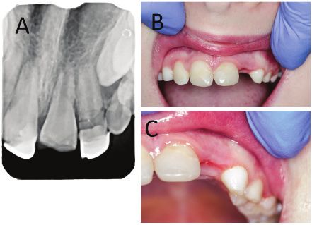
You can also read