Case Report: Allergic Bronchopulmonary Aspergillosis Revealing Asthma
←
→
Page content transcription
If your browser does not render page correctly, please read the page content below
CASE REPORT
                                                                                                                                             published: 22 June 2021
                                                                                                                                    doi: 10.3389/fimmu.2021.695954

                                          Case Report: Allergic
                                          Bronchopulmonary Aspergillosis
                                          Revealing Asthma
                                          Houda Snen 1,2*, Aicha Kallel 2,3*, Hana Blibech 1,2, Sana Jemel 2,3, Nozha Ben Salah 1,2,
                                          Sonia Marouen 3, Nadia Mehiri 1,2, Slah Belhaj 3, Bechir Louzir 1,2 and Kalthoum Kallel 2,3
                                          1 Pulmonary Department, Hospital Mongi Slim, La Marsa, Tunisia, 2 Faculty of Medicine, Tunis El Manar University, Tunis,

                                          Tunisia, 3 Parasitology and Mycology Department, La Rabta Hospital, Tunis, Tunisia

                                          Allergic bronchopulmonary aspergillosis (ABPA) is an immunological pulmonary disorder
                                          caused by hypersensitivity to Aspergillus which colonizes the airways of patients with
                                          asthma and cystic fibrosis. Its diagnosis could be difficult in some cases due to atypical
                          Edited by:
                                          presentations especially when there is no medical history of asthma. Treatment of ABPA is
                  Brian Stephen Eley,     frequently associated to side effects but cumulated drug toxicity due to different molecules
University of Cape Town, South Africa
                                          is rarely reported. An accurate choice among the different available molecules and
                      Reviewed by:
                                          effective on ABPA is crucial. We report a case of ABPA in a woman without a known
                      Shivank Singh,
  Southern Medical University, China      history of asthma. She presented an acute bronchitis with wheezing dyspnea leading to an
                   Richard B. Moss,       acute respiratory failure. She was hospitalized in the intensive care unit. The
   Stanford University, United States
                                          bronchoscopy revealed a complete obstruction of the left primary bronchus by a sticky
                  *Correspondence:
                          Houda Snen
                                          greenish material. The culture of this material isolated Aspergillus fumigatus and that of
              houda.snen@gmail.com        bronchial aspiration fluid isolated Pseudomonas aeruginosa. The diagnosis of ABPA was
                           Aicha Kallel
                                          based on elevated eosinophil count, the presence of specific IgE and IgG against
               aicha.kallel@fmt.utm.tn
                                          Aspergillus fumigatus and left segmental collapse on chest computed tomography. The
                   Specialty section:     patient received an inhaled treatment for her asthma and a high dose of oral
         This article was submitted to    corticosteroids for ABPA. Her symptoms improved but during the decrease of
               Microbial Immunology,
               a section of the journal   corticosteroids, the patient presented a relapse. She received itraconazole in addition
              Frontiers in Immunology     to corticosteroids. Four months later, she presented a drug-induced hepatitis due to
            Received: 15 April 2021       itraconazole which was immediately stopped. During the monitoring of her asthma which
           Accepted: 08 June 2021
           Published: 22 June 2021
                                          was partially controlled, the patient presented an aseptic osteonecrosis of both femoral
                            Citation:
                                          heads that required surgery. Nine months after itraconazole discontinuation, she
         Snen H, Kallel A, Blibech H,     presented a second relapse of her ABPA. She received voriconazole for nine months
     Jemel S, Salah NB, Marouen S,        associated with a low dose of systemic corticosteroid therapy with an improvement of her
          Mehiri N, Belhaj S, Louzir B
    and Kallel K (2021) Case Report:      symptoms. After discontinuation of antifungal treatment, there was no relapse for one year
          Allergic Bronchopulmonary       follow-up.
     Aspergillosis Revealing Asthma.
         Front. Immunol. 12:695954.       Keywords: allergic bronchopulmonary aspergillosis, Aspergillus fumigatus, antifungal therapy, drug toxicity,
   doi: 10.3389/fimmu.2021.695954         uncontrolled asthma

Frontiers in Immunology | www.frontiersin.org                                    1                                           June 2021 | Volume 12 | Article 695954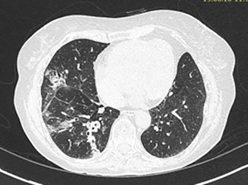
Snen et al. Case Report: Allergic Bronchopulmonary Aspergillosis Revealing Asthma
INTRODUCTION wheezing dyspnea. In her medical history she reported an allergic
rhinitis diagnosed in childhood without respiratory symptoms.
Fungal pulmonary infections are rare in immunocompetent She was sensitized to some pollen types which were not specified.
patients. These infections can be caused by several pathogens She is of French origin and living in Tunisia for thirty years. She
such as Aspergillus, Pneumocytis jirovecii and Cryptococcus. had no respiratory nor rhinitis symptoms since she came in
However, even immunocompetent patients can be affected by Tunisia and until she developed a bronchitis in December 2017.
these pathogens. In fact, they can affect patients with chronic This bronchitis was resistant to symptomatic treatment and
bronchopulmonary pathologies such as asthma, cystic fibrosis short-term systemic corticosteroid therapy. The patient was
and chronic obstructive pulmonary disease (1). The most hospitalized in an intensive care unit for an acute respiratory
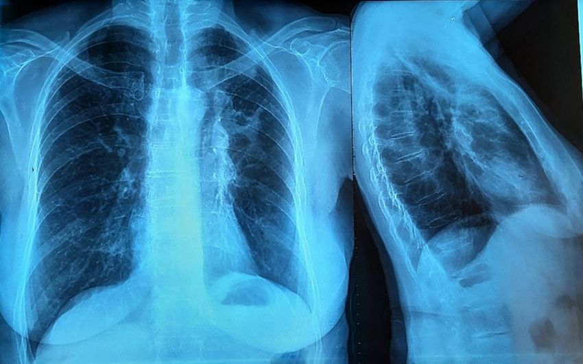
frequent pathogen associated with fungal pulmonary infections failure due to her bronchitis. A chest X-ray (face and profile,
is Aspergillus, which is a saprophytic mold isolated abundantly Figure 1) showed a left hilo-axillary linear opacity with
from soil, construction sites and hospitals (1, 2). There are three retraction signs evoking atelectasis. Her blood tests didn’t show
clinical presentations of pulmonary aspergillosis: chronic a biological inflammatory syndrome (CRP= 11mg/l; white blood
pulmonary aspergillosis (CPA), invasive pulmonary cells= 9780/mm3 with an eosinophilic count= 939/mm3). Chest
aspergillosis (IPA) and allergic bronchopulmonary aspergillosis computed tomography (CT) confirmed the diagnosis of
(ABPA) (3). Depending on the interaction between the pathogen atelectasis and showed segmental collapse of the lingula and
and its host, pulmonary aspergillosis can lead to one of these posterior segment of the left basal pyramid with no
clinical presentations (4). The estimated worldwide global rate of parenchymatous lesion (Figure 2). Flexible bronchoscopy
ABPA among asthmatic adult patients is 2,5% (5, 6), however revealed a complete obstruction of the left primary bronchus
only some cases are reported in Tunisia (7). ABPA is most often by a sticky greenish material that could be removed (Figure 3).
associated with severe uncontrolled asthma (5, 6) and drug Bacterial culture of this material isolated Pseudomonas
toxicity is frequently reported during therapeutic management. aeruginosa and the patient received consequently an
We report a case of ABPA in a patient with a medical history of antibiotherapy associating Levofloxacin and Cefpodoxime for
an allergic rhinitis, with no respiratory symptoms and who two weeks, with partial improvement in respiratory symptoms.
developed drug toxicity to corticosteroids then to itraconazole. The mycological culture isolated Aspergillus fumigatus.
Thus, we emphasize the challenges in diagnosing and treating Aspergillus serology (IgG) was positive at 12 AU/mL (ELISA,
ABPA due to its atypical clinical presentations and significant Biorad®). Total IgE count was 233 IU/ml (ELFA, Biomé rieux®).
drug toxicity associated with its therapeutic management. Aspergillus fumigatus specific IgE and Aspergillus skin testing
were not done. During pulmonary function test, the forced vital
capacity (FVC) was at 1,97 l (82%) and forced expiratory volume
CASE PRESENTATION in one second (FEV1) was at 1,60 l (81%). The diagnosis of
ABPA associated to an asthma was established. The patient
A 72-year-old woman, non-smoker, consulted in December received a high-dose regimen of oral corticosteroids for five
2017, an office-based pulmonologist for acute bronchitis with weeks (1 mg/kg/day: 80mg of prednisone for one week then
FIGURE 1 | Left hilo-axillary linear opacity associated with retraction signs evoking atelectasis on chest-ray face and profile.
Frontiers in Immunology | www.frontiersin.org 2 June 2021 | Volume 12 | Article 695954Snen et al. Case Report: Allergic Bronchopulmonary Aspergillosis Revealing Asthma
presented a relapse of her respiratory symptoms at the dose of
5mg of prednisone per day. She consulted in March in our
department. The itraconazole was prescribed in association with
corticosteroid therapy (medium-dose regimen: 0,5 mg/kg/day;
40mg of prednisone for one month then 30mg for one month
then 25mg for two weeks then 20mg for two weeks then 15mg for
two weeks, then 10mg for two weeks). After four months of anti-
fungal treatment, the patient developed a jaundice with an
intense deterioration of her general condition. Liver biological
tests showed significant hepatic cytolysis (AST= 485 IU/L, ALT=
182 IU/L) and cholestasis (Gamma-GT= 933 IU/L, Alkaline
phosphatse= 758 IU/L). After exclusion of the intake of any
other treatment associated to drug liver toxicity, the diagnosis of
an acute liver failure due to a drug-induced hepatitis associated
to the anti-fungal treatment was established. As a result, the
itraconazole was immediately stopped. At that time, the titer of
Aspergillus fumigatus specific IgE was 0.59 KUA/L (FEIA,
Thermaoscientific/Phadia®), hence the decision to continue
FIGURE 2 | Chest CT scan image showing segmental aerated collapse of inhaled asthma treatment and to continue monitoring the
the lingula.
patient. For the next seven months, the patient’s asthma was
partially controlled. The patient developed a right hip joint pain
and a lameness. The various explorations concluded to an aseptic
40mg for one week then 20mg for one week then 10mg for one osteonecrosis of both femoral heads. The patient underwent
week then 5mg for one week) and an inhaled association of long- surgery for her right hip. In May 2019, the patient presented
acting bronchodilator and a high dose of corticosteroids acute bronchitis with mucus produced by coughs. She had
(salmeterol and fluticasone). When searching for mold persistent respiratory symptoms which were resistant to
exposure in the patient’s medical history, we discovered her antibiotherapy and short-term systemic corticosteroid therapy.
gardening activities and that she had multiple indoor plants at Chest X-ray showed an atelectasis in the left lower lobe. Flexible
her house. In addition to medical treatment, it was bronchoscopy revealed a mucus plug at the left lower lobar
recommended that the patient should remove all the indoor bronchus. The middle lobar bronchus had a reduced caliber and
plants. The evolution has been marked by the disappearance of was non-catheterizable. Chest CT revealed alveolar opacities
respiratory symptoms and of the atelectasis on chest x-rays and associated with bronchiectasis in the posterior and medial
the decrease in the eosinophils’ count (282/mm3). However, segment of the right basal pyramid and lateral segment of the
during the decrease of corticosteroid therapy dosage, she middle lobe (Figure 4). During bronchoscopy, the plug has been
FIGURE 3 | Complete obstruction of the left strain bronchus by sticky FIGURE 4 | Chest CT scan image showing alveolar opacities associated to
greenish material in flexible bronchoscopy. bronchiectasis in posterior and medial segment of the right basal pyramid.
Frontiers in Immunology | www.frontiersin.org 3 June 2021 | Volume 12 | Article 695954Snen et al. Case Report: Allergic Bronchopulmonary Aspergillosis Revealing Asthma
removed and cultures done. Bacterial culture of this material was and night sweats (17). Expectoration of yellowish-green lumps of
negative. The mycological culture isolated Aspergillus fumigatus. mucus is characteristic of ABPA and can be observed in half of
In vitro susceptibility testing against voriconazole was performed the cases (8) which was the case of our patient. The chest X-ray
using E-test (Biomé rieux®). This strain was susceptible to may be normal in the early stages of the disease and can reveal
Voriconazole (MIC: 0.38µg/mL). Total IgE count was at 301 some abnormalities such as “Tramline shadows” and “finger-in-
IU/ml, eosinophilic count was at 159/mm 3 . The titer of glove opacities” which are temporary patterns corresponding to
Aspergillus fumigatus specific IgE was at 13.90 KUA/L. bronchial wall edema and thickening; “Toothpaste shadows”
Voriconazole was prescribed in addition to a step up in the which are also transient and indicate mucus plugs within
patient’s asthma treatment. The patient received antifungal bronchi; “parallel line shadows” which appear when the mucus
treatment for nine months in addition to a low dose of plugs are expectorated (18, 19). In the case of our patient, the
systemic corticosteroid therapy (prednisone 10mg/day) and an chest X-ray showed a linear opacity due to an atelectasis during
inhaled long-acting anticholinergic (tiotropium) in association to the first consultation and during the relapse. The chest CT
montelukast and an inhaled association of long-acting confirmed the diagnosis of segmental collapse during the first
bronchodilator and a high dose of corticosteroids. The consultation and showed bronchiectasis during the relapse.
Aspergillus fumigatus specific IgE decreased to 2 KUA/L. The Flexible bronchoscopy has an important place particularly in
pulmonary function test results improved [FVC=2,38 l (90%) patients with atelectasis to rule out malignant etiologies. It also
and FEV1 = 1,98 l (96%)]. After discontinuation of antifungal allows bacteriological and mycological samples. Sputum or
treatment, there was no relapse for one year follow-up. bronchial fluid cultures are positive for Aspergillus in nearly
40–60% of cases (20, 21). The presence of Aspergillus fumigatus
in the sputum culture is not sufficient to confirm the diagnosis of
ABPA as this fungus is human saprophyte and can be present in
DISCUSSION other pulmonary diseases (9, 10). Sputum cultures can contribute
to the diagnosis by isolating Aspergillus and by performing in
ABPA is caused by hypersensitivity to Aspergillus fumigatus (8). vitro antifungal-susceptibility testing and molecular testing for
It is frequently associated with severe and uncontrolled asthma resistance, of the isolated strains (9, 10, 22). In a study involving
or cystic fibrosis. The small conidia of Aspergillus fumigatus can 13 countries from four continents, 6% of the 2026 isolates of A.
easily enter the airways. Exposure to large numbers of conidia fumigatus were triazole resistant using molecular tools (23). This
may cause ABPA (9, 10), but not all asthmatics develop ABPA resistance prevalence varies in its geographic distribution. For
despite being exposed to the same environmental factors. This example, in a French study, the prevalence of Aspergillus
means that other factors play a role in the pathogenesis of ABPA fumigatus azole resistance in patients with cystic fibrosis was
(9). In a genetically predisposed individuals, inhaled conidia of detected in 6.8% of cases (24). However, in an English study, the
Aspergillus fumigatus germinate into hyphae with release azole resistance prevalence was higher. Resistance to at least one
antigens that activate the innate and adaptive immune azole antifungal drug was confirmed in 13.2% of included
responses (Th2 CD4+ T cell responses) of the lung (9, 10). patients among whom 16.2% had cystic fibrosis (TR34/L98H
When treating ABPA, exposure to molds in the patient’s was identified in 27.3% of azole-resistant isolates) (25).
environment must be investigated to indicate remediation. This Furthermore, the association of Pseudomonas aeruginosa and
is important to prevent relapse after discontinuation of Aspergillus fumigatus has been reported in the literature. Both
treatment, which was the case for our patient. The first microbes are responsible for considerable morbidity and
publication about ABPA as an entity was in 1952, from the mortality particularly in patients with cystic fibrosis, among
United Kingdom (11). A decade later, the second case of ABPA whom the co-infection accelerates the lung disease progression
was described in the United States (12, 13). The diagnosis of this (26). In fact, metabolite exchange and intermicrobial
disease is still difficult, particularly during atypical clinical competitions between both germs have been studied to better
presentations which was the case of our patient, who had an explain the important morbidity and mortality due to their
allergic rhinitis and no prior asthma history or diagnosis. In fact, association (27).
up to one third of patients with controlled asthma had a When the diagnosis of ABPA is suspected, some biological
relatively asymptomatic ABPA, and the diagnosis is discovered investigations are used for the diagnosis and monitoring of ABPA.
during routine testing (14). Moreover, some clinical The relevant tests are eosinophil count, total serum IgE level,
presentations are confusing such as the case reported by Savi serum IgE antibodies specific to Aspergillus fumigatus and serum
and al. and where the diagnosis of ABPA was established in a precipitins or specific IgG against Aspergillus fumigatus (9, 14).
previously healthy male (15). Also, in a nationwide Japan survey, First, blood eosinophil count should be checked and a level over
19% of patients diagnosed with ABPA had no medical history of 500 cell/L can help to establish the diagnosis. Our patient had an
asthma (16). ABPA is due to an inflammatory pulmonary elevated eosinophilic count (939/mm3). However, high eosinophil
disorder which often causes non-specific symptoms such as counts can be detected in many other diseases and normal levels
chronic cough, wheezing and recurrent pulmonary infiltration are reported in patients with ABPA receiving corticosteroids (14).
(8). It may be associated to other symptoms such as fever, weight It is known that the pulmonary eosinophilia is far greater than in
loss, deterioration of general condition, hemoptysis, chest pain peripheral blood; thus, a low eosinophil count does not exclude
Frontiers in Immunology | www.frontiersin.org 4 June 2021 | Volume 12 | Article 695954Snen et al. Case Report: Allergic Bronchopulmonary Aspergillosis Revealing Asthma
ABPA (9, 28). The measurement of the serum total IgE level is an total duration of three to five months (0.5 mg/kg/day for two
accurate and important test for the diagnosis and the follow-up of weeks, then on alternate days for eight weeks, then 5 mg less every
ABPA (10). Active ABPA is generally excluded when serum IgE is two weeks) (37). When a patient is on glucocorticoids and still has
normal (9, 10). For the cut-off value of IgE level that should be recurrent exacerbations or worsening pulmonary function test or
used in the diagnosis of ABPA, there is no consensus, and it become glucocorticoid-dependent, antifungal therapy could be
remains uncertain (9). In addition, the reported IgE values in added (37, 38). In our case, the patient presented a severe
different units (IU/mL, ng/mL) could lead to false interpretation complication of glucocorticoid treatment which is aseptic
(9). Some laboratories employ 417 IU/mL as a cut-off value, while osteonecrosis of both femoral heads requiring surgery.
others use a value of 1000 IU/mL (29). So, a validation of the IgE Itraconazole is usually used with or without glucocorticoids for
cutoff value across all populations is required since it could be at least six months, at a dose of 200 mg twice a day (38). It requires
influenced by both risk of exposure to Aspergillus antigens and frequent liver enzymes level monitoring because of its toxicity
ethnicity (9, 18). Despite this, the most sensitive investigation in (38). In fact, itraconazole can cause liver toxicity which was the
the diagnosis of ABPA is currently the detection of high levels of case of our patient. Other oral azoles such as voriconzaole and
serum IgE antibodies specific to Aspergillus fumigatus (>0.35 kUA/ posaconazole are also effective in ABPA and can be used when
l). This test is also considered the preferred one for screening itraconzaole is toxic or contraindicated (39). However, when there
asthmatic patients for ABPA (6, 9, 10). When our patient had the is a drug toxicity due to one molecule of azole, there is a risk of a
first relapse, the serum specific IgE were high with a value of 13.9 cross-azole toxicity. So alternative approaches to antifungal
KUA/L. Although the detection of IgE antibodies specific to treatment, in ABPA, that avoid systemic effects were tested and
Aspergillus fumigatus is useful for the diagnosis, it is less inhaled amphotericin B has been explored with varying results in
helpful in the follow up of patients (10). In addition, serum uncontrolled studies (40, 41). In our case, inhaled amphotericin B
precipitins or specific IgG against Aspergillus fumigatus are was not available and voriconazole was used without a cross-azole
detected in 69–90% of cases of ABPA (9, 30). In our case, the toxicity. It led to remission without relapse after discontinuation of
patient had positive specific IgG, but the technics used are not antifungal therapy. Furthermore, omalizumab has also proven its
equal. In fact, double gel diffusion technique for the detection of efficacy in ABPA, compared to long-term glucocorticoids and it
Aspergillus fumigatus-specific IgG has a limited sensitivity (27%) can be administered even in cases with high level of IgE (42). In
in the diagnosis of ABPA, whereas, commercial enzyme the case of acute lung collapse, broncho-alveolar lavage during
immunoassays have a sensitivity exceeding 90% (10, 30). rigid or flexible bronchoscopy helps the lung re-expansion and
The diagnosis of ABPA is confirmed when the case significant improvement of ABPA symptoms (33, 43). For patients
presentation meets the criteria established in 2013 by the with thick sputum, chest physiotherapy and nebulized hypertonic
ABPA Working Group of the International Society for Human saline solution improve the symptoms (44, 45). Patients should be
and Animal Mycology. If total IgE level are over 1000 IU/mL, examined every two months with chest radiography and total
two among three criteria are sufficient for establishing the serum IgE levels until remission (9). Exacerbation is confirmed
diagnosis of ABPA: positive serum precipitins/Aspergillus when the baseline total IgE levels doubles with clinical or
fumigatus IgG, eosinophil count >500 cell/L, chest CT radiological deterioration (9). Response to therapy is defined by
consistent with ABPA (mucus impaction, tree-in-bud pattern, a minimum of 25% decrease in total IgE levels with clinical and
centrilobular nodules, mosaic attenuation (31); high attenuation radiological improvement and remission is confirmed when the
mucus, pathognomonic for ABPA (9, 32); segmental, lobar and patient has no exacerbations for at least six months after stopping
total lung collapse due to mucus plugs (33–35); central or all therapeutics (9). However, it has not been demonstrated that
peripheral bronchiectasis). Patients with a total IgE levels there are benefits of treating ABPA diagnosed on routine
under 1000 IU/mL, who, otherwise, meet all the remaining investigation in asymptomatic patients with well controlled
criteria are also diagnosed with ABPA (9). This is the case for asthma. Long-term prognosis of patients with ABPA is still not
our patient who had a total IgE level under 1000 IU/mL. In the clear (46). But early detection of the disease and prescription of
different cases reported in Tunisian patients, total IgE level was treatments lead to a good prognosis (47). Untreated patients
also under 1000 IU/mL (7). Patients with uncontrolled asthma progress to irreversible lung fibrosis and respiratory failure (48).
and positive skin prick test to Aspergillus or IgE sensitization to
Aspergillus and who have a total IgE levels under 1000 IU/mL,
without meeting all the other criteria may be diagnosed with
severe asthma with fungal sensitization. A normal total IgE level
or a negative screening test in a glucocorticoid-naïve patient CONCLUSION
potentially excludes the diagnosis of ABPA (9).
Different therapeutics have shown their efficiency in the We report a case of ABPA occurring in a woman with a prior
treatment of ABPA. Glucocorticoids are the first molecules to be history of atopic rhinitis but without known history of asthma.
used. A randomized trial showed that the medium-dose regimen She was exposed to a high indoor and outdoor fungal load. We
and high-dose regimen are both effective against ABPA with less emphasize the importance of an early diagnosis in order to
side effects for the medium-dose regimen (36). In the medium- prevent long-term morbidity associated with the irreversible
dose regimen, prednisolone is prescribed in monotherapy for a changes that occur with untreated ABPA. This case highlights
Frontiers in Immunology | www.frontiersin.org 5 June 2021 | Volume 12 | Article 695954Snen et al. Case Report: Allergic Bronchopulmonary Aspergillosis Revealing Asthma
the challenges of establishing the diagnosis of ABPA and ETHICS STATEMENT
especially the challenges faced during its therapeutic
management due to glucocorticoids’ and triazoles’ significant Written informed consent was obtained from the individual(s)
side effects and drug toxicity. Management of ABPA must for the publication of any potentially identifiable images or data
include mandatory Aspergillus exposure remediation to prevent included in this article.
relapse after discontinuation of treatment.
AUTHOR CONTRIBUTIONS
DATA AVAILABILITY STATEMENT
Diagnostic and therapeutic management: HS, HB, NS, NM, and
The original contributions presented in the study are included in BL. Immunology and mycology investigation: AK, SJ, SM, SB,
the article/supplementary material. Further inquiries can be and KK. Writing, review and editing: all authors. All authors
directed to the corresponding authors. contributed to the article and approved the submitted version.
REFERENCES 17. Zhang M, Gao J. Clinical Analysis of 77 Patients With Allergic
Bronchopulmonary Aspergillosis in Peking Union Medical College
1. Kousha M, Tadi R, Soubani AO. Pulmonary Aspergillosis: A Clinical Review. Hospital. Zhongguo Yi Xue Ke Xue Yuan Xue Bao (2017) 39(3):352–7.
Eur Respir Rev (2011) 20(121):156–74. doi: 10.1183/09059180.00001011 doi: 10.3881/j.issn.1000-503X.2017.03.009
2. Hansen D, Blahout B, Benner D, Popp W. Environmental Sampling of 18. Shah A, Panjabi C. Allergic Bronchopulmonary Aspergillosis: A Perplexing
Particulate Matter and Fungal Spores During Demolition of a Building on a Clinical Entity. Allergy Asthma Immunol Res (2016) 8(4):282–97.
Hospital Area. J Hosp Infect (2008) 70(3):259–64. doi: 10.1016/ doi: 10.4168/aair.2016.8.4.282
j.jhin.2008.07.010 19. Agarwal R, Khan A, Garg M, Aggarwal AN, Gupta D. Pictorial Essay: Allergic
3. Kanj A, Abdallah N, Soubani AO. The Spectrum of Pulmonary Aspergillosis. Bronchopulmonary Aspergillosis. Indian J Radiol Imag (2011) 21(4):242–52.
Respir Med (2018) 141:121–31. doi: 10.1016/j.rmed.2018.06.029 doi: 10.4103/0971-3026.90680
4. Kosmidis C, Denning DW. The Clinical Spectrum of Pulmonary Aspergillosis. 20. Agarwal R, Khan A, Aggarwal AN, Varma N, Garg M, Saikia B, et al. Clinical
Thorax (2015) 70(3):270–7. doi: 10.1136/thoraxjnl-2014-206291 Relevance of Peripheral Blood Eosinophil Count in Allergic
5. Denning DW, Pleuvry A, Cole DC. Global Burden of Allergic Bronchopulmonary Aspergillosis. J Infect Public Health (2011) 4(5–6):235–
Bronchopulmonary Aspergillosis With Asthma and its Complication 43. doi: 10.1016/j.jiph.2011.08.006
Chronic Pulmonary Aspergillosis in Adults. Med Mycol (2013) 51(4):361– 21. Tashiro T, Izumikawa K, Tashiro M, Takazono T, Morinaga Y, Yamamoto K,
70. doi: 10.3109/13693786.2012.738312 et al. Diagnostic Significance of Aspergillus Species Isolated From Respiratory
6. Agarwal R, Aggarwal AN, Gupta D, Jindal SK. Aspergillus Hypersensitivity Samples in an Adult Pneumology Ward. Med Mycol (2011) 49(6):581–7.
and Allergic Bronchopulmonary Aspergillosis in Patients With Bronchial doi: 10.3109/13693786.2010.548084
Asthma: Systematic Review and Meta-Analysis. Int J Tuberc Lung Dis (2009) 22. Denning DW, Park S, Lass-Florl C, Fraczek MG, Kirwan M, Gore M, et al.
13(8):936–44. High-Frequency Triazole Resistance Found in Nonculturable Aspergillus
7. Fekih L, Boussoffara L, Ben Abdelghaffar H, Fenniche S, Akrout I, Hassene H, Fumigatus From Lungs of Patients With Chronic Fungal Disease. Clin
et al. L’aspergillose Broncho-Pulmonaire Allergique. A propos de 3 cas. Tunis Infect Dis (2011) 52:1123–9. doi: 10.1093/cid/cir179
Med (2011) 89(5):491–6. 23. Ashu EE, Hagen F, Chowdhary A, Meis JF, Xu J. Global Population Genetic
8. Agarwal R. Allergic Bronchopulmonary Aspergillosis. Chest (2009) 135 Analysis of. mSphere (2017) 2(1):e00019–17. doi: 10.1128/mSphere.00019-17
(3):805–26. doi: 10.1378/chest.08-2586 24. Lavergne R-A, Morio F, Danner-Boucher I, Horeau-Langlard D, David V,
9. Agarwal R, Chakrabarti A, Shah A, Gupta D, Meis JF, Guleria R, et al. Allergic Hagen F, et al. One-Year Prospective Survey of Azole Resistance in Aspergillus
Bronchopulmonary Aspergillosis: Review of Literature and Proposal of New Fumigatus at a French Cystic Fibrosis Reference Centre: Prevalence and
Diagnostic and Classification Criteria. Clin Exp Allergy (2013) 43(8):850–73. Mechanisms of Resistance. J Antimicrob Chemother (2019) 74(7):1884–9.
doi: 10.1111/cea.12141 doi: 10.1093/jac/dkz144
10. Agarwal R, Sehgal IS, Dhooria S, Muthu V, Prasad KT, Bal A, et al. Allergic 25. Abdolrasouli A, Scourfield A, Rhodes J, Shah A, Elborn JS, Fisher MC, et al.
Bronchopulmonary Aspergillosis. Indian J Med Res (2020) 151(6):529–59. High Prevalence of Triazole Resistance in Clinical Aspergillus Fumigatus
doi: 10.4103/ijmr.IJMR_1187_19 Isolates in a Specialist Cardiothoracic Centre. Int J Antimicrob Agents (2018)
11. Hinson K, Moon A, Plummer N. Broncho-Pulmonary Aspergillosis. Thorax 52(5):637–42. doi: 10.1016/j.ijantimicag.2018.08.004
(1952) 7:317–33. doi: 10.1136/thx.7.4.317 26. Amin R, Dupuis A, Aaron SD, Ratjen F. The Effect of Chronic Infection With
12. Patterson R, Golbert TM. Hypersensitivity Disease of the Lung. Univ Mich Aspergillus Fumigatus on Lung Function and Hospitalization in Patients With
Med Cent J (1968) 34:8–11. Cystic Fibrosis. Chest (2010) 137:171–6. doi: 10.1378/chest.09-1103
13. Slavin RG, Stanczyk DJ, Lonigro AJ, Broun GO. Allergic Bronchopulmonary 27. Chatterjee P, Sass G, Swietnicki W and Stevens DA. Review of Potential
Aspergillosis-A North American Rarity. Clinical and Immunologic Pseudomonas Weaponry, Relevant to the Pseudomonas–Aspergillus
Characteristics. Am J Med (1969) 47(2):306–13. doi: 10.1016/0002-9343(69) Interplay, for the Mycology Community. J Fungi (2020) 6:81. doi: 10.3390/
90156-9 jof6020081
14. Shah A, Panjabi C. Allergic Aspergillosis of the Respiratory Tract. Eur Respir 28. Wark PA, Saltos N, Simpson J, Slater S, Hensley MJ, Gibson PG. Induced
Rev (2014) 23(131):8–29. doi: 10.1183/09059180.00007413 Sputum Eosinophils and Neutrophils and Bronchiectasis Severity in Allergic
15. Savi D, Valente G, Iacovelli A, Olmati F, Bezzi M, Palange P. Uncommon Bronchopulmonary Aspergillosis. Eur Respir J (2000) 16(6):1095–101.
Presentation of Allergic Bronchopulmonary Aspergillosis During the doi: 10.1034/j.1399-3003.2000.16f13.x
COVID-19 Lockdown: A Case Report. BMC Pulm Med (2020) 20:325. 29. Mahdavinia M, Grammer LC. Management of Allergic Bronchopulmonary
doi: 10.1186/s12890-020-01373-7 Aspergillosis: A Review and Update. Ther Adv Respir Dis (2012) 6(3):173–87.
16. Oguma T, Taniguchi M, Shimoda T, Kamei K, Matsuse H, Hebisawa A, et al. doi: 10.1177/1753465812443094
Allergic Bronchopulmonary Aspergillosis in Japan: A Nationwide Survey. 30. Agarwal R, Dua D, Choudhary H, Aggarwal AN, Sehgal IS, Dhooria S, et al.
Allergol Int (2018) 67:79–84. doi: 10.1016/j.alit.2017.04.011 Role of Aspergillus Fumigatus-Specific IgG in Diagnosis and Monitoring
Frontiers in Immunology | www.frontiersin.org 6 June 2021 | Volume 12 | Article 695954Snen et al. Case Report: Allergic Bronchopulmonary Aspergillosis Revealing Asthma
Treatment Response in Allergic Bronchopulmonary Aspergillosis. Mycoses 41. Casciaro R, Naselli A, Cresta F, Ros M, Castagnola E, Minicucci L. Role of
(2017) 60(1):33–9. doi: 10.1111/myc.12541 Nebulized Amphotericin B in the Management of Allergic
31. Kaur M, Sudan DS. Allergic Bronchopulmonary Aspergillosis (ABPA)-The Bronchopulmonary Aspergillosis in Cystic Fibrosis: Case Report and
High-Resolution Computed Tomography (HRCT) Chest Imaging Scenario. Review of Literature. J Chemother (2015) 27:307–11. doi: 10.1179/
J Clin Diagn Res (2014) 8(6):RC05–7. doi: 10.7860/JCDR/2014/8255.4423 1973947814Y.0000000194
32. Phuyal S, Garg MK, Agarwal R, Gupta P, Chakrabarti A, Sandhu MS, et al. 42. Voskamp AL, Gillman A, Symons K, Sandrini A, Rolland JM, O’Hehir RE,
High-Attenuation Mucus Impaction in Patients With Allergic et al. Clinical Efficacy and Immunologic Effects of Omalizumab in Allergic
Bronchopulmonary Aspergillosis: Objective Criteria on High-Resolution Bronchopulmonary Aspergillosis. J Allergy Clin Immunol Pract (2015) 3
Computed Tomography and Correlation With Serologic Parameters. Curr (2):192–9. doi: 10.1016/j.jaip.2014.12.008
Probl Diagn Radiol (2016) 45(3):168–73. doi: 10.1067/j.cpradiol.2015.07.006 43. Khalil KF. Therapeutic Bronchoalveolar Lavage With Conventional
33. Kumar R, Poongadan MN, Singh M. Allergic Bronchopulmonary Treatment in Allergic Bronchopulmonary Aspergillosis. J Coll Physicians
Aspergillosis Presenting as Lobar or Total Lung Collapse. Pneumonol Surg Pak (2015) 25(5):359–62.
Alergol Pol (2015) 83(2):144–50. doi: 10.5603/PiAP.2015.0023 44. Kellett F, Redfern J, Niven RM. Evaluation of Nebulised Hypertonic Saline
34. Shah A, Behera S, Panjabi C. Middle Lobe Syndrome: A Rare Presentation of (7%) as an Adjunct to Physiotherapy in Patients With Stable Bronchiectasis.
Allergic Bronchopulmonary Aspergillosis. Eur Ann Allergy Clin Immunol Respir Med (2005) 99(1):27–31. doi: 10.1016/j.rmed.2004.05.006
(2014) 46(4):147–51. doi: 10.1136/bcr-2016-214670 45. Murray MP, Pentland JL, Hill AT. A Randomised Crossover Trial of Chest
35. Ghosh K, Sanders BE. Allergic Bronchopulmonary Aspergillosis Causing Physiotherapy in non-Cystic Fibrosis Bronchiectasis. Eur Respir J (2009) 34
Total Lung Collapse. BMJ Case Rep (2012) 2012:1–2. doi: 10.1136/ (5):1086–92. doi: 10.1183/09031936.00055509
bcr.12.2011.5349 46. Vlahakis NE, Aksamit TR. Diagnosis and Treatment of Allergic
36. Agarwal R, Aggarwal AN, Dhooria S, Sehgal IS, Garg M, Saikia B, et al. A Bronchopulmonary Aspergillosis. Mayo Clin Proc (2001) 76(9):930–8.
Randomised Trial of Glucocorticoids in Acute-Stage Allergic doi: 10.4065/76.9.930
Bronchopulmonary Aspergillosis Complicating Asthma. Eur Respir J (2016) 47. Knutsen AP, Slavin RG. Allergic Bronchopulmonary Aspergillosis in Asthma
47(2):490–8. doi: 10.1183/13993003.01816-2015 and Cystic Fibrosis. Clin Dev Immunol (2011) 2011:843763. doi: 10.1155/
37. Agarwal R, Sehgal IS, Dhooria S, Aggarwal AN. Developments in the Diagnosis 2011/843763
and Treatment of Allergic Bronchopulmonary Aspergillosis. Expert Rev Respir 48. Vaughan LM. Allergic Bronchopulmonary Aspergillosis. Clin Pharm (1993)
Med (2016) 10(12):1317–34. doi: 10.1080/17476348.2016.1249853 12(1):24–33.
38. Stevens DA, Schwartz HJ, Lee JY, Moskovitz BI, Jerome DC, Catanzaro C,
et al. A Randomized Trial of Itraconazole in Allergic Bronchopulmonary Conflict of Interest: The authors declare that the research was conducted in the
Aspergillosis. N Engl J Med (2000) 342(11):756–62. doi: 10.1056/ absence of any commercial or financial relationships that could be construed as a
NEJM200003163421102 potential conflict of interest.
39. Chishimba L, Niven RM, Cooley J, Denning DW. Voriconazole and
Posaconazole Improve Asthma Severity in Allergic Bronchopulmonary Copyright © 2021 Snen, Kallel, Blibech, Jemel, Salah, Marouen, Mehiri, Belhaj, Louzir
Aspergillosis and Severe Asthma With Fungal Sensitization. J Asthma and Kallel. This is an open-access article distributed under the terms of the Creative
(2012) 49(4):423–33. doi: 10.3109/02770903.2012.662568 Commons Attribution License (CC BY). The use, distribution or reproduction in other
40. Chishimba L, Langridge P, Powell G, M Niven R, W Denning D. Efficacy and forums is permitted, provided the original author(s) and the copyright owner(s) are
Safety of Nebulised Amphotericin B (NAB) in Severe Asthma With Fungal credited and that the original publication in this journal is cited, in accordance with
Sensitisation (SAFS) and Allergic Bronchopulmonary Aspergillosis (ABPA). accepted academic practice. No use, distribution or reproduction is permitted which
J Asthma (2015) 52:289–95. doi: 10.3109/02770903.2014.958853 does not comply with these terms.
Frontiers in Immunology | www.frontiersin.org 7 June 2021 | Volume 12 | Article 695954You can also read