Assessment of the usefulness of image reconstruction in the oblique and double-oblique sagittal planes for magnetic resonance imaging of the ...
←
→
Page content transcription
If your browser does not render page correctly, please read the page content below
J Vet Res 65, 209-216, 2021
DOI:10.2478/jvetres-2021-0024
Assessment of the usefulness of image reconstruction
in the oblique and double-oblique sagittal planes
for magnetic resonance imaging
of the canine cranial cruciate ligament
Adam Przeworski1, Zbigniew Adamiak2, Michał Nowicki3,
Marta Mieszkowska4, Angelika Tobolska4, Joanna Głodek4
1Veterinary
Clinic Tworkowski Przeworscy, 10-685 Olsztyn, Poland
2Small
Animal Clinic, 15-306 Białystok, Poland
3Vet4Pet Veterinary Clinic, 04-052 Warsaw, Poland
4Department of Surgery and Roentgenology with Clinic, Faculty of Veterinary Medicine,
University of Warmia and Mazury in Olsztyn, 10-719 Olsztyn, Poland
angelika.tobolska@uwm.edu.pl
Received: September 3, 2020 Accepted: April 21, 2021
Abstract
Introduction: The aim of the study was to determine the quality and significance of the magnetic resonance image of the
canine knee after reconstruction in the oblique and double-oblique sagittal plane. This reconstruction and 3D images are rarely
used in common protocols due to the longer study time they require. The study aimed to demonstrate significance for such
diagnostic images in specific sequences in order to stimulate consideration of their more frequent use in diagnosis of diseases
of the cruciate ligament in dogs. Material and Methods: All tests were carried out using an open magnetic resonance tomography
scanner with magnetic field induction. The images obtained from the 30 canine patients examined were reconstructed and evaluated
by independent appraisers. Statistical analysis was performed. Results: The study showed that MRI of the stifle joint using
3D sequences provides higher quality images of the cranial cruciate ligament in dogs. The results of the statistical analysis showed
that multi-faceted reconstruction allows the secondary determination of the oblique imaging planes and obtains images of adequate
quality. Conclusion: It can be concluded that multi-faceted reconstruction facilitates the secondary determination of oblique
imaging planes. This reconstruction additionally makes images available of better quality compared to the 2D sequence.
Keywords: MRI, stifle joint, imaging, dog.
Introduction is usually longer than in 2D sequences, which can
potentially increase the risk of movement artefacts and
The technique of three-dimensional signal acquisition cause the sequence to be repeated (35).
is not widely used. Although both the two- and three- Multi-plane reconstruction (MPR) of the obtained
dimensional methods obtain a series of images of the signal in order to gain a different imaging plane was used
tested object, in 3D sequences, the signal is additionally only in two works on the canine knee joint. In both cases,
received in the direction of the 3D coding phase from the they employed high-field devices (12, 13). In the case
specified volume of the object. This means that the of low-field devices, MPR reconstruction was used
obtained data can be reconstructed (by Fourier when imaging the knee joint of a marsh deer
transform) and the possibility is granted of changing the (Blastocerus dichotomous) (42).
thickness of the layer and the imaging plane. The images Particular interest in the possibility of image
obtained in 3D sequences have high tissue signal reconstruction can be observed in human medicine.
intensity, good signal-to-noise ratio (SNR), high image Although it was mainly used in research with the application
resolution, and fewer volume averaging artefacts. of high-field devices (16, 21, 30, 34, 35, 39, 47), its use has
Unfortunately, the image acquisition time in 3D sequences also been described with low-field systems in operation (31).
© 2021 A. Przeworski et al. This is an open access article distributed under the Creative Commons Attribution-
NonCommercial-NoDerivs license (http://creativecommons.org/licenses/by-nc-nd/3.0/)210 A. Przeworski et al./J Vet Res/65 (2021) 209-216
The low-field device image was characterised by lower The purpose of the present study was to determine
spatial resolution and poorer quality, but it allowed more the diagnostic value of the image obtained after multi-
layers to be obtained in a given cross-section. plane reconstruction in the oblique imaging planes. This
The suitability of MPR in diagnosis has not been report also describes the consistency in assessment
conclusively proven. Notohamiprodjo et al. (30) obtained between the technicians surveying the images and the
comparable SNR and (contrast-to-noise ratio) (CNR) internal consistency of individual technicians. The study
values for layers (of equal thickness) after MPR and 2D. also determined the values of the angles of inclination of
Lee et al. (31) obtained anterior cruciate ligament (ACL) the oblique cross-sectional planes which allow the entire
images with higher SNR and CNR values than 2D sacral ligament to be visualised.
sequences using 3D VISTA (Volume ISotropic Turbo
spin echo Acquisition) sequences; unfortunately, they
were less sharp, which did not affect the diagnostic Material and Methods
possibilities, however. The use of MPR can potentially
shorten the overall examination time and it made In the years 2014–2017, 30 mainly non-breed dogs
detection possible of small changes in the menisci and of both sexes were assessed, aged 1.5 to 12 years
ligaments that were not noticeable in the 2D sequence (average age 5.2). The average weight of the dogs was
images (47). The possibility of multi-faceted reconstruction 25.1 kg (ranging from 13.5 kg to 45 kg). There were
consisting of very thin layers of the examined object 8 females (26.7%) and 22 males (73.3%). Most often,
without the need for gaps between them was taken one joint was examined; 18 left (60%) and 12 right
advantage of image menisci in the transverse plane (31). (40%) knee joints were inspected. If there were no
Pass et al. (35) however, unlike previous authors, did not contraindications, the animal was further qualified for
recommend replacing 2D sequences in three planes with the study based on clinical, orthopaedic and radiological
images derived from MPR of 3D sequences because of examinations. In the event of suspected or established
deterioration in the accuracy of meniscus and cartilage pathological changes (degenerative changes, tumours,
damage detection. metal implants, or fractures) in the knee joint and
In order to improve the visualisation of the ligament its surrounding area, the animal was excluded from
in human medicine, it is recommended to lay the knee further study.
slightly rotated to the outside, or oblique imaging All tests were performed using an open magnetic
planes are used. In veterinary medicine, there is little resonance tomograph with magnetic field induction of
information on canine cranial cruciate ligament (CCL) 0.245 T ± 5.0 mT (Vet-MR Grande, Esaote, Genova,
imaging plane choice. Most authors defined the plane Italy) permanent magnet, gradient strength of 20 mT/m,
“according to the course of the CCL”, usually in the and gradient slew rate of 25 mT/m/s. A dedicated veterinary
sagittal plane (2, 17, 36, 37, 38, 41, 44). It was not until receiving dual-phase array knee coil with internal
2007 that Winegardner et al. (46) further described the diameter of 14.3 × 16 × 18.3 cm was used.
course of knee imaging planes used, including imaging Magnetic resonance imaging was performed on
the CCL. In 2014 in a study on anatomical preparation animals under general anaesthesia. The animals were
for imaging, Podadera et al. (36) determined the angle placed in the lateral position with the examined limb on
of bend of the knee joint and the course of the imaging top. The knee joint was placed in the coil. Using a plastic
plane which afforded the most accurate visualisation of Saehan goniometer, the angle closest to the
the CCL. Standard sagittal plane imaging was carried physiological angle, i.e., the normal standing angle, was
out parallel to the medial condyle of the femur, and fixed. The mean knee bend angle was 139° (SD 7.1°;
sagittal oblique plane imaging was also captured based range: 127°–151°).
on locating sequences in the frontal and transverse plane. The knee imaging protocol used during the study
Despite the use of skewed imaging planes in the study consisted of a) a 2D spin-echo sequence of T1-dependent
being novel, their significance was not analysed in images obtained in three planes: sagittal, frontal, and
detail. Similarly to previous authors, Fazio et al. (10) transverse and b) a 3 D spin-echo sequence of T1-dependent
planned the oblique plane of CCL imaging. They used images obtained in the sagittal plane. The detailed
the locating sequence in the frontal plane, running lines parameters of the imaging protocol used are presented
parallel to the lateral femoral condyle. in Table 1.
Most authors presented greater effectiveness of In the sagittal plane, the course of the layers was
ACL imaging or finer detection of its damage using determined thus: in the transverse image it was
oblique planes (sagittal, frontal, transverse). However, perpendicular to the tangent line to the caudal edge of
the use of both the sagittal and oblique frontal planes in the femoral condyle and in the frontal image
the protocol did not augment diagnostic effectiveness perpendicular to the tibial plateau. In the frontal plane,
(20). Oblique imaging planes were also used for the course of the layers was determined thus: in the
postoperative assessment, where additional oblique transverse image it was tangential to the caudal edge of
imaging planes (sagittal and frontal) increased the the femoral condyle, and in the sagittal image parallel to
specificity and accuracy of graft assessment after ACL the patellar ligament. Lastly, in the transverse plane, the
reconstructive surgery (19). course of the layers was determined thus: in the frontalA. Przeworski et al./J Vet Res/65 (2021) 209-216 211
image it was parallel to the tibial plateau and tangential In the first in the frontal image, the imaging plane was
to the femoral condyles, and on the sagittal image tilted according to the anatomical course of the CCL.
perpendicular to the straight patellar ligament. On the transverse image, the plane was determined
Data obtained with isotropic 3D sequences were perpendicular to the tangent line to the caudal edge of
used for MPR. It was performed after the test using the femoral condyle. The double-oblique sagittal plane
dedicated software (Opi, release 1.2B E-MRI Brio was determined in two stages: in the first one, the
BUILD_20 SP1, Esaote). oblique course was determined on the transverse
The section plane was determined during geometric image, then in the second, the oblique course was
planning using images obtained in the 3D scout process. determined on the frontal images. The imaging plane
During preparation, the function of previewing the was set until the best ligament visibility was achieved
obtained cross-section in real-time was helpful and based on the subjective visual assessment of the person
minimised the need to use localising sequences performing the procedure. In all cases it was the same
(localiser or topogram). person.
Then oblique cross-sectional planes were The detailed parameters of the sequences used are
determined to be diagonal sagittal and double-sagittal. shown in Table 2.
Table 1. Parameters of 2D and 3D spin-echo sequences of T1-dependent images used during the study
Sequence T1 Spin Echo HF Spin Echo T1 3D HF
Echo time (TE, ms) 18 (15–30) 24
Repetition time (TR, ms) 950 (750–1150) 300
Number of signal averages (NEX) 1 1
Image field size (FOV, mm × mm) 200 × 200 (200–220 × 180–200) 150 × 150 (150–200 × 130–200)
3D image field size (FOV 3D) nd 70 (50–90)
Imaging matrix (mm × mm) 192 × 115 (192–256 × 115–216) 192 × 152 (192–256 × 132– 200)
Layer thickness (mm) 3 1.9
Spacing between layers (mm) 0.3 0
Number of images (most common,
18 (15–30) 26 (26–52)
range)
nd – not detected
Table 2. Parameters of T1-dependent images after multi-plane reconstruction (MPR) used during the study
Sequence MPR
Echo time (TE, ms) 24
Repetition time (TR, ms) 300
Number of signal averages (NEX) 1
Image field size (FOV, mm × mm) 150 × 150 (150–200 × 150–200)
3D image field size (FOV 3D) N/A
Imaging matrix (mm × mm) 256 × 152 (196–256 × 150–168)
3D imaging matrix N/A
Layer thickness (mm) 0.6
Spacing between layers (mm) 0
Number of images (most common, range) 65 (23–87)
N/A – not applicable212 A. Przeworski et al./J Vet Res/65 (2021) 209-216
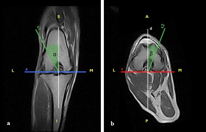
Fig. 1. The method of determining the values of the inclination angles of the inclined sagittal (α) and double-oblique (α, β) planes on the images in
the frontal (a) and transverse (b) planes. Auxiliary lines: a, b, c, d, s1, s2 (A – anterior; I – inferior; L – lateral; M – medial; P – posterior;
S – superior)
In the next stage of the study, the angle of The Mann–Whitney U test was used to test the statistical
inclination of the plane relative to the classical course of significance of differences. A P value of ≤ 0.05 meant
the imaging plane was determined. Angles were that a difference between values was statistically
measured by a person who had previously performed significant.
a multi-plane reconstruction. When determining the
angle value, images obtained in the frontal and
transverse planes were used. To determine the angle of Results
inclination of the oblique section planes, a reference line
(available in the software options and showing the The data were anonymised before the evaluation.
course of the layers in space) and auxiliary lines (a, b, c, The obtained series of images were evaluated randomly
d, s1, s2) were added to the images. Proper angles of and independently by three MRI technicians. The
inclination are marked as α and β. evaluation was carried out according to the prepared
In the frontal images, a line (a) parallel to the protocol twice with an interval of three weeks. To make
articular surface of the tibial plateau and tangential to the certain of objective interpretation, the evaluators did not
lateral and medial femoral condyles was determined. know the details of the study.
Then two lines were determined: a perpendicular (b) to First, the images were subjected to visual
line a and a parallel to the reference line (s1). The angle assessment, during which attention was paid to the
of inclination (α) was the angle between line b and image sharpness, blur, contrast between soft tissues,
line s1 (Fig. 1a). noise, and presence of artefacts or distortions. Image
In the case of double-skewed planes, the angle β quality was assessed based on a five-degree ordinal scale
was additionally determined based on images in the according to Likert (22): 1) low, 2) below average,
transverse plane. First, a line (c) was added tangentially 3) medium, 4) good, or 5) high.
to the caudal edges of the femoral condyles, then a line (d) Next, the visibility of the cranial cruciate ligament
perpendicular to line c was drawn. Finally, a line parallel was determined. The assessment scale was developed
to the reference line (s2) was drawn. The angle between based on the visual assessment score presented by
line d and line s2 was the slope of the oblique section Podadera et al. (36). It was as follows for the cranial
plane (Fig. 1b). cruciate ligament: 0 – CCL invisible; 1 – CCL partially
The statistical analysis was made using the visible in the images; 2 – CCL visible in full with
Statistica v.12.5 program (StatSoft, Tulsa, OK, USA). a blurred boundary; 3 – CCL visible in full with a clearlyA. Przeworski et al./J Vet Res/65 (2021) 209-216 213
defined boundary. The number of images in which the The study described in the article was carried out in
CCL was visible was also determined. a lateral position with the examined limb up. The joint
The data did not have a normal distribution and was placed in the coil in physiological flexion with some
therefore nonparametric tests were selected for analysis. deviations related to the size of the joints. Currently,
The statistical tests determined whether there was apart from the manufacturer’s instructions, there are no
a statistically significant difference between the results guidelines regarding the positioning of the dog for knee
of the assessments in individual sequences and to what imaging. In other articles, dogs were placed in the dorsal
extent the assessors were in agreement for individual (2, 5, 12, 23, 24, 32, 40, 43, 44) lateral (4, 11, 36, 37) or
features. The consistency of individual evaluators’ bridge position (15).
scores in the two assessments separated by the three- Both the position of the joint in the coil and the
week interval was also established. The Mann–Whitney angle of bending affect the spatial location of the
U test was used to test the statistical significance of cruciate ligament in relation to the magnetic field lines.
differences and the valid P value was as noted above. Spriet et al. (45) determined that imaging of the ligament
The consistency of assessments between the evaluators at an angle of 55° ± 10° to the magnetic field line promotes
was determined using the Kendall compliance factor the creation of a magic angle artefact, which increases
separately for each of the two assessments. Similarly, the the signal intensity of the examined structure. Most
level of accord of the individual evaluators’ assessments low-field systems have a vertical magnetic field
was determined for the two evaluations. The compliance orientation, which should be taken into account during
factor W assumes values between 0 (no agreement) and 1 resonance imaging. The current authors cannot exclude
(full agreement). the occurrence of this phenomenon in the study,
The number of images with a visible ligament especially when using the lateral arrangement.
obtained using MPR in the double-oblique sagittal plane Image quality assessment spanned a significant
was significantly higher than that in the oblique sagittal range (which was identical for all sequences). The
plane. In the second assessment, the same median lowest-rated images were defined as below average,
number of layers was obtained using MPR with the while the highest rated images were high. Only in the
oblique as with the double-oblique sagittal plane. case of images from 3D sequences was the quality was
However, the images in the double-oblique sagittal plane rated as good and high in over 50% of captures Images
were characterised by a higher rank sum (Fig 1). obtained using 3D sequences in the sagittal plane
Magnetic resonance imaging is a useful method of improved CCL visibility and a significantly higher
visualising structures within a dog’s knee joint. The number of them presented a visible CCL compared to
standard test protocol usually consists of 2D sequences. images using 2D sequences. Only two studies on
The use of an additional 3D sequence during the test imaging CCL damage in dogs have thoroughly analysed
creates the possibility of multi-plane reconstruction the diagnostic capabilities of 3D sequences (38, 41).
at any time after the test (Tables 1 and 2). This allows Both teams used high-field magnetic resonance imaging
new images to be reconstructed in any plane using with 3 T magnetic field induction. The 3D FSE (Fast
previously obtained data. It is thereby possible to Spin Echo) CUBE (GE Healthcare) sequence allowed
improve the previously defined imaging plane or to for the assessment of partial CCL tears and proper
present the structure in an additional plane. evaluation of the severity of knee synovitis, and the
VIPR-aTR (Vastly under-sampled Isotropic PRojection
with alternating length repetition times) 3D sequence
Discussion allowed better estimation of the structural properties
of CCL assessed on anatomical models (ex vivo test)
In the available literature, the authors used different (41). In human medicine, 2D and 3D sequences were
coils depending on their device and test assumptions. compared when imaging various knee structures. Most
Most often, these were rigid or flexible organ coils studies have shown the comparable diagnostic utility of
dedicated to limb imaging and included elbow (37), both sequences (1, 9, 16, 18). Ristow et al. (39) noticed
carpal (2), knee (5, 32, 40, 43), and spinal (4) varieties, that low-contrast structures were usually rated worse
which were also solenoidal (44) or dual-phased array than high-contrast structures.
coils (8, 15, 46). The selection for this study of a dual- The potential for improving the efficiency of ACL
phased array knee coil allowed for better image quality imaging using oblique planes has been carefully
due to the higher SNR. Due to the significant correlation evaluated in human medicine. Buckwalter and Pennes (7)
between the weight of dogs and the quality of images proposed using a template printed on transparent film to
from 3D sequences, it can be assumed that the determine the sagittal plane at an angle of 15° to the
differences in dog size and the closeness of fit of the coil classical plane. Nakanishi et al. (25) fixed the plane at
to the knee potentially affected the quality of the study. an angle of 10° but increased this angle to 20° if they did
Nemec et al. (26) experienced a positive effect of not originally achieve satisfactory ACL visualisation.
suitable matching of the coil diameter to the examined Barberie et al. (3) determined that planning an oblique
structure on the diagnostic quality of the obtained imaging plane using the locating sequence in the frontal
images. plane allows more accurate ACL imaging than using the214 A. Przeworski et al./J Vet Res/65 (2021) 209-216
locating sequence in the transverse plane. The intact (oblique sagittal plane) and angles α and β (double-oblique
ACL spatial arrangement and the oblique sagittal, frontal sagittal) significantly deteriorated CCL visibility. In
and transverse imaging planes were compared by another test determining the relationship between the
Breitenseher and Mayerhoefer (6), who achieved the values of these angles and weight, it was noticed that as the
best results using planes at an angle of 20° to the dog’s weight increased, these angles decreased
classical plane. Nenezić and Kocijancic (27) determined significantly (with reference to the double-oblique sagittal
that both imaging of a bent knee joint and the use of plane – 3D MPR DT). In interpretation of this, the authors
oblique sagittal plane imaging increased the sensitivity suspect that the person performing the reconstruction
of detection of partial tears of the ACL. No statistical tended to tilt the imaging plane more heavily for dogs with
difference was observed between the two techniques. a lower weight, the images of which were of lower quality.
Subsequent authors evaluated the possibilities of other In addition, the observed difference in the significance of
oblique imaging planes. Thinner layers (1 mm) and the the relationship between the dog’s weight and angle α for
use of an oblique face increased the efficiency of ACL the oblique sagittal plane (P > 0.05, median angle α 7.76°,
damage detection (14). The influence of additional sequences interquartile range 7.22°) and that between the dog’s weight
in the oblique planes on the overall effectiveness of ACL and angle α for the double-oblique plane (P < 0.05, median
imaging was also assessed. It was found that the use of angle α 6.53°, interquartile range3.51°) may have resulted
a combination of images obtained in the classical planes from the order in which oblique imaging planes were
and the oblique frontal plane during the evaluation gave performed. When determining the slope of double-skewed
higher specificity in detecting ACL damage for 3 T devices planes, the plane affecting angle β was first determined,
than 1.5 T devices (33). Ng et al. (29) obtained higher then that affecting angle α. The authors speculate that after
imaging sensitivity (71%) of intact ACL using additional determining the first plane and achieving an improvement
images in the oblique transverse plane. Also, the use of in CCL visibility (on the preview), the second plane was
additional images obtained in the oblique transverse tilted more carefully, i.e., by a smaller angle.
plane when this plane was in the protocol increased the The study supports the final conclusion that
sensitivity of ACL partial tear detection from 33% to imaging of the knee joint using 3D sequences makes
87% (27) and from 74% to 95% (17). Specificity did not possible the composition of higher quality images and
change in either study. better and more accurate visualisation of the cranial
Multi-plane reconstruction made from data cruciate ligament. The research results have determined
collected by volume in a 3D sequence caused the that multi-plane reconstruction affords an opportunity
obtained images to be of lower quality than the images for the secondary determination of the oblique imaging
from the output data. The images from reconstruction planes and assembles images of equal quality to those of
contained more distortion, artefacts, and sometimes the 2D sequence and allows better visibility of the
higher noise than the original images. cranial cruciate ligament than 2D or 3D sequences. The
In the presented study, after obtaining images in planes study determined that the use of 3D and MPR sequences
parallel to the CCL course, the authors determined the during magnetic resonance imaging visualises the
angles of inclination of the cross-section planes on the cranial cruciate ligament precisely and repeatably.
frontal image (angle α) and transverse image (angle β).
The measurement made on images from 3D sequences Conflict of Interests Statement: The authors declare
was to determine the inaccuracy of line routing during that there is no conflict of interests regarding the
geometric planning. The angle values α and β differed publication of this article.
from zero, and their medians were 0.8° and 1.36°,
respectively. The median angle α for the oblique sagittal Financial Disclosure Statement: The source of funding
plane was 7.76°, the median angle α was 6.53° for the of research and the article were: Statutory funds of the
double oblique sagittal plane, and the median angle β Department of Surgery and Radiology, Faculty of
was 9.22°. The values of the angles differed considerably. Veterinary Medicine, University of Warmia and Mazury
The values of angles α were similar to those used by in Olsztyn
Böttcher et al. (5) assuming 5° and 10°, respectively.
No information was found in the available literature on Animal Rights Statement: The examinations and all
the effect of a double-oblique cross-sectional plane on procedures on animals were conducted after the decision
CCL imaging in dogs, so it was impossible to compare of the owners.
the value of the angle β with the results of other studies.
The spatial determination of the imaging plane during
MPR planning was performed by one operator, who References
subjectively assessed the improvement of CCL visibility on
the basis of a real-time view of the obtained cross-section. 1. A Ai T., Zhang W., Priddy N.K., Li X.: Diagnostic performance
During the study, a negative correlation (P < 0.05) was of CUBE MRI sequences of the knee compared with conventional
MRI. Clin Radiol 2012, 67, e58–e63, doi: 10.1016/j.crad.
observed between the angles of the imaging plane and the 2012.07.020.
assessment of CCL visibility and the number of images in 2. Banfield C.M., Morrison W.B.: Magnetic resonance arthrography
which it can be seen. Increasing the value of angle α of the canine stifle joint technique and applications in elevenA. Przeworski et al./J Vet Res/65 (2021) 209-216 215
military dogs. Vet Radiol Ultrasound 2000, 41, 200–213, doi: 17. Kamal H.A., Abdelwahab N., El-Liethy N.E.: The role of oblique
10.1111/j.1740-8261.2000.tb01479.x. axial MR imaging in the diagnosis of ACL bundle lesions. Egypt
3. Barberie J.E., Carson B.W., Finnegan M., Wong A.D.: Oblique J Radiol Nucl Med 2015, 46, 683–693, doi: 10.1016/j.ejrnm.
sagittal view of the anterior cruciate ligament: comparison of 2015.05.007.
coronal vs. axial planes as localizing sequences. J Magn Reson 18. Kijowski R., Davis K.W., Woods M.A., Lindstrom M.J.,
Imaging 2001, 14, 203–206, doi: 10.1002/jmri.1174. De Smet A.A., Gold G.E., Busse R.F.: Knee joint: comprehensive
4. Blond L., Thrall D.E., Roe S.C., Chailleux N., Robertson I.D.: assessment with 3D isotropic resolution fast spin-echo MR
Diagnostic accuracy of magnetic resonance imaging for meniscal imaging - diagnostic performance compared with that of conventional
tears in dogs affected with naturally occuring cranial cruciate MR imaging at 3.0 T. Radiology 2009, 252, 486–495, doi:
ligament rupture. Vet Radiol Ultrasound 2008, 49, 425–431, doi: 10.1148/radiol.2523090028.
10.1111/j.1740-8261.2008.00401.x. 19. Kim S.I., Park H.J., Lee S.Y., Chung E.C., Kwon H.J., Cha J.G.,
5. Böttcher P., Armbrust L., Blond L., Brühschwein A., Gavin P.R., Kim S.: Usefulness of oblique coronal and sagittal MR images of
Gielen I., Hecht S., Jurina K., Kneissl S., Konar M., Pujol E., the knee after double-bundle and selective anterior cruciate
Robinson A., Schaefer S.L., Theyse L.F., Wigger A., Ludewig E.: ligament reconstructions. Acta Radiol 2015, 56, 312–321, doi:
Effects of observer on the diagnostic accuracy of low‐field MRI 10.1177/0284185114525952.
for detecting canine meniscal tears. Vet Radiol Ultrasound 2012, 20. Kwon J.W., Yoon Y.C., Kim Y.N., Ahn J.H., Choe B.K.: Which
53, 628–635, doi: 10.1111/j.1740-8261.2012.01967.x. oblique plane is more helpful in diagnosing an anterior cruciate
6. Breitenseher M.J., Mayerhoefer M.E.: Oblique MR imaging of the ligament tear? Clin Radiol 2009, 64, 291–297, doi: 10.1016/
anterior cruciate ligament based on three‐dimensional orientation. j.crad.2008.10.007.
J Magn Reson Imaging 2007, 26, 794–798, doi: 10.1002/ 21. Lee J.E., Park H.J., Lee S.Y., Ahn J.H., Park J.H., Park J.Y.:
jmri.20922. Evaluation of selective bundle injury to the anterior cruciate
7. Buckwalter K.A., Pennes D.R.: Anterior cruciate ligament: ligament: T2-weighted fast spin-echo 3-T MRI with reformatted
oblique sagittal MR imaging. Radiology 1990, 175, 276–277, doi: 3D oblique isotropic (VISTA) versus 2D technique. AJR Am J
10.1148/radiology.175.1.2315495. Roentgenol 2017, 209, W308–W316, doi: 10.2214/AJR.16.
8. D’Anjou M.A., Moreau M., Troncy E., Martel-Pelletier J., Abram F., 17659.
Raynauld J.P., Pelletier J.P.: Osteophytosis, subchondral bone 22. Likert R.: A technique for the measurement of attitudes. Arch
sclerosis, joint effusion and soft tissue thickening in canine Psychol 1932, 22, 55.
experimental stifle osteoarthritis: comparison between 1.5 T magnetic 23. Martig S., Konar M., Schmökel H.G., Rytz U., Spreng D.,
resonance imaging and computed radiography. Vet Surg 2008, 37, Scheidegger J., Höhl B., Kircher P.R., Boisclair J., Lang J.:
166–177, doi: 10.1111/j.1532-950X.2007.00363.x. Low‐field MRI and arthroscopy of meniscal lesions in ten dogs
9. Duc S.R., Pfirrmann C.W., Koch P.P., Zanetti M., Hodler J.: with experimentally induced cranial cruciate ligament
Internal knee derangement assessed with 3- minute three- insufficiency. Vet Radiol Ultrasound 2006, 47, 515–522, doi:
dimensional isovoxel true FISP MR sequence: preliminary study. 10.1111/j.1740-8261.2006.00179.x.
Radiology 2008, 246, 526–535, doi: 10.1148/radiol.2462062092. 24. Matsui A., Shimizu M., Beale B., Takahashi F., Yamaguchi S.:
10. Fazio C.G., Muir P., Schaefer S.L., Waller III K.R.: Accuracy of Assessment of T2 relaxation times for normal canine knee
3 Tesla magnetic resonance imaging using detection of fiber loss articular cartilage by T2 mapping using 1.5-T magnetic resonance
and a visual analog scale for diagnosing partial and complete imaging. Vet Comp Orthop Traumatol 2017, 30, 391–397, doi:
cranial cruciate ligament ruptures in dogs. Vet Radiol Ultrasound 10.3415/VCOT-17-01-0014.
2018, 59, 64–78, doi: 10.1111/vru.12567. 25. Nakanishi K., Horibe S., Shiozaki Y., Ishida T., Narumi Y., Ikezoe J.,
11. Feichtenschlager C., Gerwing M., Failing K., Peppler C., Kása A., Nakamura H.: MRI of normal anterior cruciate ligament (ACL)
Kramer M., von Pückler K.H.: Magnetic Resonance Imaging and reconstructed ACL: comparison of when the knee is extended
Assessment of Intra-Articular Structures in the Canine Stifle Joint with when the knee is flexed. Eur Radiol 1997, 7, 1020–1024, doi:
after Implantation of a Titanium Tibial Plateau Levelling 10.1007/s003300050244.
Osteotomy Plate. Vet Comp Orthop Traumatol 2018, 31, 26. Nemec S.F., Marlovits S., Trattnig S., Matzek W., Mayerhoefer M.E.,
261–272, doi: 10.1055/s-0038-1647248. Krestan C.R.: High-resolution magnetic resonance imaging and
12. Franklin S.P., Cook J.L., Cook C.R., Shaikh L.S., Clarke K.M., conventional magnetic resonance imaging on a standard field-strength
Holmes S.P.: Comparison of ultrasonography and magnetic magnetic resonance system compared to arthroscopy in patients
resonance imaging to arthroscopy for diagnosing medial meniscal with suspected meniscal tears. Acad Radiol 2008, 15, 928–933,
lesions in dogs with cranial cruciate ligament deficiency. J Am doi: 10.1016/j.acra.2008.02.007.
Vet Med Assoc 2017, 251, 71–79, doi: 10.2460/javma.251.1.71. 27. Nenezić D., Kocijancic I.: The value of the sagittal-oblique MRI
13. Galindo-Zamora V., Dziallas P., Ludwig D.C., Nolte I., Wefstaedt P.: technique for injuries of the anterior cruciate ligament in the knee.
Diagnostic accuracy of a short-duration 3 Tesla magnetic Radiol Oncol 2013, 47, 19–25, doi: 10.2478/raon-2013-0006.
resonance protocol for diagnosing stifle joint lesions in dogs with 28. Ng A.W.H., Griffith J.F., Hung E.H., Law K.Y., Yung P.S.: MRI
non-traumatic cranial cruciate ligament rupture. BMC Vet Res diagnosis of ACL bundle tears: value of oblique axial imaging.
2013, 9, 40, doi: 10.1186/1746-6148-9-40. Skeletal Radiol 2013, 42, 209–217, doi: 10.1007/s00256-012-
14. Gokalp G., Demirag B., Nas O.F., Aydemir M.F., Yazici Z.: 1372-y.
Contribution of thin slice (1 mm) oblique coronal proton density- 29. Ng A.W.H., Griffith J.F., Law K.Y., Ting J.W., Tipoe G.L.,
weighted MR images for assessment of anteromedial and Ahuja A.T., Chan K.M.: Oblique axial MR imaging of the normal
posterolateral bundle damage in anterior cruciate ligament anterior cruciate ligament bundles. Skeletal Radiol 2011, 40,
injuries. Eur J Radiol 2012, 81, 2358–2365, doi: 10.1016/ 1587–1594, doi: 10.1007/s00256-011-1208-1.
j.ejrad.2011.09.008. 30. Notohamiprodjo M., Horng A., Pietschmann M.F., Müller P.E.,
15. Harper T.A., Jones J.C., Saunders G.K., Daniel G.B., Leroith T., Horger W., Park J., Crispin A., Garcia del Olmo J.R., Weckbach S.,
Rossmeissl E.: Sensitivity of low‐field T2* images for detecting Herrmann K.A., Reiser M.F., Glaser C.: MRI of the knee at 3T:
the presence and severity of histopathologic meniscal lesions in first clinical results with an isotropic PDfs-weighted 3D-TSE-
dogs. Vet Radiol Ultrasound 2011, 52, 428–435, doi: sequence. Invest Radiol 2009, 44, 585–597, doi: 10.1097/
10.1111/j.1740-8261.2011.01818.x. RLI.0b013e3181b4c1a1.
16. Jung J.Y., Jee W.H., Park M.Y., Lee S.Y., Kim J.M.: Meniscal 31. Ohishi T., Takahashi M., Abe M., Tsuchikawa T., Mori M.,
tear configurations: categorization with 3D isotropic turbo spin-echo Nagano A.: The use of axial reconstructed images from three-
MRI compared with conventional MRI at 3 T. AJR Am J dimensional MRI datasets for morphological diagnosis of
Roentgenol 2012, 198, W173–W180, doi: 10.2214/AJR.11.6979. meniscal tears of the knee. Arch Orthop Trauma Surg 2005, 125,
622–627, doi: 10.1007/s00402-004-0792-0.216 A. Przeworski et al./J Vet Res/65 (2021) 209-216
32. Olive J., d’Anjou M.A., Cabassu J., Chailleux N., Blond L.: Fast 40. Ruoff C.M., Eichelberger B.M., Pool R.R., Griffin IV J.F.,
presurgical magnetic resonance imaging of meniscal tears and Cummings K.J., Pozzi A., Padua A., Saunders W.B.: The use of
concurrent subchondral bone marrow lesions: study of dogs with small field‐of‐view 3 Tesla magnetic resonance imaging for
naturally occurring cranial cruciate ligament rupture. Vet Comp identification of articular cartilage defects in the canine stifle:
Orthop Traumatol 2014, 27, 1–7, doi: 10.3415/VCOT-13-04- an ex vivo cadaveric study. Vet Radiol Ultrasound 2016, 57,
0054. 601–610, doi: 10.1111/vru.12420.
33. Park H.J., Kim S.S., Lee S.Y., Park N.H., Ahn J.H., Chung E.C., 41. Sample S.J., Racette M.A., Hans E.C., Volstad N.J., Holzman G.,
Park J.Y., Kim M.-S.: Comparison between arthroscopic findings Bleedorn J.A., Schaefer S.L., Waller III K.R., Hao Z., Block W.F.,
and 1.5-T and 3-T MRI of oblique coronal and sagittal planes of Muir P.: Radiographic and magnetic resonance imaging predicts
the knee for evaluation of selective bundle injury of the anterior severity of cruciate ligament fiber damage and synovitis in dogs
cruciate ligament. AJR Am J Roentgenol 2014, 203, W199– with cranial cruciate ligament rupture. PloS One, 2017, 12,
W206, doi: 10.2214/AJR.13.11571. e0178086, doi: 10.1371/journal.pone.0178086.
34. Park H.J., Lee S.Y., Park N.H., Ahn J.H., Chung E.C., Kim S.J., 42. Shigue D.A., Rahal S.C., Schimming B.C., Santos R.R., Vulcano L.C.,
Cha J.G.: Three-dimensional isotropic T2-weighted fast spin-echo Linardi J.L., Teixeira C.R.: Evaluation of the Marsh Deer Stifle
(VISTA) knee MRI at 3.0 T in the evaluation of the anterior Joint by Imaging Studies and Gross Anatomy. Anat Histol
cruciate ligament injury with additional views: comparison with Embryol 2015, 44, 468–474, doi: 10.1111/ahe.12162.
two-dimensional fast spin-echo T2-weighted sequences. Acta 43. Simpler R.E., Kerwin S.C., Eichelberger B.M., Wall C.R.,
Radiol 2016, 57, 1372–1379, doi: 10.1177/0284185114568048. Thompson J.A., Padua A., Purdy D., Griffin IV J.F.: Evaluation
35. Pass B., Robinson P., Hodgson R., Grainger A.J.: Can a single of the WARP‐turbo spin echo sequence for 3 Tesla magnetic
isotropic 3D fast spin echo sequence replace three-plane standard resonance imaging of stifle joints in dogs with stainless steel tibial
proton density fat-saturated knee MRI at 1.5 T? Br J Radiol 2015, plateau leveling osteotomy implants. Vet Radiol Ultrasound 2014,
88, 20150189, doi: 10.1259/bjr.20150189. 55, 414–419, doi: 10.1111/vru.12141.
36. Podadera J., Gavin P., Saveraid T., Hall E., Chau J., Makara M.: 44. Soler M., Murciano J., Latorre R., Belda E., Rodríguez M.J.,
Effects of stifle flexion angle and scan plane on visibility of the Agut A.: Ultrasonographic, computed tomographic and magnetic
normal canine cranial cruciate ligament using low‐field magnetic resonance imaging anatomy of the normal canine stifle joint. Vet
resonance imaging. Vet Radiol Ultrasound 2014, 55, 407–413, J 2007, 174, 351–361, doi: 10.1016/j.tvjl.2006.08.019.
doi: 10.1111/vru.12142. 45. Spriet M., Mai W., McKnight A.: Asymmetric signal intensity in
37. Pujol E., Van Bree H., Cauzinille L., Poncet C., Gielen I., Bouvy B.: normal collateral ligaments of the distal interphalangeal joint in
Anatomic study of the canine stifle using low‐field magnetic horses with a low‐field MRI system due to the magic angle effect.
resonance imaging (MRI) and MRI arthrography. Vet Surg 2011, Vet Radiol Ultrasound 2007, 48, 95–100, doi: 10.1111/j.1740-
40, 395–401, doi: 10.1111/j.1532-950X.2011.00823.x. 8261.2007.00211.x.
38. Racette M., Al Saleh H., Waller III K.R., Bleedorn J.A., McCabe R.P., 46. Winegardner K.R., Scrivani P.V., Krotscheck U., Todhunter R.J.:
Vanderby Jr R., Markel M.D., Brounts S.H., Block W.F., Muir P.: Magnetic resonance imaging of subarticular bone marrow lesions
3D FSE Cube and VIPR-aTR 3.0 Tesla magnetic resonance in dogs with stifle lameness. Vet Radiol Ultrasound 2007, 48,
imaging predicts canine cranial cruciate ligament structural 312–317, doi: 10.1111/j.1740-8261.2007.00248.x.
properties. Vet J 2016, 209, 150–155, doi: 10.1016/ 47. Yoon Y.C., Kim S.S., Chung H.W., Choe B.K., Ahn J.H.:
j.tvjl.2015.10.055. Diagnostic efficacy in knee MRI comparing conventional
39. Ristow O., Steinbach L., Sabo G., Krug R., Huber M., Rauscher I., technique and multiplanar reconstruction with one-millimeter
Ma B., Link T.M.: Isotropic 3D fast spin-echo imaging versus FSE PDW images. Acta Radiol 2007, 48, 869–874, doi:
standard 2D imaging at 3.0 T of the knee – image quality and 10.1080/02841850701459791.
diagnostic performance. Eur Radio 2009, 19, 1263–1272, doi:
10.1007/s00330-008-1260-y.You can also read