In vivo imaging of vulnerable plaque with intravascular modalities: its advantages and limitations
←
→
Page content transcription
If your browser does not render page correctly, please read the page content below
Review Article on Intracoronary Imaging

In vivo imaging of vulnerable plaque with intravascular modalities:
its advantages and limitations
Satoshi Kitahara1, Yu Kataoka1, Hiroki Sugane2, Fumiyuki Otsuka1, Yasuhide Asaumi1, Teruo Noguchi1,
Satoshi Yasuda1
1
    Department of Cardiovascular Medicine, National Cerebral & Cardiovascular Center, Suita, Osaka, Japan; 2Department of Cardiovascular
Medicine, Chikamori Hospital, Kochi, Japan
Contributions: (I) Conception and design: Y Kataoka; (II) Administrative support: S Yasuda; (III) Provision of study materials or patients: Y Kataoka, S
Kitahara; (IV) Collection and assembly of data: Y Kataoka, S Kitahara; (V) Data analysis and interpretation: Y Kataoka, S Kitahara; (VI) Manuscript
writing: All authors; (VII) Final approval of manuscript: All authors.
Correspondence to: Yu Kataoka, MD, PhD. Department of Cardiovascular Medicine, National Cerebral and Cardiovascular Center, 6-1, Shinmachi,
Kishibe, Suita, Osaka, 564-8565, Japan. Email: yu.kataoka@ncvc.go.jp.

                  Abstract: In vivo imaging of plaque instability has been considered to have a great potential to predict
                  future coronary events and evaluate the stabilization effect of novel anti-atherosclerotic medical therapies.
                  Currently, there are several intravascular imaging modalities which enable to visualize plaque components
                  associated with its vulnerability. These include virtual histology intravascular ultrasound (VH-IVUS),
                  integrated backscatter IVUS (IB-IVUS), optical coherence tomography (OCT), near-infrared spectroscopy
                  and coronary angioscopy. Recent studies have shown that these tools are applicable for risk stratification of
                  cardiovascular events as well as drug efficacy assessment. However, several limitation exists in each modality.
                  The current review paper will outline advantages and limitation of VH-IVUS, IB-IVUS, OCT, NIRS and
                  coronary angioscopy imaging.

                  Keywords: Vulnerable plaque; intravascular imaging; virtual-histology intravascular ultrasound (VH-IVUS);
                  optical coherence tomography (OCT); near-infrared spectroscopy (NIRS); integrated backscatter intravascular
                  ultrasound (IB-IVUS), coronary angioscopy

                  Submitted Feb 15, 2020. Accepted for publication Apr 20, 2020.
                  doi: 10.21037/cdt-20-238
                  View this article at: http://dx.doi.org/10.21037/cdt-20-238

Introduction                                                                      the occurrence of acute coronary syndrome (ACS) and
                                                                                  commence effective preventive therapies.
An insignificant but prone-to-rupture coronary lesion has
                                                                                     In 1990, grey-scale intravascular ultrasound (IVUS) has
been named as “vulnerable plaque” (1). To elucidate the
                                                                                  been developed and then used mainly for the guidance of
nature and mechanism of vulnerable plaque, numerous                               percutaneous coronary intervention (PCI) procedures (9).
pathohistological studies have been conducted, and findings                       One of advantages of grey-scale IVUS imaging is that it
from these studies have considerably contributed to the                           provides quantitative analysis of plaque volume in vivo. This
understanding of the pathophysiology of vulnerable plaque                         helps interventionalists to select the optimal size of devices.
(2-8). Meticulous analyses of coronary specimen have                              In addition, grey-scale IVUS has become a tool to evaluate
characterized vulnerable plaque which include a large lipid                       drug efficacy due to its quantitative and reproducible
core, thin fibrous cap, expansive vessel remodeling and                           features. With regard to the ability of grey-scale IVUS
macrophage infiltration (2-8). These collective evidences                         for visualization of vulnerable plaques, pathohistological
emerge the notion that identification of vulnerable                               and clinical studies reported that attenuation of ultrasonic
plaque with imaging modality would enable to predict                              signals corresponds to the presence of necrotic core.

© Cardiovascular Diagnosis and Therapy. All rights reserved.             Cardiovasc Diagn Ther 2020;10(5):1461-1479 | http://dx.doi.org/10.21037/cdt-20-238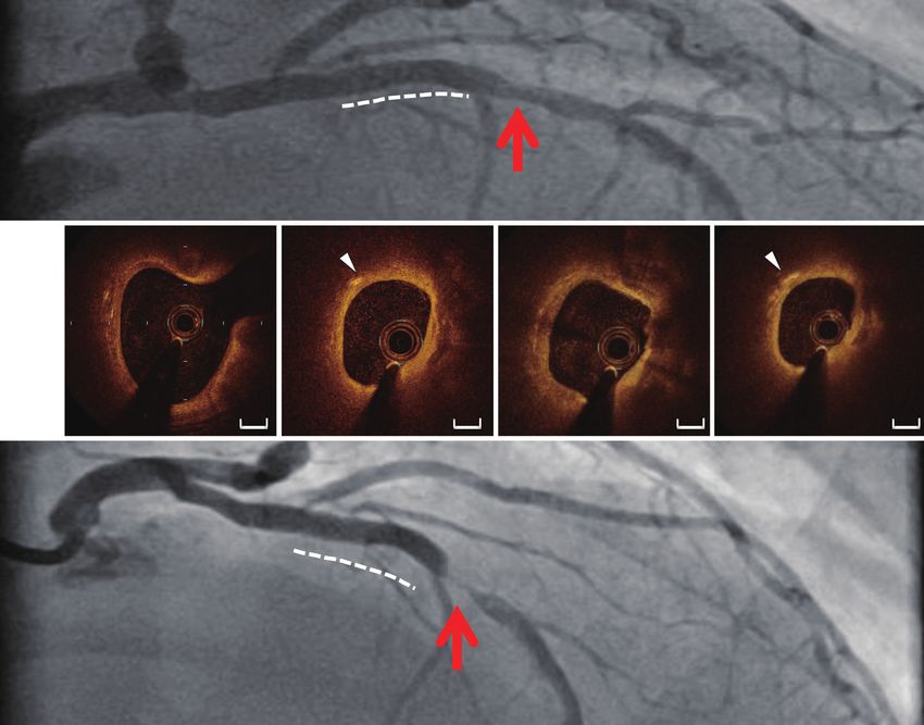
1462                                                                                           Kitahara et al. Imaging of plaque instability

However, recent study showed that the positive predictive               cardiovascular events (Tables 1,2). The PROSPECT (The
value of IVUS for detecting thin-cap fibroatheroma (TCFA)               Providing Regional Observations to Study Predictors
was only 19% (10). Due to this limitation of IVUS, another              of Events in the Coronary Tree) trial is the largest
intravascular imaging devices which visualizes plaque quality           observational study which investigated the predictive ability
has been developed. In this review, we will summarize                   of TCFA on VH-IVUS for cardiovascular events (14). This
advantages and limitation to image vulnerable plaques with              study enrolled a total of 697 ACS patients. All three major
currently available modalities: virtual-histology IVUS (VH-             coronary arteries were imaged by VH-IVUS. VH-IVUS-
IVUS), integrated backscatter IVUS (IB-IVUS), optical                   derived TCFA was defined as more than 30 degrees of the
coherence tomography (OCT), near-infrared spectroscopy                  necrotic core abutted the lumen in 3 or more consecutive
(NIRS) and coronary angioscopy.                                         frames. In this study, 596 VH-IVUS derived TCFA were
                                                                        identified in 313 patients. During a median follow-up
                                                                        period of 3.4 years, patient-level analysis demonstrated
VH-IVUS
                                                                        VH-IVUS derived TCFA as an independent predictor
Tissue characterization with VH-IVUS                                    of subsequent non-culprit lesion-related major adverse
                                                                        cardiovascular events (MACEs) [hazard ratio (HR) =3.35,
VH-IVUS utilizes radiofrequency analysis of reflected                   95% confidence interval (CI): 1.77–6.36, P70% (HR
necrotic core and dense calcium. A reconstructed color-                 =5.03, 95% CI: 2.51–10.11, P10%
   Brown et al. investigated the ability of VH-IVUS to                  plaque cross-sectional area, in contact with vessel lumen for
detect TCFA by comparing with histology (13). This study                3 consecutive frames. In this study with a median follow-up
showed that the diagnostic accuracy and sensitivity of                  of 625 days, both patient-based and lesion-based analyses
TCFA on VH-IVUS was 76.5 and 83.6%, respectively. In                    elucidated that VH-IVUS-derived TCFA was the only
this analysis, VH-IVUS classified 7 of 8 TCFAs as thick-cap             plaque phenotype associated with the occurrence of MACE
fibroatheroma. This suggests that VH-IVUS is capable of                 (patient-based analysis: HR =7.53, 95% CI: 1.12–50.55,
identifying a large necrotic core, but is limited to correctly          P=0.038, lesion-based analysis: HR =4.43, 95% CI: 1.50–
evaluate thin fibrous cap.                                              13.18, P=0.007).
                                                                           T h e AT H E R O R E M O - I V U S ( t h e E u r o p e a n
                                                                        Collaborative Project on Inflammation and Vascular Wall
The ability of VH-IVUS for future cardiovascular events
                                                                        Remodeling in Atherosclerosis-Intravascular Ultrasound)
Given that plaque containing greater quantities of both                 study is a single-center observational study of 581 ACS
necrotic and lipidic material confer an increased risk                  or stable CAD subjects (16). VH-IVUS was used to
of cardiovascular events, there has been considerable                   interrogate non-culprit vessel. The primary outcome was
interests whether VH-IVUS could predict future risks of                 the occurrence of MACE which included non-culprit lesion

© Cardiovascular Diagnosis and Therapy. All rights reserved.   Cardiovasc Diagn Ther 2020;10(5):1461-1479 | http://dx.doi.org/10.21037/cdt-20-238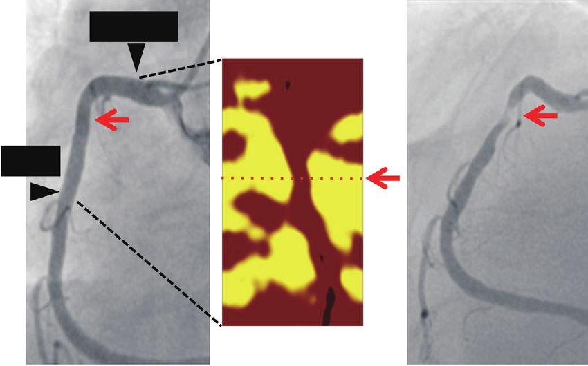
Cardiovascular Diagnosis and Therapy, Vol 10, No 5 October 2020                                                                                 1463

 Table 1 Clinical studies with VH-IVUS imaging—prediction of future cardiovascular events
 Authors Sites               Population         VH-IVUS-derived measure           Findings

 Stone     37 sites in the 697 patients with TCFA defined by VH-IVUS: more During a 3.4-year follow-up period, VH-IVUS derived TCFA
 et al.    United States ACS                 than 30 degrees of the necrotic predicted subsequent non-culprit lesion-related major adverse
 (14)      and Europe                        core abutted the lumen in 3 or cardiovascular events (HR =3.35, 95% CI: 1.77–6.36, P70%, minimum lumen area 10%; plaque           adverse cardiovascular events (patient-based analysis: HR
                                               cross-sectional area, in           =7.53, 95% CI: 1.12–50.55, P=0.038, lesion-based analysis: HR
                                               contact with vessel lumen for 3    =4.43, 95% CI: 1.50–13.18, P=0.007)
                                               consecutive frames

 Cheng One site in the 581 CAD                  TCFA defined by VH-IVUS:          TCFA and plaque burden >70% were associated with an
 et al. Netherlands    subjects (ACS:           plaque burden >40%, confluent     increased risk of MACE (TCFA: HR =1.98, 95% CI: 1.09–3.60,
 (16)                  n=318, stable            necrotic core >10%; plaque        P=0.02, plaque burden >70%: HR =2.90, 95% CI: 1.60–5.25,
                       CAD: n=263)              cross-sectional area, in          P70% predicted
                                                contact with vessel lumen for 3   the occurrence of MACE within (P=0.011) and after 6 months
                                                consecutive frames                (P70% (HR =2.90, 95% CI: 1.60–5.25,
                                                                             fluvastatin and those without it for 12 months (17). Under
P70% was associated with an elevated risk of MACE within                     98.1±12.7 mg/dL. Additionally, the use of fluvastatin was
(P=0.011) and after 6 months (P
1464 Kitahara et al. Imaging of plaque instability Table 2 Clinical studies with VH-IVUS imaging—evaluation of drug efficacy Authors Subjects Therapy Outcomes Findings Nasu 80 subjects Fluvastatin vs. Percent change in Patients treated with fluvastatin were more likely to exhibit an increase in et al. (17) with stable no statin use atheroma volume fibrous tissue volume (P=0.03) and a decrease of fibro-fatty (P
Cardiovascular Diagnosis and Therapy, Vol 10, No 5 October 2020                                                                            1465

between evolocumab and control groups. The lack of any                  Consideration of limitation of IB-IVUS imaging
demonstrable differences between the treatment groups in
                                                                        Evidence is limited to show the association of IB-IVUS
VH-IVUS analysis of the GALOGOV trial suggests the
                                                                        derived plaque features with cardiovascular events.
limitation of VH-IVUS for drug efficacy assessment trial.
   The acquisition of VH-IVUS images is gated at the
R wave of the electrocardiography signal, which fails to                OCT imaging
allow for VH-IVUS imaging to be performed upon the
                                                                        Imaging of plaque microstructures
frames acquired within each R–R interval. Variability in
a patient’s heart rate at different time points results in a            OCT uses near-infrared light which enables to provide
degree of horizontal bias during serial VH-IVUS imaging.                imaging of atherosclerotic plaques in coronary artery
These cause different numbers of frames at baseline and                 (36,37). High resolution imaging is one of the advantages
follow-up imaging, which is not adequate for analyzing the              of OCT imaging. Its resolution is up to 10 μm in an axial
exactly same segment in the clinical trials of drug efficacy            resolution and to 20 μm in a lateral resolution, which
evaluation.                                                             is approximately 10 times higher compared to that of
                                                                        IVUS. This distinct feature of OCT enables to generate
                                                                        high quality imaging of plaque microstructures such as
IB-IVUS                                                                 thin fibrous cap, microchannel, accumulation of lipid and
Tissue characterization and validation of IB-IVUS                       macrophages (36,37).
imaging                                                                    There are numerous studies which elucidated features of
                                                                        plaque microstructures in patients with CAD. In 30 subjects
IB-IVUS is another imaging modality to evaluate                         with acute myocardial infraction, thin fibrous cap, plaque
compositional features of coronary atheroma in vivo. This               rupture, thrombus and TCFA were observed, in line with
imaging technique utilizes time-domain information                      findings from pathohistological studies (38). In another
through the acquired radiofrequency signals (25). Ex vivo               study, the presence of vaso vasorum at culprit lesions was
validation study showed that IB signals could differentiate             associated with thinner fibrous cap, a higher frequency
different types of plaque tissues including fibrous,                    of TCFA and a higher c-reactive protein level (39). OCT
calcification and lipid pool. The accuracy of IB-IVUS to                has been shown to identify differences in plaque features
detect fibrous, lipid-rich and fibrocalcific plaques was 93%,           of subjects with ST-elevation myocardial infarction, non-
90% and 96%, respectively (25,26). Another study reported               ST-elevation ACS and stable angina pectoris (37). TCFA
that the positive predictive value of IB-IVUS for TCFA was              was defined as a plaque with lipid arc >90 degrees and its
50.0% (27).                                                             fibrous cap thickness
1466 Kitahara et al. Imaging of plaque instability
Table 3 Clinical studies with IB-IVUS imaging—evaluation of drug efficacy
Authors Subjects Therapy Outcomes Findings
Kawasaki 52 subjects Pravastatin vs. Percent change Compared to diet therapy (fibrous volume: +4%, P=ns vs. baseline,
et al. (28) with CAD atorvastatin vs. in IB-IVUS lipid volume: +5%, P=ns vs. baseline), a significant increase in fibrous
diet therapy derived plaque volume and a reduction of lipid volume were identified under the
composition use of pravastatin (fibrous volume: +11%, PCardiovascular Diagnosis and Therapy, Vol 10, No 5 October 2020 1467
Table 4 Clinical studies with OCT imaging—prediction of future cardiovascular events
Authors Sites Population OCT-derived measure Findings
Xing 20 sites 1,471 patients lipid plaque with its arc Subjects with lipid-rich plaque exhibited an increased risk of non-
et al. across 6 with CAD of >1 quadrant culprit lesion related major adverse cardiac events (7.2% vs. 2.6%,
(42) countries (ACS =584, P=0.033). Multivariate analysis demonstrated the presence of lipid-rich
stable CAD =887) plaque as an independent predictor of non-culprit lesion related major
adverse cardiac events included lipid-rich plaque (risk ratio =2.061,
95% CI: 1.050–4.044, P=0.036). Longer lipid lengths (9.9±3.6 vs.
7.9±4.6, P180°, fibrous Lipid arc >180° (HR =2.4, 95% CI: 1.2–4.8, P=0.017), fibrous cap
et al. with suspected cap thickness1468 Kitahara et al. Imaging of plaque instability in 33.6% of study subjects. During the follow-up period in 275 patients with CAD receiving PCI (44). Compared (median =2 years), the presence of lipid-rich plaque was to subjects who did not receive a statin, statin use with a associated with an increased risk of non-culprit lesion greater intensity was associated with thicker fibrous cap related MACE (7.2% vs. 2.6%, P=0.033). On multivariate (P=0.01) and a lower frequency of TCFA (P1 year (risk ratio =4.517, 95% CI: 1.923–10.610, thickness of fibrous cap. Moreover, in patients with on- P=0.001). Further OCT analysis elucidated that lipid-rich treatment LDL-C
Cardiovascular Diagnosis and Therapy, Vol 10, No 5 October 2020                                                                             1469

          A
                                                                          (1)
                                                                                (2) (3) (4)

                   (1)                             (2)                      (3)                           (4)
          B                             Septal
                                        Branch                                      L
                                                                                                                L
                    L

                                                         L                                                          L

                                            1 mm                   1 mm                            1 mm                           1 mm

          C

Figure 1 The recurrence of ACS due to progression of lipid-rich plaque containing cholesterol crystal (28). (A) A 60-year-old man with
ST-segment elevation myocardial infarction received DES implantation at the middle segment of LAD (dotted line). There was a mild
residual stenosis in LAD (red arrow). [1–4] correspond to OCT images in (B). (B) OCT imaging visualized the presence of lipid-rich plaque
(L) harbouring cholesterol crystal (white triangle). (C) One year later, non-ST-segment elevation myocardial infarction occurred due to
the progression of lipid-rich lesions with cholesterol crystal despite statin therapy (red arrow). ACS, acute coronary syndrome; DES, drug-
eluting stent; LAD, left anterior descending artery; OCT, optical coherence tomography.

algorithms in physicians, reproducibility of measurements                 based diagnostic approach of plaque erosion is attractive.
was improved, reflected by intraclass correlation coefficients            However, correct diagnosis of plaque erosion in vivo by
at 0.82 and 0.88, respectively. This study indicates that                 using OCT is not easy in the clinical settings. Given that
mutual discussion and consensus are mandatory for                         the presence of luminal thrombus hampers the penetration
accurate and reproducible OCT analysis. In the future,                    of light into the underlying plaque, it is difficult to
novel technologies such as machine learning and/or                        assess features of plaques behind attached thrombus. We
artificial intelligence could be a solution to improve OCT                experienced one ACS case diagnosed as plaque erosion
measurement.                                                              according to OCT-based criteria (53) (Figure 2). However,
   Jia et al. established the criteria of OCT-defined plaque              considering clinical characteristics including the presence of
erosion (52). OCT-defined plaque erosion is defined                       atrial fibrillation enables to diagnosis as coronary embolism
and categorized according to the absence of fibrous cap                   but not plaque erosion. As such, meticulous evaluation of
disruption and the presence of thrombus. This OCT-                        plaques suspected with its erosion is required.

© Cardiovascular Diagnosis and Therapy. All rights reserved.    Cardiovasc Diagn Ther 2020;10(5):1461-1479 | http://dx.doi.org/10.21037/cdt-20-238
1470 Kitahara et al. Imaging of plaque instability
A B 1 2 3 4
1 2
3 4
C 60
0 20 30 40 50
4 3 2 1
LAD LAD distal thrombus LAD proximal
D E 1’ 2’ 3’ 4’
L
1’ 2’ 3’
4’
L L
F 10 20 30 40 mm
4’ 3’ 2’ 1’
LAD distal LAD proximal
LAD
L = lipid, white arrows = macrophage
Figure 2 Limitation of OCT imaging for correct diagnosis of plaque erosion (35). (A) A 93-year-old woman was hospitalized due to anterior
STEMI. Coronary angiography identified TIMI grade II flow with severe stenosis within LAD. [1-4] correspond to OCT images in (B
and C). (B,C) The surface of culprit lesion was invisible due to its overlying thrombus (*) [2-4]. (D) After retrieving red thrombus with
thrombectomy catheter, TIMI grade III flow was achieved without any residual stenosis. 1’-4’ correspond to OCT images in (E and F). (E)
On OCT imaging after thrombectomy, an intact fibrous cap and signal attenuation were observed [1’-4’]. This lesion was accompanied by
adjacent focal signal-rich region (3’: white arrows), suggesting the small lipidic plaque with superficial macrophages. IVUS, intravascular
ultrasound; LAD, left anterior descending artery; OCT, optical coherence tomography; STEMI, ST-segment elevation myocardial
infarction; TIMI, thrombolysis in myocardial infarction.
NIRS imaging lipid core plaque is shown as the following four different
colors; red (probabilityCardiovascular Diagnosis and Therapy, Vol 10, No 5 October 2020 1471
Validation of chemogram on NIRS imaging The ability of NIRS imaging for future cardiovascular
events
The ability of NIRS imaging to reveal plaque compositions
associated with its instability has been analyzed by using 199 Several single-center studies have been conducted to
samples of 5 human aortic specimens (56). In this analysis, investigate the prognostic value of LCBI from 2014 to 2017
35 of 39 lesions with lipid pools and 56 of 60 lesions without (60-63) (Tables 6,7). Oemrawsingh et al. analyzed 203 CAD
lipid pools were identified by NIRS imaging (sensitivity patients in which their non-culprit vessel was imaged by
=90%, specificity =93%). This modality detected other NIRS (Tables 4,5) (67). Subjects with LCBI above median
unstable plaque features including thin fibrous cap and the value of LCBI (=43.0) was associated with an increased
presence of inflammatory cells (thin fibrous caps: sensitivity risk of MACE (= all-cause mortality, nonfatal ACS, stroke
=77%, specificity =93%, inflammatory cells: sensitivity and unplanned coronary revascularization) compared
=84%, specificity =91%) (56). Gardner et al. performed to those with LCBI below its median value (16.7% vs.
ex vivo validation study using human coronary specimens (57). 4.0%, log-rank, P=0.003). Even after adjusting baseline
This study analyzed 212 coronary segments of 84 autopsy clinical characteristics (age, sex, hypercholesterolemia,
hearts by NIRS imaging and histopathological evaluation of diabetes, hypertension, history of myocardial infarction,
sections taken at 2-mm intervals. The algorithm established peripheral artery disease and a history of PCI), there was
from the first 33 hearts was effective to identify lipid core a 4.04 (95% CI: 1.33–12.29, P=0.01) greater likelihood of
plaques with a receiver operating characteristics curve area experiencing MACE during one-year observational period.
of 0.80 (95% CI: 0.76–0.85). Additionally, a higher LCBI Another analysis from the Spectrum NIRS-IVUS Registry
corresponded to the presence of any fibroatheroma with an included 121 CAD patients monitored by NIRS imaging
area under the curve of 0.86 (95% CI: 0.81–0.91). Another (Tables 4,5) (60). Cox regression analysis showed
validation study of the NIRS algorithm is the SPECTACL maxLCBI 4 mm ≥400 at non-culprit segment as one-year
(SPECTroscopic Assessment of Coronary Lipid) study with occurrence of MACE (HR =10.2, 95% CI: 3.4–30.6,
106 patients with CAD (58). The investigators compared P1472 Kitahara et al. Imaging of plaque instability
Table 6 Clinical studies with NIRS imaging—prediction of future cardiovascular events
Authors Cites Population NIRS-derived measure Findings
Oemrawsingh Single 203 patients LCBI at non-culprit LCBI >43 (median) predicted the occurrence of one-year MACE
et al. (60) center with CAD (ACS vessel (at least 40 mm (= all-cause death, non-fatal ACS, stroke, unplanned coronary
=95, stable CAD in length, % diameter revascularization): HR =4.04, 95% CI: 1.33–12.29, P=0.01
=108) stenosis 400 was associated with MACCE (all-cause
(61) center with suspected stented segment mortality, non-fatal ACS, acute cerebrovascular events) during
CAD (ACS =103, follow-up: HR =10.2, 95% CI: 3.4–30.6, P400 was associated with
et al. (64) from 6 with CAD (ACS stented segment from at non-culprit lesion related MACE (= cardiac death, cardiac arrest,
countries =974, stable least two major coronary non-fatal MI, ACS, revascularization): adjusted HR =1.89, 95%
CAD =589) arteries (>50 mm in CI: 1.26–2.83, P=0.021. Segment-based analysis: Max4 mmLCBI
length) >400 was associated with non-culprit lesion related MACE:
adjusted HR =3.39, 95% CI: 1.85–6.20, P70% and −0.6%, P=0.02)
CAD fractional flow reserve 70% and reduced at lesions with LCBImax4 mm ≥500 under 20 mg rosuvastatin
CAD fractional flow reserveCardiovascular Diagnosis and Therapy, Vol 10, No 5 October 2020 1473
A B C
Proximal
Proximal
Distal
Distal
Figure 3 Progression of non-obstructive lesion harbouring high Max4 mmLCBI. (A) 70-year old gentleman received diagnostic coronary
angiography due to suspected anginal chest symptom. There was a mild stenosis at the proximal segment of RCA (red arrow). (B) NIRS
imaging identified high Max4 mmLCBI (=882) at the corresponding lesion (red arrow). (C) One year later, ACS occurred due to the
progression of the lesion with high Max4 mmLCBI (red arrow). ACS, acute coronary syndrome; LCBI, lipid core burden index; NIRS,
near-infrared spectroscopy; RCA, right coronary artery.
greater reduction of LDL-C level was observed in subjects Consideration of limitation of NIRS imaging
receiving intensive statin therapy (20 mg rosuvastatin)
Near-infrared light reaches around 3 mm far from the
(58.4±26.3 vs. 81.9±27.9 mg/dL, P1474 Kitahara et al. Imaging of plaque instability
Table 8 Clinical studies with coronary angioscopy imaging—prediction of future cardiovascular events
Authors Cites Population NIRS-derived measure Findings
Ohtani, Single 552 patients Number of yellow During the observational period (57.3±22.1 months), patients who
et al. center with suspected plaques within the experienced the occurrence of ACS were more likely to have a greater
(68) or diagnosed culprit vessel number of yellow plaques at baseline (3.1±1.8 vs. 2.2±1.5, P=0.008).
CAD Multivariate logistic regression analysis revealed number of yellow plaques
as an independent risk factors of ACS events (adjusted hazard ratio =1.23,
95% CI: 1.03–1.45, P=0.02)
ACS, acute coronary syndrome; CAD, coronary artery disease; CI, confidence interval.
Table 9 Clinical studies with coronary angioscopy imaging—evaluation of drug efficacy
Authors Population Drug Outcome Findings
Takano, 31 patients Atorvastatin vs. diet Change in the In the atorvastatin group, a reduction of the mean yellow score was
et al. (69) with CAD therapy mean yellow observed (from 2.03±0.45 to 1.13±0.33, PCardiovascular Diagnosis and Therapy, Vol 10, No 5 October 2020 1475
and therefore, this modality can not visualize all of plaques Quantitative analysis of plaque composition
within vessel wall. Yellow colour assessment is a subjective
VH-IVUS and IB-IVUS measure the volume of each
measure. This limitation suggests that yellow colour grade
plaque component. NIRS enables to provide quantitative
may be different between each physician.
measurement of lipidic plaque materials. By contrast, the
assessment of plaque component, especially lipid plaque on
Comparison of each imaging modality OCT and plaque colour grade on coronary angioscopy are
subjective.
As mentioned above, VH-IVUS, IB-UVUS, OCT, NIRS
and coronary angioscopy are similar to visualize vulnerable
plaque. In addition, it is important to understand the Future directions
following differences in each modality for better selection
While the currently available intravascular imaging
and interpretation of images.
modalities provide a variety of anatomical information of
coronary atheroma in vivo, recent studies indicate not only
Imaging procedure anatomical data but also other plaque-related characteristics
OCT and coronary angioscopy requires the continuous as another important contributor to plaque vulnerability.
infusion of contrast medium or low-molecular-weight For instance, Costopoulos et al. reported the association
dextran for its imaging, whereas others do not. This of plaque structural stress and wall shear stress with plaque
technical aspect could affect the quality of acquired images. vulnerability and progression (74). In this analysis, high
plaque structural stress and low wall shear stress were
associated with an increased vulnerability and plaque
Measurement of vessel dimensions progression, respectively.
Values of measurement about vessel and lumen diameter Another potential feature of plaques is its biological
is different between VH-IVUS, IB-IVUS and OCT, functionality (75). Intravascular near-infrared fluorescence
whereas NIRS and coronary angioscopy does not provide is a novel imaging approach which uses protease-activated
any information of vessel and lumen sizes. In detail, fluorescence agent. Pre-clinical study has shown this
although the measurements of vessel dimensions on OCT imaging technique visualized inflammation within vessel
is significantly correlated to those on VH-IVUS and IB- wall of rabbit atherosclerosis model (76). Although
IVUS, their small differences exist (11–22%). In particular, further clinical studies are warranted to elucidate the
the interventionalist has to recognize that lumen area feasibility and safety of intravascular imaging of plaque
measured by OCT is normally smaller than IVUS-derived inflammation, visualization of plaque activity will be also
one (72). important to predict future cardiovascular risks. In the
future, by collecting anatomical as well as biomechanical
and functional data of coronary plaques, it may be possible
Visualization of plaque compositions
to establish novel therapeutic approach to stabilize
VH-IVUS, IB-IVUS, OCT and coronary angioscopy shows vulnerable plaque, thereby leading to the prevention of
a variety of plaque components/structures, whereas NIRS ACS.
evaluates only the degree of lipidic plaque materials.
Conclusions
Evaluation of TCFA
The accumulating evidence from clinical studies have
One study showed the discrepancy of TCFA between VH- shown intravascular imaging of vulnerable plaques
IVUS and OCT (73). In this analysis, there were 61 VH- as a great potential tool for predicting future risk of
IVUS derived and 36 OCT-derived TCFAs. Of these, only cardiovascular events and evaluating the efficacy of novel
28 lesions fulfilled both VH-IVUS and OCT based TCFA agents. However, it is important to recognize that not only
definition. The remaining 33 VH-IVUS TCFA did not anatomical plaque features but also biomechanical factors
exhibit thin-cap on OCT, and 8 OCT TCFA exhibited less as well as functionality of plaques could play important
than 10% of necrotic core areas in contact with the lumen. roles in driving plaque instability. In addition, a variety of
© Cardiovascular Diagnosis and Therapy. All rights reserved. Cardiovasc Diagn Ther 2020;10(5):1461-1479 | http://dx.doi.org/10.21037/cdt-20-2381476 Kitahara et al. Imaging of plaque instability
biomarkers reflecting pathophysiology of atherosclerosis References
may help to assess the degree of vulnerability at coronary
1. Muller JE, Tofler GH, Stone PH. Circadian variation
lesions. Given that technological advances such as
and triggers of onset of acute cardiovascular disease.
machine learning and/or artificial intelligence are rapidly
Circulation 1989;79:733-43.
progressing, these technologies are expected to create
2. Virmani R, Kolodgie FD, Burke AP, et al. Lessons from
sophisticated tool which collects anatomical, biomechanical
sudden coronary death: a comprehensive morphological
and functional imaging data of coronary atheroma in
classification scheme for atherosclerotic lesions.
conjunction with biomarkers. This novel but ideal approach
Arterioscler Thromb Vasc Biol 2000;20:1262-75.
will be useful in refining the degree of plaque vulnerability,
3. Davies MJ, Thomas A. Thrombosis and acute coronary-
which enables to adopt individualizing therapy for further
artery lesions in sudden cardiac ischemic death. N Engl J
reduction of future coronary event’s risk.
Med 1984;310:1137-140.
4. DeWood MA, Spores J, Notske R, et al. Prevalence of total
Acknowledgments coronary occlusion during the early hours of transmural
myocardial infarction. N Engl J Med 1980;303:897-902.
Funding: None.
5. Farb A, Burke AP, Tang AL, et al. Coronary plaque
erosion without rupture into a lipid core: a frequent
Footnote cause of coronary thrombosis in sudden coronary death.
Provenance and Peer Review: This article was commissioned Circulation 1996;93:1354-63.
by the Guest Editor (Dennis T. L. Wong) for the series 6. Burke AP, Farb A, Malcom GT, et al. Coronary risk factors
“Intracoronary Imaging” published in Cardiovascular and plaque morphology in men with coronary disease who
Diagnosis and Therapy. The article was sent for external peer died suddenly. N Engl J Med 1997;336:1276-82.
review organized by the Guest Editor and the editorial 7. Glagov S, Weisenberg E, Zarins CK, et al. Compensatory
office. enlargement of human atherosclerotic coronary arteries. N
Engl J Med 1987;316:1371-5.
Conflicts of Interest: All authors have completed the 8. Burke AP, Kolodgie FD, Farb A, et al. Healed plaque
ICMJE uniform disclosure form (available at http://dx.doi. ruptures and sudden coronary death: evidence that
org/10.21037/cdt-20-238). The series “Intracoronary subclinical rupture has a role in plaque progression.
Imaging” was commissioned by the editorial office without Circulation 2001;103:934-40.
any funding or sponsorship. YK serves as an unpaid editorial 9. Mintz GS, Nissen SE, Anderson WD, et al. American
board member of Cardiovascular Diagnosis and Therapy from College of Cardiology Clinical Expert Consensus
Jul 2019 to Jun 2021. The authors have no other conflicts Document on Standards for Acquisition, Measurement
of interest to declare. and Reporting of Intravascular Ultrasound Studies (IVUS).
A report of the American College of Cardiology Task
Ethical Statement: The authors are accountable for all Force on Clinical Expert Consensus Documents. J Am
aspects of the work in ensuring that questions related Coll Cardiol 2001;37:1478-92.
to the accuracy or integrity of any part of the work are 10. Fujii K, Hao H, Shibuya M, et al. Accuracy of OCT,
appropriately investigated and resolved. Grayscale IVUS, and Their Combination for the
Diagnosis of Coronary TCFA: An Ex Vivo Validation
Open Access Statement: This is an Open Access article Study. JACC Cardiovasc Imaging 2015;8:451-60.
distributed in accordance with the Creative Commons 11. Nair A, Kuban BD, Tuzcu EM, et al. Coronary plaque
Attribution-NonCommercial-NoDerivs 4.0 International classification with intravascular ultrasound radiofrequency
License (CC BY-NC-ND 4.0), which permits the non- data analysis. Circulation 2002;106:2200-6.
commercial replication and distribution of the article with 12. Nasu K, Tsuchikane E, Katoh O, et al. Accuracy of in
the strict proviso that no changes or edits are made and the vivo coronary plaque morphology assessment: a validation
original work is properly cited (including links to both the study of in vivo virtual histology compared with in vitro
formal publication through the relevant DOI and the license). histopathology. J Am Coll Cardiol 2006;47:2405-12.
See: https://creativecommons.org/licenses/by-nc-nd/4.0/. 13. Brown AJ, Obaid DR, Costopoulos C, et al. Direct
© Cardiovascular Diagnosis and Therapy. All rights reserved. Cardiovasc Diagn Ther 2020;10(5):1461-1479 | http://dx.doi.org/10.21037/cdt-20-238Cardiovascular Diagnosis and Therapy, Vol 10, No 5 October 2020 1477
Comparison of Virtual-Histology Intravascular Cardiol 2018;72:2012-21.
Ultrasound and Optical Coherence Tomography Imaging 25. Kawasaki M, Takatsu H, Noda T,et al. In vivo quantitative
for Identification of Thin-Cap Fibroatheroma. Circ tissue characterization of human coronary arterial plaques
Cardiovasc Imaging 2015;8:e003487. by use of integrated backscatter intravascular ultrasound
14. Stone GW, Maehara A, Lansky AJ, et al; PROSPECT and com- parison with angioscopic findings. Circulation
Investigators. A prospective natural-history study of 2002;105:2487-92.
coronary atherosclerosis. N Engl J Med 2011;364:226-35. 26. Okubo M, Kawasaki M, Ishihara Y, et al. Development of
15. Calvert PA, Obaid DR, O'Sullivan M, et al. Association integrated backscatter intravascular ultrasound for tissue
between IVUS findings and adverse outcomes in patients characterization of coronary plaques. Ultrasound Med Biol
with coronary artery disease: the VIVA (VH-IVUS in 2008;34:655-63.
Vulnerable Atherosclerosis) Study. JACC Cardiovasc 27. Nakano M, Yahagi K, Yamamoto H, et al. Additive
Imaging 2011;4:894-901. Value of Integrated Backscatter IVUS for Detection of
16. Cheng JM, Garcia-Garcia HM, de Boer SP, et al. In vivo Vulnerable Plaque by Optical Frequency Domain Imaging:
detection of high-risk coronary plaques by radiofrequency An Ex Vivo Autopsy Study of Human Coronary Arteries.
intravascular ultrasound and cardiovascular outcome: JACC Cardiovasc Imaging 2016;9:163-72.
results of the ATHEROREMO-IVUS study. Eur Heart J 28. Kawasaki M, Sano K, Okubo M, et al. Volumetric
2014;35:639-47. Quantitative Analysis of Tissue Characteristics of
17. Nasu K, Tsuchikane E, Katoh O, et al. Effect of fluvastatin Coronary Plaques After Statin Therapy Using Three-
on progression of coronary atherosclerotic plaque Dimensional Integrated Backscatter Intravascular
evaluated by virtual histology intravascular ultrasound. Ultrasound. J Am Coll Cardiol 2005;45:1946-53.
JACC Cardiovasc Interv 2009;2:689-96. 29. Hattori K, Ozaki Y, Ismail TF, et al. Impact of Statin
18. Puri R, Libby P, Nissen SE, et al. Long-term effects of Therapy on Plaque Characteristics as Assessed by Serial
maximally intensive statin therapy on changes in coronary OCT, Grayscale and Integrated backscatter-IVUS. JACC
atheroma composition: insights from SATURN. Eur Cardiovasc Imaging 2012;5:169-77.
Heart J Cardiovasc Imaging 2014;15:380-8. 30. Otagiri K, Tsutsui H, Kumazaki S, et al. Early Intervention
19. Räber L, Taniwaki M, Zaugg S, et al. IBIS 4 (Integrated With Rosuvastatin Decreases the Lipid Components of
Biomarkers and Imaging Study-4) Trial Investigators the Plaque in Acute Coronary Syndrome: Analysis Using
(NCT00962416). Effect of high-intensity statin therapy Integrated Backscatter IVUS (ELAN Study). Circ J
on atherosclerosis in non-infarct-related coronary arteries 2011;75:633-41.
(IBIS-4): a serial intravascular ultrasonography study. Eur 31. Watanabe T, Ando K, Daidoji H, et al. A Randomized
Heart J 2015;36:490-500. Controlled Trial of Eicosapentaenoic Acid in Patients
20. Kubo T, Maehara A, Mintz GS, et al. The dynamic With Coronary Heart Disease on Statins. J Cardiol
nature of coronary artery lesion morphology assessed 2017;70:537-44.
by serial virtual histology intravascular ultrasound tissue 32. Niki T, Wakatsuki T, Yamaguchi K, et al. Effects of the
characterization. J Am Coll Cardiol 2010;55:1590-7. Addition of Eicosapentaenoic Acid to Strong Statin
21. Nicholls SJ, Ballantyne CM, Barter PJ, et al. Effect of Therapy on Inflammatory Cytokines and Coronary
two intensive statin regimens on progression of coronary Plaque Components Assessed by Integrated Backscatter
disease. N Engl J Med 2011;365:2078-87. Intravascular Ultrasound. Circ J 2016;80:450-60
22. Thim T, Hagensen MK, Wallace-Bradley D, et al. 33. Kuramitsu S, Miyauchi K, Yokoi H, et al. Effect of
Unreliable assessment of necrotic core by virtual histology Sitagliptin on Plaque Changes in Coronary Artery
intravascular ultrasound in porcine coronary artery disease. Following Acute Coronary Syndrome in Diabetic Patients:
Circ. Cardiovasc. Imaging 2010;3:384-91. The ESPECIAL-ACS Study. J Cardiol 2017;69:369-76.
23. Nicholls SJ, Puri R, Anderson T, et al. Effect of 34. Nozue T, Fukui K, Koyama Y, et al. Effects of Sitagliptin
Evolocumab on Progression of Coronary Disease in on Coronary Atherosclerosis in Patients With Type 2
Statin-Treated Patients: The GLAGOV Randomized diabetes-A Serial Integrated Backscatter-Intravascular
Clinical Trial. JAMA 2016;316:2373-84. Ultrasound Study. Am J Cardiovasc Dis 2016;6:153-62.
24. Nicholls SJ, Puri R, Anderson T, et al. Effect of 35. Yamaguchi K, Wakatsuki T, Soeki T, et al. Effects of
Evolocumab on Coronary Plaque Composition. J Am Coll Telmisartan on Inflammatory Cytokines and Coronary
© Cardiovascular Diagnosis and Therapy. All rights reserved. Cardiovasc Diagn Ther 2020;10(5):1461-1479 | http://dx.doi.org/10.21037/cdt-20-2381478 Kitahara et al. Imaging of plaque instability
Plaque Component as Assessed on Integrated Backscatter IT Investigators. Ezetimibe Added to Statin Therapy
Intravascular Ultrasound in Hypertensive Patients. Circ J after Acute Coronary Syndromes. N Engl J Med
2014;78:240-7. 2015;372:2387-97.
36. Jang IK, Bouma BE, Kang DH, et al. Visualization of 48. Xing L, Higuma T, Wang Z, et al. Clinical Significance
coronary atheroscle- rotic plaques in patients using optical of Lipid-Rich Plaque Detected by Optical Coherence
coherence tomography: comparison with intravascular Tomography: A 4-Year Follow-Up Study. J Am Coll
ultrasound. J Am Coll Cardiol 2002;39:604-9. Cardiol 2017;69:2502-13.
37. Jang IK, Tearney GJ, MacNeill B, et al. In vivo 49. Komukai K, Kubo T, Kitabata H, et al. Effect of
characterization of coronary atherosclerotic plaque atorvastatin therapy on fibrous cap thickness in coronary
by use of optical coherence tomography. Circulation atherosclerotic plaque as assessed by optical coherence
2005;111:1551-5. tomography: the EASY-FIT study. J Am Coll Cardiol
38. Kubo T, Imanishi T, Takarada S, et al. Assessment of 2014;64:2207-17.
culprit lesion morphology in acute myocardial infarction: 50. Barlis P, Schmitt JM. Current and future developments
ability of optical coherence tomography compared with in intracoronary optical coherence tomography imaging.
intravascular ultrasound and coronary angioscopy. J Am EuroIntervention 2009;4:529-33.
Coll Cardiol 2007;50:933-9. 51. Kini AS, Vengrenyuk Y, Yoshimura T, et al. Fibrous Cap
39. Kitabata H, Tanaka A, Kubo T, et al. Relation of Thickness by Optical Coherence Tomography In Vivo. J
microchannel structure identified by optical coherence Am Coll Cardiol 2017;69:644-657.
tomography to plaque vulnerability in patients with 52. Jia H, Abtahian F, Aguirre AD, et al. In vivo diagnosis of
coronary artery disease. Am J Cardiol 2010;105:1673-8. plaque erosion and calcified nodule in patients with acute
40. Yabushita H, Bouma BE, Houser SL, et al. coronary syndrome by intravascular optical coherence
Characterization of human atherosclerosis by optical tomography. J Am Coll Cardiol 2013;62:1748-58.
coherence tomography. Circulation 2002;106:1640-5. 53. Kitahara S, Kataoka Y, Otsuka F, et al. Plaque erosion
41. Kume T, Akasaka T, Kawamoto T, et al. Measurement or coronary artery embolism? Findings from clinical
of the thickness of the fibrous cap by optical coherence presentation, optical coherence tomographic and
tomography. Am Heart J 2006;152:755.e1-4. histopathological analysis in a case with acute coronary
42. Prati F, Romagnoli E, Gatto L, et al. Relationship syndrome. Int J Cardiovasc Imaging 2019;35:1791-2.
between coronary plaque morphology of the left anterior 54. Caplan JD, Waxman S, Nesto RW, et al. Near-infrared
descending artery and 12 months clinical outcome: the spectroscopy for the detection of vulnerable coronary
CLIMA study. Eur Heart J 2020;41:383-91. artery plaques. J Am Coll Cardiol 2006;47(8 Suppl):C92-6.
43. Sugane H, Kataoka Y, Otsuka F, Yasuda S. Cholesterol- 55. Kilic ID, Caiazzo G, Fabris E, et al. Near-infrared
crystalized coronary atheroma as a potential precursor spectroscopy-intravascular ultrasound: scientific basis and
lesion causing acute coronary syndrome: a case report. Eur clinical applications. Eur Heart J Cardiovasc Imaging
Heart J Case Rep 2019;3:ytz128. 2015;16:1299-306.
44. Kataoka Y, Puri R, Hammadah M, et al. Frequency- 56. Moreno PR, Lodder RA, Purushothaman KR, et al.
domain optical coherence tomographic analysis of Detection of lipid pool, thin fibrous cap, and inflammatory
plaque microstructures at nonculprit narrowings in cells in human aortic atherosclerotic plaques by near-
patients receiving potent statin therapy. Am J Cardiol infrared spectroscopy. Circulation 2002;105:923-7.
2014;114:549-54. 57. Gardner CM, Tan H, Hull EL, et al. Detection of lipid
45. Kataoka Y, Hammadah M, Puri R, et al. Plaque core coronary plaques in autopsy specimens with a novel
microstructures in patients with coronary artery disease catheter-based near- infrared spectroscopy system. JACC
who achieved very low low-density lipoprotein cholesterol Cardiovasc Imaging 2008;1:638-48.
levels. Atherosclerosis 2015;242:490-5. 58. Waxman S, Dixon SR, L'Allier P, et al. In vivo validation
46. Habara M, Nasu K, Terashima M, et al. Impact on optical of a catheter-based near-infrared spectroscopy system for
coherence tomographic coronary findings of fluvastatin detection of lipid core coronary plaques: initial results
alone versus fluvastatin + ezetimibe. Am J Cardiol of the SPECTACL study. JACC Cardiovasc Imaging
2014;113:580-7. 2009;2:858-68.
47. Cannon CP, Blazing MA, Giugliano RP, et al; IMPROVE- 59. Puri R, Madder RD, Madden SP, et al. Near-Infrared
© Cardiovascular Diagnosis and Therapy. All rights reserved. Cardiovasc Diagn Ther 2020;10(5):1461-1479 | http://dx.doi.org/10.21037/cdt-20-238Cardiovascular Diagnosis and Therapy, Vol 10, No 5 October 2020 1479
Spectroscopy Enhances Intravascular Ultrasound plaques detected in a coronary artery is associated with
Assessment of Vulnerable Coronary Plaque: A Combined future risk of acute coronary syndrome: detection of
Pathological and In Vivo Study. Arterioscler Thromb Vasc vulnerable patients by angioscopy. J Am Coll Cardiol
Biol 2015;35:2423-31. 2006;47:2194-200.
60. Madder RD, Husaini M, Davis AT, et al. Large lipid-rich 69. Takano M, Mizuno K, Yokoyama S, et al. Changes in
coronary plaques detected by near-infrared spectroscopy at coronary plaque color and morphology by lipid-lowering
non-stented sites in the target artery identify patients likely therapy with atorvastatin: serial evaluation by coronary
to experience future major adverse cardiovascular events. angioscopy. J Am Coll Cardiol 2003;42:680-6.
Eur Heart J Cardiovasc Imaging 2016;17:393-9. 70. Kodama K, Komatsu S, Ueda Y, et al. Stabilization and
61. Schuurman AS, Vroegindewey M, Kardys I, et al. Near- regression of coronary plaques treated with pitavastatin
infrared spectroscopy-derived lipid core burden index proven by angioscopy and intravascular ultrasound--the
predicts adverse cardiovascular outcome in patients with TOGETHAR trial. Circ J 2010;74:1922-8.
coronary artery disease during long-term follow-up. Eur 71. Ueda Y, Hiro T, Hirayama A, et al. Effect of Ezetimibe
Heart J 2018;39:295-302. on Stabilization and Regression of Intracoronary Plaque -
62. Danek BA, Karatasakis A, Karacsonyi J, Alet al. Long- The ZIPANGU Study. Circ J 2017;81:1611-9.
term follow-up after near-infrared spectroscopy coronary 72. Okamura T, Onuma Y, Garcia-Garcia HM, et al. First-
imaging: Insights from the lipid cORe plaque association in-man evaluation of intravascular optical frequency
with CLinical events (ORACLE-NIRS) registry. domain imaging (OFDI) of Terumo: A comparison
Cardiovasc Revasc Med 2017;18:177-81. with intravascular ultrasound and quantitative coronary
63. Waksman R, Di Mario C, Torguson R, et al; LRP angiography. EuroIntervention 2011;6:1037-45.
Investigators. Identification of patients and plaques 73. Sawada T, Shite J, Garcia-Garcia HM, Shinke T, Watanabe
vulnerable to future coronary events with near-infrared S, Otake H, Matsumoto D, Tanino Y, Ogasawara
spectroscopy intravascular ultrasound imaging: a D, Kawamori H, Kato H, Miyoshi N, Yokoyama M,
prospective, cohort study. Lancet 2019;394:1629-37. Serruys PW, Hirata K. Feasibility of combined use of
64. Kini AS, Baber U, Kovacic JC, et al. Changes in plaque intravascular ultrasound radiofrequency data analysis and
lipid content after short-term intensive versus standard optical coherence tomography for detecting thin-cap
statin therapy: the YELLOW trial (reduction in yellow fibroatheroma. Eur Heart J 2008;29:1136-46.
plaque by aggressive lipid-lowering therapy). J Am Coll 74. Costopoulos C, Timmins LH, Huang Y, et al. Impact
Cardiol 2013;62:21-9. of Combined Plaque Structural Stress and Wall Shear
65. Dohi T, Maehara A, Moreno PR, et al. The relationship Stress on Coronary Plaque Progression, Regression, and
among extent of lipid-rich plaque, lesion characteristics, Changes in Composition. Eur Heart J 2019;40:1411-22.
and plaque progression/regression in patients with 75. Tarkin JM, Dweck MR, Evans NR, et al. Imaging
coronary artery disease: a serial near-infrared spectroscopy Atherosclerosis. Circ Res 2016;118:750-69.
and intravascular ultrasound study. Eur Heart J Cardiovasc 76. Jaffer FA, Calfon MA, Rosenthal A, et al. Two-dimensional
Imaging 2015;16:81-7. intravascular near-infrared fluorescence molecular imaging
66. Thieme T, Wernecke KD, Meyer R, et al. Angioscopic of inflammation in atherosclerosis and stent-induced
evaluation of atherosclerotic plaques: validation by vascular injury. J Am Coll Cardiol 2011;57:2516-26.
histomorphologic analysis and association with stable
and unstable coronary syndrome. J Am Coll Cardiol
1996;28:1-6.
Cite this article as: Kitahara S, Kataoka Y, Sugane H,
67. Oemrawsingh RM, Cheng JM, García-García HM, et
Otsuka F, Asaumi Y, Noguchi T, Yasuda S. In vivo imaging of
al. Near-infrared spectroscopy predicts cardiovascular
vulnerable plaque with intravascular modalities: its advantages
outcome in patients with coronary artery disease. J Am
and limitations. Cardiovasc Diagn Ther 2020;10(5):1461-1479. doi:
Coll Cardiol 2014;64:2510-8.
10.21037/cdt-20-238
68. Ohtani T, Ueda Y, Mizote I, et al. Number of yellow
© Cardiovascular Diagnosis and Therapy. All rights reserved. Cardiovasc Diagn Ther 2020;10(5):1461-1479 | http://dx.doi.org/10.21037/cdt-20-238You can also read