Intravitreal versus sub-Tenon triamcinolone acetonide for refractory diffuse diabetic macular oedema - University of KwaZulu-Natal - Anthony G ...
←
→
Page content transcription
If your browser does not render page correctly, please read the page content below
University of KwaZulu-Natal

Intravitreal versus sub-Tenon triamcinolone
  acetonide for refractory diffuse diabetic
              macular oedema

          Anthony G. Zaborowski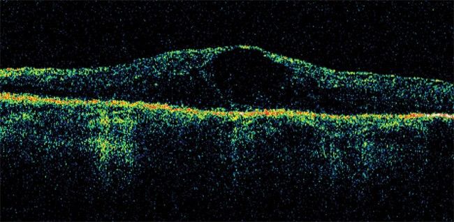
Intravitreal versus sub-Tenon triamcinolone
  acetonide for refractory diffuse diabetic
              macular oedema

                               By

                   Anthony Grant Zaborowski

Submitted in partial fulfilment of the requirements for the degree
                                of

                      Masters of Medicine

                            In the
                Department of Ophthalmology
            Nelson R. Mandela School of Medicine
                University of KwaZulu-Natal
                           Durban

                              2008

                                                                     1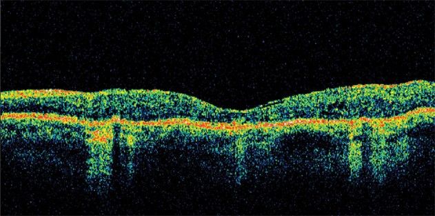
Abstract

Purpose: To compare the safety and efficacy of intravitreal (IVT) and sub-
Tenon (ST) triamcinolone acetonide for the treatment of refractory diffuse
diabetic macular oedema.

Method: 29 eyes of 22 patients with long-standing, diffuse diabetic
macular oedema refractory to argon laser treatment were randomly assigned
to a single 4mg injection of IVT triamcinolone acetonide or a 40mg sub-
Tenon injection. Patients were subsequently monitored for six to nine
months. Outcome measures were visual acuity, intraocular pressure, macular
thickness on optical coherence tomography and adverse effects.

Results: There was no significant improvement in visual acuity in either
group. A transient decrease in macular thickness was found in the IVT group
but not in the ST group. There were no significant adverse effects apart from
a mild to moderate intra-ocular pressure rise found more frequently in the
IVT group.

Conclusion: IVT and ST triamcinolone acetonide injections for refractory
diffuse diabetic macular oedema appear relatively safe and well-tolerated.
IVT injection produces a significant temporary decrease in macular
thickness in patients with long-standing diffuse diabetic macular oedema
while ST injection does not. Neither intervention was shown to significantly
improve visual acuity in this group of patients.

                                                                             2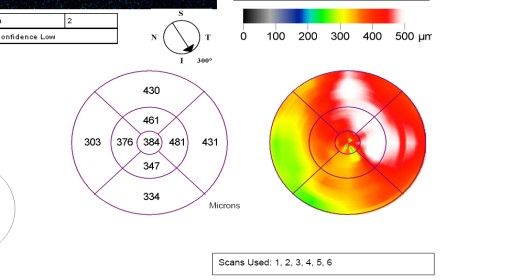
Preface

I hereby declare that this dissertation has been my own work and has not
been submitted in any form to any other university. The research work was
carried out in the Department of Ophthalmology, Nelson R. Mandela School
of Medicine, University of KwaZulu-Natal, under the supervision of Dr
Linda Visser.

_______________________

______ day of ________________ 2009

Acknowledgements:

Thank you to Visicare for donating the triamcinolone acetonide used in this
study and for paying the fee levied by the Medicine Control Council for each
application to use triamcinolone off label.

Presentations of the results of this study:

Ophthalmic Society of South Africa (OSSA) congress – 2006

Nelson R. Mandela School of Medicine faculty research day - 2006

                                                                          3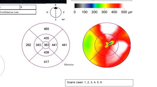
Contents

Chapter 1    Introduction and literature review   6

Chapter 2    Materials and methods                22

Chapter 3    Results                              27

Chapter 4    Discussion                           39

Chapter 5    Conclusions                          50

References                                        52

                                                       4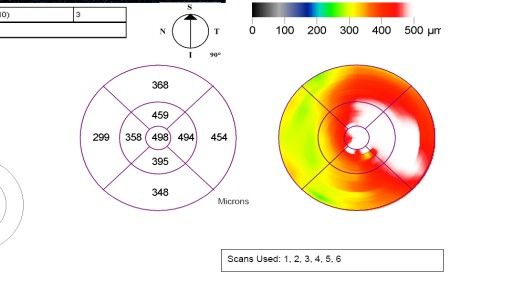
List of Illustrations
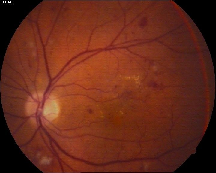
Figure 1 Fundus photograph - diabetic maculopathy with clinically
significant macular oedema. 7
Figure 2 Fluorescein angiogram - diffuse diabetic macular oedema and
previous argon laser retinal scars. 8
Figure 3 Fluorescein angiogram - chronic diffuse cystoid diabetic macular
oedema. 8
Figure 4 Optical coherence tomography maps of macular thickness following
administration of intravitreal triamcinolone acetonide. 34-35
Figure 5 Optical coherence tomography maps of macular thickness following
sub-Tenon triamcinolone acetonide. 36-37
Figure 6 Optical coherence tomography image - diabetic macular oedema 38
Figure 7 Optical coherence tomography image – diabetic macular oedema
post 4mg intravitreal triamcinolone acetonide. 38
Table I Study inclusion and exclusion criteria. 23
Table II Study outcome measures. 24
Table III Independent Samples Test – comparing macular thickness and visual
acuity 28
Table IV Changes in macular thickness after intravitreal triamcinolone. 29
Table V Changes in macular thickness after sub-Tenon triamcinolone. 30
Table VI Percentage change in central macular thickness after intravitreal
triamcinolone. 31
Table VII Change in visual acuity after intravitreal triamcinolone. 31
Table VIII Percentage change in central macular thickness after sub-Tenon
triamcinolone. 32
Table IX Change in visual acuity after sub-Tenon triamcinolone. 32
Table X A comparison of the results of the 4 previous intravitreal versus
sub-Tenon injection studies. 42
Table XI Changes in central macular thickness and visual acuity following
triamcinolone treatment. 46
Graph I Change in macular thickness after intravitreal and sub-Tenon
triamcinolone. 33
Graph II Change in visual acuity after intravitreal and sub-Tenon
triamcinolone. 33
5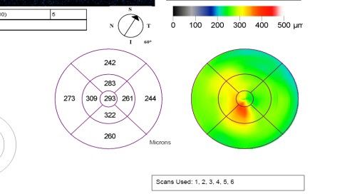
Chapter 1

                  Introduction and literature review

The greater Durban metropolitan region of KwaZulu-Natal has a large

diabetic population with a full spectrum of diabetic-related eye disease,

posing a considerable public health problem. Macular oedema is a common

ocular complication of diabetes worldwide, affecting 29% of diabetics with

disease duration of 20 years or more.1 It remains the most common reason

for loss of vision in diabetics. The treatment of diffuse diabetic macular

oedema is at present limited and often unsatisfactory, leaving many patients

severely debilitated with compromised macular vision.

The current standard of care in the treatment of diffuse diabetic macular

oedema is the use of argon laser grid photocoagulation. The Early Treatment

Diabetic Retinopathy Study (ETDRS - 1985) showed the benefit of focal

laser treatment for clinically significant macular oedema, which reduced the

risk of further visual loss by 50%.2 However only 17% of patients were

shown to have an improvement in vision with laser treatment and only 3%

                                                                               6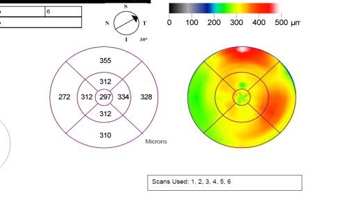
had an improvement of 3 or more Snellen chart lines. Diffuse diabetic

macular oedema is notoriously difficult to treat and studies have shown

disappointing results from grid argon laser treatment.3 The experience of the

ophthalmology department of the University of KwaZulu-Natal reflects

published findings - one of satisfactory results with focal argon laser

treatment for focal clinically significant macular oedema but disappointing

results with grid laser in diffuse oedema. Diffuse oedema and visual acuity

seldom improve despite laser treatment and many patients are considered to

have refractory disease.

Figure 1 - Diabetic maculopathy with clinically significant macular oedema.

                                                                              7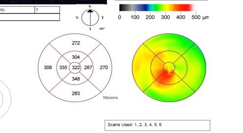
Figure 2 – Fluorescein angiogram - diffuse diabetic macular oedema and previous argon
laser retinal scarring.

Figure 3 – Fluorescein angiogram - chronic diffuse cystoid diabetic macular oedema.

                                                                                      8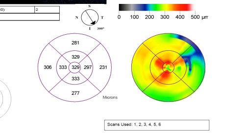
It is in this context that alternative treatments to laser have been sought.

Intravitreal crystalline corticosteroid suspension in the form of triamcinolone

acetonide (TA) has been investigated for the treatment of diabetic macular

oedema refractory to conventional therapy at several ophthalmology centres

worldwide.4,5,6 Its use proliferated worldwide after Martidis, Duker et al

(2002) first published promising results of its use in the treatment of diffuse

diabetic macular oedema. They injected 4mg of triamcinolone intravitreally

in sixteen eyes with clinically significant diabetic macular oedema that

failed to respond to two previous sessions of laser photocoagulation. They

found a significant mean improvement in visual acuity of 2.4 Snellen lines at

the 1 month follow up, 2.4 lines at 3 months and 1.3 lines at 6 months. The

central macular thickness was measured by Optical Coherence Tomography

(OCT) and decreased by 55%, 57.5%, and 38% respectively over the same

intervals.5

Jonas JB, Kreissig I et al (2003) studied 26 eyes which received an

intravitreal injection of 25 mg of triamcinolone acetonide for diffuse diabetic

macular oedema and followed them for 6 months. Mean visual acuity

improved from 0.12 at baseline to a maximum of 0.19 during follow-up.

                                                                                  9
This was compared to a control group of 16 eyes that underwent macular
grid laser and had no significant improvement in visual acuity.4
Massim P, Audren F et al (2004) injected 4mg of triamcinolone acetonide
into one eye of twelve patients with diffuse diabetic macular oedema
unresponsive to laser treatment, the other eye serving as a control.6 Central
macular thickness on OCT improved from an average of 509µ before
injection to 207µ at 4 weeks and 207µ at 12 weeks. In the control group
central macular thickness was 474 pre-injection, 506µ at 4 weeks and 469µ
at 12 weeks. At 24 weeks the oedema had recurred in all eyes and there was
no longer a statistically significant reduction.
Triamcinolone has also been used for a variety of other retinal pathologies
including cystoid macular oedema secondary to uveitis7, central and branch
retinal vein occlusions8,9 and the treatment of choroidal neovascular
membranes in combination with photodynamic therapy10.
10The pathophysiology of diabetic macular oedema is multifactorial and
appears to have an inflammatory component. 20 There is a generalised
breakdown in the blood-retinal barrier due to changes in tight junction
proteins and leukocyte adherence to vascular endothelium, resulting in
increased vascular permeability and accumulation of fluid in the outer
plexiform layer of the retina - diffuse oedema. Vascular endothelial growth
factor (VEGF) is stimulated by interleukins and other inflammatory
mediators and has been shown to increase vascular permeability by
phosphorylating the tight junction proteins occludin and zonula occludens-
1.11 The specific mechanisms by which intravitreal steroids achieve their
effects are not entirely understood but it has been postulated that
triamcinolone is effective in several areas to decrease oedema: Firstly, it
decreases the activity of inflammatory mediators - interleukins 5,6 and 8,
prostaglandins, interferon-γ and tumour necrosis factor – inhibiting VEGF
expression and vascular permeability. Secondly, it has a positive effect on
cellular calcium channels, aiding in the active diffusion of fluid across cells
and therefore decreasing oedema. Thirdly, triamcinolone has been shown to
have a generally favourable effect on the integrity of the blood-retinal
barrier, decreasing the osmotic component of oedema.20
11The results of investigations into the use of intravitreal triamcinolone
acetonide for diabetic macular oedema have been largely positive with
significant improvements seen in visual acuity and, initially, with a
seemingly low incidence of irreversible side effects.4,5,6 Further retrospective
studies of larger numbers of injections have concluded that morbidity is, in
fact, quite significant. The most frequent side effect has been shown to be a
steroid-induced rise in intraocular pressure in about one third to one half of
patients, controllable by topical pressure lowering agents in the majority of
cases.12 However, irreversible steroid-induced glaucoma refractory to
medical treatment and requiring filtration surgery is well documented.13
Corticosteroid-induced glaucoma in general has, in fact, been shown to be
irreversible in 3% and has to be managed like primary chronic open angle
glaucoma.14 Endophthalmitis has been shown to occur at a rate of 0.3% per
injection and retinal detachment, uveitis, intraocular haemorrhage and
vascular occlusions have all been described. Formation or acceleration of
cataract within two years is considered universal.12,13
12The appropriate concentration of intravitreal triamcinolone required for
macular oedema treatments has also been the topic of much interest. Jonas
JB et al have published a number of papers on the topic of intravitreal
triamcinolone using a 25mg intravitreal dosage.4,17 This dosage was based
on the concentration of 0,1ml of the commercially available product and was
continued in numerous subsequent studies by Jonas based on this arbitrary
fact. (Personal communication). Over time, 4mg has become the generally
accepted intravitreal dose as the therapeutic effects are noted to be
equivalent to 25mg while possibly causing less steroid-response glaucoma.6
Recently, Audren F, Lecleire-Collet A et al (2006) showed that there was no
statistical difference at 24 weeks post intravitreal injection between 2mg and
4mg with regard to visual acuity, central macular thickness and intraocular
pressure, although macular oedema recurred at 16 weeks in the 2mg group
but only at 20 weeks in the 4mg group.15 Lam DS, Chan CK et al (2007) in a
randomised prospective trial studied the differences in efficacy and adverse
effects of 4mg, 6mg and 8mg doses intravitreally.16 Visual acuity at 6
months was significantly higher in the 8mg group compared to the 4mg
group. The reduction in central macular thickness at 6 months for the 4, 6
and 8mg groups was 28%, 42% and 60% respectively, while ocular
13hypertension occurred in 39%, 30% and 55%. These studies suggest that a
higher dosage of triamcinolone has a longer duration of effect but may be
associated with a higher incidence of glaucoma. At present, a 4mg, 0.1ml
injection remains the generally accepted and most widely used intravitreal
dosage.
The benefits of intravitreal triamcinolone therapy have been noted to be
transient in most patients, usually lasting between 4 and 6 months. A small
group of patients appear to have a lasting response to just one injection.
Repeated injections have been shown to effectively maintain visual and
anatomical results in those patients with recurrent macular oedema.17
Sub-Tenon steroid injections are well described for a variety of ophthalmic
conditions, including posterior uveitis and cystoid macular oedema.18,19,20
Their use in diabetic macular oedema is less well accepted although some
reports in the literature show modest initial improvements in visual acuity.
20,21
Bakri SJ and Kaiser PK (2005) looked retrospectively at 63 eyes (73
injections) that received 40mg of sub-Tenon triamcinolone for persistent
clinically significant diabetic macular oedema. At 1 month 46% of eyes
showed no change in vision and 50% showed 1 line or more improvement.
14At 3, 6 and 12 months there was a less pronounced improvement from
baseline but without statistical significance. No OCT analysis was included
in this paper.20
Triamcinolone has recently been shown to enter the vitreous trans-sclerally
by sub-Tenon injection, however the same study showed the steroid
concentration in the vitreous to be six times greater by intravitreal
injection.22 Later, Thomas ER, Wang J et al (2006) showed that the vitreous
steroid concentration attained by sub-Tenon injection can vary substantially
in each individual, ranging from a clinically significant concentration to
zero.23 These studies suggest that attaining appropriate vitreous
concentrations of steroid are less reliable with sub-Tenon injections than
direct placement with intravitreal injections.
Peri-ocular steroid injections are not without risk and they have been
associated with ptosis, globe perforation, retinal and choroidal vascular
occlusions, raised intraocular pressure and cataracts.24,25 However, the risk
of complication remains very low and in a retrospective review of 73 sub-
Tenon triamcinolone injections, Bakari and Kaiser (2005) found few
complications with only a 4% incidence of ptosis at 3 months post-injection
15and in 10% a minor, transient increase in intraocular pressure that was easily
controlled with topical hypotensive agents. Fifty percent had the
development or progression of cataract at 1 year.20
Intravitreal injections are effective in delivering appropriate quantities of
steroid close to the target tissue, the retina. One of the major disadvantages
of intravitreal steroid injections for diabetic macular oedema is the
temporary nature of the therapeutic response and the need for repeated
injections every 4 to 6 months in most patients.17 The incidence of adverse
effects in a patient requiring multiple intravitreal injections becomes far
more significant with each injection and in this context sub-Tenon injections
intuitively have a more favourable risk profile.
At the time of commencement of the design and data collection of this study
in 2004, there were no published reports comparing intravitreal and sub-
Tenon injections of triamcinolone acetonide. Subsequently four reports have
been published in the peer-reviewed literature:
Cardillo JA, Melo LA Jr et al (2005) conducted a prospective, double-
masked, randomized controlled trial of twelve patients (24 eyes) with
16bilateral diffuse diabetic macular oedema.26 One eye of each patient
randomly received a single 4mg triamcinolone acetonide intravitreal
injection and the fellow eye received a 40mg triamcinolone acetonide
posterior sub-Tenon's capsule injection. They looked at visual acuity, central
macular thickness and complications over a 6 month follow-up period and
compared the 2 interventional arms. Significant but transient improvements
in central macular thickness and visual acuity were found in both groups,
although results were found to be statistically better in the intravitreal group.
The mean central macular thickness in eyes with intravitreal injection was
found to be significantly thinner than in the sub-Tenon's capsule-injected
eyes at 1 month (226µ versus 431µ) and 3 months (242µ and 364µ) The
mean visual acuity in the intravitreally injected eyes was significantly better
than in the sub-Tenon's capsule-injected eyes at 3 months post injection (0.8
and 1.1) Neither group were found to have any significant side effects in the
6 month follow-up period. The authors suggested a better therapeutic result
from intravitreal triamcinolone. This was a particularly informative result,
despite the small study size since the two methods of injection were
performed on the same patient, thus allowing an excellent comparative
analysis.
17Bonini- Filho MA, Jorge R, Barbosa JC et al. (2005) looked prospectively at
28 patients with refractory diffuse diabetic macular oedema.27 Patients
randomly received 40mg sub-Tenon triamcinolone acetonide or 4mg
intravitreally. Central macular thickness was measured by OCT and found to
be significantly reduced in the intravitreal group at several follow-up
intervals up to 24 weeks while there was no statistically significant decrease
in the sub-Tenon group. Visual acuities were significantly higher in the
intravitreal group at each time point. A significant increase in intraocular
pressure was found in the sub-Tenon group at weeks 4 and 8 and at week 8
in the intravitreal group. The authors concluded that intravitreal injections
were more effective in improving central macular thickness and visual
acuity than sub-Tenon injections. Twenty of the 28 eyes had been previously
treated with pan retinal photocoagulation for proliferative diabetic
retinopathy.
Ozdek S, Bahceci UA et al (2006) did a retrospective analysis of 95 patients
(126 eyes) with diffuse diabetic macular oedema refractory to argon laser
treatment.28 Eighty five eyes of 60 patients had received 20mg posterior
sub-Tenon TA and 41 eyes of 35 patients had received 4mg intravitreal TA.
In the IVT group, 24 eyes were from a primary treatment and 17 eyes were
18from subsequent treatments. Sub-Tenon patients were followed for a mean
of 4.1 months and IVT patients for 4.6 months. In the sub-Tenon group
mean visual acuity improved from 0.19 to 0.22 and the mean central macular
thickness decreased from 413µ to 312µ. In the IVT group visual acuity
improved from 0.15 to 0.20 and central macular thickness from 494.5µ to
288µ. The authors concluded that sub-Tenon and IVT injections were both
effective treatments for diffuse diabetic macular oedema with IVT injections
being more efficacious. Importantly, 20% of sub-Tenon injections were
found to have no effect at all, supporting the Thomas ER, Wang J et al
(2006) paper which showed that some sub-Tenon triamcinolone injections
result in minimal or no intravitreal steroid concentration.23 Of note was that
a significant rise in intraocular pressure was found in only 8.2% of the sub-
Tenon group and in 24.3% of the IVT injection group.
Finally, Choi YJ, Oh IK et al (2006) did a prospective study of 60 eyes of 60
patients with diffuse diabetic macular oedema.29 Each patient was assigned
to receive a single 4mg intravitreal injection or a single 40mg posterior sub-
Tenon injection of triamcinolone acetonide. Visual acuity, intraocular
pressure and OCT measurement of central macular thickness were recorded
at 1 and 3 months. Both groups showed statistically significant
19improvements in visual acuity and central macular thickness from baseline
at 1 month and 3 months after injection. The intravitreal group went from a
baseline macular thickness of 428µ to 256µ at 1 month and 230µ at 3
months and in the sub-Tenon group from 480µ to 318µ and 271µ. There
were no statistically significant differences between the two groups. In the
intravitreal group visual acuity measured by the LogMAR system improved
from a baseline of 0.73 to 0.57 at 1 month and 0.53 at 3 months and in the
sub-Tenon group from 0.78 to 0.65 and 0.62. Again, there were no
statistically significant differences between the two groups. Ten eyes (33%)
in the intravitreal group had an intraocular pressure rise to 21mmHg or
more, while this occurred in only 1 eye (3%) in the sub-Tenon group. The
authors found that the posterior sub-Tenon injection of triamcinolone
acetonide had a comparable effect to the intravitreal injection and showed a
lower risk of elevated IOP. They concluded that sub-Tenon injection of
triamcinolone acetonide might be a good alternative to IVT injection for the
treatment of diffuse diabetic macular oedema. They also conceded that their
results might have been good because the macular oedema was not
refractory at baseline.
20This study is a prospective, interventional case series to investigate the use
of triamcinolone acetonide in the treatment of patients in the Durban
metropolitan area of KwaZulu-Natal, South Africa, with diffuse diabetic
macular oedema refractory to conventional laser therapy. The drug was
made available through Visicare as Kenacort (Bristol Meyers Squibb). Each
millilitre contains 40mg triamcinolone acetonide with sodium chloride,
0.99% benzyl alcohol preservative, 0.75% carboxymethylcellulose sodium
and 0.04% polysorbate 80.
How efficacious is intravitreal and sub-Tenon triamcinolone acetonide for
the treatment of diffuse diabetic macular oedema in our patient population
with respect to macular thickness and visual acuity? What is the incidence
of complication of these interventions? How do these two modes of delivery
compare with regard to efficacy and incidence of complications?
21Chapter 2
Materials and methods
A prospective, randomised comparative study was undertaken. The study
had approval from the Ethics Committee of the Nelson R Mandela School of
Medicine.
Nineteen patients with symptomatic, clinically significant diffuse diabetic
macular oedema based on assessment by fundoscopy with a 90 dioptre lens
and showing no response to two or more macular grid laser therapies were
included in the study. Patients excluded from the study: Those with pre-
existing glaucoma or ocular hypertension, known steroid responders,
concomitant macular pathology affecting visual acuity, macular ischaemia,
an unclear fundal view (e.g. significant cataract, vitreous haemorrhage) and
proliferative diabetic retinopathy or severe non-proliferative diabetic
retinopathy requiring pan retinal argon laser.
22INCLUSION: • Diffuse diabetic macular oedema - 2 previous
failed grid argon laser treatments
• Pre-existing glaucoma/ocular hypertension
• Steroid responders
• Poor fundal view
• Macular ischaemia (FFA)
EXCLUSION:
• Other macular pathology causing low VA
• Cataract surgery within the last 6 months
• Severe non-proliferative diabetic retinopathy
or proliferative diabetic retinopathy
Table I – Study inclusion and exclusion criteria.
After obtaining written consent, patients underwent fluorescein fundal
angiography to exclude macular ischaemia as a cause of reduced visual
acuity. A proforma with demographic and clinical information (age, sex,
race, diabetic history etc) was completed. Patients were examined with
respect to their visual acuity, as measured by logarithm of minimal angle of
resolution (Logmar) test, intraocular pressure and macular thickness by
optical coherence tomography (OCT – Zeiss-Humphrey instruments, San
23Leandro, California) measurement. OCT examinations consisted of six
radial cuts centred on the fovea. The central macular thickness was taken as
the 1000µ circle centred on the foveola and was automatically calculated by
the OCT software.
Patients’ eyes were then alternately assigned to two experimental groups.
Eyes assigned to group A received a single pars plana intravitreal injection
of 4mg triamcinolone acetonide in the eye clinic by conventional sterile
technique. Group B received a single 40mg sub-Tenon triamcinolone
injection.
• Visual acuity (Logmar)
• Fundoscopy
OUTCOME MEASURES:
• OCT
• IOP
• Observation of complications
Table II – Study outcome measures.
24The intravitreal injection technique:
The injection was preceded by softening of the globe by gentle massage. The
eye was anaesthetised with topical benoxinate and sterilised with 5%
povidone iodine into the conjunctival sac. The periorbital area was cleaned
with a betadine solution followed by sterile draping of the head and neck and
the use of a sterile wire speculum to keep the lids open and the eyelashes
away from the injection site. A sterile cotton tipped applicator soaked in
benoxinate was applied to the injection site for 5 minutes and the injection
site was identified with a calliper - 4mm posterior to the limbus in phakic
patients and 3.5mm in pseudophakic patients. Thereafter, 4mg (0.1ml) of
triamcinolone acetonide (Kenacort, Bristol Myers Squibb) was injected via
the pars plana, posterior to the superior limbus. Patients then received one
drop of chloramphenicol immediately after the procedure and four times
daily for the following 5 days. Correct placement of the steroid and normal
retinal arterial circulation was confirmed on indirect fundoscopy.
The sub-Tenon injection technique:
Benoxinate drops were used to anaesthetise the eye and 5% povidone iodine
was instilled into the conjunctival sac. The periorbital area was cleaned with
25a betadine solution followed by sterile draping of the head and neck and the
use of a sterile wire speculum to keep the lids open. A small bleb of
lignocaine was injected inferonasally under the conjunctiva and a cut-down
made with Westcott scissors through conjunctiva and Tenon’s capsule to
bare sclera, 8mm posterior to the limbus. Forty milligrams (1ml) of
triamcinolone acetonide (Kenacort) was injected into the sub-Tenon's space
using an olive-tipped canula, with particular attention to avoiding reflux of
steroid.
Follow up examinations were done at day 1, 2 weeks, 6 weeks, 12 weeks, 24
weeks and 36 weeks after injection. At each visit visual acuity, macular
thickness by OCT scan, intraocular pressure and complications arising from
the intervention were recorded, as well as a clinical evaluation of the macula
and optic disc by fundoscopy. Appropriate intraocular pressure lowering
measures were undertaken if there was any clinically significant rise in
intraocular pressure.
All examinations, investigations and treatments were performed by the
author.
26Chapter 3
Results
Twenty nine eyes of 22 patients were initially enrolled in the study. Fifteen
eyes received intravitreal triamcinolone and 14 eyes received sub-Tenon
injections. One patient absconded after the 6 week follow up visit (2 study
eyes), one patient died of renal failure after the 6 week follow up (2 study
eyes) and one patient died of cardiac disease after the 6 week follow up (1
study eye). Thus 24 eyes of 19 patients were followed for 6 to 9 months post
injection with 13 eyes in the intravitreal group and 11 eyes in the sub-Tenon
group.
There were no statistically significant differences between the two groups at
baseline in terms of patient macular thickness as measured by OCT
(p=0.914) and visual acuity (p=0.459).
27Levene's Test for
Equality of t-test for Equality of Means
Variances
Mean Std. Error 95% Confidence Interval of
F Sig. t df Sig. (2-tailed)
Difference Difference the Difference
Lower Upper
Equal variances
OCT .232 .634 .109 23 .914 7.96 73.322 -143.717 159.640
assumed
Equal variances
.107 20.744 .915 7.96 74.088 -146.229 162.152
not assumed
Equal variances
VA .083 .776 .754 23 .459 .0929 .12335 -.16222 .34812
assumed
Equal variances
.764 21.673 .453 .0929 .12168 -.15962 .34552
not assumed
Table III - Independent Samples Test – comparing macular thickness and visual acuity
at baseline between the 2 treatment groups.
For OCT p=0.914 and for VA p=0.459, therefore no significant differences between the
groups at baseline.
Eighteen patients were Indian, 3 African, and 1 Coloured. The mean patient
age was 65 (range 52 to 80). All patients had type 2, non-insulin dependent
diabetes mellitus. Macular oedema was present in all patients for at least two
years and was considered longstanding and refractory to treatment.
The mean central macular thickness at baseline in the intravitreal group was
458µ. Macular thickness at the follow up intervals was 368 (n=14), 370
(n=14), 408 (n=13), 512 (n=13) and 508 (n=10) at 2, 6, 12, 24 and 36 weeks
post-injection respectively. Mean visual acuity at the follow up intervals was
0.764 at baseline and 0.68, 0.66, 0.60, 0.70 and 0.88 at 2, 6, 12, 24 and 36
weeks post-injection respectively. The mean central macular thickness at
28baseline in the sub-Tenon group was 450µ. At follow up macular thickness
was 480 (n=14), 462 (n=12), 530 (n=10), 563 (n=11) and 471 (n=7) at 2, 6,
12, 24 and 36 weeks post-injection respectively. Mean visual acuity was
0.66 at baseline and on follow up, 0.70, 0.72, 0.64, 0.78 and 0.80 at 2, 6, 12,
24 and 36 weeks post-injection respectively. There were statistically no
significant differences between the changes in mean value for macular
thickness or visual acuity between the intravitreal and sub-Tenon groups
using the ANOVA repeated measures test.
2 6 12 24 36
Intravitreal Baseline:
weeks weeks weeks weeks weeks
Macular 458 368 370 408 512 508
thickness: (µ)
No. of eyes: 14 14 14 13 13 10
Table IV - Changes in macular thickness after intravitreal triamcinolone
292 6 12 24 36
Sub-Tenon Baseline:
weeks weeks weeks weeks weeks
Macular 450 480 462 530 563 471
thickness: (µ)
No. of eyes: 15 14 12 10 11 7
Table V - Changes in macular thickness after sub-Tenon triamcinolone
In the intravitreal group 9 of the 13 eyes that were followed to 6 months had
a rise in intraocular pressure above 21mmHg. In 1 eye this was above
30mmHg. Four eyes (31%) required pressure lowering treatment. In 2 eyes,
1 topical pressure lowering drop was needed and in 2 eyes 2 medications
were needed. In the sub-Tenon group, 3 of the 13 eyes that were followed to
6 months had a rise in intraocular pressure above 21mmHg with only 1 eye
(8%) requiring 1 topical medication. In both groups, pressures returned to
normal by 6 months and all drops were discontinued.
There were no other significant adverse events in either group.
30Macular thickness
Intravitreal group
Macular thickness: Change in thickness from baseline:
Baseline: 458µ
Week 2: 368µ 20% decrease
Week 6: 370µ 26% decrease
Week 12: 408µ 11% decrease
Week 24: 512µ 12% increase
Week 36: 508µ 11% increase
Table VI - Percentage change in central macular thickness after intravitreal
triamcinolone.
Visual Acuity (LogMAR)
Intravitreal group
Baseline: 0.76
Week 2: 0.68 Gained 4 letters
Week 6: 0.66 Gained 5 letters
Week 12: 0.6 Gained 8 letters
Week 24: 0.7 Gained 3 letters
Week 36: 0.88 Lost 6 letters
Table VII - Change in visual acuity after intravitreal triamcinolone.
31Macular thickness
Sub-Tenon group
Macular thickness: Change in thickness from baseline:
Baseline: 450µ
Week 2: 480µ 7% increase
Week 6: 462µ 3% increase
Week 12: 530µ 18% increase
Week 24: 563µ 25% increase
Week 36: 471µ 5% increase
Table VIII - Percentage change in central macular thickness after sub-Tenon
triamcinolone.
Visual Acuity (LogMar)
Sub-Tenon Group
Baseline: 0.66
Week 2: 0.70 Lost 2 letters
Week 6: 0.72 Lost 3 letters
Week 12: 0.64 Gained 1 letters
Week 24: 0.78 Lost 6 letters
Week 36: 0.80 Lost 7 letters
Table IX - Change in visual acuity after sub-Tenon triamcinolone.
32600
Macular thickness 550
500
450 IVT
400 ST
350
300
250
0 2 6 12 24 36
Time
Graph I - Change in macular thickness after IVT and sub-Tenon triamcinolone.
1
0.9
0.8
Visual Acuity
0.7 IVT
0.6 ST
0.5
0.4
0.3
0 2 6 12 24 36
Weeks
Graph II - Change in visual acuity after IVT and sub-Tenon triamcinolone.
33Intravitreal - Baseline
Intravitreal - Week 2
Intravitreal - Week 6
34Intravitreal - Week 12
Intravitreal - Week 24
Intravitreal - Week 36
Figure 4 - OCT map of macular thickness – intravitreal triamcinolone
acetonide. Macular thickness reduced after triamcinolone injection at weeks
2 and 6. Recurrence of oedema starting again at week 12.
35Sub-Tenon - Baseline
Sub-Tenon – Week 2
Sub-Tenon – Week 6
36Sub-Tenon – Week 12
Sub-Tenon – Week 24
Sub-Tenon – Week 36
Figure 5 - OCT map of macular thickness – sub-Tenon triamcinolone
acetonide. Macular thickness does not reduce after triamcinolone injection.
Progressively worsens over the follow-up period.
37Figure 6: Optical coherence tomography image - diabetic macular oedema.
Figure 7: Optical coherence tomography image – oedema resolved 6 weeks
post 4mg intravitreal triamcinolone acetonide.
38Chapter 4
Discussion
In this study, intravitreal triamcinolone for longstanding diffuse diabetic
macular oedema refractory to laser treatment resulted in a small and
transient but clinically significant improvement in visual acuity and central
macular thickness. It was superior to sub-Tenon injections which had no
apparent effect on macular thickness or visual acuity. Neither intervention
was found to have any serious adverse side effects over the 9 month follow-
up period. A modest increase in intraocular pressure, seen with greater
frequency in the intravitreal group, was transient and well controlled with
topical medications.
There were no statistically significant differences between the sub-Tenon
and intravitreal groups but the improvements in macular thickness and visual
acuity in the intravitreal group were clinically superior
In the intravitreal group there was a modest decrease in macular thickness
for the first 12 weeks post-injection – to a maximum mean decrease of 26%
at week 6. A modest temporary improvement in visual acuity was also seen
39for the first six months - with a maximum mean improvement of 8 letters
(1.5 Snellen lines) at week 12. By week 24 the macular oedema had
recurred. Martidis et al5 reported 55%, 58% and 38% reductions in mean
macular thickness at 1, 3 and 6 months follow up in their groundbreaking
intravitreal triamcinolone study. They also saw a mean improvement of 2.5
Snellen lines at 1 and 3 months. Massin et al6 reported similar transient
reductions in central macular thickness with macular oedema recurring by
week 24. Jonas et al4 reported a mean visual acuity improvement from 0.12
at baseline to a maximum of 0.19 during follow-up.
In this study, therefore, the transient improvements in visual acuity and
central macular thickness seen with intravitreal injections, although far more
modest, reflect findings from previous studies of intravitreal triamcinolone
injections for refractory diffuse diabetic macular oedema. There is a clear
trend towards an initial improvement in visual acuity and a reduction in
central macular thickness, followed by recurrence of oedema and
deterioration in visual acuity by 6 months post-injection.
40In the sub-Tenon group there was no mean improvement in macular
thickness in the 9 months following injection and this did not clinically
appear to be a useful intervention. In fact, there was a mean loss of visual
acuity at each time point, except at week 12 where there was a gain of just 1
letter. In contrast, Bakri and Kaiser20 reported improvements in visual acuity
from a mean baseline of 20/80 to 20/50 at 1 month, stabilizing to 20/65 at 3
months, 20/68 at 6 months and 20/63 at 12 months. OCT was not performed
in this study. There do not appear to be any other studies in the literature
looking at the effect of posterior sub-Tenon injections for diffuse diabetic
macular oedema alone.
Four previous studies have looked at the difference between the effect and
safety of intravitreal and sub-Tenon injections for diffuse diabetic macular
oedema.26-29
41Bonini-Filho, Jorge et
Cardillo, Melo et al al Ozdek, Bahceci et al Choi, Oh et al Zaborowski
IVT ST IVT ST IVT ST IVT ST IVT ST
Central macular
thickness (%)
1 month 56 16 41 6 40 18 41 34 26 3
3 months 53 29 31 5 42 24 46 44 11 18
6 months 15 16 23 0 25 25 12 25
Visual acuity
(letters)
1 month 7 1 10 6 3 1 8 7 5 3
3 months 12 0 5 0 3 0 10 8 8 1
6 months 2 0 0 0 0 0 3 6
Decreased central macular thickness/improved visual acuity IVT = intravitreal
Increased central macular thickness/worse visual acuity ST = sub-Tenon
Table X – A comparison of the results of the 4 previous intravitreal versus
sub-Tenon injection studies.
In all four studies, the reductions in central macular thickness with
intravitreal injections are comparable to the results of Martidis and Duker 5.
In this study a trend towards improvement was seen but was far less
impressive than the previous four studies. Reported improvements in visual
acuity were reasonably modest. Cardillo, Melo et al26 reported a maximum
mean improvement in visual acuity of 12 letters (2.5 Snellen lines) at 3
months. Bonini-Filho, Jorge et al27 reported a maximum of 10 letters (2
42Snellen lines) at 1 month. Choi, Oh et al29 reported a maximum of 10 letters
(2 Snellen lines) at 3 months. Ozdek, Bahceci et al28 had the least
impressive improvements reporting a 3 letter improvement at 1 and 3
months.
In this study, the visual acuity improvements with intravitreal injections
were surprisingly good and comparable to the 4 previous studies despite the
unimpressive reductions in central macular thickness. A 5 letter
improvement was found at 1 month (1 Snellen line) and an 8 letter
improvement (1.5 Snellen lines) was found at 3 months.
With sub-Tenon injections, Cardillo, Melo et al and Ozdek, Bahceci et al
reported no real improvement in mean visual acuity, while Bonini-Filho,
Jorge et al reported a 6 letter improvement at 1month and Choi, Oh et al
reported 7 letters at 1 month and 8 letters at 3 months.
Cardillo, Melo et al, Ozdek, Bahceci et al and Choi, Oh et al all reported
improvements in central macular thickness which were statistically inferior
to the intravitreal injections. Bonini-Filho, Jorge et al found no improvement
from baseline.
43In this study, sub-Tenon’s injections were more in keeping with the results
of Bonini-Filho et al and there was no significant mean improvement in
central macular thickness or visual acuity.
Choi, Oh et al found that there was statistically no difference between the
sub-Tenon and intravitreal groups. Sub-Tenon injections were considered a
good and safer alternative to intravitreal injections. Their patients differed
from other studies in that the diffuse diabetic macular oedema was not
refractory. Cardillo, Melo et al concluded that there was a clear short-term
trend favouring intravitreal injection although both interventions had a
favourable transient benefit. Ozdek, Bahceci et al found a pronounced effect
with intravitreal injections and less effective but significant benefits with
posterior sub-Tenon’s. Bonini-Filho, Jorge et al found significant benefits
with intravitreal steroid but not with sub-Tenon injections. They believed
that triamcinolone reflux might be partly to blame for the poorer sub-Tenon
results, as well as an inadequate positioning of the steroid next to the
macular area. They felt that the sclera and choroid acted as a significant
barrier to triamcinolone diffusion into the vitreous.
44There are several possible explanations for the poor results in this study:
1. The group of patients studied in the Department of Ophthalmology of
the University of KwaZulu-Natal have notoriously poor metabolic and
hypertensive control. There may actually be degrees of treatment
resistance with diabetic macular oedema and these metabolic factors
may limit the efficacy of steroid treatment. Metabolic factors were not
quantified but may warrant further study as to their role in the efficacy
of steroid treatment. A subsequent study might exclude patients with
poor glycaemic and blood pressure control.
2. All the studied patients had diabetic macular oedema for at least 2
years and in many cases for much longer. Again, there might be
degrees of treatment resistance in diabetic macular oedema and the
duration of oedema may play a role in the sensitivity to steroid
treatment.
3. There was a definite study bias towards a more severe, refractory
disease. Referred patients from fellow clinicians to the study project
typically had profound macular oedema and did not necessarily
represent the typical patient population of diabetics with macular
oedema refractory to 2 macular grid laser treatments.
45Unlike the 4 previous studies comparing intravitreal and sub-Tenon
injections, this study continued to 9 months of follow up in most patients.
An interesting finding was that at 9 months, central macular thickness and
visual acuity was worse than baseline in both the intravitreal and sub-Tenon
groups.
Zaborowski
IVT ST
Central macular thickness
(%)
11% 5%
9 months increase increase
Visual acuity (letters)
6 letters 7 letters
9 months decrease decrease
Table XI – Changes in central macular thickness and visual acuity following
triamcinolone treatment.
This suggests the interesting possibility of a steroid withdrawal, rebound
type effect. No study has yet looked at the 9 month outcome of patients who
were treated with triamcinolone acetonide injections but not re-treated. This
data is suggestive of a deleterious effect of once-off triamcinolone injections
and may support continued injections for the recurrence of oedema. The
possibility of a steroid withdrawal, rebound effect warrants further study.
46Most previous studies have shown either very poor efficacy with sub-Tenon
triamcinolone as a treatment for diffuse diabetic macular oedema or modest
benefits statistically inferior to intravitreal injections. The results of this
study do not support the use of sub-Tenon triamcinolone acetonide for
longstanding diffuse diabetic macular oedema refractory to laser treatment.
Why is intravitreal delivery apparently more effective than sub-Tenon?
The most likely reason is poor localisation of steroid adjacent to the macula
due to reflux or poor injection technique. Intravitreal injections deliver a
very predictable dose of steroid into the vitreous but it is unpredictable with
sub-Tenon injections. This was seen experimentally by Thomas ER, Wang J
et al (2006)23 where sub-Tenon injections of triamcinolone produced a wide
variety of vitreous steroid concentrations ranging from levels comparable to
intravitreal injections to no detectable steroid at all. On average, the vitreous
concentration with sub-Tenon injections was much lower than with
intravitreal injections. There is certainly a degree of technical skill in
ensuring that the full 40mg bolus of steroid is successfully delivered sub-
Tenon and adjacent to the macula. Intravitreal injections are technically
simple and there is little doubt about the ease of achieving a high
concentration of the drug in the vitreous. Intuitively one also feels that the
47sclera and choroid pose a natural barrier to diffusion of the steroid into the
vitreous. This is overcome to a degree by injecting ten times the intravitreal
dose in sub-Tenon’s injections.
One of the main outcome measures of this study was to report any adverse
effects of the triamcinolone treatments. In both study groups there were no
major adverse effects although there was a rise in intraocular pressure above
21mmHg in 9 of the 13 eyes in the intravitreal group. In 4 eyes (31%)
topical pressure lowering treatment was required. In the sub-Tenon group, 3
of 11 had a rise in intraocular pressure above 21mmHg with only 1 eye (8%)
requiring topical medication. In both groups, pressure lowering was
successful and pressures returned to normal by 6 months with all drops
discontinued. The increased frequency of ocular hypertension in the
intravitreal group would be expected if intravitreal injections caused a higher
intraocular concentration of steroid. In a meta-analysis of 272 patients who
received 20mg intravitreal injections, 41.2% had pressures >21mmHg,
11.4% >30mmHg, 5.5% >35mmHg and 1.8% >40mmHg.
The effect of the steroid on cataract formation was not specifically studied
but posterior sub-capsular cataracts are a well-recognised complication and
48were shown to have a prevalence of 45% after 1 year after a single 4mg
intravitreal injection of triamcinolone.30 No endophthalmitis, sterile or
infective, was encountered although the study size was small and other
studies have found the incidence of infective endophthalmitis to be up to
0.87%, with sterile endophthalmitis up to 1.6%.31
49Chapter 5
Conclusions
The results of this study should be viewed with caution as the study size was
small with a large number lost to follow up. Patient eyes were alternately
assigned to either treatment arm and therefore this was not a randomised
controlled study allowing potential selection bias.
Triamcinolone acetonide appears to be of limited value in the treatment of
longstanding diabetic macular oedema refractory to laser treatment. While
intravitreal injections showed a clinically significant trend towards a
transient improvement in visual acuity and central macular thickness, this
effect was modest. Sub-Tenon injections had no effect on visual acuity or
central macular thickness. Intravitreal injections appear to be more effective
than sub-Tenon injections, probably due to a more predictable delivery of
steroid into the vitreous. The results of this study do not support the use of
sub-Tenon injections for longstanding, refractory disease.
Poor clinical results may be due to permanent damage to the retinal pigment
epithelium and photoreceptors due to disease chronicity, making
50improvements in visual acuity modest at best. The macular oedema may also
become more refractory with time. Intravitreal triamcinolone may be more
effective earlier in the disease process. Poor metabolic control may play a
role in a weak response to steroid treatment.
Once the decision has been made to treat, the clinician should be prepared to
repeat injections if and when oedema returns. Single injections may actually
worsen the disease in the long-term, possibly through a rebound-type effect
of steroid withdrawal and should be used with caution. This effect warrants
further study.
Single injections of sub-Tenon and intravitreal triamcinolone for diffuse
diabetic macular oedema refractory to laser are relatively safe short to
medium term, but patients will probably require management of secondary
ocular hypertension which is usually self-limiting.
51References
1. Klein R, Klein BE, Moss SE, et al. The Wisconsin epidemiologic
study of diabetic retinopathy. IV. Diabetic macular oedema.
Ophthalmology 1984;91:1464-74
2. Early Treatment Diabetic Retinopathy Study Research Group.
Photocoagulation for diabetic macular oedema: ETDRS report
number 1. Arch Ophthalmol. 1985;103:1796-1806.
3. Bresnick GH. Diabetic macular oedema: a review. Ophthalmology
1986;93:989-97
4. Jonas JB, Kreissig I, Sofker A et al. Intravitreal injection of
triamcinolone for diffuse diabetic macular oedema. Arch Ophthalmol.
2003;121:57-61
5. Martidis A, Duker JS, Greenberg PB et al. Intravitreal triamcinolone
for refractory diabetic macular oedema. Ophthalmology
2002;109:920-27
6. Massin P, Audren F, Hauchine B. Intravitreal triamcinolone acetonide
for diffuse diabetic macular oedema. Ophthalmology 2004;111:218-
225
527. Young S, Larkin G, Branley M et al. Safety and efficacy of
intravitreal triamcinolone for cystoid macular oedema in uveitis. Clin
Exp Ophthalmol 2001;29:2-6
8. Greenberg PB, Martidis A, Rogers AH et al. Intravitreal triamcinolone
acetonide for macular oedema due to central retinal vein occlusion. Br
J Ophthalmol 2002;86:247-248.
9. Chen SD, Lochhead J, Patel CK et al. Intravitreal triamcinolone
acetonide for ischaemic macular oedema caused by branch retinal
vein occlusion. Br J Ophthalmol 2004:88:154-155
10. Spaide RF, Sorenson J, Maranan L et al. Combined PDT with
verteporforin and intravitreal triamcinolone for choroidal
neovascularisation. Ophthalmology 2003;110:1517-1525.
11.Funuatsu H, Yamashita H, Nakamura s et al. Vitreous levels of
pigment epithelial-derived factor and vascular endothelial growth
factor are related to diabetic macular oedema. Ophthalmology
2006;113:294-301.
12.Konstantopolous A, Williams CP, Newsom RS et al. Ocular morbidity
associated with intravitreal triamcinolone acetonide. Eye 2006;May 19
(E publication ahead of print)
5313.Jager RD, Aiello LP, Patel SC et al. Risks of intravitreous injection.
Retina 2004;24:676-698.
14.Francois J. Corticosteroid glaucoma. Ann Ophthalmol 1977;9:1075-
1080.
15.Audren F, Lecleire-Collet A, Erginay A et al. Intravitreal
triamcinolone acetonide for diffuse diabetic macular edema: phase 2
trial comparing 4 mg vs 2 mg. Am J Ophthalmol 2006.
Nov;142(5):794-99
16. Lam DS, Chan CK, Mohamed S, Lai TY et al. A prospective
randomised trial of different doses of intravitreal triamcinolone for
diabetic macular oedema. Br J Ophthalmol 2007 Feb;91(2):199-203
17.Jonas JB, Kreissig I, Degenring RF et al. Repeated intravitreal
injection of triamcinolone acetonide for diffuse diabetic macular
oedema. Br J Ophthalmol 2005;89;122
18.Helm CJ, Holland GN. The effects of posterior subtenon injection of
triamcinolone acetonide in patients with intermediate uveitis. Am J
Ophthalmol 1995 Jul;120(1):55-64.
19.Yoshikawa K, Kotake S, Ichiishi A et al. Posterior sub-Tenon
injections of repository corticosteroids in uveitis patients with cystoid
macular edema. Jpn J Ophthalmol 1995;39(1):71-6.
5420.Bakri SJ, Kaiser PK. Posterior subtenon triamcinolone acetonide for
refractory diabetic macular edema. Am J Ophthalmol 2005
Feb;139(2):290-4.
21.Shimura M, Nakazawa T, Yasuda K et al. Pre-treatment of posterior
subtenon injection of triamcinolone acetonide has beneficial effects
for grid pattern photocoagulation against diffuse diabetic macular
oedema. Br J Ophthalmol 2006;Oct 31 (E publication ahead of print)
22.Inoue M, Takeda K, Morita K et al.Vitreous concentrations of
triamcinolone acetonide in human eyes after intravitreal or subtenon
injection. Am J Ophthalmol. 2004 Dec;138(6):1046-8
23.Thomas ER, Wang J, Ege E et al. Intravitreal triamcinolone acetonide
concentration after subtenon injection. Am J Ophthalmol 2006
Nov;142(5):860-1.
24. Abel AD, Carlson JA, Bakri S et al. Sclerosing lipogranuloma of the
orbit after periocular steroid injection. Ophthalmology
2003;110:1841-1845
25. Moshfeghi DM, Lowder CY, Roth DB et al. Retinal and choroidal
vascular occlusion after posterior sub-Tenon triamcinolone injection.
Am J Ophthalmol 2002:134:132-134
5526.Cardillo JA, Melo LA Jr, Costa RA et al. Comparison of intravitreal
versus posterior sub-Tenon's capsule injection of triamcinolone
acetonide for diffuse diabetic macular edema. Ophthalmology. 2005
Sep;112(9):1557-63.
27. Bonini- Filho MA, Jorge R, Barbosa JC et al. Intravitreal injection
versus sub-Tenons infusion of triamcinolone acetonide for refractory
diabetic macular oedema: a randomised clinical trial. Invest
Ophthalmol Vis Sci 2005;46:3845-9
28.Ozdek S, Bahceci UA, Gurelik G et al. Posterior subtenon and
intravitreal triamcinolone for diabetic macular oedema. J Diabetes
Complications 2006;20(4):246-51
29.Choi YJ, Oh IK, Oh JR et al. Intravitreal versus posterior subtenon
injection of triamcinolone acetonide for diabetic macular oedema.
Korean J Ophthalmol. 2006;20(4):205-9
30. Thompson JT. Cataract formation and other complications of
intravitreal triamcinolone for macular oedema. Am J Ophthalmol
2006;141;629-37
31. Westfall AC, Osborn A, Kuhl et al. Acute endophthalmitis incidence:
intravitreal triamcinolone. Arch Ophthalmol 2005;123:1075-7
56You can also read