Effects of Diethylstilbestrol on Zebrafish Gonad Development and Endocrine Disruption Mechanism - MDPI
←
→
Page content transcription
If your browser does not render page correctly, please read the page content below
biomolecules

Article
Effects of Diethylstilbestrol on Zebrafish Gonad Development
and Endocrine Disruption Mechanism
Xuan Liu, Xianyi Xie and Hongling Liu *

                                          State Key Laboratory of Pollution Control and Resource Reuse, School of the Environment, Nanjing University,
                                          Nanjing 210023, China; mg20250024@smail.nju.edu.cn (X.L.); mf1725069@smail.nju.edu.cn (X.X.)
                                          * Correspondence: hlliu@nju.edu.cn; Tel.: +86-25-89680356

                                          Abstract: Environmental estrogen is a substance that functions as an endocrine hormone in organisms
                                          and can cause endocrine system disruption. A typical environmental estrogen, diethylstilbestrol
                                          (DES), can affect normal sexual function and organism development. However, even though the
                                          effects of different exposure stages of DES on the endocrine system and gonadal development of
                                          zebrafish juveniles are unknown, sex determination is strongly influenced by endocrine-disrupting
                                          chemicals (EDCs). From 10–90 days post fertilization (dpf), juvenile zebrafish were exposed to
                                          DES (100 and 1000 ng/L) in three different stages (initial development stage (IDS), 10–25 dpf;
                                          gonadal differentiation stage (GDS), 25–45 dpf and gonadal maturity stage (GMS), 45–60 dpf).
                                          Compared with that of IDS and GMS, the growth indicators (body length, body weight, and others)
                                          decreased significantly at GDS, and the proportion of zebrafish females exposed to 100 ng/L DES
                                          was significantly higher (by 59.65%) than that of the control; in addition, the zebrafish were biased
                                          towards female differentiation. The GDS is a critical period for sex differentiation. Our results show
                                that exposure to environmental estrogen during the critical gonadal differentiation period not only

                                          affects the development of zebrafish, but also affects the population development.
Citation: Liu, X.; Xie, X.; Liu, H.
Effects of Diethylstilbestrol on
                                          Keywords: environmental estrogen; diethylstilbestrol (DES); zebrafish gonad development; en-
Zebrafish Gonad Development and
                                          docrine disruption-chemicals (EDCs)
Endocrine Disruption Mechanism.
Biomolecules 2021, 11, 941. https://
doi.org/10.3390/biom11070941

Academic Editors: Jung-Ren Chen
                                          1. Introduction
and Chung-Der Hsiao                             In recent decades, plenty of evidence from laboratory animals, in vitro studies, and
                                          epidemiology showed that many synthetic environmental estrogens that are released
Received: 31 May 2021                     into the environment have the capacity to imitate the actions of the natural hormone
Accepted: 22 June 2021                    17β-estradiol (E2), which then causes adverse outcomes for humans and animals [1–3].
Published: 25 June 2021                   Diethylstilbestrol (DES), a nonsteroidal estrogen, was the first synthetic estrogen used in
                                          clinical treatment to prevent miscarriages. In 1971, the U.S. Food and Drug Administration
Publisher’s Note: MDPI stays neutral      (FDA) issued the ”Drug Bulletin”, advising doctors to stop prescribing DES to pregnant
with regard to jurisdictional claims in   women because it was related to a rare vaginal cancer in female offspring [4]. However,
published maps and institutional affil-   DES is still used in Asia and other regions as a growth promoter for livestock [5,6]. It
iations.                                  is also used in aquaculture for promoting growth and for producing monosex (female)
                                          populations of fish [7]. According to reports, the detectable concentration of DES in surface
                                          water ranges from slightly less than 1 to about 20 ng/L in different locations [8–10]. In
                                          China, the concentration of DES in some wastewaters, as well as in wastewater from
Copyright: © 2021 by the authors.         wastewater treatment plants, reached 421 and 268 ng/L, respectively [11].
Licensee MDPI, Basel, Switzerland.              Recently, a toxicology study found some adverse developmental effects caused by DES.
This article is an open access article    Some studies [12–15] found that DES had a damaging impact on male Sprague–Dawley
distributed under the terms and           rattos’ reproductive systems through decreasing the numbers of spermatogonia, Sertoli cells,
conditions of the Creative Commons        and Leydig cells, which affect steroidogenesis and spermatogenesis. Estrogenic endocrine-
Attribution (CC BY) license (https://
                                          disrupting chemical (EDC) exposure, including to E2, DES, mestranol, and 4-n-nonyphenol,
creativecommons.org/licenses/by/
                                          disrupts testis differentiation and causes xenopus laevis feminization to different degrees [16].
4.0/).

Biomolecules 2021, 11, 941. https://doi.org/10.3390/biom11070941                                       https://www.mdpi.com/journal/biomolecules
Biomolecules 2021, 11, 941                                                                                            2 of 12

                             Air-breathing catfish exposed to 17α-ethynylestradiol (EE2) and DES presented with a devel-
                             opmental delay, as well as premature morphological and oocyte development during the early
                             stages of growth and gonadal differentiation [17]. Wang et al. [18] observed that the oogenesis
                             of yellow catfish juveniles were affected by DES by disrupting gonad development and the
                             hypothalamic-pituitary-gonadal (HPG) axis. Pan et al. [19] found that mature male zebrafish
                             exposed to DES and flutamide for 30 days had damaged spermatogenesis. In the present
                             study, we attempted to determine when the sensitive development and endocrine-disrupting
                             effect stage of environmental estrogen DES occurred for aquatic organisms. A previous
                             study [18] found that 10 days of DES exposure could disrupt the HPG axis, promote apoptosis
                             and meiosis, and impair the oogenesis of female yellow catfish juveniles. However, despite
                             the available research [5,18,19] based on experiments involving DES exposure in mature
                             fish, there is a lack of evidence about the specific stages of DES exposure. The zebrafish is
                             an ideal model organism and is used in a large number of toxicological studies [20], as its
                             gonadal differentiation style is dioecious and its sex is not determined at birth, as zebrafish
                             gradually differentiate to have male or female gonads during their individual growth and
                             development. Environmental sex determination (ESD) plays a crucial role in zebrafish
                             sex differentiation [21]. According to previous research, we can divide zebrafish gonad
                             development into three phases: (1) the initial development stage (IDS), which is about
                             10–25 days post fertilization (dpf), where the zebrafish gonad develops into an ovarian
                             gonad and primordial germ cells begin to undergo mitosis [22]; (2) the gonadal differen-
                             tiation stage (GDS), which is at 25–45 dpf and is an important turning point in gonadal
                             differentiation [23], and (3) the gonadal maturity stage (GMS), which occurs at 45–60 dpf,
                             after which complete gonad differentiation can be observed at 60 dpf and the gonad will
                             be completely sex mature [24]. Therefore, to better understand the gonad development
                             toxicity of DES in zebrafish, three phases, according to their development characteristics,
                             were chosen for exposure applying two concentrations (100 and 1000 ng/L) until complete
                             maturity was maintained at 90 dpf.

                             2. Materials and Methods
                             2.1. Materials
                             2.1.1. Chemicals
                                 DES (CAS number 56-53-1) was purchased from Shanghai J&K Scientific (Shanghai,
                             China) and the purity was above 95%. Acetone (CAS number 67-64-1) was purchased from
                             Shanghai Generay Biotech (Shanghai, China) and the purity was above 99%.

                             2.1.2. Zebrafish Maintenance
                                  Zebrafish (Danio rerio) embryos were obtained from the same batch of 4-month-old
                             wild AB-type adult zebrafish, raised in a laboratory zebrafish breeding system, through
                             natural mating at a 1:2 ratio of female to male. The embryos were maintained at 28 ± 1 ◦ C
                             in 60 mg/L of sea salt water (pH = 6.5–8.5, dissolved oxygen ≥ 60% saturation) with a
                             14/10 h light/dark cycle. The fish were fed thrice daily with fairy shrimp (Tianjin Red
                             Sun Aquaculture Co. Ltd. Tianjin, China). The amount of food did not exceed 2% of the
                             body weight.

                             2.2. Methods
                             2.2.1. Eighty-Day Exposure Experiments
                                  We investigated the concentration of DES on the surface and in wastewater [8–11],
                             and then we did a pretest to find an appropriate concentration that would affect the
                             development of zebrafish, but with no serious outcomes. Consequently, two doses (100 and
                             1000 ng/L) of DES were chosen and used for the exposure in this study. To assess the effects
                             of DES exposure in different gonad development stages, we conducted experiments in
                             three exposure periods (as illustrated in Figure A1 in Appendix A) according to the gonad
                             development process of zebrafish [24]. Specifically, zebrafish larvae were only exposed to
                             DES in IDS, GDS, and GMS separately, and were maintained in 0.01% acetone for 90 dpf,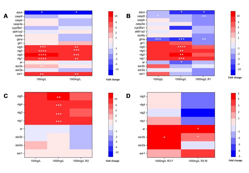
Biomolecules 2021, 11, 941                                                                                            3 of 12

                             after the exposure phase (as illustrated in Figure A1). The exposure started on 10 dpf after
                             adult fish mating. The treatment and control groups were prepared in triplicate. Each
                             replication had 50 zebrafish larvae, which were randomly selected and placed in 2 L glass
                             beakers with a DES solution (0, 100, and 1000 ng/L, dissolved in 0.01% acetone) from 10
                             to 25 dpf, and then moved into a 5 L glass beaker to ensure that they had enough living
                             space after 25 dpf (age < 30 dpf, space < 30 fish/L; age > 30 dpf, space < 5 fish/L). During
                             the exposure, the water in each glass beaker was replaced every two days with freshly
                             exposed water containing the corresponding drug concentrations. To avoid the evaporation
                             of the solution, each glass beaker was covered with a breathable membrane during the
                             experiment. We measured some of the morphological parameters (body length (L), body
                             weight (W), and sex) of the zebrafish exposed at 90 dpf and also the body length of the
                             larvae at 25, 45, and 60 dpf in the experimental period. The body length and body weight
                             were measured according to the Organization for Economic Cooperation and Development
                             (OECD) 236 guidelines and the Fish Early-Life Stage Toxicology Test (OECD 210, 2013). In
                             the below equation, K indicates plumpness, which was calculated as described in Formula
                             (1), which was used to estimate the true growth rates.

                                                                   K = W/L3 × 100                                        (1)

                                  In addition, we also recorded the sexual distribution in different groups to observe the
                             effect of DES on the sex ratio. At 90 dpf, the zebrafish were placed in ice water to euthanize
                             them and confirm the sex of each zebrafish by the dissection of the gonads, thereby
                             enabling us to determine the sex ratio statistics. The gonads were weighed (Wgonad ), and
                             the gonadosomatic index (GSI) was calculated as described in Formula (2).

                                                               GSI = Wgonad /W × 100                                     (2)

                             2.2.2. Observation of Gonadal Tissue
                                  To study the effects of exposure to DES over three different periods on zebrafish gonad
                             development and to understand the maturity of zebrafish gonad development at 90 dpf,
                             we observed a gonadal tissue section and judged the sex of the fish according to the OECD
                             No.123 Guidance Document on the Diagnosis of Endocrine-Related Histopathology in Fish
                             Gonads (2010). We also counted the proportion of germ cells in the gonads of male and
                             female zebrafish at various developmental stages.
                                  The gonadal tissues sampled from 4 fish for every replicate at 90 dpf were randomly
                             selected and dissected. We rinsed the gonads with normal saline, immersed them in a 10%
                             formaldehyde solution and placed them in a cool environment at room temperature for
                             more than 48 h, and then conducted tissue slice observations on the gonads by paraffin
                             sectioning and hematoxylin-eosin (HE) staining. The female gonadal section was divided
                             into several areas 1000 × 1000 µm in size; we took 2–3 areas of each slice from repeat
                             experiments of each exposure and control groups to count the number of egg cells. When
                             there was an atresia follicle, a degenerated oocyte with a separated cell content and vitelline
                             membrane, the statistics were not calculated but a record was made. Egg cells in females
                             mainly include perinuclear oocytes (PO), cortical granular oocytes (CO), early vitellogen-
                             esis oocytes (EV), and late vitellogenesis oocytes (LV), and sperm cells in males include
                             spermatogonia (sg), spermatocytes (sc), spermatids (st), and sperm (sp). As male germ cells
                             are too small to be counted, we counted the area of the sperm cells at each stage. Besides
                             the male gonad slices, which were divided into several 200 × 200 µm areas, the sizes of
                             these slices were the same as those mentioned before.

                             2.2.3. Analysis of Genes and Proteins
                                  The gene expression analyses were conducted using the qRT-PCR method. A qRT-PCR
                             assay of the genes on the HPG axis and the genes related to ER (the estrogen receptor) and
                             AR (the androgen receptor) was conducted to assess the triggers of reproductive toxicity
                             in zebrafish. Three fish from each replicate were selected to extract RNA from the control
Biomolecules 2021, 11, 941 4 of 12
group (at 25, 45, 60, and 90 dpf), and the IDS (at 25 dpf, 45 dpf (only 1000 ng/L)), GDS
(at 45 dpf and 60 dpf (only 1000 ng/L)), and GMS (at 60 dpf and 90 dpf (only 1000 ng/L))
exposure groups, and the samples were then stored in RNAlater (Qiagen, Hilden, Germany)
at −80 ◦ C in a refrigerator. Three zebrafish larvae were pooled and homogenized as one
biological sample. The total RNA was extracted using an RNeasy Mini kit (Qiagen, Hilden,
Germany), and the cDNA was transcribed with an Omniscript RT kit (Qiagen, Germany)
according to the manufacturer’s instructions. A SYBR real-time quantitative PCR kit from
Toyobo (Shanghai, China) Biotechnology (Shanghai, China) was used to amplify the gene
expressions related to the HPG axis, ER, and AR. The sequences of the selected genes and
primers were characterized previously [25] and are listed in Table 1. The housekeeping
gene beta-actin was used as an internal control for the mRNA expression, and the gene
expression was calculated with the 2 −∆∆Ct method [26]. At 25 and 45 dpf, the measured
genes included those related to growth, the zebrafish HPG axis, the sex hormone receptor,
sex differentiation, and gonad development. At 60 and 90 dpf, because the gonadal
differentiation process ended, the tested genes included those related to the HPG axis and
the zebrafish sex hormone receptor.
Table 1. Primer sequences for receptor-associated genes for RT-PCR.
Gene Name Forward Primer (50 -30 ) Reverse Primer (50 -30 ) Gene ID
ddx4 ctcaaccacaagcatcca agcgtccagttcttccta ENSDARG00000014373
casp9 acgagatggacgctattc ttgagtaggacaccaggat ENSDARG00000004325
caspb ggacagagacgaggagaa agttcacagagttcagatgg ENSDARG00000052039
casp3a gcggatacggagactact gattgaggcttggcatca ENSDARG00000017905
cyp26a1 tgttctccttgccaatcg tcacttcttctgctgttctc ENSDARG00000033999
aldh1a2 tcttcaataacggtcaatgc ttcctcctctgtgctctc ENSDARG00000053493
sox9a gacaccagcagacaacaa gcatcagacagacacttct ENSDARG00000003293
ghra ccttcttcacaaccattctg ctccaccacttctgattcc ENSDARG00000054771
gh1, cagttggtggtggttagtt gcgttcctcaggcataag ENSDARG00000038185
vtg5 agctaatgctctgcccgtta gttcagcctcaaacagcaca ENSDARG00000092126
vtg4 ctacaaggtggaggctctgc ggaggacaaatcaccagcat ENSDARG00000078429
vtg2 tactttgggcactgatgcaa agacttcgtgaagcccaaga ENSDARG00000055809
vtg1 ctgcgtgaagttgtcatgct gaccagcattgcccataact ENSDARG00000092233
ar gtctattaagagccgcctat caccgcaacaagttcatc ENSDARG00000067976
esr2b ttgtgttctccagcatgagc ccacatatggggaaggaatg ENSDARG00000034181
esr2a agcattagccttgttagca atgttgtcacggatgtcat ENSDARG00000016454
esr1 ggtccagtgtggtgtcctct cacacgaccagactccgtaa ENSDARG00000004111
2.2.4. Statistical Analysis
The toxicology data in this study were statistically analyzed using Graph Pad Prism5
(GraphPad, San Diego, CA, USA). There was no significant difference between the water
control and the solvent control for all investigated endpoints, and thus, the solvent control
was used as the control in the following analysis. A one-way analysis of variance (one-way
ANOVA) was applied to analyze the significance of differences between the exposed group
and the control group at each key timepoint. Values are shown as the mean ± standard
deviation (SD). Significant differences were found when p < 0.05.
3. Results
3.1. Morphology Parameters
3.1.1. 25,45 and 60 dpf Body Length
The body length after every exposure stage was significantly reduced compared with
that of the control, which demonstrated that DES exposure in the development stage had
a significant inhibitory effect on zebrafish body development, as shown in Figure 1. At
25 dpf, the body length of the fish who were in the IDS when they were exposed was
significantly lower than in that of the control groups (0 ng/L; lower by 20.91% (p < 0.0001)
and 35.29% (p < 0.0001)) for the 100 ng/L and 1000 ng/L treatments, respectively. At 45 dpf,3.1.1. 25,45 and 60 dpf Body Length
The body length after every exposure stage was significantly reduced compared with
that of the control, which demonstrated that DES exposure in the development stage had
Biomolecules 2021, 11, 941 a significant inhibitory effect on zebrafish body development, as shown in Figure 1. 5At 25
of 12
dpf, the body length of the fish who were in the IDS when they were exposed was signif-
icantly lower than in that of the control groups (0 ng/L; lower by 20.91% (p < 0.0001) and
35.29% (p < 0.0001)) for the 100 ng/L and 1000 ng/L treatments, respectively. At 45 dpf, the
the
body body length
length of fish
of fish subjected
subjected to GDS
to GDS exposure
exposure waswas significantly
significantly lowerlower (by 16.74%
(by 16.74% (p <
(p < 0.001)
0.001) and and 31.57%
31.57% (p < 0.0001))
(p < 0.0001)) in thein100
theng/L
100 ng/L andng/L
and 1000 1000 treatment
ng/L treatment
groupsgroups
than inthan
that
in
of that of the control
the control groups, groups, respectively.
respectively. At 60 At 60the
dpf, dpf, the body
body lengthlength
of fishofthat
fishunderwent
that underwent
GMS
GMS exposure was significantly lower (by 12.79% (p < 0.01) and 15.40%
exposure was significantly lower (by 12.79% (p < 0.01) and 15.40% (p < 0.001)) (p < 0.001)) for the
for the 100
100
ng/L ng/L
and and
10001000
ng/Lng/L treatments,
treatments, respectively.
respectively.
2
5
d
p
f
4
5
d
p
f
6
0
d
p
f
(A)
(B)
(C)
Figure 1. (A) Body length measured after diethylstilbestrol (DES) exposure at 25 dpf, 45 dpf, and
60 dpf, respectively. (B) Body length and body weight of zebrafish maintained after exposure,
measured at 90 dpf. Different shapes of dots are body weight, and histogram is the body length
(yellow is initial development stage (IDS), blue is gonadal differentiation stage (GDS), and pink
is gonadal maturity stage (GMS)). (C) The plumpness (K) of zebrafish maintained after exposure,
measured at 90 dpf. Values are shown as the mean ± SD (n = 3 replicates per treatment, n = 11 fish
per replicate at 25, 45, and 60 dpf, and n = 15 fish per replicate at 90 dpf). Asterisks denote significant
differences between control and treatments (* p < 0.05, ** p < 0.01, *** p < 0.005, and **** p < 0.0001,
according to Dunnett’s multiple comparisons test). CK-control group.maturity stage (GMS)). (C) The plumpness (K) of zebrafish maintained after exposure, measured a
90 dpf. Values are shown as the mean ± SD (n = 3 replicates per treatment, n = 11 fish per replicate
at 25, 45, and 60 dpf, and n = 15 fish per replicate at 90 dpf). Asterisks denote significant difference
between control and treatments (* p < 0.05, ** p < 0.01, *** p < 0.005, and **** p < 0.0001, according to
Biomolecules 2021, 11, 941 Dunnett’s multiple comparisons test). CK-control group. 6 of 12
3.1.2. Body Length, Body Weight, and K at 90 dpf
3.1.2. We found
Body that
Length, exposure
Body Weight, to
andDES
K at in
90 different
dpf development stages affected multiple
aspects
We of zebrafish
found development,
that exposure to DES inincluding
differentbody length, body
development stagesweight,
affected K, and sex (as il
multiple
lustrated
aspects of in Figure development,
zebrafish 1C and Figureincluding
2A). Thebody zebrafish
length,that
bodywere exposed
weight, to DES
K, and in the IDS
sex (as
showed ainsignificant
illustrated Figures 1C decrease in body
and 2A). The zebrafishlength at 25exposed
that were dpf (as to
illustrated
DES in thein
IDSFigure
showed 1B). At 90
a significant decrease in body length at 25 dpf (as illustrated in Figure
dpf, the body length and body weight were still significantly decreased when the 1B). At 90 dpf, the
body length
zebrafish andkept
were bodyin weight were stillenvironment
a freshwater significantly decreased
for 65 days when
afterthe zebrafish
exposure inwere
the IDS (100
kept in a freshwater environment for 65 days after exposure in the IDS (100 ng/L) and were
ng/L) and were sexually mature. However, the body length of females exposed during the
sexually mature. However, the body length of females exposed during the IDS (1000 ng/L)
IDS (1000 ng/L) returned to the control level after 65 days of freshwater treatment, with
returned to the control level after 65 days of freshwater treatment, with the female body
the female
length beingbody length higher
significantly being significantly
than that of the higher
controlthan that while
groups, of thethe
control groups, while
body length
of males was significantly lower. Besides the K of the females exposed during the IDSexposed
the body length of males was significantly lower. Besides the K of the females
during
(100 thewhich
ng/L), IDS (100ng/L), whichdecreased,
was significantly was significantly
there weredecreased, there
no significant were no
differences significan
after
differences
exposure afterthe
during exposure during the
IDS (as illustrated in IDS (as1C).
Figure illustrated in Figure 1C).
(A)
(B)
Figure2.2.(A)
Figure (A)Gonadosomatic
Gonadosomatic index (GSI)
index of zebrafish
(GSI) at 90 dpf
of zebrafish fordpf
at 90 every
forgroup.
every (n = 15 fish
group. (n =per
15 fish pe
replicate at 90 dpf). (B) Sex ratio of zebrafish at 90 dpf for every group. (n = 6 fish per replicate
replicate at 90 dpf). (B) Sex ratio of zebrafish at 90 dpf for every group. (n = 6 fish per replicate at at 90
90 dpf).
dpf). Valuesare
Values are shown
shown as mean± ±SD
asmean SD(n (n
= 3=replicates per treatment).
3 replicates Asterisks
per treatment). denote significant
Asterisks denote significan
differences between control and treatments (* p < 0.05, ** p < 0.01, and *** p < 0.005, according to
Dunnett’s multiple comparisons test). CK-control group.
The zebrafish that were exposed to DES in the GDS showed a significant decrease in
body length at 45 dpf. However, after 45 days of freshwater maintenance, at 90 dpf, the
body length and body weight of females were still significantly decreased (100 ng/L), asBiomolecules 2021, 11, 941 7 of 12
well as those of males exposed in the GDS (100 ng/L and 1000 ng/L). K was significantly
decreased in females and males exposed during the GDS (100 ng/L).
The zebrafish that were exposed to DES in the GMS showed a significant decrease in
body length at 60 dpf. However, after 30 days of freshwater maintenance, at 90 dpf, the
growth parameters of all the groups returned to control levels. However, the female body
length for the group exposed in the GMS (1000 ng/L) was significantly higher than that of
the control groups, while K was lower for the control groups because body length increases
faster than body weight.
3.1.3. GSI
Although zebrafish had some time to recover after the exposure stage, the effects
continued when they reached sexual maturity. Exposure led to various reductions in the
GSI in females compared with that of the control groups, while the IDS and GDS exposure
effects were stronger for the GMS. Exposure led to a significant decrease in the GSI during
the IDS (1000 ng/L) and GDS (100 ng/L), while GMS exposure had no effect on the GSI
in males. DES resulted in a more significant GSI decrease in females compared to that
of males (as illustrated in Figure 2A). In general, in the first two phases, IDS and GDS,
exposure to DES had stronger effects than during the GMS, and the effect on females was
greater than for males.
3.1.4. Sex Ratio
We observed the sex ratio of zebrafish at 90 dpf, and only the GDS (100 ng/L) exposure
group was significantly higher (by 59.65%, p < 0.05) than the control, by about twice as
many females as males (as illustrated in Figure 2B), so the results of this group were biased
towards female differentiation. There was no significant difference between the zebrafish
sex ratio in other exposure groups compared with that of the control group.
3.2. Gonad Tissues Observation
We counted the egg and sperm cells at 90 dpf for the control group and the DES
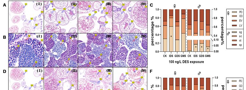
exposure group exposed during the IDS, GDS, and GMS (100 ng/L (as illustrated in
Figure 3A,B) and 1000 ng/L (as illustrated in Figure 3D,E)). In females, the number of PO
in the early gonad development stage were approximately 59.42%, 27.93%, and 43.21%,
respectively, higher than that of the control (by 30.55%), and significantly higher (p < 0.05)
in that of the IDS 100 ng/L exposure group (as illustrated in Figure 3C). The proportion of
LV at the stage of near developmental maturity decreased accordingly, and there was no
significant difference due to large individual differences, to a certain extent. There was no
significant difference in the other groups for 100 ng/L and 1000 ng/L DES exposure. For
males, the percentage of “sg” in the IDS, GDS, and GMS groups was significantly higher
(by 6.26% (p < 0.05), 6.96% (p < 0.05), and 4.17% (p < 0.05), respectively, in 100 ng/L) than
for the control group (by 0.88%; Figure 3C). In addition, the percentage of “sg” was higher
(by 7.31% in 1000ng/L (p < 0.05)) than in that of the control group (by 0.88%; Figure 3F).
3.3. Gene Expression Changes
The expressions of the genes involved in growth, the zebrafish HPG axis, sex hormone
receptors, gonadal differentiation, and gonad development were determined for zebrafish
exposed to DES in every phase (as illustrated in Figure 4). At 25 dpf, the IDS exposure
group (100 ng/L and 1000 ng/L) displayed significantly downregulated expression of
ddx4, which is linked to gonadal maturation and dramatically upregulated (more than
1000-folds) expression of vtg1, vtg2, vtg4, and vtg5, which are estrogen receptor downstream
genes. Among the genes involved in the HPG axis, the genes related to estrogen signaling
in the brain were significantly upregulated and the genes for sex hormone synthesis were
downregulated (as illustrated in Figure 4A). At 45 dpf, the GDS exposure group (1000 ng/L)
showed significant downregulation of the expression of ar by approximately three-fold,
and ddx4 and ghra by approximately four-fold, and dramatically upregulated expressionBiomolecules 2021, 11, 941 8 of 12
of vtg1, vtg2, vtg4, and vtg5 (only significantly for 1000 ng/L). The GDS exposure group
(100 ng/L) showed significant downregulation of the expression of casp9 by approximately
two-fold, and ghra by approximately three-fold. After a period of recovery, the IDS exposure
group (1000 ng/L) showed significant downregulation of ddx4 by approximately five-fold,
and casp9 and ghra by approximately two-fold; in addition, the growth and sex hormone
receptor genes were downregulated after recovery (as illustrated in Figure 4B). At 60 dpf,
the expression of vtg1, vtg2, vtg4, and vtg5 was dramatically upregulated by the exposure,
while the expression of the vtg genes did not significantly change in the other exposure
group. The sex hormone synthesis genes for the HPG axis, overall, showed a trend of
downregulation (as illustrated in Figure 4C). At 90 dpf, exposure in the GMS (1000 ng/L)
significantly upregulated the expression of cyp19a1b and esr2b in females and ar in males,
and the expression of ghra in the pituitary was also upregulated in females and males (as
illustrated in Figure 4D). In addition, we discovered that the increases or decreases in the
gene expression induced by 1000 ng/L DES showed different degrees of recovery for the
fish that were maintained in 0.01% acetone for a period of time after exposure. In the IDS
exposure group, the significant impact on the genes vtgs and ar at 25 dpf diminished at
45 dpf. The significant gene impact at 45 dpf in the GDS exposure group mostly diminished
at 60 dpf. However, the effects caused by exposure during the GMS did not seem 8to
Biomolecules 2021, 11, x FOR PEER REVIEW of be
13
alleviated at 90 dpf in the IDS and GDS groups.
Figure
Figure 3. (A,B)
(A,B) are tissue slices from zebrafish
zebrafish exposed
exposed toto 100
100 ng/L
ng/L DES, (D,E) are tissue
tissue slices
slices for
for 1000
1000 ng/L Ⅰ-Ⅳ
ng/L DES. I–IV
stand
stand for
for different exposure groups
different exposure groups (control,
(control,IDS,
IDS,GDS
GDSandandGMS,
GMS,respectively).
respectively).Percentages
Percentagesofofegg
eggcells
cells and
and sperm
sperm cells
cells in
in different development stages in females and males at 90 dpf in (C) (100 ng/L) and (D) (1000 ng/L), respectively.
different development stages in females and males at 90 dpf in (C) (100 ng/L) and (D) (1000 ng/L), respectively. (C) and (C) and
(F) are percentage of eggs cells and sperm cells exposed to 100 ng/L and 1000 ng/L, respectively. Values are shown as mean
(F) are percentage of eggs cells and sperm cells exposed to 100 ng/L and 1000 ng/L, respectively. Values are shown as mean
(n = 3 replicates per treatment, n = 2 fish per gender of each replicate with 2 cross sections). Asterisks denote significant
(n = 3 replicates per treatment, n = 2 fish per gender of each replicate with 2 cross sections). Asterisks denote significant
differences between control and treatments (* p < 0.05, according to Dunnett’s test).LV: late vitellogenesis oocytes; EV: early
differences between
vitellogenesis oocytes;control and treatments
CO: cortical (* p < 0.05,
granular oocytes; according to
PO: perinuclear Dunnett’s
oocytes; test).LV: late vitellogenesis
sp: spermatogonia; oocytes;
sc: spermatocytes; EV:
st:sper-
early vitellogenesis
matids; sp: sperm. oocytes; CO: cortical granular oocytes; PO: perinuclear oocytes; sp: spermatogonia; sc: spermatocytes;
st:spermatids; sp: sperm.
3.3. Gene Expression Changes
The expressions of the genes involved in growth, the zebrafish HPG axis, sex hor-
mone receptors, gonadal differentiation, and gonad development were determined for
zebrafish exposed to DES in every phase (as illustrated in Figure 4). At 25 dpf, the IDS
exposure group (100 ng/L and 1000 ng/L) displayed significantly downregulated expres-
sion of ddx4, which is linked to gonadal maturation and dramatically upregulated (more
than 1000-folds) expression of vtg1, vtg2, vtg4, and vtg5, which are estrogen receptor down-
stream genes. Among the genes involved in the HPG axis, the genes related to estrogen
signaling in the brain were significantly upregulated and the genes for sex hormone syn-increases or decreases in the gene expression induced by 1000 ng/L DES showed different
degrees of recovery for the fish that were maintained in 0.01% acetone for a period of time
after exposure. In the IDS exposure group, the significant impact on the genes vtgs and ar
at 25 dpf diminished at 45 dpf. The significant gene impact at 45 dpf in the GDS exposure
group mostly diminished at 60 dpf. However, the effects caused by exposure during the
Biomolecules 2021, 11, 941 9 of 12
GMS did not seem to be alleviated at 90 dpf in the IDS and GDS groups.
Figure 4. Genes for growth, HPG axis, sex hormone receptor, and gonadal differentiation and development) expression in
every
Figureexposure
4. Genesstage. (A) IDSHPG
for growth, exposure
axis,group at 25 dpf;
sex hormone (B) GDSand
receptor, exposure
gonadalgroup and the highest
differentiation and concentration
development)(1000 ng/L)
expression
IDS exposure
in every groupstage.
exposure at 45 dpf;
(A) (C)
IDSGMS exposure
exposure groupgroup
at 25and
dpf;highest concentration
(B) GDS GDS exposure
exposure group group atconcentration
and the highest 60 dpf; (D) highest
(1000
ng/L) IDS exposure
concentration group at group
GMS exposure 45 dpf;at(C)
90 GMS exposure
dpf. Mean group
values and highest
of normalized concentration
transcript levels GDS exposure
in each treatmentgroup
wereatused
60 dpf;
to
(D) highest
draw heat mapconcentration GMS exposure
(n = 3 replicates groupn at
per treatment, = 390fish
dpf.per
Mean values
replicate atof25normalized
dpf, and n =1 transcript
fish perlevels in each
replicate treatment
at 45, 60, and
were
90 used
dpf). to draw
Asterisks heat map
denote (n = 3 replicates
significant differencesper treatment,
between n = 3 fish
the control andper replicate (*
treatments at p25< dpf,
0.05,and
** p n< =1 fish
0.01, ***per
p IDS > GMS, which proved that theconcentrations. The
significant
gonads wereDES
moreinhibition
sensitiveeffect ranking
during was GDS
the gonadal > IDS > GMS,
differentiation which
stage. We proved thatthat
also found the
gonads
the DES were moreeffect
inhibition sensitive
was during
strongerthe
ingonadal
females differentiation
than in males. In stage. We also
addition, thefound
impactthat
of
the DES inhibition effect was stronger in females than in males. In addition,
DES inhibition on zebrafish gonad development may have a connection to the inhibition of the impact of
DES inhibition on zebrafish gonad development may have a connection to
growth, especially for females. Previous studies [24,27–29] found that the degree of gonad the inhibition
of growth, especially
development for females.
and maturity may not Previous
depend studies [24,27–29]
on the current age,found thathave
but may the degree of
a certain
gonad development
relationship and maturity
with growth. The growth may not depend
indicators on the
include bodycurrent
lengthage,
and but mayIfhave
weight. bodya
certainand/or
length relationship
weight with
aregrowth.
affected,The growth
gonad indicatorsmay
development include body length
be affected. and weight.
In general, DES
If body length
exposure may and/or weight are
affect zebrafish affected,
gonad gonad development
development by influencing maybody
be affected.
lengthIn general,
and body
DES exposure
weight, althoughmay affect
body zebrafish
length gonad
and body development
weight did recoverby influencing
in fish keptbody length and
in a freshwater
environment after a period of exposure. The mating mode of male and female zebrafish
comprises one-to-one mating, and changes in the sex ratio may affect the mating of males
and females, resulting in a decline in the development of the population and even leading
to population collapse [30]. The GDS (25–45 dpf) is a key stage for the sex differentia-
tion of zebrafish, and DES—as a compound with estrogenic effects—could contribute to
the percentage of females being higher during GDS exposure, which could decrease the
development of the population [31]. In this study, DES affects the sex ratio at 100 ng/L,
but not at 1000 ng/L compared with that of the control group, which may be a typicalBiomolecules 2021, 11, 941 10 of 12
hormesis effect (overcompensation stimulation) [32]. The sex ratio in the GDS (100 ng/L) is
significantly different to that of the control group, with the proportion of female and male
zebrafish being 59.65 ± 9.924% and 40.35 ± 9.924% (as illustrated in Figure 2B). The degree
of feminization reached approximately 70%. White, J.W et al. [31] reported that even if
there was no additional EDC effect on fecundity, the sex ratio was different from 0.5, and a
zebrafish population size < 60% (at best) would be unimpacted size due to feminization,
resulting in population decline.
From the apparent results, DES affects the overall size of zebrafish gonads. From
the tissue-level research, we found that DES may slow the maturation of egg cells in
the ovary and sperm cells in the testis, and may especially inhibit testis development in
IDS. The egg cells in the early stages of development are small, and the vitellogenesis
oocytes are the main components of the ovary. The DES exposure group of female zebrafish
ovaries had fewer LV and more oocytes in the early stages, which possibly caused the
lower GSI. Compared with that of ovarian development, DES relatively inhibited testicular
development, such as the number of sperm cells and their development, which may affect
sustainable population development because of the sex ratio imbalance.
The GDS is a vital period for gonadal differentiation, and gonadal differentiation
is influenced by many of factors. When detecting gene expressions in zebrafish, we
found vtg series genes were highly upregulated, as there were some studies that showed
that vitellogenin concentrations could be applied to the estrogen exposure endpoint, and
vitellogenin concentrations and sex ratio changes could become effective biomarkers
of estrone and E2 [33,34]. Sex hormones determined the sex differentiation direction.
At the same time, estrogen receptors were upregulated and androgen receptors were
downregulated in the zebrafish, and were biased toward differentiation in females. Dang
et al. [35] reported a reversible sex ratio after depuration. The sex ratio is likely affected by
the duration of exposure and depuration. We found that the GDS was easily affected by
the environment, and this stage induced a stronger gene expression in DES, leading to the
fish recovering more quickly in water (0.01% acetone). The GDS is an important stage to
identify the potential adverse effects of DES.
5. Conclusions
Through our exposure experiments and depuration processes, we found obvious
evidence that showed that 25–45 dpf was the most sensitive stage for zebrafish gonadal
differentiation. DES exposure in zebrafish larvae, especially in the gonadal differentiation
stage, caused morphological development retardation and apparently caused endocrine
disruption, leading to feminization. In the future, closer attention should be paid to the
impact of estrogen chemicals on the sex ratio of the zebrafish population to maintain the
appropriate biological quantity and ecological balance.
Author Contributions: Conceptualization, X.L. and X.X.; methodology, X.X.; software, X.X.; valida-
tion, X.X.; formal analysis, H.L.; investigation, X.L.; resources, H.L.; data curation, X.L.; writing—
original draft preparation, X.L.; writing—review and editing, H.L.; visualization, X.L.; supervision,
H.L.; project administration, H.L.; funding acquisition, H.L. All authors have read and agreed to the
published version of the manuscript.
Funding: This work was financially supported by the National Natural Science Foundation of China
(No. 21677073), the National Key Research and Development Program of China (2018YFC1801505), and
Major National Science and Technology Project of China (Nos. 2018ZX07208001 and 2017ZX07301002).
Institutional Review Board Statement: In this study, all adult zebrafish holding and culture was
conducted in accordance with the Nanjing University Animal Care and Use (IACUC) Committee
(IACUC) guidance (IACUC-D2103091).
Informed Consent Statement: Not applicable.
Conflicts of Interest: The authors declare no conflict of interest.and Major National Science and Technology Project of China (Nos. 2018ZX07208001 and
2017ZX07301002).
Institutional Review Board Statement: In this study, all adult zebrafish holding and culture was
conducted in accordance with the Nanjing University Animal Care and Use (IACUC) Committee
(IACUC) guidance (IACUC-D2103091).
Biomolecules 2021, 11, 941 11 of 12
Informed Consent Statement: Not applicable.
Conflicts of Interest: The authors declare no conflict of interest.
Appendix A
Appendix A
DES Exposure stage
IDS
a DES Vehicle
10 25 45 60 90dpf
GDS
b Vehicle DES Vehicle
10 25 45 60 90dpf
GMS
c Vehicle DES Vehicle
10 25 45 60 90dpf
d Vehicle
10 25 45 60 90dpf
Figure
FigureA1.A1.Different
Differentstages
stagesofofDES
DESexposure:
exposure:(a)
(a)zebrafish
zebrafishexposed
exposedtotoDES
DESfrom
from1010to
to25
25dpf,
dpf,which
whichisisinitial
initialdevelopment
development
stage
stage (IDS);
(IDS); (b)
(b)zebrafish
zebrafish exposed
exposed to
to DES
DES from
from2525to
to45
45dpf,
dpf,which
whichisisgonadal
gonadaldifferentiation
differentiationstage
stage(GDS);
(GDS);(c)(c)zebrafish
zebrafish
exposed
exposedtotoDES
DESfromfrom4545toto
6060
dpf, which
dpf, is gonadal
which maturity
is gonadal stage
maturity (GMS);
stage (d) zebrafish
(GMS); exposed
(d) zebrafish to 0.01%
exposed acetone.
to 0.01% We only
acetone. We
exposed zebrafish
only exposed at IDS,atGDS,
zebrafish IDS, and
GDS,GMS,
and individually, and maintained
GMS, individually, them inthem
and maintained freshwater for 90 dpf
in freshwater for after
90 dpfexposure phase.
after exposure
phase.
References
1.References
Hayes, T.; Haston, K.; Tsui, M.; Hoang, A.; Haeffele, C.; Vonk, A. Herbicides: Feminization of Male Frogs in the Wild. Nature
1. 2002,
Hayes,419,
T.;895–896.
Haston, [CrossRef]
K.; Tsui, M.; [PubMed]
Hoang, A.; Haeffele, C.; Vonk, A. Herbicides: Feminization of Male Frogs in the Wild. Nature
2. Hunt, P.A.; Koehler, K.E.; Susiarjo,
2002, 419, 895–896, doi:10.1038/419895a. M.; Hodges, C.A.; Ilagan, A.; Voigt, R.C.; Thomas, S.; Thomas, B.F.; Hassold, T.J. Bisphenol A
2. Exposure
Hunt, P.A.; Koehler, K.E.; Susiarjo, M.;inHodges,
Causes Meiotic Aneuploidy the Female Ilagan,Curr.
Mouse.
C.A.; Biol. 2003,
A.; Voigt, R.C.;13, 546–553.
Thomas, S.;[CrossRef]
Thomas, B.F.; Hassold, T.J. Bisphenol
3. Witorsch,
A Exposure R.J.Causes
Low-Dose in Utero
Meiotic Effects ofinXenoestrogens
Aneuploidy the Female Mouse.in Mice andBiol.
Curr. Their Relevance
2003, to Humans:
13, 546–553, An Analytical Review of the
doi:10.1016/S0960-9822(03)00189-1.
3. Literature. FoodLow-Dose
Witorsch, R.J. Chem. Toxicol. 40, 905–912.
2002,Effects
in Utero [CrossRef] in Mice and Their Relevance to Humans: An Analytical Review of
of Xenoestrogens
4. Herbst, A.L.; Ulfelder,
the Literature. Food Chem. H.; Toxicol.
Poskanzer,2002, D.C. Adenocarcinoma
40, 905–912, of the Vagina: Association of Maternal Stilbestrol Therapy with
doi:10.1016/S0278-6915(02)00069-8.
4. Tumor in Young Women. N. Engl. J. Med.
Herbst, A.L.; Ulfelder, H.; Poskanzer, D.C. Adenocarcinoma of878–881.
Appearance 1971, 284, [CrossRef]
the Vagina: Association of Maternal Stilbestrol Therapy with
5. Adedeji, O.B.; Durhan, E.J.; Garcia-Reyero, N.; Kahl, M.D.; Jensen, K.M.; LaLone, C.A.; Makynen, E.A.; Perkins, E.J.; Thomas, L.;
Tumor Appearance in Young Women. N. Engl. J. Med. 1971, 284, 878–881, doi:10.1056/NEJM197104222841604.
5. Villeneuve, D.L.; et al. Short-Term Study Investigating the Estrogenic
Adedeji, O.B.; Durhan, E.J.; Garcia-Reyero, N.; Kahl, M.D.; Jensen, K.M.; LaLone, C.A.; Potency of Diethylstilbesterol
Makynen, E.A.; in Perkins,
the Fathead
E.J.;Minnow
Thomas,
(Pimephales Promelas). Environ. Sci. Technol. 2012, 46, 7826–7835. [CrossRef]
L.; Villeneuve, D.L.; et al. Short-Term Study Investigating the Estrogenic Potency of Diethylstilbesterol in the Fathead Minnow
6. Chen, T.-S.; Chen,
(Pimephales T.-C.; Yeh,
Promelas). K.-J.C.;
Environ. Sci. Chao,
Technol.H.-R.;
2012,Liaw, E.-T.; Hsieh,
46, 7826–7835, C.-Y.; Chen, K.-C.; Hsieh, L.-T.; Yeh, Y.-L. High Estrogen
doi:10.1021/es301043b.
6. Concentrations
Chen, T.-S.; Chen, in Receiving
T.-C.; Yeh,River Discharge
K.-J.C.; fromLiaw,
Chao, H.-R.; a Concentrated
E.-T.; Hsieh,Livestock Feedlot.
C.-Y.; Chen, Sci.
K.-C.; Total L.-T.;
Hsieh, Environ. Y.-L.408,
Yeh,2010, High3223–3230.
Estrogen
[CrossRef]
Concentrations in Receiving River Discharge from a Concentrated Livestock Feedlot. Sci. Total Environ. 2010, 408, 3223–3230,
7. Potts, A.C.; Phelps, R.P. Use of Diethylstilbestrol and Ethynylestradiol to Feminize Nile Tilapia Oreochromis niloticus (L.) in an
doi:10.1016/j.scitotenv.2010.03.054.
7. Outdoor
Potts, A.C.; Phelps, R.P.J. Use
Environment. Appl.ofIchthyol. 1995, 11, 111–117.
Diethylstilbestrol [CrossRef] to Feminize Nile Tilapia Oreochromis niloticus (L.) in an
and Ethynylestradiol
8. Lei, B.; Peng, W.; Li, W.; Yu, Y.; Xu, J.; Wang, Y. Diethylstilbestrol at Environmental Levels Affects the Development of Early Life
Outdoor Environment. J. Appl. Ichthyol. 1995, 11, 111–117, doi:10.1111/j.1439-0426.1995.tb00011.x.
Stage and Target Gene Expression in Japanese Medaka (Oryzias latipes). Ecotoxicology 2016, 25, 563–573. [CrossRef]
9. Lei, B.; Huang, S.; Zhou, Y.; Wang, D.; Wang, Z. Levels of Six Estrogens in Water and Sediment from Three Rivers in Tianjin Area,
China. Chemosphere 2009, 76, 36–42. [CrossRef]
10. Solé, M.; López de Alda, M.J.; Castillo, M.; Porte, C.; Ladegaard-Pedersen, K.; Barceló, D. Estrogenicity Determination in Sewage
Treatment Plants and Surface Waters from the Catalonian Area (NE Spain). Environ. Sci. Technol. 2000, 34, 5076–5083. [CrossRef]
11. He, Y.; Chen, W.; Zheng, X.; Wang, X.; Huang, X. Fate and Removal of Typical Pharmaceuticals and Personal Care Products by
Three Different Treatment Processes. Sci. Total Environ. 2013, 447, 248–254. [CrossRef]
12. Rahman, M.M.; Wie, J.; Cho, J.-H.; Tae, H.-J.; Ahn, D.; Lee, S.W.; Kim, I.-S.; Park, B.-Y. Diethylstilbestrol Induces Morphological
Changes in the Spermatogonia, Sertoli Cells and Leydig Cells of Adult Rat. Res. Vet. Sci. 2019, 124, 433–438. [CrossRef]
13. Goyal, H.O.; Robateau, A.; Braden, T.D.; Williams, C.S.; Srivastava, K.K.; Ali, K. Neonatal Estrogen Exposure of Male Rats Alters
Reproductive Functions at Adulthood1. Biol. Reprod. 2003, 68, 2081–2091. [CrossRef]Biomolecules 2021, 11, 941 12 of 12
14. Hosoi, I.; Toyama, Y.; Maekawa, M.; Ito, H.; Yuasa, S. Development of the Blood-Testis Barrier in the Mouse Is Delayed by
Neonatally Administered Diethylstilbestrol but Not by Beta-Estradiol 3-Benzoate. Andrologia 2002, 34, 255–262. [CrossRef]
15. Odum, J. Comparison of the Developmental and Reproductive Toxicity of Diethylstilbestrol Administered to Rats in Utero,
Lactationally, Preweaning, or Postweaning. Toxicol. Sci. 2002, 68, 147–163. [CrossRef]
16. Shen, Y.; Li, Y.; Zhu, M.; Li, J.; Qin, Z. Transcriptional Changes Caused by Estrogenic Endocrine Disrupting Chemicals in Gonad-
Mesonephros Complexes of Genetic Male Xenopus Laevis: Multiple Biomarkers for Early Detection of Testis Differentiation
Disruption. Sci. Total Environ. 2020, 726, 138522. [CrossRef]
17. Sridevi, P.; Chaitanya, R.K.; Prathibha, Y.; Balakrishna, S.L.; Dutta-Gupta, A.; Senthilokumaran, B. Early Exposure of 17α-
Ethynylestradiol and Diethylstilbestrol Induces Morphological Changes and Alters Ovarian Steroidogenic Pathway Enzyme
Gene Expression in Catfish, Clarias gariepinus: EE 2 and Des Affect Ovarian Steroidogenesis. Environ. Toxicol. 2015, 30, 439–451.
[CrossRef]
18. Wang, Y.; Li, Y.; Chen, Q.; Liu, Z. Diethylstilbestrol Impaired Oogenesis of Yellow Catfish Juveniles through Disrupting
Hypothalamic-Pituitary-Gonadal Axis and Germ Cell Development. J. Appl. Toxicol. 2018, 38, 308–317. [CrossRef]
19. Yin, P.; Li, Y.-W.; Chen, Q.-L.; Liu, Z.-H. Diethylstilbestrol, Flutamide and Their Combination Impaired the Spermatogenesis of
Male Adult Zebrafish through Disrupting HPG Axis, Meiosis and Apoptosis. Aquat. Toxicol. 2017, 185, 129–137. [CrossRef]
20. Dai, Y.-J.; Jia, Y.-F.; Chen, N.; Bian, W.-P.; Li, Q.-K.; Ma, Y.-B.; Chen, Y.-L.; Pei, D.-S. Zebrafish as a Model System to Study
Toxicology: Zebrafish Toxicology Monitoring. Environ. Toxicol. Chem. 2014, 33, 11–17. [CrossRef]
21. Santos, D.; Luzio, A.; Coimbra, A.M. Zebrafish Sex Differentiation and Gonad Development: A Review on the Impact of
Environmental Factors. Aquat. Toxicol. 2017, 191, 141–163. [CrossRef]
22. Maack, G.; Segner, H. Morphological Development of the Gonads in Zebrafish: Zebrafish. J. Fish Biol. 2003, 62, 895–906. [CrossRef]
23. Wang, X.G.; Bartfai, R.; Sleptsova-Freidrich, I.; Orban, L. The Timing and Extent of “juvenile Ovary” Phase Are Highly Variable
during Zebrafish Testis Differentiation. J. Fish Biol. 2007, 70, 33–44. [CrossRef]
24. Chen, W.; Ge, W. Gonad Differentiation and Puberty Onset in the Zebrafish: Evidence for the Dependence of Puberty Onset on
Body Growth but Not Age in Females: G Rowth and P Uberty in the Z Ebrafish. Mol. Reprod. Dev. 2013, 80, 384–392. [CrossRef]
25. Ma, Z.; Tang, S.; Su, G.; Miao, Y.; Liu, H.; Xie, Y. Effects of Tris (2-Butoxyethyl) Phosphate (TBOEP) on Endocrine Axes during
Development of Early Life Stages of Zebrafish (Danio Rerio). Chemosphere 2016, 144, 1920–1927. [CrossRef]
26. Livak, K.J.; Schmittgen, T.D. Analysis of Relative Gene Expression Data Using Real-Time Quantitative PCR and the 2(T)(-Delta
Delta C) Method. Methods 2001, 25, 402–408. [CrossRef]
27. Kennedy, G.C.; Mitra, J. Body Weight and Food Intake as Initiating Factors for Puberty in the Rat. J. Physiol. 1963, 166, 408–418.
[CrossRef]
28. Ramaley, J.A.; Phares, C.K. Delay of Puberty Onset in Females Due to Suppression of Growth Hormone. Endocrinology
1980, 106, 1989–1993. [CrossRef]
29. Taranger, G.L.; Carrillo, M.; Schulz, R.W.; Fontaine, P.; Zanuy, S.; Felip, A.; Weltzien, F.-A.; Dufour, S.; Karlsen, Ø.; Norberg, B.; et al.
Control of Puberty in Farmed Fish. Gen. Comp. Endocrinol. 2010, 165, 483–515. [CrossRef]
30. Hazlerigg, C.R.E.; Tyler, C.R.; Lorenzen, K.; Wheeler, J.R.; Thorbek, P. Population Relevance of Toxicant Mediated Changes in Sex
Ratio in Fish: An Assessment Using an Individual-Based Zebrafish (Danio Rerio) Model. Ecol. Model. 2014, 280, 76–88. [CrossRef]
31. White, J.W.; Cole, B.J.; Cherr, G.N.; Connon, R.E.; Brander, S.M. Scaling Up Endocrine Disruption Effects from Individuals to
Populations: Outcomes Depend on How Many Males a Population Needs. Environ. Sci. Technol. 2017, 51, 1802–1810. [CrossRef]
[PubMed]
32. Calabrese, E.J.; Baldwin, L.A. A Quantitatively-based Methodology for the Evaluation of Chemical Hormesis. Hum. Ecol. Risk
Assess. Int. J. 1997, 3, 545–554. [CrossRef]
33. Holbech, H.; Kinnberg, K.; Petersen, G.I.; Jackson, P.; Hylland, K.; Norrgren, L.; Bjerregaard, P. Detection of Endocrine Disrupters:
Evaluation of a Fish Sexual Development Test (FSDT). Comp. Biochem. Physiol. Part C Toxicol. Pharmacol. 2006, 144, 57–66.
[CrossRef] [PubMed]
34. Ma, Y.; Han, J.; Guo, Y.; Lam, P.K.S.; Wu, R.S.S.; Giesy, J.P.; Zhang, X.; Zhou, B. Disruption of Endocrine Function in in Vitro
H295R Cell-Based and in in Vivo Assay in Zebrafish by 2,4-Dichlorophenol. Aquat. Toxicol. 2012, 106–107, 173–181. [CrossRef]
35. Dang, Z.; Kienzler, A. Changes in Fish Sex Ratio as a Basis for Regulating Endocrine Disruptors. Environ. Int. 2019, 130, 104928.
[CrossRef]You can also read


