The Impact of Using Kinesio Tape on Non-Infectious Complications after Impacted Mandibular Third Molar Surgery - MDPI
←
→
Page content transcription
If your browser does not render page correctly, please read the page content below
International Journal of
               Environmental Research
               and Public Health

Article
The Impact of Using Kinesio Tape on Non-Infectious
Complications after Impacted Mandibular Third Molar Surgery
Aleksandra Jaroń *,† , Olga Preuss † , Elżbieta Grzywacz and Grzegorz Trybek

                                           Department of Oral Surgery, Pomeranian Medical University in Szczecin, Powstańców Wielkopolskich 72/18,
                                           70-111 Szczecin, Poland; olga.preuss@pum.edu.pl (O.P.); 50422@student.pum.edu.pl (E.G.);
                                           grzegorz.trybek@pum.edu.pl (G.T.)
                                           * Correspondence: aleksandra.jaron@pum.edu.pl
                                           † These authors made equal contributions as first author.

                                           Abstract: Non-infectious complications such as post-extraction pain, trismus, and swelling are
                                           extremely common after impacted wisdom tooth removal. The aim of the study was to assess the
                                           impact of using kinesio tape on the level of the postoperative swelling of soft tissues, trismus, and
                                           pain in patients undergoing the surgical extraction of an impacted mandibular third molar. One
                                           hundred patients undergoing the surgical extraction of a lower wisdom tooth were randomly divided
                                           into two groups: a study group with kinesio taping (KT) (n = 50) and a control group without kinesio
                                           taping (NKT) (n = 50). The surgical procedure was performed according to the same repeatable
                                           scheme. Kinesio tape was applied immediately after surgery in the KT group. In both groups,
                                           measurements of swelling, trismus, and pain were performed before the surgery and on the third and
                                           seventh postprocedural days. Kinesio tape had a significant effect on the decrease in facial swelling
                                           on the third day after surgery and a decrease in trismus and pain severity levels on the third and
                                           seventh days after surgery. The kinesio tape method is non-invasive, continuously active throughout
                                 the entire application period, and requires no additional patient appointments. KT application is an

                                           effective method for reducing postoperative edema, pain, and trismus after impacted mandibular
Citation: Jaroń, A.; Preuss, O.;          wisdom teeth surgery.
Grzywacz, E.; Trybek, G. The Impact
of Using Kinesio Tape on                   Keywords: third molar removal; kinesio taping; pain; swelling; edema; trismus; morbidity; complications
Non-Infectious Complications after
Impacted Mandibular Third Molar
Surgery. Int. J. Environ. Res. Public
Health 2021, 18, 399. https://doi.org/
                                           1. Introduction
10.3390/ijerph18020399
                                                The removal of an impacted third molar in the mandible is the most common surgical
Received: 17 November 2020                 procedure performed in the field of oral surgery [1–4]. The decision to perform surgery to
Accepted: 31 December 2020                 remove an impacted lower wisdom tooth should always be carefully considered, and all
Published: 6 January 2021                  contraindications and possible postoperative complications should be analyzed [5]. The
                                           surgical removal of an impacted third molar is one of the most complicated procedures in
Publisher’s Note: MDPI stays neu-          the field of oral surgery [5,6]. The degree of difficulty depends, inter alia, on the degree
tral with regard to jurisdictional clai-   of tooth impaction, the tooth’s anatomical structure, and the tooth’s location in relation
ms in published maps and institutio-       to adjacent structures [7]. The procedure is associated with the risk of infectious and
nal affiliations.                          non-infectious postoperative complications.
                                                The triad of the most common non-infectious complications, i.e., post-extraction
                                           pain, trismus, and the swelling of soft tissues, results from surgical trauma and related
Copyright: © 2021 by the authors. Li-
                                           procedures traumatizing periodontal tissues. Traumatizing procedures include: the incision
censee MDPI, Basel, Switzerland.
                                           of the mucosa, the detachment of the mucoperiosteal flap, the removal of the bone casing,
This article is an open access article
                                           and the retraction of the surgical field by the surgical hook, which usually causes the
distributed under the terms and con-       additional local stagnation of the lymph [3]. There are few methods of preventing these
ditions of the Creative Commons At-        complications. The effectiveness of some of them is questionable, while others might exert
tribution (CC BY) license (https://        side effects. Therefore, there is currently no effective and safe method that can significantly
creativecommons.org/licenses/by/           lower the risk of these complications. The latest reports have indicated that the use of
4.0/).                                     kinesio tape could have a significant effect on reducing pain, swelling, and post-operative

Int. J. Environ. Res. Public Health 2021, 18, 399. https://doi.org/10.3390/ijerph18020399                    https://www.mdpi.com/journal/ijerph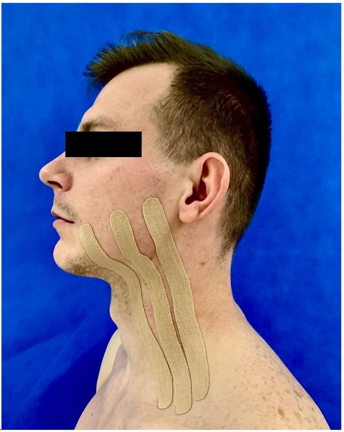
Int. J. Environ. Res. Public Health 2021, 18, 399                                                                                 2 of 13

                                        trismus. The results of previous observations in this area are promising; however, the
                                        number of available studies is very small, as is the size of the patient groups discussed in
                                        them [8].
                                              A physical method of treating selected postoperative complications using so-called
                                        dynamic taping (kinesio taping—KT) has been recently postulated in oral and maxillo-
                                        facial surgery. It is a therapeutic method that is widely used in sports medicine and the
                                        rehabilitation of the musculoskeletal system. It is used to prevent injuries, reduce pain
                                        after an injury, and increase the range of motion in joints, as well as to functionally increase
                                        performance [9,10].
                                              Kinesio tapes are thin, flexible tapes that can be stretched by up to 30–40% of their
                                        original length. After their application to the skin, they do not limit mobility of the body
                                        area receiving treatment. They are made of cotton covered with hypoallergenic glue. The
                                        thickness, specific weight, and extensibility of the tapes are similar to the properties of the
                                        epidermis [11–13]. The mechanism of the tape is based on supporting the body’s natural
                                        self-healing processes [14]. The therapeutic effect of the tape is to improve the flow of
                                        blood and lymph at the site of its application by pulling the skin away from the muscles
                                        and subcutaneous tissue, as well as by causing pressure and stretching the skin, which lead
                                        to the activation of mechanoreceptors through the central nervous system, which leads to
                                        increased excitability muscles [15]. A decrease in the level of perceived pain occurs due
                                        to the reduction of pressure on nociceptors [16]. The tapes are waterproof, air-permeable,
                                        and do not disturb thermoregulation. They should be kept on the skin for four-to-five
                                        days [17]. Kinesiology taping is a non-invasive and cheap method. Another advantage is
                                        its continuity of therapy, which allows it to exert a 24 h therapeutic effect.
                                              In the literature, there have been few reports on the effectiveness of kinesio taping
                                        after surgical tooth extraction [8]. The positive results of preliminary reports on the use of
                                        kinesio tape in the surgery of the lower wisdom tooth became an inspiration to research
                                        our clinical material. The aim of the study was to assess the impact of kinesio taping on the
                                        level of postoperative swelling of soft tissues, trismus, and pain in patients undergoing a
                                        surgical extraction of an impacted mandibular third molar.

                                        2. Materials and Methods
                                             The research was carried out at the Department of Oral Surgery after obtaining a
                                        positive opinion from the Bioethics Committee (opinion no. KB-0012/152/13, as amended.
                                        KB-0012/135/15)

                                        2.1. Patient Recruitment and Study Groups
                                             Participation in the research project was offered to 100 consecutive Caucasian patients
                                        (male—26; female—74). The study group included 36 women and 14 men, while the control
                                        group included 38 women and 12 men undergoing the surgical removal of an asymptomatic,
                                        impacted mandibular third molar, usually for orthodontic reasons. The exclusion criteria
                                        included: age below 18 years; unstable arterial hypertension; pregnancy; allergy to local
                                        anesthetics; patients with contraindications for outpatient surgery; pericoronitis; and those
                                        who, based on a preoperative radiological examination, needed no bones removed during
                                        surgery.
                                             The eligible patients were randomly assigned to one of the two groups:
                                        1.     Study group (n = 50): kinesio tape applied for five days after surgery
                                        2.     Control group (n = 50): non-kinesio tape applied.
                                               Patients in each group were asked about their age and gender.

                                        2.2. Surgery
                                             The surgical procedure was performed according to the same repeatable scheme for
                                        each patient by one of the two oral surgery specialists with the same degree of professional
                                        experience. The surgical procedure was performed under the local anesthesia of the inferior
                                        alveolar nerve and the buccal nerve using 2% lidocaine with 1:80,000 noradrenaline. The
Int. J. Environ. Res. Public Health 2021, 18, 399 3 of 13
incision was made on the ridge of the mandibular alveolus lateral from the linea obliqua
towards the second molar. Then, a release incision was made in the distal part of second
molar, downward and in mesial direction for approximately 1 cm according to the same
repeatable scheme for each patient. The bone casing of the mandible was removed using
a drill mounted on the handpiece with cooling including a sterile physiological saline
solution according to the same protocol for each patient. After the surgery, the wound was
closed with sutures for a period of 7 days. A risk of increased bleeding was not expected.
No additional hemostatic materials were used to exclude and minimize the influence of
other factors on the test results. The time of each wisdom tooth removal was measured in
minutes. In addition to the standard recommendations for the procedure after the tooth
extraction, patients in both groups were treated with an antiseptic rinse based on 0.1%
chlorhexidine solution, as well as ketoprofen in a 100 mg dose taken twice daily, were on a
semi-liquid diet, and avoid physical exertion for seven days after treatment.
2.3. Kinesiotaping
Kinesio Tape K-Active Tape Classic (Nitto Denko Corporation, Osaka, Japan; K-Active
Europe GmbH, Hösbach, Germany), 50 mm × 5 m was applied immediately after the
wisdom tooth removal in the KT group. The skin before application was cleaned with
sterile gas soaked in saline. For every particular patient, the tape was prepared before its
application. In order to apply the tapes, a technique mapped from the technique used by
Ristow et al. was employed [18]. The 50 mm-wide tape was cut into 3 equal pieces forming
smaller stripes. The application of the tape was started in the area of supraclavicular lymph
nodes. The tape was then advanced to line A on the patient’s face where the greatest edema
was expected. The method of application is shown in Figure 1. The degree of tension in the
tape was approximately 15% of the maximum stretch. Tape length (x) was calculated by
Equation (1). The distance from the area of supraclavicular lymph nodes to line A on the
patient’s face (y) was 115%. The tape length needed to create 15% tension was calculated
from the formula:
100% × y [cm]
. (1)
115%
The tape was prepared according to the calculations, which was about 86.96% of
the y-distance. During application, the tape was stretched in such a way that it spanned
the entire y-distance. The tapes were kept for five days after surgery. In both groups,
measurements of swelling, trismus, and pain were performed before the surgery and on
the 3rd and 7th postprocedural days.
2.4. Data Collection
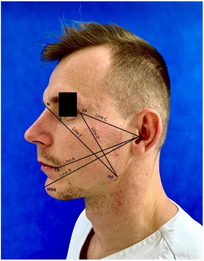
2.4.1. Edema
For detailed evaluations of the post-operative edema of the facial soft tissues, appro-
priate measurements of the patient’s face were taken before each procedure (Figure 2). Each
measurement was performed with an elastic measuring tape. All patients were located in
the same position. The measurements of swelling were performed using five lines mapped
out on the patient’s face. All points were connected with the lines.
1. Line A—from tragus (T) to cheilon (Ch)
2. Line B—from tragus (T) to pogonion (WPg)
3. Line C—from tragus (T) to exocanthion (Ex)
4. Line D—from exocanthion (Ex) to gonion (Go)
5. Line E—from gonion (Go) to nasion (N)Int. J. Environ. Res. Public Health 2021, 18, 399 4 of 13
Figure 1. Method of kinesio tape application.Int. J. Environ. Res. Public Health 2021, 18, 399 5 of 13
Figure 2. Measurement points and lines.
2.4.2. Trismus
The maximum jaw opening—the distance between the incisal edges of the upper
medial incisors and the incisal edges of the lower medial incisors in the midline of the
body—was measured with a caliper.
2.4.3. Pain
To assess the impact of the method on the postoperative pain level, each participant
determined the pain level according to the VAS (Visual Analogue Scale). The patient
marked a point which, in their opinion, corresponded to their pain level. The patient was
asked to mark a point on a 100 mm horizontal line, with the left pole showing no symptoms
at all and the right pole showing “unbearable” symptoms.Int. J. Environ. Res. Public Health 2021, 18, 399 6 of 13
2.5. Statistical Methodology
Statistical analysis was performed with the R statistical package (The R Foundation
for Statistical Computing, Indianapolis, IN, USA, 2012). The variables were described
using position measures, i.e., arithmetic mean and quartiles with the median, standard
deviation (SD), and the minimum and maximum values. In all analyses, the level of
statistical significance was assumed to be 0.05. The qualitative variables were compared in
both groups using the chi-quadrant test. A comparative analysis of edema between the two
groups was performed using the Mann–Whitney test after the prior confirmation of the
non-normality of the data distribution using the Shapiro–Wilk test. The differences between
the groups with normal distributions of the variable were analyzed using Student’s t-test.
On the other hand, the Mann–Whitney test was used if the values of the maximum jaw
opening and pain symptoms did not show a normal distribution in the analyzed groups.
3. Results
3.1. Baseline Characteristics
The study group included a total of 100 patients: 50 patients with KT and 50 patients
without KT. The study group included 36 women and 14 men, while the control group in-
cluded 38 women and 12 men. The chi-square test did not show any significant differences
in this respect between the two groups (p = 0.82). The age of the patients in the study group
fluctuated in the range of 19–59 years (median of 26.5 years), while in the control group,
it was 18–38 years (median of 25 years). Both groups showed no statistically significant
differences for age (p = 0.221; Mann–Whitney test). The duration of the treatment in both
groups was similar; in the study group, it ranged from 10 to 60 min, whereas in the control
group, it ranged from 6 to 60 min. A statistical analysis showed no statistically significant
differences in this respect between the two groups (p = 0.801; Mann–Whitney test). In
the study group, a total of thirty teeth—38 and twenty teeth—48 were removed, while
in the control group, it was 28 and 22, respectively. The chi-square test did not show
any statistically significant differences between the two groups in this respect (p = 0.839).
The complete characteristics of the sample with the results of statistical calculations are
presented in Table 1.
Table 1. Characteristics of the study group.
Study Group (n = 50) Control Group (n = 50) Total (n = 100)
Feature p*
Median Min–Max Median Min–Max Median Min–Max
Age 26.5 19–59 25 18–38 25.5 18–59 0.221
Operation time [min] 21 10–60 24.5 6–60 23 6–60 0.801
Feature n % n % n % p **
Woman 36 72.00% 38 76.00% 74 74.00% 0.82
Sex
Man 14 28.00% 12 24.00% 26 26.00%
Tooth 38 30 60.00% 28 56.00% 58 58.00% 0.839
number 48 20 40.00% 22 44.00% 42 42.00%
* Not normal variable distribution, p value from the Mann–Whitney test; ** chi-square test; n—number of patients; Min—minimum value;
Max—maximum value; and p—significance level.
3.2. Analysis of Postoperative Edema Measurements
The conducted analysis showed that on the third day after surgery, there was a
statistically significant difference in the length of the D and E lines between both groups.
The mean length of the D line in the control group was 10.35 cm, and for the E line it was
15.44 cm, whereas in the study group the mean lengths were 9.99 and 14.89 cm, respectively.
The significance levels were lower than 0.05 and were p = 0.008 and p = 0.005 for the D and
E lines, respectively. The analysis of the differences in the measurements of the lines from
A to C showed no statistical significance. The detailed results of the statistical analysis in
this regard are summarized in Table 2.Int. J. Environ. Res. Public Health 2021, 18, 399 7 of 13
Table 2. Measurement results of A–E line on the 3rd day between the two groups.
Measurement on
Group n Mean SD Median Min Max Q1 Q3 p*
the 3rd Day (cm)
study 50 11.75 0.78 11.55 10.45 13.5 11.1 12.4
Line A p = 0.336
control 50 11.89 0.91 11.78 10 14.5 11.45 12.5
study 50 15.42 1.07 15.07 13.6 18 14.56 16
Line B p = 0.304
control 50 15.52 0.84 15.43 14 18.05 15 16
study 50 8.42 0.89 8.35 6.95 11.25 7.78 8.91
Line C p = 0.724
control 50 8.45 0.67 8.22 7 10 8 9
study 50 9.99 0.75 10 8.5 12 9.5 10.45
Line D p = 0.008
control 50 10.35 0.79 10.22 8 12 10 10.97
study 50 14.89 0.9 15 12.5 16.6 14.16 15.5
Line E p = 0.005
control 50 15.44 0.9 15.5 13.5 16.9 14.75 16.24
* Mann–Whitney test; n—number of patients; SD—standard deviation; Min—minimum value; Max—maximum value; Q1—first quartile;
Q3—third quartile; p—significance level.
It was shown that the E line was significantly longer in the control group (p = 0.029;
Mann–Whitney test). Both the median and the arithmetic mean were higher compared
to the study group. Concerning the remaining lines (A–D), no statistically significant
differences were found between the two groups on seventh day. The detailed results of the
statistical analysis in this regard are summarized in Table 3.
Table 3. Measurement results of line A–E on 7th day between the two groups.
Measurement on
Group n Mean SD Median Min Max Q1 Q3 p*
the 7th Day (cm)
study 50 11.52 0.76 11.5 10 13.4 11 12
Line A p = 0.964
control 50 11.53 0.87 11.5 9.75 13.75 10.96 12.04
study 50 15.15 1.13 15 12 18 14.5 15.76
Line B p = 0.714
control 50 15.02 0.86 15 13.25 17.05 14.5 15.57
study 50 8.2 0.74 8.15 7 10 7.55 8.66
Line C p = 0.732
control 50 8.22 0.54 8.07 7 9.25 8 8.5
study 50 9.8 0.77 10 8.25 11.5 9.25 10.15
Line D p = 0.227
control 50 9.93 0.93 10 7.75 11.5 9.51 10.57
study 50 14.57 0.95 14.53 12.1 16.6 14 15.24
Line E p = 0.029
control 50 15 0.94 15 12.8 16.5 14.26 16
* Mann–Whitney test; n—number of patients; SD—standard deviation; Min—minimum value; Max—maximum value; Q1—first quartile;
Q3—third quartile; p—significance level.
3.3. Analysis of Postoperative Trismus Measurements
The measurement of the baseline size (before the procedure) of the jaw opening did
not show any statistically significant differences between the two groups. The mean value
in the study group was 4.73 cm (±0.72), whereas in the control group, it was 4.5 cm (±0.56).
It was shown that both on the third and seventh days after the procedure, the jaw opening
level was significantly higher in the study group. The mean jaw opening value on the
third day after surgery among patients with tapes applied was 2.93 cm (±1.08), whereas
in the group without tapes, it was 2.42 cm (±0.69). On the seventh day after surgery, the
degree of trismus disappeared in both groups. It was reduced, but, in the study group, the
average degree of jaw opening was still significantly higher (3.97 cm ±1.03) than in the
control group, where it was 3.55 cm (±0.81).
The detailed results of the statistical analysis in this regard are summarized in Table 4.Int. J. Environ. Res. Public Health 2021, 18, 399 8 of 13
Table 4. The results of comparative analysis of the jaw opening before the procedure on the 3rd and 7th days.
Jaw Opening (cm) Group n Mean SD Median Min Max Q1 Q3 p*
study 50 4.73 0.72 4.62 3.5 6.5 4.01 5
Baseline p = 0.135
control 50 4.5 0.56 4.43 3.75 6.1 4 4.93
study 50 2.93 1.08 3 1 5 2.02 3.73
3rd day p = 0.012
control 50 2.42 0.69 2.5 1.25 4 1.85 2.79
study 50 3.97 1.03 4.08 1.5 6.35 3.06 4.74
7th day p = 0.02
control 50 3.55 0.81 3.6 1.5 6 3 4
* Mann–Whitney test; n—number of patients; SD—standard deviation; Min—minimum value; Max—maximum value; Q1—first quartile;
Q3—third quartile; p—significance level.
3.4. Analysis of Postoperative Pain Measurements
An analysis of the initial (before the procedure) pain intensity level measured with
the VAS showed no statistically significant differences between the two groups (p = 0.065;
Mann–Whitney test). The mean value in the study group was 5.2 (±10.74), whereas in
the control group, it was 1.6 (±5.48). A statistically significant difference in the level of
pain was demonstrated between the study groups on the third day after the procedure
(p = 0.003; Mann–Whitney test). In the study group, the average pain level measured by
the VAS was 37.6 (±25.36), whereas in the control group, it was 52 (±23.82). However, no
statistically significant differences concerning the discussed parameter were found between
the patients in both groups on the seventh day after surgery. Table 5 presents the detailed
results of the statistical analysis in this regard.
Table 5. The results of the comparative analysis of the pain intensity level measured with the Visual Analogue Scale (VAS)
before the procedure, on the 3rd and 7th days after the procedure between the control group and the study group.
Level of Pain
Group n Mean SD Median Min Max Q1 Q3 p*
Intensity (VAS)
study 50 5.2 10.74 0 0 30 0 0
Baseline p = 0.065
control 50 1.6 5.48 0 0 20 0 0
study 50 37.6 25.36 30 0 90 20 50
3rd day p = 0.003
control 50 52 23.82 50 0 100 40 70
study 50 16.8 20.04 20 0 80 0 27.5
7th day p = 0.06
control 50 25 22.7 20 0 80 0 47.5
* Mann–Whitney test; n—number of patients; SD—standard deviation; Min—minimum value; Max—maximum value; Q1—first quartile;
Q3—third quartile; p—significance level.
4. Discussion
Nowadays, the surgical extraction of a lower third molar is the most commonly
performed oral surgery procedure. Due to phylogenetic changes in the jawbones, the
incidence of retention of third molars is constantly increasing. This has contributed to
an increase in the medical needs of the surgical extraction of wisdom teeth [1–3]. This
procedure very often leads to postsurgical, non-infectious complications, such as facial
swelling, trismus, and post-extraction pain.
While the risk of infectious complications in immunocompetent patients is low, the
occurrence of certain non-infectious complications such as post-extraction pain, trismus,
and the swelling of the soft facial tissues is extremely common in the postoperative period.
Infectious complications in immunocompetent patients develop in less than 4% of cases and
are mainly related to infections of the surgical wound [19]. They may appear in the form of
an abscess [20] or osteitis, and they are much less frequently systemic complications [21];
therefore, they usually affect immunocompetent patients. Incidentally, there are long-term
infectious complications associated with intraoperative bacteremia, but this applies to
a small group of patients predisposed to, for example, infective endocarditis [22] or an
infection of the implanted artificial joint [23].Int. J. Environ. Res. Public Health 2021, 18, 399 9 of 13
Though non-infectious complications most often do not pose a threat to a patient’s
health and are very common, they constitute a major therapeutic problem and significantly
reduce the patient’s postoperative quality of life [1]. They are much more common in the
extraction of lower wisdom teeth and are normally post-extraction pains, as well as trismus
and the swelling of the surrounding soft tissues (edema). Among the much less common
complications from the group of non-infectious agents, one should also mention post-
extraction bleeding (sanguinatio post extractionem) and so-called dry alveolitis (alveolitis
sicca) [1–3].
The methods currently used for these indications often lead to side effects associated
with discomfort for the patient, especially in the case of pharmacotherapy (antibiotic,
non-opioid analgesic, or steroid) [5]. Post-extraction pain is often an indication for taking
analgesics, especially from the group of non-steroidal anti-inflammatory drugs (NSAIDs),
which, in turn, are one of the most toxic groups of drugs to many systems and organs,
especially the gastrointestinal tract [24]. The matter is further complicated by the fact that
these drugs are often used in so-called self-medication (self-treatment carried out without
medical supervision), which results in their abuse, an exceeding of maximum doses, and a
risk of dangerous interactions with other medications taken by patients. Trismus, on the
other hand, usually impedes speech and food intake, further reducing the postoperative
quality of life in patients [25]. The location of edema in the visible area of the face means
that after the surgical extraction of an impacted lower wisdom tooth, patients are often on
sick leave and avoid contact with the environment. All this results in the fact that patients
feel a great fear of possible treatments of this type.
There is a great need to introduce a minimally invasive method to prevent the oc-
currence of non-infectious complications after surgical wisdom tooth extraction. One of
them may be the dynamic taping method (kinesiology taping), which, until recently, was
only used in sports medicine [10,26,27]. Currently, it is assumed that the above-mentioned
non-infectious complications constitute a physiological response of the body to surgical
trauma [3]. According to Bortoluzzi et al., more than 50% of patients after the surgical
extraction of an impacted lower wisdom tooth on the first days after surgery experience
moderate-severe to severe pain [28]. Trismus of varying severity is observed in the majority
of patients after surgery, and in an analysis of Osunde and Saheeb, it occurred in all patients
under study [24].
Initially, KT was used mainly in physiotherapy practice and sports. Its action was
mainly used to heal injuries. Currently, a small amount of research in the literature deals
with KT in the maxillofacial area, especially with the third molars in the mandible [8].
Ristow [18] was the first author to introduce dynamic taping methods to the surgery
of the mandibular third molar.
Authors have provided different taping techniques in their research centers, and some
of them have not provided their technique [29]. Some authors have not provided the degree
of tape tension. They applied tapes by pulling the skin away from the subcutaneous tissue,
thus facilitating lymph flow (anti-edema effect) and reducing pressure on nociceptors
(analgesic effect) [8].
Similarly to the report by Ristow et al. [18], in our study, the method of applying three
tapes with a width of about 1.5 cm was used. In studies by Genc and Erdil, their tension
was reduced to 15%, which is currently used in the lymphatic technique [14,30,31]. All
the techniques mentioned by the authors contributed to the reduction of postoperative
edema. It would seem reasonable to carry out studies comparing different techniques of
KT application.
Researchers have disagreed about the length of time that patient should be taped.
Genc and Erdil removed tapes after two days [32,33]; Tatli [34], Gözlüklü [35], de Rocha
Heras [36], and Ristow [18] removed tapes after five days; and Yurttutan [37] removed
tapes after seven days.
In this study, as in most publications, the tapes were removed after five days. Fur-
ther research on the effectiveness of kinesio taping in counteracting post-ascending com-Int. J. Environ. Res. Public Health 2021, 18, 399 10 of 13
plications should be conducted because the literature in regard to the study groups
(13–76 patients) is limited in number. In the original study, as many as 100 patients were
analyzed. 50 of them were in the study group and 50 in the control group. In the literature,
the authors have unanimously agreed that the most severe swelling occurs after two-to-
three days following the surgical removal of the third molar in the mandible [38]. Most
authors also agree on the seven-day follow up [18,29,32–35,37,39,40].
In our study, the removal of the third molar was performed unilaterally. A study of
the application of two therapies or a comparison of the application of KT therapy to a
procedure without the use of KT in one patient in split-mouth studies could give the best
results due to the possibility of comparing the therapeutic effect. Each patient feels pain
differently, which may cause inaccuracies in results. In our study, as in most of the studies
presented in the literature, the pain sensation was determined with the VAS to standardize
this subjective feeling.
Ristow showed significantly less pain, trismus, and swelling after the surgical extrac-
tion of the wisdom tooth on the third day after surgery in the group with KT complications
than in the control group [18]. Similar conclusions in the split-mouth randomized con-
trolled trial (RCT )study were reached by da Rocha Heras et al. [36]. The authors also
used other methods of applying the tapes. Gozuklu et al. used a modified method of KT
application, the effectiveness of which they compared with the classic application [35]. The
new method was more effective in reducing postoperative complications than the classic
method [35].
The cited data show that the kinesiology taping method significantly reduces the level
of soft tissue swelling after maxillofacial surgery, and it is also likely to reduce postoperative
pain and trismus [8].
Some of the articles concerned the influence of patients’ age and the duration of the
surgical removal of wisdom teeth on the severity of oedema, jaw compression, and pain
after surgery [40]. A study by Bello et al. did not show any significant statistical effect of
the duration of surgery on postoperative swelling and maxillofacial pain (p > 0.05). It was
also revealed that the level of postoperative pain was statistically significant, depending on
the duration of the procedure, as a longer time was associated with more severe pain [41].
In our study, due to the fact that the study and control groups did not differ in terms
of age and duration, it can be concluded that only the use of kinesio tape patches had an
effect on parameters such as maxilla, pain, and swelling.
Though the study group was not very large (n = 100), to the authors’ knowledge, it
was the largest of the reported studies so far. In addition, the study could be expanded to
include the determination of the degree of retention of the third molar in the mandible, as
well as the classification of the position of the impacted third molar in relation to inferior
alveolar nerve (IAN).
It should also be taken into account that few authors have reported the results of edema
measurement with and without KT as a mean of all linear measurements [32,33,35–37,39,40].
In our study, as well as in Tatli’s study [34], the results were related to each linear mea-
surement separately, which allowed us to separately determine the effect of KT on the
occurrence and size of edema in each area. The effect of KT on the occurrence of postop-
erative pain was also emphasized: KT applications reduced the pressure on nociceptors
(analgesic effect). Because flexible KT bands are not invisible, the placebo effect in the assess-
ment of post-treatment pain cannot be ruled out. Patients’ lack of credibility was excluded
in the Tatli study on a group of 60 patients. The authors in the study group used the placebo
effect to rule out patients’ unreliability [34]. In our study, the edema was measured with
an elastic band, but establishing precise measurement points increased the reliability of the
results. Introducing volumetric measurements using 3D scanners and by comparing the
color maps of stereolithography (STL) files obtained during scanning would allow one to
achieve an even greater minimization of the risk of measurement errors [42–44].Int. J. Environ. Res. Public Health 2021, 18, 399 11 of 13
5. Conclusions
The kinesio tape method is non-invasive, continuously active throughout the entire ap-
plication period, and requires no additional patient appointments. Kinesio tape application
after the surgical extraction of the third lower molar has a significant effect on decreasing
facial swelling on the third day after surgery, decreasing the trismus level on the third and
seventh days after surgery and decreasing pain severity on the third and seventh days after
surgery. The KT application is an effective method for reducing postoperative edema, pain,
and trismus after impacted mandibular wisdom teeth surgery.
Author Contributions: Conceptualization, G.T. and O.P.; methodology, G.T. and O.P.; software,
A.J. and E.G.; validation, G.T. and A.J.; formal analysis, O.P. and A.J.; investigation, G.T. and O.P.;
resources, A.J. and E.G.; data curation, O.P. and A.J.; writing—original draft preparation, O.P. and
A.J.; writing—review and editing, G.T.; visualization, O.P., E.G., and A.J.; supervision, G.T.; project
administration, G.T. All authors have read and agreed to the published version of the manuscript.
Funding: This research received no external funding.
Institutional Review Board Statement: The study was conducted according to the guidelines of the
Declaration of Helsinki, and approved by the Ethics Committee of Pomeranian Medical University
in Szczecin, Poland (KB-0012/152/13 with changes KB-0012/135/15).
Informed Consent Statement: The study was conducted according to the guidelines of the Decla-
ration of Helsinki, and approved by the Ethics Committee of Pomeranian Medical University in
Szczecin, Poland (KB-0012/152/13 with changes KB-0012/135/15).
Data Availability Statement: The data presented in this study are available on request from the
corresponding author.
Conflicts of Interest: The authors declare no conflict of interest.
References
1. Bui, C.H.I.; Seldin, E.; Dodson, T. Types, frequencies, and risk factors for complications after third molar extraction. J. Oral.
Maxillofac. Surg. 2003, 61, 1379–1389. [CrossRef]
2. Yuasa, H.; Sugiura, M. Clinical postoperative findings after removal of impacted mandibular third molars: Prediction of
postoperative facial swelling and pain based on preoperative variables. J. Oral. Maxillofac. Surg. 2004, 42, 209–214. [CrossRef]
3. Zandi, M.; Amini, P.; Keshavarz, A. Effectiveness of cold therapy in reducing pain, trismus, and edema after impacted mandibular
third molar surgery: A randomized, self-controlled, observer-blind, split-mouth clinical trial. J. Oral. Maxillofac. Surg. 2015, 45,
118–123. [CrossRef]
4. Osunde, O.; Saheeb, B.; Bassey, G. Indications and risk factors for complications of lower third molar surgery in a nigerian
teaching hospital. Ann. Med. Health Sci. Res. 2014, 4, 938–942. [CrossRef]
5. Miloro, M.; Ghali, G.E.; Larsen, P.E.; Waite, P.D. Peterson’s Principles of Oral and Maxillofacial Surgery, 2nd ed.; BC Decker:
London, UK, 2004.
6. Gbotolorum, O.M.; Arotiba, G.T.; Ladeine, A.L. Assessment of factors associated with surgical difficulty in impacted mandibular
third molar extraction. J. Oral. Maxillofac. Surg. 2007, 65, 1977–1983. [CrossRef]
7. Jaroń, A.; Aniko-Włodarczyk, M.; Preuss, O.; Trybek, G. Radiological assessment of the type of retention and the degree
of difficulty of surgical removal of an impacted wisdom tooth in the mandible extended with three-dimensional modeling.
(Ocena radiologiczna typu retencji oraz stopnia trudności operacyjnego usuni˛ecia zatrzymanego z˛eba madrości ˛ w żuchwie
poszerzona o modelowanie trójwymiarowe.). In Proceedings of the Kongres Polskiego Towarzystwa Chirurgii Stomatologicznej i
Szcz˛ekowo-Twarzowej, Lublin, Poland, 9–11 May 2019.
8. Jaroń, A.; Jedliński, M.; Grzywacz, E.; Mazur, M.; Trybek, G. Kinesiology Taping as an Innovative Measure against Post-Operative
Complications after Third Molar Extraction—Systematic Review. J. Clin. Med. 2020, 9, 3988. [CrossRef]
9. Osorio, J.A.; Vairo, G.L.; Rozea, G.D.; Bosha, P.J.; Millard, R.L.; Aukerman, D.F.; Sebastianelli, W.J. The effects of two therapeutic
patellofemoral taping techniques on strength, endurance, and pain responses. Phys. Ther. Sport 2013, 14, 199–206. [CrossRef]
10. Lee, M.S.; Lee, J.H. Ankle inversion taping using kinesiology tape for treating medial ankle sprain in an amatour soccer player.
J. Phys. Ther. Sci. 2015, 27, 2407–2408. [CrossRef]
11. Kiebzak, W.; Kowalski, I.M.; Pawłowski, M.; Gasior,
˛ J.; Zaborowska-Sapeta, K.; Wolska, O.; Śliwiński, Z. Wykorzystanie metody
Kinesiology Tapingu w praktyce fizjoterapeutycznej: Przeglad ˛ literatury. Fizjoter Pol. 2012, 12, 1–11.
12. Hałas, I.; Senderek, T.; Krupa, L. Wykorzystanie kinesiotapingu w usprawnianiu pacjentki po rekonstrukcji nerwu twarzowego.
Fizjoter Pol. 2005, 5, 272–276.Int. J. Environ. Res. Public Health 2021, 18, 399 12 of 13
13. Tozzi, U.; Santaga, M.; Sellitto, A.; Tartaro, G.P. Influence of Kinesiologic Tape on postoperative swelling after orthognatic surgery.
J. Oral. Maxillofac. Surg. 2015, 15, 52–58. [CrossRef]
14. Mikołajewska, E. Kinesiotaping; Wydawnictwo Lekarskie PZWL: Warszawa, Poland, 2011.
15. Gómez-Soriano, J.; Abián-Vicén, J.; Aparicio-García, C.; Ruiz-Lázaro, P.; Simón-Martínez, C.; Bravo-Esteban, E.; Fernández-
Rodríguez, J.M. The effects of Kinesio taping on muscle tone in healthy subjects: A double-blind, placebo-controlled crossover
trial. Man. Ther. 2014, 19, 131–136. [CrossRef]
16. Zhang, S.; Fu, W.; Pan, J.; Wang, L.; Xia, R.; Liu, Y. Acute effects of Kinesio taping on muscle strength and fatigue in the forearm of
tennis players. J. Sci. Med. Sport 2015, 19, 459–464. [CrossRef]
17. Śliwiński, Z.; Krajczy, M. Dynamiczne Plastrowanie, Podr˛ecznik Kinesiology Taping; Markmed: Wrocław, Poland, 2014.
18. Ristow, O.; Hohlweg-Majert, B.; Stürzenbaum, S.R.; Kehl, V.; Koerdt, S.; Hahnefeld, L.; Pautke, C. Therapeutic elastic tape reduces
morbidity after wisdom teeth removal—A clinical trial. Clin. Oral. Investig. 2014, 18, 1205–1212. [CrossRef]
19. Adeyemo, W.L.; Ladeinde, A.L.; Ogunlewe, M.O. Clinical evaluation of post-extraction site wound healing. J. Contemp. Dent.
Pract. 2006, 7, 40–49. [CrossRef]
20. Trybek, G.; Chamarczuk, A.; Falkowska, J.; Grzegorzewska, M.; Preuss, O.; Aniko-Włodarczyk, M. Intraoral odontogenic
abscesses in patients of The Department of Oral Surgery at the Pomeranian Medical University in Szczecin: 7 years of observation.
Postep. Hig. Med. Dosw. 2018, 72, 491–498. [CrossRef]
21. Trybek, G.; Chruściel-Nogalska, M.; Machnio, M.; Smektała, T.; Malinowski, J.; Tutak, M.; Sporniak-Tutak, K. Surgical extraction of
impacted teeth in elderly patients. A retrospective analysis of perioperative complications—The experience of a single institution.
Gerodontology 2016, 33, 410–415. [CrossRef]
22. Chen, P.C.; Tung, Y.C.; Wu, P.W.; Wu, L.S.; Lin, Y.S.; Chang, C.J.; Kung, S.; Chu, P.H. Dental procedures and the risk of infective
endocarditis. Medicine 2015, 94, 1826–1832. [CrossRef]
23. Little, J.W.; Jacobson, J.J.; Lockhart, P.B. The dental treatment of patients with joint replacements: A position paper from American
Academy of Oral Medicine. JADA 2010, 141, 667–671. [CrossRef]
24. Osunde, O.D.; Saheeb, B.D. Effect of age, sex and level of surgical difficulty on inflammatory complications after third molar
surgery. J. Maxillofac. Oral. Surg. 2015, 14, 7–12. [CrossRef]
25. Al-Khateeb, T.H.; Nusair, Y. Effect of the proteolytic enzyme serrapeptase on swelling, pain and trismus after surgical extraction
of mandibular third molars. Int. J. Oral. Maxillofac. Surg. 2008, 37, 264–268. [CrossRef]
26. Garczyński, W.; Lubkowska, A.; Dobek, A.; Andryszczyk, M. Wpływ aplikacji kinesiology tapingu technika˛ mi˛eśniowa˛ na
zakres ruchomości l˛edźwiowego odcinka kr˛egosłupa oraz subiektywne odczuwanie nat˛eżenia bólu u chorych z dolegliwościami
bólowymi kr˛egosłupa. Ann. Acad. Med. Stetin. 2014, 2, 19–24. [CrossRef]
27. Świerczyńska, A.; Kłusek, R.; Czachor, T.; Gajda, B. Kinezjotaping jako jedna z metod leczenia urazów rdzenia kr˛egowego. Prz.
Lek. 2011, 68, 1144–1148.
28. Bortoluzzi, M.C.; Guollo, A.; Capella, D.L.; Manfro, R. Pain levels after third molar surgical removal: An evaluation of predictive
variables. J. Contemp. Dent. Pract. 2011, 12, 239–244. [CrossRef]
29. Mohammed, I.A.; Delemi, Z.H. Kinesiology tape in comparison with oral Diclofenac sodium in reducing swelling after surgical
removal of lower wisdom teeth. Al Rafidain Dent. J. 2019, 19, 90–97. [CrossRef]
30. Kase, K.; Wallis, J.; Kase, T. Clinical Therapeutic Applications of the Kinesio Taping Method, 2nd ed.; Ken Ikai: Tokyo, Japan, 2003.
31. Markowski, A. Kinesiotaping; Wydawnictwo SBM: Warszawa, Poland, 2015.
32. Genc, A.; Cakarer, S.; Yalcin, B.K.; Kilic, B.B.; Isler, S.C.; Keskin, C. A comparative study of surgical drain placement and the use
of kinesiologic tape to reduce postoperative morbidity after third molar surgery. Clin. Oral. Investig. 2019, 23, 345–350. [CrossRef]
33. Erdil, A.; Akbulut, N.; Altan, A.; Demirsoy, M.S. Comparison of the effect of therapeutic elastic bandage, submucosal dexametha-
sone, or dexketoprofen trometamol on inflammatory symptoms and quality of life following third molar surgery: A randomized
clinical trial. Clin. Oral. Investig. 2020, 15. [CrossRef]
34. Tatli, U.; Benlidayi, I.C.; Salimov, F.; Guzel, R. Effectiveness of kinesio taping onpostoperative morbidity after impacted
mandibular third molar surgery: A prospective, randomized, placebo-controlled clinical study. J. Appl. Oral. Sci. 2020, 28,
e20200159. [CrossRef]
35. Gözlüklü, Ö.; Ulu, M.; Gözlüklü, H.Ö.; Yilmaz, N. Comparison of Different Kinesio Taping Techniques After Third Molar Surgery.
J. Oral. Maxillofac. Surg. 2020, 78, 695–704. [CrossRef]
36. Da Rocha Heras, A.C.T.; De Oliveira, D.M.S.; Guskuma, M.H.; De Araújo, M.C.; Fernandes, K.B.P.; Da Silva Junior, R.A.;
Andraus, R.A.C.; Maia, L.P.; Fernandes, T.M.F. Kinesio taping use to reduce pain and edema after third molar extraction surgery:
A randomized controlled split-mouth study. J. Craniomaxillofac. Surg. 2020, 48, 127–131. [CrossRef]
37. Yurttutan, M.E.; Sancak, K.T. The effect of kinesio taping with the web strip technique on pain, edema, and trismus after impacted
mandibular third molar surgery. Niger. J. Clin. Pract. 2020, 23, 1260–1265. [CrossRef]
38. Xue, P.; Wang, J.; Wu, B.; Ma, Y.; Wu, F.; Hou, R. Efficacy of antibiotic prophylaxis on postoperative inflammatory complications
in Chinese patients having impacted mandibular third molars removed: A split-mouth, double-blind, self-controlled, clinical
trial. Br. J. Oral. Maxillofac. Surg. 2015, 53, 416–420. [CrossRef]
39. Mohammed, I.A.; Delemi, Z.H. Kinesiology Tape in Comparison with Submucosal Injection of Dexamethasone in Reducing Pain
and Swelling After Surgical Removal of Impacted Lower Wisdom Teeth. Al Rafidain Dent. J. 2020, 20, 18–24. [CrossRef]Int. J. Environ. Res. Public Health 2021, 18, 399 13 of 13
40. Chiang, K.C.; Bhushan, N.S.; Kalyan, U.; Bhavana, R. Use of kinesiologic therapeutic tape on pain, trismus, swelling and its
influence on quality of life after mandibular third molar surgery. J. Clin. Diagn. Res. 2020, 14, 13–17. [CrossRef]
41. Bello, S.A.; Adeyemo, W.L.; Bamgbose, B.O.; Obi, E.V.; Adeyinka, A.A. Effect of age, impaction types and operative time on
inflammatory tissue reactions following lower third molar surgery. Head Face Med. 2011, 7, 1–8. [CrossRef]
42. Kau, C.H.; Cronin, A.; Durning, P.; Zhurov, A.I.; Sandham, A.; Richmond, S. A new method for the 3D measurement of
postoperative swelling following orthognathic surgery. Orthod. Craniofac. Res. 2006, 9, 31–37. [CrossRef]
43. Yip, E.; Smith, A.; Yoshino, M. Volumetric evaluation of facial swelling utilizing a 3-D range camera. Int. J. Oral. Maxillofac. Surg.
2004, 33, 179–182. [CrossRef]
44. Metlerski, M.; Grocholewicz, K.; Jaroń, A.; Lipski, M.; Trybek, G. Comparison of Presurgical Dental Models Manufactured with
Two Different Three-Dimensional Printing Techniques. J. Healthc. Eng. 2020, 2020, 8893338. [CrossRef]You can also read