The complex karyotype landscape in chronic lymphocytic leukemia allows to refine the risk of Richter syndrome transformation
←
→
Page content transcription
If your browser does not render page correctly, please read the page content below

The complex karyotype landscape in chronic lymphocytic
leukemia allows to refine the risk of Richter syndrome
transformation

by Andrea Visentin, Laura Bonaldi, Gian Matteo Rigolin, Francesca Romana Mauro,
Annalisa Martines, Federica Frezzato, Stefano Pravato, Leila Romano Gargarella, Maria
Antonella Bardi, Maurizio Cavallari, Eleonora Volta, Francesco Cavazzini, Mauro Nanni,
Monica Facco, Francesco Piazza, Anna Guarini, Robin Foà, Gianpietro Semenzato,
Antonio Cuneo, and Livio Trentin

Haematologica 2021 [Epub ahead of print]

Citation: Andrea Visentin, Laura Bonaldi, Gian Matteo Rigolin, Francesca Romana Mauro, Annalisa
Martines, Federica Frezzato, Stefano Pravato, Leila Romano Gargarella, Maria Antonella Bardi,
Maurizio Cavallari, Eleonora Volta, Francesco Cavazzini, Mauro Nanni, Monica Facco, Francesco
Piazza, Anna Guarini, Robin Foà, Gianpietro Semenzato, Antonio Cuneo, and Livio Trentin.
The complex karyotype landscape in chronic lymphocytic leukemia allows to refine the risk of Richter
syndrome transformation.
Haematologica. 2021; 106:xxx
doi:10.3324/haematol.2021.278304

Publisher's Disclaimer.
E-publishing ahead of print is increasingly important for the rapid dissemination of science.
Haematologica is, therefore, E-publishing PDF files of an early version of manuscripts that
have completed a regular peer review and have been accepted for publication. E-publishing
of this PDF file has been approved by the authors. After having E-published Ahead of Print,
manuscripts will then undergo technical and English editing, typesetting, proof correction and
be presented for the authors' final approval; the final version of the manuscript will then
appear in print on a regular issue of the journal. All legal disclaimers that apply to the
journal also pertain to this production process.
Visentin A. et al.                                                                               Complex karyotype subtypes and RS

The complex karyotype landscape in chronic lymphocytic
leukemia allows to refine the risk of Richter syndrome
transformation
Andrea Visentin
                        1,2,                        3                               4
                               Laura Bonaldi , Gian Matteo Rigolin , Francesca Romana Mauro , Annalisa
                                                                                                                        5
        3
Martines ,      Federica            Frezzato
                                             1,2,       Stefano       Pravato
                                                                             1,2,       Leila   Romano       Gargarella
                                                                                                                          1,2,   Maria

Antonella
                   4
              Bardi ,        Maurizio
                                                   4
                                          Cavallari ,          Eleonora
                                                                                4
                                                                           Volta ,      Francesco
                                                                                                                4
                                                                                                       Cavazzini ,   Mauro Nanni ,
                                                                                                                                    5
Monica     Facco
                     1,2,    Francesco      Piazza
                                                        1,2,   Anna   Guarini ,
                                                                               5    Robin
                                                                                                   5
                                                                                                Foà ,   Gianpietro    Semenzato
                                                                                                                                   1,2,
Antonio
                4
           Cuneo ,          Livio   Trentin
                                           1,2,+.
1 Hematology and Clinical Immunology Unit, Department of Medicine, University of Padua, Padua, Italy. 2 Veneto
Institute of Molecular Medicine, Padua, Italy. 3 Immunology and Molecular Oncology Unit, Veneto Institute of
Oncology IOV-IRCSS, Padua, Italy. 4 Hematology section, Department of Medical Sciences, Azienda Ospedaliera-
Universitaria, Arcispedale S. Anna, University of Ferrara, Ferrara. 5 Hematology division, Department of Precision
and Translational Medicine, "Sapienza" University, Rome, Italy.

Running Title        : Complex karyotype subtypes and RS

Keywords      : Richter syndrome, chronic lymphocytic leukaemia, complex karyotype, prognostic

factors, scoring system.

+Corresponding author:
Prof. Livio Trentin, M.D.

Hematology and Clinical Immunology Unit

Department of Medicine

University of Padua

Via Giustiniani, 2 - 35128 Padova

e-mail: livio.trentin@unipd.it

phone: 0039 049 821 2298

fax: 0039 049 821 1970

Abstract: 195 words

Main text: 3,634 words

Figures: 3

Tables: 2

Supplementary files: 2 tables and 5 figures

                                                                       1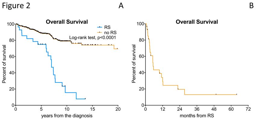
Visentin A. et al. Complex karyotype subtypes and RS
ACKNOWLEDGEMENTS
This work was supported by funds from Associazione Italiana per la Ricerca sul Cancro
(A.I.R.C.) projects to LT (IG-25024), Gilead fellowship program 2018 to LT, Special Program
‘Metastatic disease: the key unmet need in oncology’, AIRC 5x1000 (No. 21198) to RF, Fondo
di Ateneo per la Ricerca 2016, 2017 of the University of Ferrara to GMR and FC, Fondo di
Incentivazione alla Ricerca 2017 of the University of Ferrara to GMR, Ministero
dell’Istruzione, dell’Università e della Ricerca PRIN 2015 to AC (2015ZMRFEA). AV received a
research fellowship from the University of Padua supported by ONLUS Ricerca per Credere
nella Vita (RCV) odv, Padua, Italy.
ETHICAL APPROVAL AND CONSENT TO PARTICIPATE
This study was approved by the local research ethics committee of Padua hospital and
informed consent was obtained from all patients.
CONFLICT OF INTEREST
AV received honoraria from Janssen, Abbvie, Italfarmaco. LT received research funding by
Gilead, Roche, Janssen and Takeda, advisory board for Roche, Shire and Abbvie, Astrazeneca.
GMR received research funding by Gilead. FRM advisory board for Janssen, Shire and Abbvie.
AC advisory board and speaker bureau for Roche, Abbvie, Gilead and Janssen. GS board
member of Abbvie, Roche, Janssen and Celgene. RF advisory board or speaker bureau for
Roche, Abbvie, Celgene, Incyte, Amgen, Janssen, Gilead and Novartis.
DATA AVAILABILITY
The datasets generated and analyzed during the current study are not publicly
available due to the data protection and lack of consent from the patients. Access to data is
strictly limited to the researchers who have obtained permission for data processing.
AUTHORS' CONTRIBUTIONS
AV designed the study, performed statistical analysis, visited patients and wrote the
article; SP, LRG, MC, EV and FC and provided intellectual inputs and visited patients; LB, AM,
MAB and MN performed cytogenetic tests; FF, MF and AG performed cytofluorimetric and
IGHV analysis; FRM, GMR, PF, GS, RF, AC and LT visited patients, provided intellectual inputs
and reviewed the article.
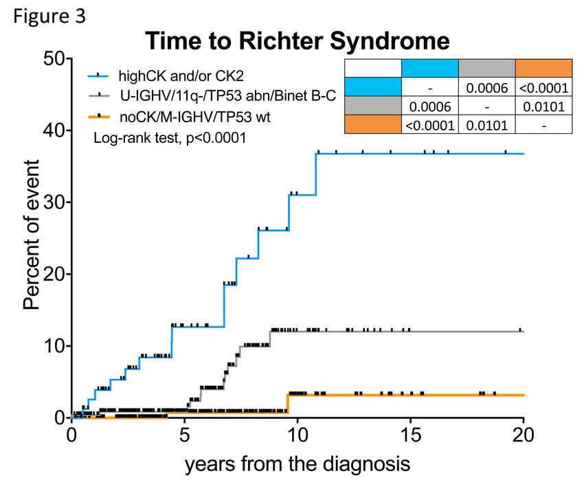
2Visentin A. et al. Complex karyotype subtypes and RS ABSTRACT Complex karyotype (CK) at chronic lymphocytic leukemia (CLL) diagnosis is a negative biomarker of adverse outcome. Since the impact of CK and its subtypes, namely type-2 CK (CK with major structural abnormalities) or high-CK (CK with ≥5 chromosome abnormalities), on the risk of developing Richter syndrome (RS) is unknown, we carried out a multicenter real- life retrospective study to test its prognostic impact. Among 540 CLL patients, 107 harbored a CK at CLL diagnosis, 78 were classified as CK2 and 52 as high-CK. Twenty-eight patients developed RS during a median follow-up of 6.7 years. At the time of CLL diagnosis, CK2 and high-CK were more common and predicted the highest risk of RS transformation, together with advanced Binet stage, unmutated (U)-IGHV, 11q-, TP53 abnormalities. We integrated these variables into a hierarchical model: high-CK and/or CK2 patients showed a 10-year time to RS (TTRS) of 31%; U-IGHV/11q-/TP53 abnormalities/Binet stage B-C patients had a 10-year TTRS of 12%; while mutated (M)-IGHV without CK and TP53 disruption a 10-year TTRS of 3% (p
Visentin A. et al.                                                                                 Complex karyotype subtypes and RS

INTRODUCTION
         Chronic lymphocytic leukemia (CLL), the most common leukemia in western countries,

is a remarkably heterogeneous disease, with some patients never requiring treatments and

others    with       a    highly          aggressive     and/or      rapidly         progressive     clinical    course(1,      2).    Richter

syndrome       (RS)           is    the   transformation       of      CLL      into   an   aggressive lymphoma, most commonly
resembling diffuse large B-cell lymphoma (DLBCL) or Hodgkin                                     lymphoma          (HL) variants(3, 4).

It is characterized by fast growing lymphadenopathies, 18-fluorodeoxyglucose (FDG) positron

emission       tomography                  computerized         tomography              (PET-CT)-avid        masses,       B    symptoms,

worsening       performance                  status    and    increased          lactate     dehydrogenase         levels      (5).    It    is   a

challenging task to distinguish RS from progressive CLL and it is even more difficult to study

prognostic markers since the frequency of RS transformation affects 2-10% of CLL patients

(5).

         Several studies have proven that chromosome banding analysis is able to refine the

prognostic      stratification              of   CLL   as    compared           to   fluorescence    in   situ   hybridization         (FISH)

analysis.     In fact, 22-36% of CLL cases with “normal”                                    FISH carry chromosomal aberration

following stimulated karyotypic analyses. In particular, complex karyotype (CK), defined by

the presence of at least 3 chromosome lesions in the same clone, is detectable in 14-34% of

CLL cases(6-9). The presence of a CK is both a negative prognostic and predictive biomarker

associated with an adverse outcome(6, 10) and worse response to chemoimmunotherapy(7,

11) as well as to novel agents(12, 13), regardless of the CLL-IPI score or IGHV mutational

status(8). However, the CK itself is a heterogeneous quantitative and qualitative cytogenetic

category       that           includes        numerical        (i.e.    monosomies             and    trisomies)        and      structural

abnormalities             (i.e.       balanced         and    unbalanced               translocations,       marker        chromosomes,

isochromosomes,                    deletions, insertions and additions). Recently, collaborative studies                                    have

demonstrated that among CK cases assessed at CLL diagnosis, those harboring 5 or more

chromosome abnormalities (high-CK)(14) or those with major structural abnormalities, also

called   type-2          CK        (CK2)(15,      16),   identify      highly         aggressive   disease       subsets    with       a    poor

outcome;      the        latter      is   also   characterized         by    a   peculiar    mRNA      expression       profile(15,          17).

Indeed,    most          of    the    patients     included     in     these         retrospective   studies     were      managed          with

chemoimmunotherapy(14-16). However, the presence of CK has been rarely associated to the

development of RS(18) and to date it is unknown whether CK subtypes, namely high-CK or

CK2, could help to identify patients at a higher risk of developing a RS at CLL diagnosis.

         In   this       multicenter          retrospective study,               we    documented      for   the   first   time       that    the

presence of a CK at CLL diagnosis is associated with an increased risk of developing a RS. In

                                                                            4
Visentin A. et al. Complex karyotype subtypes and RS
particular, patients with CK2 and high-CK had the highest likelihood of RS transformation.
Finally, we recapitulated clinico-biological variables associated with RS into a prognostic
model defining 3 statistically different classes of risk of developing RS, being the lowest risk
for IGHV gene mutated (M-IGHV) patients without any CK subtypes and absence of TP53
abnormalities, and the highest risk for patients harboring highCK and/or CK2 subtypes.
METHODS
Study design
Inclusion criteria for this study were diagnosis of CLL according to the 2008 iwCLL
guidelines(19), histologically confirmed diagnosis of RS (diffuse large B-cell lymphoma or
high-grade B-cell lymphoma), age >18 years and chromosome banding analysis performed
within 1 year from diagnosis in patients without features of disease progression. Data
included in the comparative analysis were gender, age, Binet stage(19), CLL treatment prior
to RS, 11q22-23 deletion by FISH(20), IGHV gene mutational analysis(21) and TP53
abnormalities including gene deletions (deletion 17p13) or mutations(22), β2-microglobulin
level >3.5mg/L. The primary endpoint was the impact of overall CK, CK2 and highCK on the
time to Richter syndrome (TTRS) transformation. The correlation of RS with clinical and
biologic variables and their impact on TTRS were secondary endpoints. This study was
approved by the local research ethic committee and informed consents were obtained from
all patients.
Chromosome banding analysis
Cytogenetic analysis was performed on peripheral blood after a 72h exposure to
500µM CpG ODN DSP30 (Roche, Risch, CH) mitogen + 20U/mL IL2 (Roche). Cultures were
exposed overnight to 0.1 µg/mL colcemid (Gibco® Karyomax Colcemid, ThermoFisher,
Waltham, MA USA) to obtain metaphases and then harvested following standard procedures.
Karyotype was described after the analysis of at least 20 G-banded metaphases using the
IKAROS software (MetasYstems, Altlhusseim, Germany), according to International guidelines
(ISCN 2016). The definition of a Complex karyotype (CK) was based on defined by the
presence of 3 or more chromosome abnormalities in the same clone(6, 8, 23, 24). According to
the literature, CK2 is represented by CK cases with major structural rearrangements that are
unbalanced translocations, chromosome additions, insertion, duplications, ring, dicentric and
marker chromosomes, whereas, complex karyotypes with balanced translocations, deletions,
monosomies or trisomies is defined as type-1 (CK1)(16). High-CK cases were those
5Visentin A. et al. Complex karyotype subtypes and RS
presenting at least 5 chromosome abnormalities(14). Chromosome abnormalities found in
only 1 metaphase were not considered as clonal, and were excluded. Karyotype were
reported by local cytogeneticist (AM, MAB and MN) and reviewed by LB and AV.
Detail description of IGHV mutational status (25-29), assessment of stereotyped B-cell
receptor (BCR)(30, 31), cytogenetics by fluorescence in situ hybridization (FISH)(26, 32),
TP53 gene mutation (22), and NOTCH1 c.7544_7545delCT analysis (33) are available in the
supplementary methods.
Statistical analysis
Categorical variables were compared by the Chi-square test (for Binet stages and FISH)
or the Fisher exact test (gender, treatment, TP53 and IGHV), when appropriate. Continuous
variables (median age) were compared using the Mann-Whitney test. TTRS was calculated
starting from the date of CLL diagnosis to RS transformation (event) or last known follow-up
(censored)(19, 34). OS was calculated starting from the date of CLL or RS diagnosis, when
specified, to death for any cause, or last known follow-up. Survival analyses were performed
by the Kaplan-Meier method and the Log-rank test was used to compare survival curves
between groups. The Cox regression model was employed to estimate hazard ratios (HR). The
Cox proportional hazard assumption was assessed based on the scaled Schoenfeld residuals.
The stability of our model was internally validated by the bootstrap .632 method with B=540.
The Harrell concordance index (c-index; 1.0 indicates a perfect discrimination, while a value
of 0.5 indicates equivalence to chance) was used to compare our prognostic model(35). The
prediction error was calculated as 1 - c-index, corrected for optimism and estimated using the
.632 bootstrap method(36). Akaike information criterium (AIC) was calculated using the AIC
function with R (an open source statistical package downloadable from http://www.r-
project.org)(37). A p value >0.05 was considered as not significant.
RESULTS
Patients’ characteristics.
We gathered data from 540 treatment-naive CLL patients with chromosome banding
analysis assessed within 12 months from diagnosis in three Italian centers (Table 1). The
median age at diagnosis of the whole case series was 63 ±12 years, 61% were males, 75%
showed Binet A stage, the median β2-microglobulin was 2.93mg/L, 57% of patients were U-
6Visentin A. et al. Complex karyotype subtypes and RS
IGHV, 11% harbored TP53 abnormalities (8% 17p13 deletion and 3% only TP53 mutation)
and 20% a CK (Figure S1A). NOTCH1 mutation was assessed in 47 patients at CLL diagnosis
and it was found in 2 subjects who further developed RS. Two hundred and fifty-two patients
subsequently received at least one line of therapy - 31% FCR (fludarabine, cyclophosphamide,
rituximab), 17% BR (bendamustine, rituximab), 10% ibrutinib, 5% chlorambucil plus an anti-
CD20 monoclonal antibody, 2% venetoclax, 35% other treatments such as FC or chlorambucil
single agent as first line therapy - and 90 died during the follow-up.
According to the qualitative CK subtype, 29 out of 107 (27%) patients displayed a CK1
and 78 (73%) a CK2 (Figure S1A, Table 1). Whereas according to the number of chromosome
lesions 165 (30%) patients had a normal karyotype (i.e. 46,(XX) or 46,(XY) for females and
males, respectively), 268 (50%) had 1 or 2 lesions, 54 (10%) 3 or 4 abnormalities and 52
(10%) were classifies as high-CK (i.e. ≥5 chromosome lesions) (Figures S1A and S2A, Table 1).
In particular a high-CK was more common in CK2 than in CK1 patients, being present in 63%
of patients harboring a CK2 subtype but only in 10% of CK1 patients (pVisentin A. et al. Complex karyotype subtypes and RS
(11% and 16%), β2-microglobulin (median levels 3.2mg/L and 2.9mg/L) and stereotyped
BCR (10.7% vs 9.8%) had a superimposable distribution among patients with and without a
RS transformation (Table 1).
Prognosticators of Richter Syndrome
The cumulative incidence of RS slowly increases over time. As shown in Figure 1A, the
2.6%, 12% and 13% of patients developed a RS within 5, 10 and 15 years after CLL diagnosis,
respectively. We observed that patients with a CK, overall (Figure 1B) and its subtypes
(Figure 1C-D), had a very high risk of developing a RS.
The estimated 10-year TTRS was 25% vs 8% (pVisentin A. et al. Complex karyotype subtypes and RS diagnoses. The median OS from RS transformation was 5.3 months and the 2-year OS was only 20% (Figure 2B). The OS from RS was not affected by the presence of a CK at CLL diagnosis nor its subtypes (Figure S5B-C). The 2-year OS from RS was 28% vs 10% for CK2 cases and other patients (i.e. CK1 and no-CK) (p=0.3317) and 24% vs 16% for high-CK cases and other patients, respectively (i.e.
Visentin A. et al.                                                                     Complex karyotype subtypes and RS

highest    risk      of   developing     a   RS    transformation.     Subsequently,      by   integrating     data   of   CK

subtypes with other clinical and biologic variables associated with the risk of RS we were able

to   define   a   RS      prognostic     model.     To     minimize   selection   and    attrition   biases    as   well   as

imprecise reporting of data inherent to observational studies, we asked to the clinicians to

report all patients who performed stimulated cytogenetic within the first year from diagnosis.

We analyzed the reported data, performed computerized and manual consistency checks on

each case report form.

         RS is a rare and an aggressive complication of CLL patients, affecting between 2% and

10% of CLLs(34). Most RS patients are elderly, have a poor performance status and suffer

from several comorbidities which limit the use of intensive chemoimmunotherapy(40). Since

the majority of patients are primary refractory to first-line treatment and only a few of them

can undergo allogenic stem cell transplantation procedures, the reported estimated survival

after a diagnosis of RS is usually less than 1 year even with the introduction of targeted-

therapy(41, 42) and immune checkpoint inhibitors(43). For these reasons, the standard of

care of patients with RS remains a primary unmet need. Known biologic risk factors for the

development of RS are TP53 and CDKN2A aberrations, NOTCH1 mutation and a stereotype

BCR subset #8(44, 45). So far, the impact of CK at CLL diagnosis on the risk of developing RS

has been investigated in only a few studies(46, 47).

         The German CLL study group has recently reviewed the clinical features of RS patients

within their clinical trials(34). In this study 3.5% of CLL developed a RS transformation after a

median observation time of 4.4 years. The median age at RS was 65 years and the median OS

after RS was 9.4 months, which was significantly longer for HL as compared to the DLBCL

variant    (median OS         83   months         vs 8.7   months,    respectively).    Adverse   risk   factors    at   trial

enrollment, such as 17p13 deletion by FISH, high                      β2-microglobulin      and CLL-IPI scores were

more common in patients who developed a RS(34). While NOTCH1 mutations and stereotype

#8 were not recurrent in RS cases (34). Conversely, among the 204 RS from the Mayo clinic

the   median      OS      after   RS   diagnosis     was    12   months(48).   In   a   multivariate     Cox   regression

analysis, prior CLL treatment and older age, but not TP53 disruption, were associated with a

shorter OS(48). The results of our real-life study are in line with the GCLLSG and Mayo clinic

reports, even though our patients were slightly older, and this could explain the shorter OS in

our RS cohort (median survival after RS is 5.3 months). Comparable survival rates, between 6

and 12 months, have been observed in other retrospective analyses(34, 44, 48). In addition,

advanced Binet stage, U-IGHV and 11q- were also significantly associated with the risk of RS

in our patients.

                                                                 10
Visentin A. et al. Complex karyotype subtypes and RS
Chromosome banding analysis in CLL is capable of identifying chromosomal
abnormalities than are missed by FISH analysis, sometimes fulfilling the criteria of CK(6, 24,
49, 50). Genomic microarrays also have emerged as a valuable tool for genome-wide studies
in CLL. However, in a recent study no significant differences emerged in patients’
classification, time to first treatment, OS and prediction accuracy between chromosome
banding analysis and genomic microarrays(51). The prognostic and predictive role of CK,
defined by the presence of at least 3 chromosomal lesions, is evident at diagnosis(6, 8), as well
as at disease progression(7) and in relapsed/refractory patients treated with ibrutinib(13,
52) or venetoclax(12). Of note, CK was not a prognostic marker of survival on multivariate
analysis for patients treated frontline with ibrutinib ±rituximab(53). While treatment with
idelalisib plus rituximab seems to have a comparable efficacy in R/R patients with and
without CK (54-56). CK has been found in 14-35% of CLL depending on the studies(6, 10), and
identifies a heterogeneous cytogenetic category in terms of quantitative and qualitative
characteristics. Data from the literature documented that the presence of at least 5
chromosomal aberrations is associated with a very aggressive clinical course independently
of the IGHV status and TP53 lesions(14). Our collaborative group has previously
demonstrated that almost 70% of CK cases harbor major structural aberrations such as
unbalanced translocations, ring or marker chromosomes(15). This subset, called CK2, was
associated with a higher incidence of TP53 aberrations, chemo-refractoriness, early relapse
after chemoimmunotherapy, and a shorter OS at multivariate analysis(15). In addition, the
prognostic and predictive accuracy of CK subtypes is enhanced when it is combined with the
IGHV mutational status(16). Interestingly, a recent analysis of the international CLL14 clinical
trial suggests that the fixed-duration combination of obinutuzumab plus venetoclax seems to
overcome the negative predictive impact of CK, both in terms of undetectable minimal
residual disease rates and progression-free survival(57).
The presence of CK has been sporadically linked to the development of RS(18). In a
retrospective study on CLL patients treated with FCR, 1 out of 4 cases with RS had a CK(9).
Anderson et al(12) found a CK in 48% of the 25 patients who progressed on venetoclax,
including 8 of 17 patients with RS. Rogers et al(47), reported a CK in 67% patients who
developed a RS and found that a CK had an adverse impact on the R-EPOCH regimen. A recent
study from the Ohio State University found that 6 out of 9 patients with a near-tetraploidy (4
copies of most chromosomes) karyotype developed a RS(46). At multivariate analysis, near-
tetraploidy and CK predicted ibrutinib discontinuation due to transformation(46). Although,
the exact mechanism that favor the development of a RS in patients with CK is unknown, the
11Visentin A. et al. Complex karyotype subtypes and RS
strong association between CK and TP53 abnormalities, short telomeres length and,
consequently, the increase chromosome instability could play a relevant role(18, 58).
Thanks to stimulated chromosome banding analysis we were able to identify a CK in
20% of 540 CLL patients and could demonstrate that patients harboring a CK2 or a high-CK
had a 6 and 7-fold increased risk of developing RS. We therefore suggest that the integration
of CK subtypes together with the IGHV mutational status, TP53 abnormalities, 11q22-23
deletion and Binet stage can allow to refine the prognostic risk of RS transformation (Figure
3). Indeed, we could show that M-IGHV patients without any CK subtypes and a wild-type
TP53 gene are characterized by a very low risk of developing RS, being only 0.7% in 5 years
from CLL diagnosis. On the other hand, patients with CK subtypes, both CK2 and/or high-CK,
are characterized by the highest risk of developing RS, with 31% of them experiencing a
disease transformation within 10 years from diagnosis. In addition, our model seems to better
predict the risk of RS transformation than the available scoring systems. Our results, as most
data found from the literature, derived from a cohort of patients mainly treated with
chemoimmunotherapy also due to their longer follow-up. Although the cumulative incidence
of RS among patients treated with chemo/chemoimmunotherapy seems to be higher that
patients treated with BTK or BCL2 inhibitors, this difference was not statistically significant
(p=0.3337, Figure S5E). In addition, after validation by an independent cohort of patients
treated frontline with targeted drugs and in a prospective study, our prognostic model might
be used in the follow-up management of patients with CLL. In particular, patients with a CK2
and/or high-CK should be carefully monitored for the development of a RS during their
follow-up.
12Visentin A. et al. Complex karyotype subtypes and RS
REFERENCES
1. Scarfo L, Ferreri AJ, Ghia P. Chronic lymphocytic leukaemia. Crit Rev Oncol Hematol.
2016;104:169-182.
2. Visentin A, Facco M, Frezzato F, et al. Integrated CLL Scoring System, a New and Simple
Index to Predict Time to Treatment and Overall Survival in Patients With Chronic
Lymphocytic Leukemia. Clin Lymphoma Myeloma Leuk. 2015;15(10):612-620.
3. Mauro FR, Galieni P, Tedeschi A, et al. Factors predicting survival in chronic
lymphocytic leukemia patients developing Richter syndrome transformation into Hodgkin
lymphoma. Am J Hematol. 2017;92(6):529-535.
4. Visentin A, Imbergamo S, Gurrieri C, et al. Major infections, secondary cancers and
autoimmune diseases occur in different clinical subsets of chronic lymphocytic leukaemia
patients. Eur J Cancer. 2017;72:103-111.
5. Vitale C, Ferrajoli A. Richter Syndrome in Chronic Lymphocytic Leukemia. Curr
hematol Malig Rep. 2016;11(1):43-51.
6. Baliakas P, Iskas M, Gardiner A, et al. Chromosomal translocations and karyotype
complexity in chronic lymphocytic leukemia: a systematic reappraisal of classic cytogenetic
data. Am J Hematol. 2014;89(3):249-255.
7. Herling CD, Klaumunzer M, Rocha CK, et al. Complex karyotypes and KRAS and POT1
mutations impact outcome in CLL after chlorambucil-based chemotherapy or
chemoimmunotherapy. Blood. 2016;128(3):395-404.
8. Rigolin GM, Cavallari M, Quaglia FM, et al. In CLL, comorbidities and the complex
karyotype are associated with an inferior outcome independently of CLL-IPI. Blood.
2017;129(26):3495-2498.
9. Le Bris Y, Struski S, Guieze R, et al. Major prognostic value of complex karyotype in
addition to TP53 and IGHV mutational status in first-line chronic lymphocytic leukemia.
Hematol Oncol. 2017;35(4):664-670.
10. Rigolin GM, del Giudice I, Formigaro L, et al. Chromosome aberrations detected by
conventional karyotyping using novel mitogens in chronic lymphocytic leukemia: Clinical and
biologic correlations. Genes Chromosomes Cancer. 2015;54(12):818-826.
11. Badoux XC, Keating MJ, Wang X, et al. Fludarabine, cyclophosphamide, and rituximab
chemoimmunotherapy is highly effective treatment for relapsed patients with CLL. Blood.
2011;117(11):3016-3024.
12. Anderson MA, Tam C, Lew TE, et al. Clinicopathological features and outcomes of
progression of CLL on the BCL2 inhibitor venetoclax. Blood. 2017;129(25):3362-3370.
13. Thompson PA, O'Brien SM, Wierda WG, et al. Complex karyotype is a stronger
predictor than del(17p) for an inferior outcome in relapsed or refractory chronic lymphocytic
leukemia patients treated with ibrutinib-based regimens. Cancer. 2015;121(20):3612-3621.
14. Baliakas P, Jeromin S, Iskas M, F, et al. Cytogenetic complexity in chronic lymphocytic
leukemia: definitions, associations, and clinical impact. Blood. 2019;133(11):1205-1216.
15. Rigolin GM, Saccenti E, Guardalben E, et al. In chronic lymphocytic leukaemia with
complex karyotype, major structural abnormalities identify a subset of patients with inferior
outcome and distinct biological characteristics. Br J Haematol. 2018;181(2):229-233.
16. Visentin A, Bonaldi L, Rigolin GM, et al. The combination of complex karyotype
subtypes and IGHV mutational status identifies new prognostic and predictive groups in
chronic lymphocytic leukaemia. Br J Cancer. 2019;121(2):150-156.
17. Rigolin GM, Saccenti E, Melandri A, et al. In chronic lymphocytic leukaemia, SLAMF1
deregulation is associated with genomic complexity and independently predicts a worse
outcome. Br J Haematol. 2021;192(6):1068-1072.
13Visentin A. et al. Complex karyotype subtypes and RS
18. Cavallari M, Cavazzini F, Bardi A, et al. Biological significance and
prognostic/predictive impact of complex karyotype in chronic lymphocytic leukemia.
Oncotarget. 2018;9(76):34398-34412.
19. Hallek M, Cheson BD, Catovsky D, et al. Guidelines for the diagnosis and treatment of
chronic lymphocytic leukemia: a report from the International Workshop on Chronic
Lymphocytic Leukemia updating the National Cancer Institute-Working Group 1996
guidelines. Blood. 2008;111(12):5446-5456.
20. Hallek M. Chronic lymphocytic leukemia: 2015 Update on diagnosis, risk stratification,
and treatment. Am J Hematol. 2015;90(5):446-460.
21. Langerak AW, Davi F, Ghia P, et al. Immunoglobulin sequence analysis and
prognostication in CLL: guidelines from the ERIC review board for reliable interpretation of
problematic cases. Leukemia. 2011;25(6):979-984.
22. Malcikova J, Tausch E, Rossi D, et al. ERIC recommendations for TP53 mutation
analysis in chronic lymphocytic leukemia-update on methodological approaches and results
interpretation. Leukemia. 2018;32(5):1070-1080.
23. Blanco G, Puiggros A, Baliakas P, et al. Karyotypic complexity rather than chromosome
8 abnormalities aggravates the outcome of chronic lymphocytic leukemia patients with TP53
aberrations. Oncotarget. 2016;7(49):80916-80924.
24. Kreinitz N, Polliack A, Tadmor T. Chronic lymphocytic leukemia is becoming more
complex: how to define complex karyotype? Leuk Lymphoma. 2018;59(3):521-522.
25. Terrin L, Trentin L, Degan M, et al. Telomerase expression in B-cell chronic
lymphocytic leukemia predicts survival and delineates subgroups of patients with the same
igVH mutation status and different outcome. Leukemia. 2007;21(5):965-972.
26. Raponi S, Del Giudice I, Marinelli M, et al. Genetic landscape of ultra-stable chronic
lymphocytic leukemia patients. Ann Oncol. 2018;29(4):966-972.
27. Brochet X, Lefranc MP, Giudicelli V. IMGT/V-QUEST: the highly customized and
integrated system for IG and TR standardized V-J and V-D-J sequence analysis. Nucl Acids Res.
2008;36(Web Server issue):W503-508.
28. Hamblin TJ, Davis Z, Gardiner A, Oscier DG, Stevenson FK. Unmutated Ig V(H) genes are
associated with a more aggressive form of chronic lymphocytic leukemia. Blood.
1999;94(6):1848-1854.
29. Visentin A, Facco M, Gurrieri C, et al. Prognostic and Predictive Effect of IGHV
Mutational Status and Load in Chronic Lymphocytic Leukemia: Focus on FCR and BR
Treatments. Clin Lymphoma Myeloma Leuk. 2019;19(10):678-685.
30. Bystry V, Agathangelidis A, Bikos V, Set al. ARResT/AssignSubsets: a novel application
for robust subclassification of chronic lymphocytic leukemia based on B cell receptor IG
stereotypy. Bioinformatics. 2015;31(23):3844-3846.
31. Agathangelidis A, Chatzidimitriou A, Gemenetzi K, et al. Higher-order connections
between stereotyped subsets: implications for improved patient classification in CLL. Blood.
2021;137(10):1365-1376.
32. Dohner H, Stilgenbauer S, Benner A, et al. Genomic aberrations and survival in chronic
lymphocytic leukemia. N Engl J Med. 2000;343(26):1910-1916.
33. Rossi D, Rasi S, Fabbri G, et al. Mutations of NOTCH1 are an independent predictor of
survival in chronic lymphocytic leukemia. Blood. 2012;119(2):521-529.
34. Al-Sawaf O, Robrecht S, Bahlo J, et al. Richter transformation in chronic lymphocytic
leukemia (CLL)-a pooled analysis of German CLL Study Group (GCLLSG) front line treatment
trials. Leukemia. 2021;35(1):169-176.
35. Harrell FE, Jr., Lee KL, Mark DB. Multivariable prognostic models: issues in developing
models, evaluating assumptions and adequacy, and measuring and reducing errors. Stat Med.
1996;15(4):361-387.
14Visentin A. et al. Complex karyotype subtypes and RS
36. Iba K, Shinozaki T, Maruo K, Noma H. Re-evaluation of the comparative effectiveness of
bootstrap-based optimism correction methods in the development of multivariable clinical
prediction models. BMC Med Res Methodol. 2021;21(1):9.
37. Cohen JA, Rossi FM, Zucchetto A, et al. A laboratory-based scoring system predicts
early treatment in Rai 0 chronic lymphocytic leukemia. Haematologica. 2020;105(6):1613-
1620.
38. International CLLIPIwg. An international prognostic index for patients with chronic
lymphocytic leukaemia (CLL-IPI): a meta-analysis of individual patient data. Lancet Oncol.
2016;17(6):779-790.
39. Delgado J, Doubek M, Baumann T, et al. Chronic lymphocytic leukemia: A prognostic
model comprising only two biomarkers (IGHV mutational status and FISH cytogenetics)
separates patients with different outcome and simplifies the CLL-IPI. Am J Hematol.
2017;92(4):375-380.
40. Condoluci A, Rossi D. Richter Syndrome. Curr Oncol Rep. 2021;23(3):26.
41. Ayers EC, Mato AR. Richter's Transformation in the Era of Kinase Inhibitor Therapy: A
Review. Clin Lymphoma Myeloma Leuk. 2017;17(1):1-6.
42. Visentin A, Imbergamo S, Scomazzon E, et al. BCR kinase inhibitors, idelalisib and
ibrutinib, are active and effective in Richter syndrome. Br J Haematol. 2019;185(1):193-197.
43. Ding W, LaPlant BR, Call TG, et al. Pembrolizumab in patients with CLL and Richter
transformation or with relapsed CLL. Blood. 2017;129(26):3419-3427.
44. Rossi D, Spina V, Gaidano G. Biology and treatment of Richter syndrome. Blood.
2018;131(25):2761-2772.
45. Fabbri G, Khiabanian H, Holmes AB, et al. Genetic lesions associated with chronic
lymphocytic leukemia transformation to Richter syndrome. J Exp Med. 2013;210(11):2273-
2288.
46. Miller CR, Ruppert AS, Heerema NA, et al. Near-tetraploidy is associated with Richter
transformation in chronic lymphocytic leukemia patients receiving ibrutinib. Blood Adv.
2017;1(19):1584-1588.
47. Rogers KA, Huang Y, Ruppert AS, et al. A single-institution retrospective cohort study of
first-line R-EPOCH chemoimmunotherapy for Richter syndrome demonstrating complex
chronic lymphocytic leukaemia karyotype as an adverse prognostic factor. Br J Haematol.
2018;180(2):259-266.
48. Wang Y, Tschautscher MA, Rabe KG, et al. Clinical characteristics and outcomes of
Richter transformation: experience of 204 patients from a single center. Haematologica.
2020;105(3):765-773.
49. Baliakas P, Puiggros A, Xochelli A, et al. Additional trisomies amongst patients with
chronic lymphocytic leukemia carrying trisomy 12: the accompanying chromosome makes a
difference. Haematologica. 2016;101(7):e299-302.
50. Puiggros A, Collado R, Calasanz MJ, et al. Patients with chronic lymphocytic leukemia
and complex karyotype show an adverse outcome even in absence of TP53/ATM FISH
deletions. Oncotarget. 2017;8(33):54297-54303.
51. Ramos-Campoy S, Puiggros A, Bea S, et al. Chromosome banding analysis and genomic
microarrays are both useful but not equivalent methods for genomic complexity risk
stratification in chronic lymphocytic leukemia patients. Haematologica. 2021 Mar 11. [Epub
ahead of print]
52. Morabito F, Del Poeta G, Mauro FR, et al. TP53 disruption as a risk factor in the era of
targeted therapies: a multicenter retrospective study of 525 chronic lymphocytic leukemia
cases. Am J Hematol. 2021 May 14. [Epub ahead of print]
15Visentin A. et al. Complex karyotype subtypes and RS
53. Woyach JA, Ruppert AS, Heerema NA, et al. Ibrutinib Regimens versus
Chemoimmunotherapy in Older Patients with Untreated CLL. N Engl J Med.
2018;379(26):2517-2528.
54. Kreuzer KA, Furman RR, Stilgenbauer S, et al. The impact of complex karyotype on the
overall survival of patients with relapsed chronic lymphocytic leukemia treated with idelalisib
plus rituximab. Leukemia. 2020;34(1):296-300.
55. Rigolin GM, Cavazzini F, Piciocchi A, et al. Efficacy of Idelalisib and Rituximab in
Relapsed/Refractory Chronic Lymphocytic Leukemia Treated Outside of Clinical Trials. A
Report of the Gimema Working Group. Hematol Oncol. 2021 Mar 19. [Epub ahead of print]
56. Visentin A, Frezzato F, Severin F, et al. Lights and Shade of Next-Generation Pi3k
Inhibitors in Chronic Lymphocytic Leukemia. Onco Targets Ther. 2020;13:9679-9688.
57. Al-Sawaf O, Lilienweiss E, Bahlo J, et al. High efficacy of venetoclax plus obinutuzumab
in patients with complex karyotype and chronic lymphocytic leukemia. Blood.
2020;135(11):866-870.
58. Jebaraj BMC, Tausch E, Landau DA, et al. Short telomeres are associated with inferior
outcome, genomic complexity, and clonal evolution in chronic lymphocytic leukemia.
Leukemia. 2019;33(9):2183-2194.
16Visentin A. et al. Complex karyotype subtypes and RS
Table 1. Clinical and biological features of patients
Population RS no RS p values
(n=540) (n= 28) (n=512)
Age (years)
median±sd 63±12 63±9.8 63±12 0.8793
Age at RS (years)
median±sd 68±12 68±12 - n.a.
Gender
Female 211 (39%) 11 (39%) 200 (39%) >0.9999
Male 329 (61%) 17 (61%) 312 (61%)
Binet stage
A 407 (75%) 15 (54%) 392 (77%) 0.0113
B - C 133 (25%) 13 (46%) 120 (23%)
β2-microglobulin (mg/L)*
median±sd 2.9±1.5 3.2±0.98 2.9±1.6 0.1216
IGHV status
M-IGHV 232 (43%) 6 (21%) 225 (44%) 0.0191
U-IGHV 309 (57%) 22 (79%) 287 (56%)
FISH
13q or Normal 404 (75%) 19 (68%) 385 (75%) 0.0861
+12 84 (15%) 3 (11%) 81 (16%) 0.0415
+
11q- 52 (10%) 6 (21%) 46 (9%)
TP53 abn
Normal 482 (89%) 19 (68%) 463 (90%) 0.0043
Disrupted 58 (11%) 9 (32%) 49 (10%)
KARYOTYPE
no CK 433 (80%) 14 (50%) 419 (82%) 0.0002
CK 107 (20%) 14 (50%) 93 (18%)
QUALITATIVE
no CK 433 (80%) 14 (50%) 419 (82%)Visentin A. et al. Complex karyotype subtypes and RS
Table 2. Hazard ratios (HR) for the time to Richter syndrome.
UNIVARIATE ANALYSIS MULTIVARIATE ANALYSIS
HR 95% C.I. p values HR 95% C.I. p values
TTRS
≥65 years 1.4 0.6-2.9 0.4289 - - -
Age+ 1.02 0.9-1.1 0.2120 - - -
Male 1.0 0.5-2.1 0.9379 - - -
β2MG high* 1.8 0.6-5.6 0.2925 - - -
Binet B-C 3.9 1.6-9.6 0.0024 2.9 1.4-6.3 0.0039
U-IGHV 4.0 1.9-8.6 0.0004 4.5 1.8-11.3 0.0011
+12 0.8 0.3-2.4 0.6675 - - -
11q- 4.6 1.3-16.7 0.0215 2.8 1.1-6.9 0.0285
TP53 abn 9.5 2.9-31.4 0.0002 3.9 1.8-8.7 0.0008
CK 7.4 3.0-18.3Visentin A. et al. Complex karyotype subtypes and RS LEGEND TO FIGURES Figure 1. Kaplan Meyer curves of time to Richter syndrome. The upper-left (A) panel shows the time to Richter syndrome (RS) transformation for the whole population. Patients with a CK overall (B), CK2 (C) or high-CK (D) have a risk of developing a RS significantly increased compared to the other patients (Log-rank test, p
Visentin A. et al.                                                         Complex karyotype and Richter syndrome

The complex karyotype landscape in chronic lymphocytic leukemia
allows to refine the risk of Richter syndrome transformation
Andrea Visentin1,2, Laura Bonaldi3, Gian Matteo Rigolin4, Francesca Romana Mauro5, Annalisa
Martines3, Federica Frezzato1,2, Stefano Pravato1,2, Romano Gargarella Leila1,2, Maria Antonella
Bardi4, Maurizio Cavallari4, Eleonora Volta4, Francesco Cavazzini4, Mauro Nanni5, Monica Facco1,2,
Francesco Piazza1,2, Anna Guarini5, Robin Foà5, Gianpietro Semenzato1,2, Antonio Cuneo4, Livio
Trentin1,2,+.
1 Hematology and Clinical Immunology Unit, Department of Medicine, University of Padua, Padua, Italy. 2 Veneto
Institute of Molecular Medicine, Padua, Italy. 3 Immunology and Molecular Oncology Unit, Veneto Institute of Oncology
IOV-IRCSS, Padua, Italy. 4 Hematology section, Department of Medical Sciences, Azienda Ospedaliera-Universitaria,
Arcispedale S. Anna, University of Ferrara, Ferrara. 5 Hematology division, Department of Translational and Precision
Medicine, "Sapienza" University, Rome, Italy.

SUPPLEMENTARY FILES

Supplementary methods                                               pag. 2
Legends to figures                                                  pag. 3
Supplementary table S1                                              pag. 4
Supplementary table S2                                              pag. 4

                                                         1
Visentin A. et al. Complex karyotype and Richter syndrome SUPPLEMENTARY METHODS IGHV mutational status Analysis of the IGHV mutational status was performed within 12 months from diagnosis on peripheral blood CLL cells from fresh samples or frozen purified CLL cells harvested in DMSO. RNA was extracted from 2x10⁶ B cells using the RNeasy™ Total RNA kit (Qiagen, Hilgen, Germany) and reverse transcribed using the SuperScript™ Preamplification System for first-strand cDNA synthesis (Life Technologies, Carlsbad, CA). The CLL B-cell HV gene family was assigned as previously described(25, 26). HV gene sequences were determined by amplifying 5μl of the original cDNA using the appropriate HV leader and HC primers. PCR products were directly sequenced after purification with the Wizard PCR Preps (Promega, Madison, WI) using an automated genetic analyzer (3130 ABI Applied Biosystems, Foster City, CA, USA). Sequences were analyzed using the IMGT/VQUEST and BLAST softwares(27) to detect VDJ junction. Cases with a sequence homology
Visentin A. et al.                                               Complex karyotype and Richter syndrome

LEGENDS TO FIGURES

Figure S1. Prevalence of CK subtypes and RS. In the upper panel (A) there are apple-pie graphics of
the complex karyotype (CK) rate in the whole population, on the left, and CK qualitative and
quantitative subtypes on the right. The percentage of CK subtypes refers to 20% of all patients. In
the middle panel (B), there is an apple pie-graphic showing the prevalence of Richter syndrome (RS)
in our study population. In the bottom panel, there is a bar graph comparing clinico-biological
features of patients who developed RS and those who did not transform (no RS). Fisher exact-T test,
* = p
Visentin A. et al.                                                                   Complex karyotype and Richter syndrome

syndrome for patients who were untreated during the follow-up, those treated with chemo-
chemoimmunotherapy (CT or CIT), a those treated with BTK or BCL2 inhibitors.

TABLES

Table S1. Univariate and multivariate analysis for overall survival from CLL diagnosis
                                 UNIVARIATE ANALYSIS                                     MULTIVARIATE ANALYSIS
                            HR          95% C.I    p values                           HR        95% C.I    p values
 MALE                      1.48       0.97-2.26     0.0711                             -           -           -
 AGE>65yy                  4.87       3.11-7.63
Figure S1 A
Qualitative CK subtypes
27%
CK1
Distribution of CK CK2
73%
20%
CK
no CK Quantitative CK subtypes
80%
31% 3
49% 4
≥5
21%
Cases of Richter syndrome B
5%
RS
no RS
C
Clinico-biological features
100%
RS no RS
80% * * ** *** **** ****
60%
40%
20%
0%
Male Binet B-C U-IGHV TP53 abn CK CK2 highCKFigure S2 A
Number of Chromosome Abnormalities
100% ****
80%
0
60%
1-2
40% 3-4
20% ≥5
0%
Whole population no CK CK1 CK2
B C
100 Overall Survival 100 Overall Survival
CK CK2
no CK CK1
no CK
Log-rank test, pFigure S3 A B
50 Time to Richter Syndrome 50 Time to Richter Syndrome
male ≥ 65 years
female < 65 years
40 Log-rank test, p=0.9379 40 Log-rank test, pFigure S4 A B
Time to Richter Syndrome Time to Richter Syndrome
CK2 and/or highCK patients CK2 and/or highCK patients
50 50
TP53 dis U-IGHV
TP53 wt M-IGHV
Log-rank test, p=0.1405
Log-rank test, p=0.1150
40 40
Percent of event
Percent of event
30 30
20 20
10 10
0 0
0 5 10 15 20 0 5 10 15 20
years from the diagnosis years from the diagnosis
C D
CLL-IPI score Barcellona-Brno score
Time to Richter Syndrome Time to Richter Syndrome
50 50
≥7 U-IGHV and 11q- / 17p-
4-6 U-IGHV or 11q- / 17p-
2-3 M-IGHV without 11q- / 17p-
40 0-1 40 Log-rank test, pFigure S5 A
100 Overall Survival
80
Percent survival
60
40
20
0
0 5 10 15 20
years from the diagnosis
B C
100 Overall Survival 100
Overall Survival
CK2 ≥5
others others
Log-rank test, p=0.3317 Log-rank test, p=0.9864
80 80
Percent survival
Percent survival
60 60
40 40
20 20
0 0
0 12 24 36 48 60 72 0 12 24 36 48 60 72
months from RS months from RS
D E
100 Overall Survival 50
Time to Richter Syndrome
high-risk CT or CIT
Int.-risk BTKi or BCL2i
Low-risk Untreated
80 Log-rank test, p=0.6195 40 Log-rank test, pYou can also read