Effects of COVID-19 on in-hospital cardiac arrest: incidence, causes, and outcome - a retrospective cohort study
←
→
Page content transcription
If your browser does not render page correctly, please read the page content below
Roedl et al. Scandinavian Journal of Trauma, Resuscitation and Emergency Medicine
     (2021) 29:30
https://doi.org/10.1186/s13049-021-00846-w

 ORIGINAL RESEARCH                                                                                                                                    Open Access

Effects of COVID-19 on in-hospital cardiac
arrest: incidence, causes, and outcome – a
retrospective cohort study
Kevin Roedl1* , Gerold Söffker1, Dominik Fischer2, Jakob Müller1,3, Dirk Westermann4, Malte Issleib2,
Stefan Kluge1 and Dominik Jarczak1

  Abstract
  Background: Severe acute respiratory syndrome coronavirus-2 (SARS-CoV-2), an emerging virus, has caused a
  global pandemic. Coronavirus disease 2019 (COVID-19), caused by SARS-CoV-2, has led to high hospitalization rates
  worldwide. Little is known about the occurrence of in-hospital cardiac arrest (IHCA) and high mortality rates have
  been proposed. The aim of this study was to investigate the incidence, characteristics and outcome of IHCA during
  the pandemic in comparison to an earlier period.
  Methods: This was a retrospective analysis of data prospectively recorded during 3-month-periods 2019 and 2020
  at the University Medical Centre Hamburg-Eppendorf (Germany). All consecutive adult patients with IHCA were
  included. Clinical parameters, neurological outcomes and organ failure/support were assessed.
  Results: During the study period hospital admissions declined from 18,262 (2019) to 13,994 (2020) (− 23%). The
  IHCA incidence increased from 4.6 (2019: 84 IHCA cases) to 6.6 (2020: 93 IHCA cases)/1000 hospital admissions.
  Median stay before IHCA was 4 (1–9) days. Demographic characteristics were comparable in both periods. IHCA
  location shifted towards the ICU (56% vs 37%, p < 0.01); shockable rhythm (VT/VF) (18% vs 29%, p = 0.05) and
  defibrillation were more frequent in the pandemic period (20% vs 35%, p < 0.05). Resuscitation times, rates of ROSC
  and post-CA characteristics were comparable in both periods. The severity of illness (SAPS II/SOFA), frequency of
  mechanical ventilation and frequency of vasopressor therapy after IHCA were higher during the 2020 period.
  Overall, 43 patients (12 with & 31 without COVID-19), presented with respiratory failure at the time of IHCA. The
  Horowitz index and resuscitation time were significantly lower in patients with COVID-19 (each p < 0.01). Favourable
  outcomes were observed in 42 and 10% of patients with and without COVID-19-related respiratory failure,
  respectively.
  Conclusion: Hospital admissions declined during the pandemic, but a higher incidence of IHCA was observed.
  IHCA in patients with COVID-19 was a common finding. Compared to patients with non-COVID-19-related
  respiratory failure, the outcome was improved.
  Keywords: COVID-19, Corona virus disease, Multiple organ failure, Intensive care unit, SARS-COV-2, Cardiac arrest,
  Cardiopulmonary resuscitation, In-hospital cardiac arrest

* Correspondence: k.roedl@uke.de
1
 Department of Intensive Care Medicine, University Medical Centre
Hamburg-Eppendorf, Martinistraße 52, 20246 Hamburg, Germany
Full list of author information is available at the end of the article

                                          © The Author(s). 2021 Open Access This article is licensed under a Creative Commons Attribution 4.0 International License,
                                          which permits use, sharing, adaptation, distribution and reproduction in any medium or format, as long as you give
                                          appropriate credit to the original author(s) and the source, provide a link to the Creative Commons licence, and indicate if
                                          changes were made. The images or other third party material in this article are included in the article's Creative Commons
                                          licence, unless indicated otherwise in a credit line to the material. If material is not included in the article's Creative Commons
                                          licence and your intended use is not permitted by statutory regulation or exceeds the permitted use, you will need to obtain
                                          permission directly from the copyright holder. To view a copy of this licence, visit http://creativecommons.org/licenses/by/4.0/.
                                          The Creative Commons Public Domain Dedication waiver (http://creativecommons.org/publicdomain/zero/1.0/) applies to the
                                          data made available in this article, unless otherwise stated in a credit line to the data.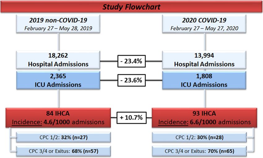
Roedl et al. Scandinavian Journal of Trauma, Resuscitation and Emergency Medicine   (2021) 29:30                        Page 2 of 11

Background                                                               hospital and includes 12 ICUs (total capacity: 142 ICU
Originating from Wuhan, China, a series of pneumonias                    beds). The study complied with the Declaration of
of initially unknown cause emerged in December 2019                      Helsinki. The Ethics Committee of the Hamburg
[1, 2]. A novel coronavirus (severe acute respiratory syn-               Chamber of Physicians was informed about the study
drome coronavirus 2 (SARS-CoV-2)) spread and caused                      (No.: WF-152/20). The requirement for informed patient
a pandemic [3, 4]. Although many patients have a mild                    consent was waived due to the use of only anonymized
course of disease, a considerable number of patients suf-                data collected during routine clinical care. The last day
fer from severe illness with rapid progression to acute                  of follow-up was September 30, 2020.
respiratory distress syndrome (ARDS) or/and end-organ
failure [1–3].                                                           Inclusion and exclusion criteria
  COVID-19 has resulted in high rates of hospitalization                 We included all consecutive adult patients (≥ 18 years)
and a high number of patients requiring intensive care                   with an IHCA event. Patients < 18 years of age and pa-
unit (ICU) treatment [5, 6]. The course of disease can be                tients or with a prior OHCA event and/or re-arrest after
complicated, and can potentially lead to cardiac arrest                  hospital admission were not considered as an incident
(CA) for several reasons, as shown by various studies                    IHCA and were therefore excluded.
[7–10]. An increase in out-of-hospital cardiac arrest
(OHCA) cases was observed during the COVID-19 pan-                       Study definitions and patient management
demic [11, 12]. However, little is known about the CA                    IHCA was defined as cessation of circulation, and there-
risk in hospitalized patients with COVID-19 [10, 13–15].                 fore, an indication for chest compression and/or cardiac
Poor in-hospital survival following in-hospital cardiac ar-              defibrillation in patients who had a pulse and circulation
rest (IHCA) in patients with COVID-19 has been de-                       at the time of hospital admission. Sustained return of
scribed, and mortality ranged from 88 to 100% [9, 10,                    spontaneous circulation (ROSC) was defined as stable
13, 15]. However, data on in-hospital cardiac arrest                     circulation for at least 20 min. Assessment of neuro-
(IHCA) in patients with respiratory failure with and                     logical outcome was performed within routine clinical
without COVID-19 are scarce.                                             practice using cerebral performance categories (CPCs)
  In general, an estimated 290,000 adults suffer from                    after the IHCA and during follow-up. A CPC score of
IHCA in the United States annually [16, 17]. IHCA is                     1–2 was defined as a favourable neurological outcome,
often unexpected and presents as an acute event; every                   and a score of 3–5 was defined as an unfavourable
hospitalized patient can potentially be affected. Different              neurological outcome. Survival was assessed through the
studies have shown abnormal vital signs as predictors of                 end of the ICU stay. Cardiopulmonary resuscitation and
IHCA [18, 19]. Therefore, rapid response teams and the                   post-CA care were performed in accordance with the
use of warning scores have been established [20]. Al-                    European Resuscitation Council guidelines [25]. Data
though most IHCAs occur in general wards [21, 22], a                     were collected according to Utstein-style guidelines [26].
considerable number of IHCAs occur in the ICU [23].                      Cardiac failure was defined as the need for inotrope/va-
The incidence of IHCA varies greatly in the literature                   sopressors (dobutamine, epi−/norepinephrine) during
(1–5/1000 hospital admissions) [16, 17]. Rates of survival               the first 72 h after CA [27]. Hypoxic liver injury (HLI)
to hospital discharge range from 13 to 22% [24].                         was diagnosed according to established criteria [28].
  However, data on IHCA during the COVID-19 pan-                         COVID-19 was defined as a positive result on a reverse
demic are very limited. In the present study we aimed to                 transcriptase-polymerase chain reaction, and only
investigate the occurrence, determinants, outcome and                    laboratory-confirmed cases were counted as COVID-19.
post-CA course of patients suffering from IHCA during                    ARDS was defined using the PaO2/FiO2 ratio (Horowitz
the COVID-19 pandemic and before.                                        index) according to the Berlin definition [29–31]. The
                                                                         severity of illness was evaluated by the sequential organ
Methods                                                                  failure assessment (SOFA) score [32] and simplified
Study population, design and ethics                                      acute physiology (SAPS II) [33] score. The Charlson co-
This was a retrospective analysis of data prospectively                  morbidity index (CCI) [34] was calculated in all patients.
recorded at the University Medical Centre Hamburg-
Eppendorf (Germany). All consecutive adult patients                      Data collection
suffering an IHCA during a 3-month period in 2019 and                    Data were collected through electronic patient data
2020 were included. The following time periods were                      management systems (PDMS, Integrated Care Manager®
compared: 2019 (February 27–May 28) and 2020                             (ICM), Version 9.1 – Draeger Medical, Luebeck,
(February 27–May 27). For post-CA care all patients                      Germany; Soarian Clinicals, Version 4.3.200 – Cerner
were treated at the Department of Intensive Care Medi-                   Health Service, Inc.) and consisted of age, sex, comor-
cine, which cares for all critically ill adult patients of the           bidities, admission diagnosis, length of ICU-stay,
Roedl et al. Scandinavian Journal of Trauma, Resuscitation and Emergency Medicine (2021) 29:30 Page 3 of 11
treatment modalities, organ support (mechanical ventila- (2019-non-COVID-19 period) and 93 (2020-COVID-19
tion, vasopressor, renal replacement therapy (RRT), period) patients suffering from IHCA during the two
blood transfusions, antibiotics, antivirals, etc.), laboratory study periods; these patients were included in the
parameters and further clinical parameters of interest present study (see Study Flow-Chart Fig. 1).
through the end of ICU-stay. Pre-existing medication
was recorded based on known regular medications and Baseline and cardiac arrest characteristics of the study
medication on admission. Laboratory assessment was population
performed daily as part of the clinical routine. Detailed baseline and IHCA characteristics are shown in
Tables 1 and 2. Patients were predominantly male (68%,
Statistical analysis n = 120); the median age was 70 (57–78) years. In this
The results are presented as counts and relative frequen- study, comorbidities were frequent, and a median CCI of
cies or medians and 25–75% interquartile ranges (IQRs). 3 (2–5) was observed. Arterial hypertension (67%, n =
Binary variables were compared via chi-square analysis/ 118) was the leading comorbidity. Furthermore, com-
Fisher’s exact test, as appropriate. Metric variables were mon comorbidities were history of malignant condition
compared via the Mann-Whitney U test. We used multi- (tumour, haematologic malignancy) (33%, n = 58), coron-
variable Cox regression to investigate factors associated ary heart disease (35%, n = 62), diabetes mellitus type II
with mortality and unfavourable outcomes. Factors of (21%, n = 38), chronic respiratory disease (20%, n = 36)
clinical relevance were selected and included. A p-value and chronic kidney disease (18%, n = 32). The reasons
< 0.05 was considered statistically significant. Statistical for hospital admission were medical in 74% (n = 131) of
analysis was conducted using IBM SPSS Statistics Ver- the patients, unplanned surgery in 14% (n = 24) and
sion 24.0 (IBM Corp., Armonk, NY). The study was pre- planned surgical in 13% (n = 22). The median duration
pared in accordance with the STROBE (STrengthening from hospital admission to IHCA was 4 (1–9) days. The
the Reporting of OBservational studies in Epidemiology) IHCA location was non-ICU in 53% (n = 94). The initial
recommendations. cardiac rhythm was shockable (VT/VF) in 24% (n = 42);
defibrillation during CPR was performed in 28% (n = 50).
Results The median total resuscitation time was 5 (2–17) mi-
Study population nutes. Sustained ROSC was observed in 80% (n = 142),
During the two study time periods, namely 2019-non- and cardiac re-arrest was observed in 30% (n = 53). A
COVID-19 (February 27–May 28) and 2020-COVID-19 mechanical chest compression system was used in 11%
(February 27–May 27), a total of 18,262 and 13,994 in- (n = 19). Aetiology of the IHCA was presumed cardiac in
patients were treated at the University Medical Centre 37% (n = 66). Due to refractory IHCA 5% (n = 9) received
Hamburg-Eppendorf, respectively. We identified 84 extracorporeal-CPR (E-CPR).
Fig. 1 Study flow chartRoedl et al. Scandinavian Journal of Trauma, Resuscitation and Emergency Medicine (2021) 29:30 Page 4 of 11
Table 1 Baseline characteristics of patients with in-hospital cardiac arrest stratified according the 2019 (Non-COVID-19) and 2020
(COVID-19) period
Parameters All patients 2019 – Non-COVID-19 Period 2020 – COVID-19 Period p-value
(n = 177) (n = 84) (n = 93)
Demographics
Age, years median (IQR) 70 (57–78) 72 (57–78) 68 (57–78) 0.721
Sex, male n (%) 120 (68) 60 (71) 60 (65) 0.206
Height, cm median (IQR) 172 (165–180) 175 (168–180) 170 (165–179) 0.109
Weight, kg median (IQR) 76 (65–85) 73 (65–84) 78 (67–86) 0.207
BMI, kg/m2 median (IQR) 25 (23–29) 25 (23–27) 26 (24–29) 0.077
Comorbidities
Charlson comorbidity index, pts.; median (IQR) 3 (2–5) 3 (2–5) 3 (2–5) 0.802
Arterial hypertension, n (%) 118 (67) 49 (58) 69 (74) 0.019
Coronary heart disease, n (%) 66 (37) 29 (35) 33 (35) 0.510
Chronic kidney disease, n (%) 32 (18) 13 (15) 19 (20) 0.255
Chronic respiratory disease, n (%) 36 (20) 20 (24) 16 (17) 0.183
Diabetes, n (%) 38 (21) 20 (24) 18 (19) 0.312
Malignant condition, n (%) 58 (33) 23 (27) 35 (38) 0.098
COVID-19
Confirmed COVID-19, n (%) 12 (7) – 12 (13)
Positive test to ICU, days median (IQR) 10 (3–17) – 10 (3–17)
Positive test to IHCA, days median (IQR) 17 (14–28) – 17 (14–28)
Cough, n (%) – – 7 (58)a
Shortness of breath, n (%) – – 3 (25)a
Fever, n (%) – – 6 (50)a
Fatigue, n (%) – – 3 (25)a
Myalgia, n (%) – – 1 (8)a
Reason of hospital admission
Surgical
planned, n (%) 22 (13) 7 (8) 15 (16) 0.089
unplanned, n (%) 24 (14) 14 (17) 10 (11) 0.177
Medical, n (%) 131 (74) 63 (75) 68 (73) 0.455
Characteristics – before CA
Heart rate /min; median (IQR) 91 (77–111) 91 (80–108) 91 (75–111) 0.922
MAP mmHg; median (IQR) 70 (62–82) 72 (66–82) 70 (60–82) 0.549
Outcome
Overall mortality, n (%) 95 (54) 41 (49) 54 (58) 0.140
Discharged from ICU alive, n (%) 82 (46) 43 (51) 39 (42) 0.140
Length of stay – ICU, days median (IQR) 5 (2–17) 6 (2–17) 5 (2–16) 0.592
Abbreviations: cm Centimeter; BMI Body mass index; kg Kilogram; ICU Intensive care unit; IQR Interquartile range; n Number; pts. Points; min Minute; MAP Mean
arterial pressure; COVID-19 Coronavirus disease 2019; IHCA In-hospital cardiac arrest; a in relation to positive tested patients (n = 12)
Differences during the pandemic period and before to 6.6/1000 hospital admissions. Demographic character-
Tables 1 and 2 show a comparison of the detailed base- istics (age, sex and BMI) and comorbidities (as measured
line and IHCA characteristics comparing between the by CCI) were comparable between the groups. Arterial
study periods. During the 2020-COVID-19 period hos- hypertension was significantly more common in patients
pital admissions and ICU admissions declined from 18, during the COVID-19 period (2019 Non-COVID-19
262 to 13,994 (− 23%) and from 2365 to 1808 (− 24%), period: 58% vs 2020 COVID-19 period: 74%). The most
respectively. The incidence of IHCA increased from 4.6 common reason for hospital admission was medical care,Roedl et al. Scandinavian Journal of Trauma, Resuscitation and Emergency Medicine (2021) 29:30 Page 5 of 11
Table 2 Cardiac arrest and ICU-characteristics of the study cohort stratified in the 2019 (No-COVID-19) and 2020 (COVID-19) period
Parameters All patients 2019 – No-COVID-19 2020 – COVID-19 p-value
(n = 177) (n = 84) (n = 93)
Cardiac arrest Characteristics
Location of IHCA 0.009
ICU, n (%) 83 (47) 31 (37) 52 (56)
Non-ICU, n (%) 94 (53) 53 (63) 41 (44)
Initial rhythm - shockable (VT/VF), n (%) 42 (24) 15 (18) 27 (29) 0.058
Defibrillation, n (%) 50 (28) 17 (20) 33 (35) 0.018
Sustained ROSC, n (%) 142 (80) 65 (77) 77 (83) 0.237
Cardiac re-arrest, n (%) 53 (30) 27 (32) 26 (30) 0.329
Presumed cardiac cause, n (%) 66 (37) 35 (42) 31 (33) 0.161
Epinephrine – total dose, mg, median (IQR) 2 (1–4) 2 (1–4) 2 (1–4) 0.978
Resuscitation time, min; median (IQR)
No-flow 0 (0–0) 0 (0–0) 0 (0–0) 0.300
Total resuscitation time 5 (2–17) 4 (1.5–14) 5 (2–20) 0.204
Targeted temperature management, n (%) 56 (32) 27 (32) 29 (31) 0.509
Use of mechanical compression system, n (%) 19 (11) 12 (14) 7 (8) 0.114
E-CPR, n (%) 9 (5) 5 (6) 4 (4) 0.436
ICU – Characteristics
Severity of illness
SAPS II (pts.) median (IQR) 45 (35–55) 44 (35–56) 47 (35–54) 0.837
SOFA – after CA (pts.) median (IQR) 12 (9–14) 11 (8–13) 12 (10–14) 0.060
SOFA – 24 h after CA (pts.) median (IQR) 11 (7–14) 11 (7–14) 11 (8–14) 0.923
Physiological parameters – post CA
Heart rate – after CA median (IQR) 96 (77–115) 94 (77–110) 96 (77–125) 0.232
MAP – after CA median (IQR) 73 (63–88) 76 (62–92) 72 (63–82) 0.324
Lab values – post CA median (IQR)
Lactate – highest after CA, mmol/l 4.5 (1.9–9.1) 4.8 (2–9.1) 4.4 (1.9–8.2) 0.965
pH – lowest after CA 7.26 (7.07–7.36) 7.26 (7.07–7.34) 7.26 (7.10–7.36) 0.755
Procedures/Complications – post CA
Mechanical ventilation, n (%) 124 (70) 55 (65) 69 (74) 0.031
Vasopressor therapy, n (%) 120 (68) 49 (58) 71 (76) 0.025
Renal replacement therapy, n (%) 26 (15) 11 (13) 15 (16) 0.243
Coronary angiography, n (%) 21 (12) 14 (17) 7 (8) 0.050
Hypoxic liver injury, n (%) 35 (20) 16 (19) 19 (20) 0.484
Cholestasis – bilirubin > 2 mg/dl, n (%) 45 (25) 22 (26) 23 (25) 0.480
Abbreviations: CA Cardiac arrest; E-CPR Extracorporeal cardiopulmonary resuscitation; ICU Intensive care unit; IQR Inter quartile range; n Number; min Minute; mg
Milligram; mmol/l Millimole per liter; pts. Points; ROSC Return of spontaneous circulation; SAPS Simplified Acute Physiology Score; SOFA Sequential Organ Failure
Assessment; VF Ventricular Fibrillation; VT Ventricular Tachycardia; MAP Mean arterial pressure; COVID-19 Coronavirus disease 2019; IHCA In-hospital cardiac arrest
which did not differ between the time periods. The total epinephrine use (2 mg vs 2 mg) were comparable in
IHCA location was primarily non-ICU during 2019-non- both study periods. The median resuscitation time was
COVID-19 period and primarily in the ICU during the 4 min vs. 5 min and did not differ significantly between
2020-COVID-19 period (p < 0.01). A shockable rhythm the groups. Mechanical compression systems were used
(18% vs 29%) was more frequently observed during the more frequently during the 2019 period (14% vs 8%).
COVID-19 period, and the use of defibrillation (20% vs Targeted temperature management post-CA was used in
35%) was significantly higher. The rates of sustained 32% of patients in the whole cohort, and the frequency
ROSC (77% vs 83%), cardiac re-arrest (32% vs 30%) and was similar in both study periods. The SAPS II andRoedl et al. Scandinavian Journal of Trauma, Resuscitation and Emergency Medicine (2021) 29:30 Page 6 of 11
SOFA score post-CA were higher during the 2020- severe respiratory failure. The most common initial
COVID-19 period. During the ICU stay mechanical rhythm was non-shockable in both groups. The use of
ventilation was performed more frequently during the epinephrine was comparable in both groups. The total
2020-COVID-19 period (65% vs 74%, p < 0.05). Vaso- resuscitation time was longer in patients with non-
pressor therapy was more commonly used during the COVID-19 related severe respiratory failure (median 5
2020-COVID-19 period (58% vs 76%). Liver dysfunction vs 1.5 min; p < 0.01). The severity of illness at ICU ad-
was frequent during both study periods; 20% suffered mission and after IHCA was comparable between the
from hypoxic liver injury and 25% suffered from groups. During the ICU stay, RRT was more frequent
cholestasis. (p < 0.01) in patients with COVID-19. Laboratory values
before and after IHCA were comparable between the
IHCA and COVID-19 groups. Furthermore, physiological parameters before
During the aforementioned 2020 time period, 144 pa- and after IHCA did not differ significantly.
tients with COVID-19 were treated as inpatients at our
centre. Of these, 75 patients were treated in the normal Survival and functional outcome
ward, and 69 patients were critically ill and therefore Of the 177 included patients who had an IHCA event, 99
treated in the ICU. Twelve patients (10%) with COVID- (54%) did not survive the ICU-stay. Fifty-six patients
19 treated at our hospital suffered from IHCA. All (32%) died within 24 h after the IHCA. At ICU discharge
patients had severe respiratory failure either due to 31% (n = 55) had favourable neurological outcomes (CPC
pneumonia or due to the development of ARDS. The I/II). Rates did not differ significantly between the two
median times from the first positive SARS-CoV-2 test to study periods (2019: 32% - 2020: 30%). In patients with
the ICU and to IHCA were 10 (3–17) days and 17 (14– COVID-19, the rates of favourable neurological outcomes
28) days, respectively. The most common symptoms of (CPC I/II) were higher than those in patients with non-
COVID-19 were cough (n = 7; 58%), fever (n = 6; 50%), COVID-19-related severe respiratory failure (42% vs 10%).
shortness of breath and fatigue (n = 3 for each, 25%). Cox regression analysis revealed that the SOFA-score after
None of the IHCAs occurred outside the ICU. All pa- IHCA [HR 1.17, 95% CI (1.00–1.36); p < 0.05], CCI [HR
tients had a primary non-shockable rhythm (PEA/Asys- 1.13, 95% CI (1.01–1.26); p < 0.05] and low-flow time [HR
tole) and ROSC. The median resuscitation time was1.5 1.07, 95% CI (1.01–1.12); p < 0.05] were significantly asso-
(0.5–3.5) minutes. For detailed characteristics of patients ciated with unfavourable neurological outcome or ICU-
with COVID-19, see Tables 1, 2, 3 and Supplementary mortality within patients with severe respiratory failure
Tables 1 and 2. (see Supplementary Table 3).
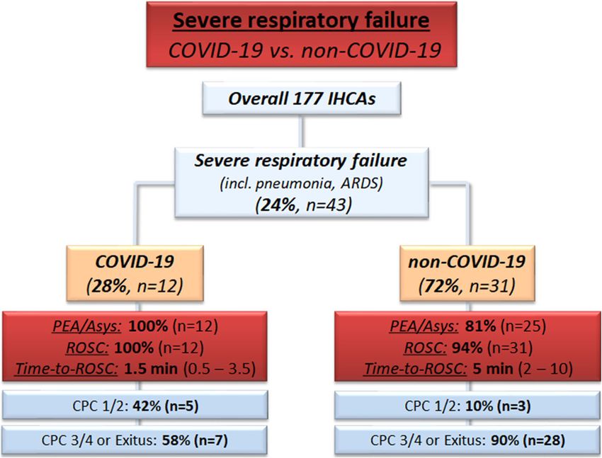
Characteristics of IHCA in patients with or without COVID- Discussion
19 related severe respiratory failure In this study investigating the effects of the COVID-19
Detailed characteristics are shown in Table 3 and Sup- pandemic on IHCA, we found that the incidence of IHCA
plementary Table 1 and 2. Overall, 25% (n = 43) of pa- was increased, the location of IHCA shifted towards the
tients had severe respiratory failure at the time of IHCA ICU and CA-characteristics were changed. To our know-
and were selected. Of those 28% (n = 12) suffered from ledge, this is the first study evaluating the effect of the
COVID-19 pneumonia. Demographic characteristics COVID-19 pandemic on IHCA. Furthermore, this is the
(age, sex, BMI) were comparable between patients with first study comparing the IHCA characteristics of patients
severe respiratory failure who did not have COVID-19. suffering from severe respiratory failure that was and was
Comorbidities, represented by the CCI were significantly not related to COVID-19 at time the of IHCA.
lower (4 vs 2 points; p < 0.01) in patients with non- The COVID-19 pandemic led to a higher incidence of
COVID-19 related severe respiratory failure. In total, OHCA and worse short-term outcomes [11, 12]. Differ-
68% received non-invasive or invasive mechanical venti- ent mechanisms suggesting direct effects of COVID-19
lation prior to IHCA. Overall, 56% (n = 24) of patients and effects from lockdown were proposed [35]. How-
suffered from ARDS at the time of IHCA and ARDS was ever, to date, no data on how the pandemic has affected
more frequently observed in patients with COVID-19. In IHCA exist. Due to the rapid spread and surge of pa-
addition, the Horowitz index after IHCA was signifi- tients with COVID-19, elective admissions to hospitals
cantly lower in patients with COVID-19. ARDS manage- were cancelled to create more capacity for patients suf-
ment, including prone positioning, neuromuscular fering from COVID-19. This was impressively demon-
blockage, corticosteroids and inhaled vasodilatory treat- strated by a 23% decrease in hospital admissions during
ment, was comparable in both groups. IHCA within the the COVID-19 period. Although hospital admissions de-
ICU was significantly more frequent in patients with creased substantially, an 11% increased incidence of
COVID-19 than in those without COVID-19-related IHCA was observed. The reported incidence of IHCA inRoedl et al. Scandinavian Journal of Trauma, Resuscitation and Emergency Medicine (2021) 29:30 Page 7 of 11
Table 3 Cardiac arrest characteristics of patients with severe respiratory failure with and without COVID-19
Parameters All patients Severe respiratory Severe respiratory p-value
(n = 43) failure no-COVID-19 failure – COVID-19
(n = 31) (n = 12)
Demographics
Age, years median (IQR) 65 (50–75) 65 (50–77) 65 (56–74) 0.565
Sex, male, n (%) 34 (79) 25 (81) 9 (75) 0.471
BMI, kg/m2 median (IQR) 27 (24–31) 26 (24–30) 28 (26–33) 0.314
Charlson comorbidity index, pts. median (IQR) 3 (1.5–6) 4 (2.5–6) 2 (1–2) 0.003
Characteristics of respiratory failure
Respiratory support (before CA)
Non-invasive ventilation n (%) 7 (16) 3 (10) 4 (33) 0.125
Mechanical ventilation n (%) 22 (51) 17 (55) 5 (42) 0.148
Cause of respiratory failure (at CA)
Pneumonia n (%) 37 (86) 25 (81) 12 (100) 0.001
ARDS n (%) 24 (56) 12 (39) 10 (83) 0.000
Horowitz index (PaO2/FiO2-ratio)
Worst Horowitz index, mmHg, median (IQR) 84 (57–148) 90 (57–149) 82 (59–107) 0.503
Horowitz index after CA, mmHg, median (IQR) 97 (76–145) 101 (78–152) 89 (69–19) 0.007
ARDS Management
Prone Positioning n (%) 8 (19) 2 (6) 6 (50) 0.437
Neuromuscular Blockage n (%) 6 (14) 1 (3) 5 (42) 0.306
Corticosteroids n (%) 11 (26) 4 (13) 7 (58) 0.563
Inhaled Vasodilators n (%) 9 (21) 3 (10) 6 (50) 0.437
Cardiac arrest Characteristics
Location 0.009
ICU, n (%) 37 (86) 25 (81) 12 (100)
Non-ICU, n (%) 6 (14) 6 (19) 0 (0)
Initial Rhythm - Shockable (VT/VF), n (%) 6 (14) 6 (19) 0 (0) 0.255
Sustained ROSC, n (%) 40 (93) 28 (90) 12 (100) 0.364
Epinephrine – total dose, mg, median (IQR) 1 (1–2) 2 (1–2.5) 1 (1–1.3) 0.310
Resuscitation time, min; median (IQR)
No-Flow 0 (0–0) 0 (0–0) 0 (0–0) 1
Total resuscitation time 4 (1.8–8.5) 5 (2–10) 1.5 (0.5–3.5) 0.008
Targeted temperature management, n (%) 10 (23) 8 (26) 2 (17) 0.339
E-CPR, n (%) 0 (0) 0 (0) 0 (0) 1
ICU – Characteristics
Severity of illness
SAPS II (pts.) median (IQR) 44 (36–52) 42 (35–49) 50 (40–56) 0.485
SOFA – after CA (pts.) median (IQR) 14 (12–16) 14 (12–17) 15 (13–16) 0.202
SOFA – 24 h after CA (pts.) median (IQR) 13 (11–16) 13 (11–15) 14 (10–16) 0.145
Lab values – post CA median (IQR)
Lactate – highest after CA, mmol/l 4.6 (1.6–8.5) 4.8 (1.5–10) 4.2 (3.1–4.8) 0.765
pH – lowest after CA 7.21 (7.15–7.32) 7.22 (7.06–7.32) 7.2 (7.19–7.3) 0.889Roedl et al. Scandinavian Journal of Trauma, Resuscitation and Emergency Medicine (2021) 29:30 Page 8 of 11
Table 3 Cardiac arrest characteristics of patients with severe respiratory failure with and without COVID-19 (Continued)
Parameters All patients Severe respiratory Severe respiratory p-value
(n = 43) failure no-COVID-19 failure – COVID-19
(n = 31) (n = 12)
Procedures/Complications – post CA
Vasopressor therapy, n (%) 40 (93) 29 (94) 11 (92) 0.505
Renal replacement therapy, n (%) 22 (51) 12 (39) 10 (83) 0.009
Coronary angiography, n (%) 0 (0) 0 (0) 0 (0) 1
Hypoxic liver injury, n (%) 11 (26) 7 (23) 4 (33) 0.201
Cholestasis – bilirubin > 2 mg/dl, n (%) 15 (58) 10 (32) 5 (42) 0.190
Abbreviations: CA Cardiac arrest; cm Centimeter; E-CPR Extracorporeal cardiopulmonary resuscitation; ICU Intensive care unit; IQR Inter quartile range; kg Kilogram;
n Number; min Minute; mg Milligram; mmol/l Millimole per liter; pts. Points; ROSC Return of spontaneous circulation; SAPS Simplified Acute Physiology Score; SOFA
Sequential Organ Failure Assessment; VF Ventricular Fibrillation; VT Ventricular Tachycardia; MAP Mean arterial pressure; COVID-19 Coronavirus disease 2019; IHCA
In-hospital cardiac arrest; BMI Body mass index
the literature is 1–5/1000 hospital admissions; during the cardiac aetiology of the IHCA. However, half of the
pandemic period, the incidence of 6.6/1000 hospital ad- patients were in the ICU before IHCA and suffered
missions exceeded reported rates [16, 17]. Different fac- from high severity of illness, and the rate of MV and
tors could explain this finding. First, patients with vasopressor support was associated with a non-
COVID-19 are at high risk of IHCA due to rapidly wors- shockable rhythm [38, 39].
ening respiratory failure eventually leading to IHCA if not The occurrence of IHCA among hospitalized patients
promptly treated. Of interest, explainable deterioration of with COVID-19 commonly ranges from 6 to 14% [9,
SpO2 and high FiO2, but only minor abnormalities in 10]. We confirmed these results and found an incidence
other vital signs, as well as higher early warning scores, of 8%. The outcome after IHCA in patients with
have recently been described as predictors for outcome COVID-19 is worse, and high mortality rates, ranging
[36, 37]. Second, the severity of illness at ICU admission from 88 to 100%, have been reported [9, 10, 13, 15, 40].
was substantially higher than that in the non-COVID-19 These reports led to a controversial discussion about fu-
period. Although an early ICU admission strategy was tility and appropriateness of care in patients suffering
followed, this may be explained by delayed or disrupted from COVID-19. However, in our small cohort, we ob-
contact with the healthcare system due to lockdown mea- served a distinctly lower mortality than previously re-
sures, leading to delayed hospital admission in general. ported, although we observed comparable IHCA
Furthermore, we observed substantial differences ac- characteristics, including similar rates of non-shockable
cording to IHCA characteristics during the study pe- initial rhythm, resuscitation time and occurrence of
riods. During the pandemic period, the IHCA location IHCA in the ICU. The lower mortality in our cohort can
shifted more towards the ICU, which may be explained be a consequence of several reasons. First, a considerably
by earlier ICU admission of deteriorating patients. More- lower number of patients were on MV or RRT before
over, the rate of shockable rhythm and defibrillation in- CA, demonstrating a lower severity of illness. Moreover,
creased and we observed high rates of ROSC. These we followed a strategy of early admission to the ICU in
observed differences are potentially explained by higher patients with COVID-19 for closer monitoring and early
rates of IHCA occurring in the ICU and a faster re- initiation of supportive care. This could also correspond
sponse to deterioration due to higher nurse/doctor to the high rate of ROSC observed in our cohort and is
staffing. Interestingly, the duration of resuscitation was probably related to continuous monitoring and higher
slightly longer during the pandemic period. CPR, an nurse/doctor staffing. Second, earlier reports originated
aerosol-generating procedure, exposes healthcare from regions with an excessive case load which poten-
workers to a risk of viral transmission. Therefore, the tially led to overwhelmed healthcare systems playing an
use of personal-protective equipment is of central im- important role in appropriate patient care [13, 15]. How-
portance but could have contributed to the delayed initi- ever, decisions on futility and withholding CPR are diffi-
ation of CPR. Furthermore, the lower rate of presumed cult and must be based on a multifactorial approach that
cardiac aetiology is important. However, resuscitation takes the severity of illness, current organ support and
times were shorter than those in previous studies [23]. the patient’s directive into account.
Overall, one quarter of patients presented with an SARS-CoV-2 primarily affects the respiratory system
initial shockable rhythm, which is in line with previ- which can lead to rapid deterioration and severe respira-
ous studies and can be explained by the low rate of tory failure. Recent clinical studies reported highRoedl et al. Scandinavian Journal of Trauma, Resuscitation and Emergency Medicine (2021) 29:30 Page 9 of 11 Fig. 2 Outcome of patients with severe respiratory failure – stratified to COIVD-19 and non-COVID-19 mortality following IHCA events in patients with Larger future studies must confirm these results and COVID-19 [9, 10, 13]. As patients with COVID-19 pri- their implications on outcome. marily suffer from respiratory failure, comparing the This study has several limitations. First, our study in- characteristics and outcomes of IHCA to patients suffer- cluded a small number of patients. Larger cohorts are ing from respiratory failure related and not related to needed to confirm our findings. Second, the data were de- COVID-19 seems reasonable. However, this is the first rived from a single centre and were collected retrospect- study comparing patients with severe respiratory failure ively. However, the data were documented prospectively not related to COVID-19 at the time of IHCA with pa- in the PDMS by trained ICU staff. Third, we show the re- tients suffering from COVID-19. We observed that pa- sults of an experienced high-volume CA centre. Thus, the tients with COVID-19 had a lower comorbidity rate and results might not generally be transferable to other, less substantially lower Horowitz index before and after experienced, settings. Fourth, the study was conducted IHCA. In patients suffering from ARDS, we observed early during the pandemic. Changes in clinical practice, comparable therapeutic approaches. Furthermore, a high due to more experience with COVID-19, could have chan- rate of IHCA occurring in the ICU was observed, and ged affecting the incidence and outcome of IHCA espe- correspondingly, a substantially lower resuscitation time cially in critically ill patients. was observed. In our cohort, we observed that IHCA often occurred during tracheal intubation. This may be a consequence of delayed decisions for tracheal intubation. Conclusions However, this should lead to higher awareness of the In conclusion, this is the first study evaluating IHCA oc- timing of intubation in patients with progredient respira- currence and outcomes during the COVID-19 pandemic tory failure. A higher number of patients with COVID- in comparison to recent years. Hospital admissions de- 19 required RRT and had liver injury (HLI/cholestasis) clined during the pandemic, but a higher incidence of contributing to the higher severity of illness after IHCA. IHCA was observed, which could be attributed to multi- However, direct viral effects cannot be entirely excluded. factorial influences and must be further evaluated. Ap- Moreover, CCI, SOFA scores and resuscitation time proximately 10% of hospitalized patients with COVID- were identified as mortality predictors in these patients. 19 suffered from IHCA, and outcomes were improved Of interest, a substantially higher number of patients compared with those previously reported and compar- with COVID-19 had a favourable outcome compared to able to those of patients with other aetiologies of respira- other patients with severe respiratory failure (Fig. 2). tory failure not related to COVID-19.
Roedl et al. Scandinavian Journal of Trauma, Resuscitation and Emergency Medicine                 (2021) 29:30                                     Page 10 of 11

Supplementary Information                                                         Tabea Hospital, Hamburg, Germany. 4Department of Interventional and
The online version contains supplementary material available at https://doi.      General Cardiology, University Heart Centre Hamburg, Hamburg, Germany.
org/10.1186/s13049-021-00846-w.
                                                                                  Received: 13 November 2020 Accepted: 1 February 2021
 Additional file 1: Supplementary Table 1. Pre-existing comorbidities
 of patients with severe respiratory failure at time of cardiac arrest strati-
 fied according patients with and without COVID-19. Supplementary                 References
 Table 2. Characteristics of patients with severe respiratory failure before      1. Huang C, Wang Y, Li X, Ren L, Zhao J, Hu Y, et al. Clinical features of
 and after cardiac arrest stratified according with and without COVID-19.             patients infected with 2019 novel coronavirus in Wuhan, China. Lancet.
 Supplementary Table 3. Cox regression model for factors associated                   2020;395(10223):497–506.
 with ICU-mortality and unfavorable neurological outcome (CPC III/IV) of          2. Chen N, Zhou M, Dong X, Qu J, Gong F, Han Y, et al. Epidemiological and
 patients with IHCA and severe respiratory failure.                                   clinical characteristics of 99 cases of 2019 novel coronavirus pneumonia in
                                                                                      Wuhan, China: a descriptive study. Lancet. 2020;395(10223):507–13.
                                                                                  3. Guan WJ, Ni ZY, Hu Y, Liang WH, Ou CQ, He JX, et al. Clinical characteristics
Abbreviations                                                                         of coronavirus disease 2019 in China. N Engl J Med. 2020;382(18):1708–20.
ARDS: Acute respiratory distress syndrome; Asys: Asystole; BMI: Body mass         4. WHO. WHO - World Map - COVID-19. 2020 [https://covid19.who.int/ -
index; CA: Cardiac arrest; CCI: Charlson comorbidity index; COVID-                    Accessed online: October, 31st 2020] [Available from: https://covid19.who.
19: Coronavirus disease 2019; CPC: Cerebral performance categories; E-                int/ - Accessed online: October, 19th 2020].
CPR: Extracorporeal cardiopulmonary resuscitation; HLI: Hypoxic liver injury;     5. Grasselli G, Pesenti A, Cecconi M. Critical care utilization for the COVID-19
ICU: Intensive care unit; IHCA: In-hospital cardiac arrest; IQR: Interquartile        outbreak in Lombardy, Italy: early experience and forecast during an
range; MV: Mechanical ventilation; OHCA: Out-of-hospital cardiac arrest;              emergency response. JAMA. 2020;323(16):1545–6.
PEA: Pulseless electrical activity; ROSC: Return of spontaneous circulation;      6. Richardson S, Hirsch JS, Narasimhan M, Crawford JM, McGinn T, Davidson
RRT: Renal replacement therapy; SAPS: Simplified acute physiology score;              KW, et al. Presenting characteristics, comorbidities, and outcomes among
SARS-CoV-2: Severe acute respiratory syndrome coronavirus 2;                          5700 patients hospitalized with COVID-19 in the New York City area. JAMA.
SOFA: Sequential organ failure assessment score; VT: Ventricular tachycardia;         2020;323(20):2052–9.
VF: Ventricular fibrillation                                                      7. Ly A, Alessandri C, Skripkina E, Meffert A, Clariot S, de Roux Q, et al. Rescue
                                                                                      fibrinolysis in suspected massive pulmonary embolism during SARS-CoV-2
Acknowledgements                                                                      pandemic. Resuscitation. 2020;152:86–8.
None.                                                                             8. Creel-Bulos C, Hockstein M, Amin N, Melhem S, Truong A, Sharifpour M.
                                                                                      Acute Cor Pulmonale in critically ill patients with Covid-19. N Engl J Med.
Authors’ contributions                                                                2020;382(21):e70.
KR, GS, DJ and SK participated in study conception and design. KR, GS, JM,        9. Shah P, Smith H, Olarewaju A, Jani Y, Cobb A, Owens J, et al. Is
DF, DW, MI were involved in acquisition of data. KR, DJ and SK contributed            cardiopulmonary resuscitation futile in coronavirus disease 2019 patients
to analysis and interpretation of data. KR drafted the manuscript. SK and DJ          experiencing in-hospital cardiac arrest? Crit Care Med. 2021;49(2):201–8.
were involved in critical revision of the manuscript for important intellectual   10. Hayek SS, Brenner SK, Azam TU, Shadid HR, Anderson E, Berlin H, et al. In-
content. SK, GS and DJ participated in supervision. All authors read and              hospital cardiac arrest in critically ill patients with covid-19: multicenter
approved the final manuscript.                                                        cohort study. BMJ. 2020;371:m3513.
                                                                                  11. Baldi E, Sechi GM, Mare C, Canevari F, Brancaglione A, Primi R, et al. Out-of-
Funding                                                                               hospital cardiac arrest during the Covid-19 outbreak in Italy. N Engl J Med.
No financial support has been received conducting this study.                         2020.
                                                                                  12. Marijon E, Karam N, Jost D, Perrot D, Frattini B, Derkenne C, et al. Out-of-
Availability of data and materials                                                    hospital cardiac arrest during the COVID-19 pandemic in Paris, France: a
The datasets supporting the conclusions of this article are included within           population-based, observational study. Lancet Public Health. 2020.
the article.                                                                      13. Shao F, Xu S, Ma X, Xu Z, Lyu J, Ng M, et al. In-hospital cardiac arrest
                                                                                      outcomes among patients with COVID-19 pneumonia in Wuhan, China.
                                                                                      Resuscitation. 2020;151:18–23.
Ethics approval and consent to participate                                        14. Sung C-W, Lu T-C, Fang C-C, Huang C-H, Chen W-J, Chen S-C, et al. Impact
The Ethics Committee of the Hamburg Chamber of Physicians was informed                of COVID-19 pandemic on emergency department services acuity and
about the study (No.: WF-152/20). The requirement for informed patient con-           possible collateral damage. Resuscitation. Resuscitation. 2020;153:185–6.
sent was waived due to the use of anonymized data from routine clinical           15. Sheth V, Chishti I, Rothman A, Redlener M, Liang J, Pan D, et al. Outcomes
care only. The study was approved by the local clinical institutional review          of in-hospital cardiac arrest in patients with COVID-19 in New York City.
board and complied with the Declaration of Helsinki.                                  Resuscitation. 2020;155:3–5.
                                                                                  16. Andersen LW, Holmberg MJ, Berg KM, Donnino MW, Granfeldt A. In-hospital
Consent for publication                                                               cardiac arrest: a review. Jama. 2019;321(12):1200–10.
Not applicable.                                                                   17. Benjamin EJ, Muntner P, Alonso A, Bittencourt MS, Callaway CW, Carson AP,
                                                                                      et al. Heart disease and stroke Statistics-2019 update: a report from the
Competing interests                                                                   American Heart Association. Circulation. 2019;139(10):e56–e528.
KR, GS, DF, JM, MI, DW and DJ do not report any conflicts of interest related     18. Nurmi J, Harjola VP, Nolan J, Castrén M. Observations and warning signs
to this article. SK received research support by Ambu, E.T.View Ltd., Fisher &        prior to cardiac arrest. Should a medical emergency team intervene earlier?
Paykel, Pfizer and Xenios, lecture honorarium from ArjoHuntleigh, Astellas,           Acta Anaesthesiol Scand. 2005;49(5):702–6.
Astra, Basilea, Bard, Baxter, Biotest, CSL Behring, Cytosorbents, Fresenius,      19. Andersen LW, Kim WY, Chase M, Berg KM, Mortensen SJ, Moskowitz A, et al.
Gilead, MSD, Orion, Pfizer, Philips, Sedana, Sorin, Xenios and Zoll, and              The prevalence and significance of abnormal vital signs prior to in-hospital
consultant honorarium from AMOMED, Astellas, Baxter, Bayer, Fresenius,                cardiac arrest. Resuscitation. 2016;98:112–7.
Gilead, MSD, Pfizer and Xenios. No other potential conflict of interest           20. Jones DA, DeVita MA, Bellomo R. Rapid-response teams. N Engl J Med.
relevant to this article was reported.                                                2011;365(2):139–46.
                                                                                  21. Sandroni C, Nolan J, Cavallaro F, Antonelli M. In-hospital cardiac arrest:
Author details                                                                        incidence, prognosis and possible measures to improve survival. Intensive
1
 Department of Intensive Care Medicine, University Medical Centre                     Care Med. 2007;33(2):237–45.
Hamburg-Eppendorf, Martinistraße 52, 20246 Hamburg, Germany.                      22. Nolan JP, Soar J, Smith GB, Gwinnutt C, Parrott F, Power S, et al. Incidence
2
 Department of Anaesthesiology, University Medical Centre                             and outcome of in-hospital cardiac arrest in the United Kingdom National
Hamburg-Eppendorf, Hamburg, Germany. 3Department of Anaesthesia,                      Cardiac Arrest Audit. Resuscitation. 2014;85(8):987–92.
Roedl et al. Scandinavian Journal of Trauma, Resuscitation and Emergency Medicine (2021) 29:30 Page 11 of 11
23. Efendijev I, Nurmi J, Castrén M, Skrifvars MB. Incidence and outcome from
adult cardiac arrest occurring in the intensive care unit: a systematic review
of the literature. Resuscitation. 2014;85(4):472–9.
24. Schluep M, Gravesteijn BY, Stolker RJ, Endeman H, Hoeks SE. One-year
survival after in-hospital cardiac arrest: a systematic review and meta-
analysis. Resuscitation. 2018;132:90–100.
25. Nolan JP, Soar J, Cariou A, Cronberg T, Moulaert VRM, Deakin CD, et al.
European resuscitation council and European Society of Intensive Care
Medicine Guidelines for post-resuscitation care 2015: section 5 of the
European resuscitation council guidelines for resuscitation 2015.
Resuscitation. 2015;95:202–22.
26. Nolan JP, Berg RA, Andersen LW, Bhanji F, Chan PS, Donnino MW, et al.
Cardiac Arrest and Cardiopulmonary Resuscitation Outcome Reports:
Update of the Utstein Resuscitation Registry Template for In-Hospital
Cardiac Arrest: A Consensus Report From a Task Force of the International
Liaison Committee on Resuscitation (American Heart Association, European
Resuscitation Council, Australian and New Zealand Council on Resuscitation,
Heart and Stroke Foundation of Canada, InterAmerican Heart Foundation,
Resuscitation Council of Southern Africa, Resuscitation Council of Asia).
Resuscitation. 2019;144:166–77.
27. Laurent I, Monchi M, Chiche JD, Joly LM, Spaulding C, Bourgeois B, et al.
Reversible myocardial dysfunction in survivors of out-of-hospital cardiac
arrest. J Am Coll Cardiol. 2002;40(12):2110–6.
28. Fuhrmann V, Jäger B, Zubkova A, Drolz A. Hypoxic hepatitis - epidemiology,
pathophysiology and clinical management. Wien Klin Wochenschr. 2010;
122(5–6):129–39.
29. Ranieri VM, Rubenfeld GD, Thompson BT, Ferguson ND, Caldwell E, Fan E,
et al. Acute respiratory distress syndrome: the Berlin definition. JAMA. 2012;
307(23):2526–33.
30. Alhazzani W, Møller MH, Arabi YM, Loeb M, Gong MN, Fan E, et al. Surviving
Sepsis campaign: guidelines on the management of critically ill adults with
coronavirus disease 2019 (COVID-19). Intensive Care Med. 2020;46(5):854–87.
31. Kluge S, Janssens U, Welte T, Weber-Carstens S, Marx G, Karagiannidis C.
German recommendations for critically ill patients with COVID-19.
Medizinische Klinik, Intensivmedizin und Notfallmedizin. 2020:1–4.
32. Vincent JL, Moreno R, Takala J, Willatts S, De Mendonça A, Bruining H, et al.
The SOFA (Sepsis-related organ failure assessment) score to describe organ
dysfunction/failure. On behalf of the working group on Sepsis-related
problems of the European Society of Intensive Care Medicine. Intensive
Care Med. 1996;22(7):707–10.
33. Le Gall JR, Lemeshow S, Saulnier F. A new simplified acute physiology score
(SAPS II) based on a European/north American multicenter study. JAMA.
1993;270(24):2957–63.
34. Charlson ME, Pompei P, Ales KL, MacKenzie CR. A new method of classifying
prognostic comorbidity in longitudinal studies: development and validation.
J Chronic Dis. 1987;40(5):373–83.
35. Lim ZJ, Reddy MP, Afroz A, Billah B, Shekar K, Subramaniam A. Incidence
and outcome of out-of-hospital cardiac arrests in the COVID-19 era: a
systematic review and meta-analysis. Resuscitation. 2020;157:248–58.
36. Pimentel MAF, Redfern OC, Hatch R, Young JD, Tarassenko L, Watkinson PJ.
Trajectories of vital signs in patients with COVID-19. Resuscitation. 2020;156:
99–106.
37. Covino M, Sandroni C, Santoro M, Sabia L, Simeoni B, Bocci MG, et al.
Predicting intensive care unit admission and death for COVID-19 patients in
the emergency department using early warning scores. Resuscitation. 2020;
156:84–91.
38. Roedl K, Jarczak D, Blohm R, Winterland S, Müller J, Fuhrmann V, et al.
Epidemiology of intensive care unit cardiac arrest: characteristics,
comorbidities, and post-cardiac arrest organ failure - a prospective
observational study. Resuscitation. 2020;156:92–8.
39. Leloup M, Briatte I, Langlois A, Cariou A, Lesieur O. Unexpected cardiac
arrests occurring inside the ICU: outcomes of a French prospective
multicenter study. Intensive Care Med. 2020.
40. Thapa SB, Kakar TS, Mayer C, Khanal D. Clinical outcomes of in-hospital
cardiac arrest in COVID-19. JAMA Intern Med. 2021;181(2):279–81.
Publisher’s Note
Springer Nature remains neutral with regard to jurisdictional claims in
published maps and institutional affiliations.You can also read