Management of Oligosymptomatic Patients with Respiratory Infection in the Era of SARS-Cov2. Experience from Rural German General Practitioners and ...
←
→
Page content transcription
If your browser does not render page correctly, please read the page content below
Management of Oligosymptomatic Patients with
Respiratory Infection in the Era of SARS-Cov2. Experience
from Rural German General Practitioners and Proposition
of a New Screening Model
Simon Wernhart ( simon.wernhart@gmx.de )
Sauerlandpraxis Medebach, Hallenberg, Winterberg https://orcid.org/0000-0001-6732-0939
Tim Henning Förster
Hochsauerlandpraxis Medebach, Hallenberg, Winterberg
Eberhard Weihe
Western University Department of Anatomy and Cell Biology
Research article
Keywords: General practitioner, COVID-19, risk strati cation model
DOI: https://doi.org/10.21203/rs.3.rs-26768/v1
License: This work is licensed under a Creative Commons Attribution 4.0 International License. Read Full License
Page 1/13
Abstract
Background: Covid-19 is causing a pandemic and forces physicians to restructure their daily work. We want to share
our successful experience in the outpatient management of potentially infected patients with colleagues from other
countries.

Methods: We analyzed all patients with respiratory symptoms reporting to our three rural general practioner (GP) o ces
in North Rhine Westphalia, Germany, from 27.01-20.04.2020 (n=489 from a total of 6090 patients). A history of
symptoms was taken at the doorstep following a speci c questionnaire. Patients with respiratory symptoms were
examined in a separated isolation area, while the others were allowed to enter the o ce.

Results: Eighty patients (16.36%, mean age: 47.03years+-18.08) were sent to a nasopharyngeal smear following the
screening algorithm of the Robert-Koch Institute, Berlin, Germany. 5 patients (6.25%) turned out to be positive, 4 of
which had established risk factors for COVID-19. Overall, the most common symptoms were cough (83.75%), sore
throat (71.25%), as well as myalgia and fatigue (66.25%). The most common diagnoses were rhinopharyngitis
(37.22%) and acute bronchitis (30.27%). Clinically, it was impossible to differentiate ordinary respiratory infections
from COVID-19 patients in our low-risk population, although a sore throat was more common in positively tested
patients (80% vs. 12%). None of our employees called in sick during this period, which emphasizes the e cacy and
safety of our screening methods. We suggest a novel risk score integrating patient history, symptoms, pulmonary
ultrasound and environmental risks to stratify patients as candidates for SARS-Cov2-PCR.

Conclusion: A clinical distinction between ordinary respiratory infections and COVID-19 is not possible in a low-risk
population. Our model to prevent unprotected physical contact, screen patients in front of the o ce with protective
equipment, and to examine respiratory infections in separated areas works in the GP setting without overt health risks
for employees. Thus, this approach should be used as a GP standard to uphold patient care without major health risks
for the personnel. We introduce a new combinatorial scoring model to assess COVID-19 probability in the GP setting.

Background
Originating from Wuhan, China, coronavirus disease 2019 (COVID-19) has spread around the world as a pandemic and
created enormous health, political, economical problems(1, 2)with 3366714 con rmed cases worldwide and 239345
deaths as per 02.05.2020(3).

Clinical predictors of mortality have been suggested from a cohort of patients from Wuhan(4–6) and
recommendations for outpatient(7) and inpatient care(6) as well as intensive care treatment(8) have been proposed.
Patients admitted to intensive care units are older, and tend to have higher leukocyte counts, D-Dimers, LDH, creatinine
and troponin levels(5). Elevated troponin as a marker for myocardial injury heralds a poor prognosis(9).

The most common symptoms, depending on the time windows during the course of infection, are symptoms
associated with respiratory dieseases, such as fever, cough, sore throat, headache, chills, fatigue and myalgia, smell
and taste dysfunction and gastrointestinal problems(10, 11), but recently more severe cases have been associated with
neurological symptoms, such as acute cerebral vascular disease, skeletal muscle injury and impaired
consciousness(12). Perniosis- like skin symptoms may als be present(13, 14). Risk factors for more severe cases of
COVID-19 are hypertension, coronary artery disease, immunosuppression, chronic lung disease(15).

Less severe cases often present to the general practicioner (GP) in the outpatient setting, which requires precautions to
avoid infection in medical personel. In Germany, testing for SARS-Cov2 infection has been widely established(16, 17).
Additionally, containment as a means to reduce exponential growth has been implemented at an early stage(18), which
                                                        Page 2/13
may account for the relatively low German case-mortality rate (4.1%) compared to other European countries(19, 20). As
per 02.05.2020 164077 patients had been infected und 6736 had died in Germany(3). Furthermore, standard and
elective examinations have been postponed to limit physical physician-patient contact to the necessary minimum(21).
The German government as well as national and federal medical institutions have made considerable efforts to prevent
less critical but potenially infective patients from showing up in the GP o ce by installing telephone and video
conferences to provide medical council without physical contact. In practice, however, concerned patients fearing to be
infected keep showing up and need to be screened in isolation rooms in case of symptoms suggestive of COVID-19.
This requires o ce re-organisation and efforts to receive personel protective equipment (PPE) which, however, has been
di cult to get for some time.
The German Robert Koch Institute (RKI) has issued recommendations for COVID-19 screening in the outpatient
setting(22). However, differentiation between oligosymptomatic COVID-positives and ordinary infections seems almost
impossible. Here, we present real-life data from our three large rural GP o ces in North Rhine Westphalia, Germany,
between January 27th and April 20th 2020 to demonstrate the di culty to lter oligosymtomatic patients with low pre-
test probability but to aim at the same time to decipher a new window of opportunity to better stratify highly suspected
infected individuals. Our aim is to share our experience as GP’s with colleagues from other countries, where infection
rates are still rising and viral doubling time is very low.
Methods
We analyzed data from our three GP o ces in rural North Rhine Westphalia, Germany, from 27.01.2020 until
20.04.2020 and selected all patients reporting symptoms of respiratory tract infection. According to the current RKI
guidelines to screen patients for potential COVID-19 infection, we either chose to send patients to a nasopharyngeal
smear or to treat them conservatively. Due to limited availability of smear testing in our rural area we are not able to
perform the smears in our o ce, but had to transfer patients to the local hospital. Patients were put into isolation until
the results of the tests are available. We have made satisfactory experience with the following procedures and
algorithm: One GP with PPE consisting of a gown, goggles, caps, gloves and an FFP ( ltering face piece)-3 mask
screens every patient in front of our o ce asking the following questions:
1. Are you currently suffering from a cough or sore throat?
2. Have you measured a temperature > 38.5 degrees Celsius in the last 5 days?
3. Have you had direct contact to a person tested positive for COVID-19?
4. Are you employed in a medical profession?
5. Are you suffering from loss of smell or taste?
6. Are you suffering from myalgia, fatigue and headache?
7. Are you suffering from diarrhea or vomiting?
8. Are you suffering from immunosuppressive disease?
The question: „Have you been travelling to a risk area in the last 2 weeks (initially, we explicitly named the risk areas)?“
has been withdrawn, because contact restrictions have restrained travel.
If two of these questions were answered with yes, patients were directed to an isolation room, which was supplied with
all basic medical devices to provide a fast clinical exam. Patient history was documented vigorously. In case of
persisting suspicion following the diagnostic algorithm of the RKI for COVID-19(23), patients were directly referred to
our smear centre. If an ordinary respiratory infection was given as a diagnosis, people were instructed on the general
Page 3/13hygiene recommendations and treated conservatively. Only patients with symptoms not suggestive of respiratory
disease were allowed to enter the regular o ce.
Results
The mean age of all tested patients (n = 80) was 47.03 years +-18.08 (mean age of positively tested, 50.20years+-13.76;
n = 5), while the mean age of all symptomatic patients (n = 489) was 52.69years+-14.75. Symptoms across all
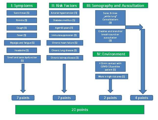
respiratory infections are provided in Table 1; Fig. 1 illustrates data collection. 13.75% of patients with respiratory tract
infections were recent returners from currently risk areas of transmission de ned by the RKI (mainly from Austria and
the Netherlands). 8.75% had signi cant (at least 15 minutes) contact with a person tested positive for COVID-19.
Table 1
Symptoms of patients with respiratory tract infections
Symptoms in all patients (n = 489) Symptoms- negatively tested (n = Symptoms- positively tested (n = 5)
75)
Cough n = 407 (83.23%) Cough n = 63 (84.00%) Cough n = 4 (80.00%)
Sore throat n = 70 (14.35%) Sore throat n = 9 (12.00%) Sore throat n = 4 (80.00%)
Myalgia and fatigue n = 309 Myalgia and fatigue n = 50 (66.67%) Myalgia and fatigue n = 3 (60.00%)
(63.19%)
Headache n = 158 (32.31%) Headache n = 22 (29.33%) Headache n = 2 (40.00%)
Rhinitis n = 245 (50.10%) Rhinitis n = 20 (26.67%) Rhinitis n = 3 (60.00%)
Fever > 38.5 degree Celsius n = 70 Fever > 38.5 degree Celsius n = 9 Fever > 38.5 degree Celsius n = 1
(14.31%) (12.00%) (20.00%)
Smell and taste dysfunction n = 51 Smell and taste dysfunction n = 9 Smell and taste dysfunction n = 0
(10.43%) (12.00%) (0.00%)
Chills n = 69 (14.11%) Chills n = 8 (10.67%) Chills n = 1 (20.00%)
Earache n = 41 (8.38%) Earache n = 4 (5.33%) Earache n = 0 (0.00%)
Table 1. Symptoms associated with respiratory tract infection between 27.01.2020 and 20.04.2020 in all patients (n =
489), patients negatively (n = 75) and positively tested (n = 5).
By far the most common diagnoses in the entire clientel were rhinopharyngitis and acute bronchitis; pneumonia was
less often found (Table 2.). Due to the low number of positive tests we did not perform mean comparison tests, data
were depicted as absolute values and percentages.
Page 4/13Table 2
Diagnoses of patients reporting with respiratory tract infections
Diagnosis in all patients (n = 489) Diagnosis- negatively tested(n = 75) Diagnosis- positively tested (n = 5)
Rhinopharyngitis: n = 182 (37.22%) Rhinopharyngitis n = 23 (30.67%) Rhinopharyngitis n = 2 (40%)
Acute bronchitis n = 148 (30.27%) Acute bronchitis n = 25 (33.33%) Acute bronchitis n = 3 (60%)
Acute sinusitis n = 52 (10.64%) Acute sinusitis n = 19 (25.33%) Acute sinusitis n = 0 (0%)
Tonsillitis n = 34 (6.95%) Tonsillitis n = 3(4.00%) Tonsillitis n = 0 (0%)
Otitis media n = 34 (6.95%) Otitis media n = 2(2.67%) Otitis media n = 0 (0%)
Pneumonia n = 31 (6.33%) Pneumonia n = 3(4.00%) Pneumonia n = 0 (0%)
Laryngitis n = 8 (1.64%) Laryngitis n = 0 (0%) Laryngitis n = 0 (0%)
Table 2. Distribution of diagnoses in all patients reporting with signs of respiratory infection (n = 489), patients
negatively tested (n = 75) and positively tested (n = 5) between 27.01.2020 and 20.04.2020. A diagnosis of pneumonia
was established in case of signi cant auscultation and one additional symptom, such as fever or productive sputum.
Chest X-ray was not available.
From 80 patients who met the RKI criteria(23) and were sent to the smear centre, only 5 turned out to be positive. The
most common symptoms in positive patients were cough (4/5), sore throat (4/5), myalgia and fatigue (3/5) and rhinitis
(3/5). Headache (2/5), chills (1/5) and fever (1/5) were less common. None of these positively tested patients suffered
from smell or taste dysfunction or earache. Table 3 illustrates the comorbidities of positively and negatively tested
patients. In both groups no skin alterations were detected.
Table 3
Prevalence of comorbidities in patients tested for SARS Cov2
Tested positive Tested negative
Diabetes: n = 2 (40.00%) Diabetes: n = 2 (2.67%)
Arterial hypertension: n = 3 (60.00%) Arterial hypertension: n = 23 (30.67%)
Hypothyroidism: n = 0 (0%) Hypothyroidism: n = 11 (14.67%)
Immunosuppression: n = 1 (20.00%) Immunosuppression: n = 6 (8.00%)
Atrial brillation: n = 1 (20.00%) Atrial brillation: n = 5 (6.67%)
Coronary artery disease: n = 0 (0%) Coronary artery disease: n = 3 (4.00%)
Lung disease: n = 2 (40.00%) Lung disease: n = 10 (13.33%)
Depression: n = 1 (20.00%) Depression: n = 11 (14.67%)
Chronic kidney disease: n = 0 (0%) Chronic kidney disease: n = 2 (2.67%)
RAAS inhibitors: n = 2 (40.00%) RAAS inhibitors: n = 15 (20.00%)
Oral anticoagulation: n = 1 (20.00%) Oral anticoagulation: n = 5 (6.67%)
Platelet Inhibitors: n = 1 (20.00%) Platelet Inhibitors: n = 1 (1.33%)
Page 5/13Table 3. Prevalence of comorbidities in patients tested positive (n = 5) and negative (n = 75) for COVID-19.
Immunospuppression was de ned as autoimmune disease or cancer in patient history. Lung disease was de ned as
chronic obstructive lung disease or asthma under medical treatment. Chronic kidney disease was de ned as a
glomerular ltration rate (GFR) < 60 ml/min for at least three months. Oral anticoagulants included vitamine K-
anatagonists (VKA) and new oral anticoagulants (NOAK.), platelet inhibitors included Aspirine, Clopidogrel, Ticagrelor
or Prasugrel. RAAS inhibitors: renin-angiotensin-aldosterone system inhibitors.
Discussion
We analyzed data from our three GP o ces in rural Germany between the onset of Covid-19 in our country on
27.01.2020 until 20.04.2020. The mean age of our patients was 47.03 years, which is quite young considering that
mortality seems to increase in COVID-19 patients beyond 65 years; patients less than 65 with little predisposing factors
may be at a low risk of severe disease(5). Although we also treat more elderly patients in our o ces, this observation
may mean that oligosymptomatic patients directly stay at home to reduce their physical contacts, or in case of
progressing symptoms, directly report to the clinic.
Only ve out of 80 tested patients were positive for COVID-19 (positives). Due to this low number and a potential
reporting bias of symptoms, we refrained from using mean comparison tests and only depicted absolute values.
However, we noticed that almost all patients in the positives suffered from a sore throat (4/5; 80%), while only 12% in
the negatively tested group (negatives) showed this symptom (9/75). Furthermore, rhinitis was more prominent in the
positives (60% vs. 26.67%). Although results have to be interpreted with caution, these two symptoms may be clinically
particularly relevant to assess probability for COVID-19 positivity.
Known comorbidities, especially pre-existing lung and cardiovascular disease, in positives and negatives were quite low
(see Table 3). The most common cardiovascular risk factor was arterial hypertension, which has already been
published(5). SARS-Cov2 uses ACE-2 as a cellular entry point(24) and has raised concern about continuation of RAAS
inhibitor intake in patients with chronic heart failure(25). However, recent data has shown that there is no evidence of
increased disease severity or mortality in hospitalized patients on RAAS blockers(26). Additionally, pharamcological
data suggests that ACE-2 expression is not increased in patients on RAAS blockers(27). Thus, current
recommendations support continuation of RAAS blockers in patients with arterial hypertension and chronic heart
failure(25). In our study two out of ve positively tested patients were on RAAS blockers and did not display more
severe symptoms than the others. Also in the negatives RAAS blockers were the most commonly prescribed
antihypertensive drugs (15 out of 23 patients received RAAS blockers) showing no difference in clinical severity.
We constantly tried to apply the algorithm provided by the RKI(23) to decide which patient needed a smear. However, it
seems quite problematic to handle patients with persistent symptoms (mainly unproductive cough), who have been
isolated and treated conservatively and who report to the o ce several times. A rather liberal management of smear-
taking could be applied in persistent symptoms refractory to conservative methods, such as inhalation, analgetics and
antiphlogistics. Although we would wish for a nationwide testing to get maximal clarity on the real number of positive,
oligo- or asymptomatic COVID-19 patients, this vision is still hampered by limited availability of tests, especially in rural
areas. Additionally, since a median incubation period of 5 days was estimated(28), the general practitioner will have
di culties to retrieve a patient’s contacts in the asymptomatic phase, which makes restriction of viral spread even more
di cult.
Due to a reliable recall system from our o ces, the clinic with the smear centre and the health department, we were
able to con rm that none of the patients negatively tested for COVID-19 progressed to COVID-19 positivity later on.
Those ve patients tested positive were kept in quarantine for at least two weeks. If symptoms had resided by then,
Page 6/13patients were allowed to take part in public life again. In case of persisting symptoms quarantine was upheld and
ambulatory medical services were sent to examine the patients whenever necessary. Moroever, due to regular
communication with the local health department we can con rm that none of the patients who were sent home without
a smear and treated conservatively, experienced symptom progression, which would have forced us to organize a
smear. In summary, the communication between GP o ces, hospitals with smear centres and the health department is
satisfactory to ensure the best possible patient care, despite the limited smear capacity. In the future, wearables, such
as smartwatches, may improve patient surveillance by constantly recording vitals and providing feedback about
potential health deteriorations at home. For GPs this would be a great opportunity to improve patient care.
In Germany we have a health system based on solidarity, in which most people have health insurance and thus have
easy access to health care. GP’s, usually as the rst medical contacts, have to lter many patients directly in the o ce.
The Center for Disease Control (CDC) has issued similar recommendations for the public as the RKI in Germany,
namely (1) to cover mouth and nose with a cloth, (2) call the GP’s o ce rst instead of showing up directly, (3) not to
get in close physical contact to others and (4) engage in regular desinfection of hands and surfaces, (5) and self-
monitor symptoms(29).
The death rate of SARS-Cov2 is currently 4.1%, almost 90% are over 70 years of age. The rate of infection in the high
risk cohort of elderly (> 80) patients is still rising in Germany (approximately 300 per 100.000 inhabitants in the group
80–89 years and 475 in the group 90-99years, as per 23.04.2020)(30), but testing frequency is declining again (peak:
30.03–05.04.2020 > 400.000 tests in Germany, 13.04–19.04.2020: 320.000 tests)(30). This generates fear of a „second
wave“ of infection. Similar to Germany, the CDC reported that 80% of deaths occurred in the age group > 65 years(29).
A fast incline of infection rates has occurred in Germany in February 2020, while in the US infections have risen in April
(as per 21.04.2020: 802.583 total cases, 44.575 total deaths)(29) and GP’s in the States will be facing the same
enormous logistic problems as in Germany earlier in the year. The paramount aim of medical personnel in this crisis is
to maintain optimal medical care and personal health in a high-risk environment. Thus, stringent algorithms for GP’s
and other medical specialties need to be introduced to achieve this goal. By segregating patients suggestive of
respiratory infection and treating them under high standards of hygiene and protection, we believe that we have
achieved this goal in a practicable and e cient manner. Until the date of submission, none of our employees called in
sick, which may suggest that our stringent selection process prior to entering the o ce is a success. Our experience
could now help colleagues in other countries with a later onset of COVID- 19 than Germany to organize their o ces
with the available resources. Transparency of strategies from different countries on how to deal with COVID-19 in
inpatient and ambulatory settings is of paramount importance to optimize further patient care and improve educational
measures(31).
Lung ultrasound seems to be a very promising tool to detect COVID-19, since it has been shown that B-lines are present
in early stages of the disease. Later hyperechoic images, called „white lung“, and nally consolidation can be found in
lung imaging(32). Although B-lines are not speci c for COVID-19 they can easily be assessed by trained
sonogrophaphers and can and should be be established in emergency departments and outpatient (GP) care.
Our data shows that it is not possible to accurately differentiate between oligosymptomatic COVID-19 patients and
ordinary respiratory infection by analyzing symptoms alone. A few days ago, Arons et al. published the spread of
COVID- 19 in a US nursing home, in which more than half of positively tested patients were asymptomatic(33, 34). This
clearly demonstrates that strategies focusing only on symptoms fail to prevent further transmission. Since the capacity
of testing is limited, we suggest the introduction of a new scoring system to stratify patients for COVID-19 that should
be tested. This scoring system should embrace the established clinical signs of (unspeci c) respiratory symptoms. The
two symptoms sore throat and rhinitis clearly prevailing in positively tested patients should be used as essential
Page 7/13symptomatic markers to stratify for obligatory testing. Known risk factors, such as arterial hypertension, chronic
obstructive lung disease and immunosuppression should be additional selection criteria for essential testing. Contact
to infected patients and working in high risk areas (such as the medical profession itself) and sonographic signs of
lung damage (e.g. B-lines, consolidations) should also be essential components of the score.
We provide a combinatorial score to pre-clinically estimate the risk of SARS-Cov2 infection (Fig. 2). Given the recent
observations that sonography is an easy and reliable method to assess suspicion of COVID19, we here add pulmonary
sonography as an integral part in our proposed scorings system to stratify patients with unclear respiratory infections
for obligatory Cov2 PCR testing.
We awarded 2 points each for sonographic signs of parenchymal or interstitial pulmonary in ltration, suggestive of
pneumonia or edema/congestion. 65 years as a cut-off is arbitrary, but Wang et al. (2020) showed that severe COVID-
19 infection is rare below 65, which may justify this value(5). We suggest that a total score > 5 points justi es to send
patients to Cov2-PCR testing. This means that, theoretically, an asymptomatic patient could qualify for PCR, if there are
enough risk factors (disease and environment); this was also demonstated by Arons et al.(34) We suggest to verify our
20 point-model in a larger cohort of suspected COVID-19 patients. The development and improvement of such a new
combinatorial score as proposed here will help GP’s to better stratify patients for necessary testing than current
suggestions.
Conclusion
In summary, we provide real-life data from rural GP o ces in Germany that demonstrate the di culty to distinguish
oligosymptomatic COVID-19 patients from ordinary respiratory tract infection. We provide a well-working example on
how to re-organize a GP’s o ce to separate potentially infectious patients from the rest with minimal risk of further
spreading the disease. We propose a concise screening score which integrates clinical symptoms, sonographic data
and history of previous diseases. Such a score is needed to stratify patients that should be obligatorily tested.
Abbreviations
ACE-2
Angiotensin converting enzyme
CDC
Center for Disease Control
COVID-19
Corona Virus Disease 2019
FFP-3
Filtering face piece
GFR
Glomerular ltration rate
GP
General practitioner
PPE
Personal protective equipment
RAAS
Renin-angiotensine-aldosterone system
RKI
Page 8/13Robert Koch Institut
SARS-Cov2
Severe acute respiratory syndrome- Corona virus 2
VKA
Vitamine K antagonist
Declarations
Ethics approval and consent to participate: No ethics committee was involved in the study. This was no clinical trial
according to WHO de nitions, data analysis was done according to WHO guidelines
(https://www.who.int/about/ethics/code-of-conduct-for-responsible-research). Data collection was done within the
daily clincial routine and analysis was performed anonymously. All patients or their legal guardians have signed a form
that their results may be used for medical purposes and analyses.
Consent for publication: not applicable.
Availability of data and materials:The datasets analyzed during the current study are not publicly available, but will be
made available from the corresponding author on reasonable request.
Competing interests: The authors declare that they have no competing interests.
Funding: there was no funding to this study.
The results have not been presented elsewhere.
Authors‘ contribution: All authors read and approved the nal manuscript. SW coined the idea to systematically analyze
symptoms and risk factors of patients with potential COVID-19. He created the risk score, wrote the manuscript (toether
with SW) and performed the systematic analysis of data. TF collected the data from all o ces and was responsible for
the compilation of the database. EW had the idea to integrate ultrasound as a modality into our score, meticulously
analyzed the literature and wrote the manuscript (together with SW).
Acknowledgments: not applicable.
Funding: no funding was obtained for this study.
Authors‘ information: SW is a preventive cardiologist who focuses on primary and secondary disease prevention, he
has profound experience in the intensive care management of cardiological and pneumological patients and is an
active emergency doctor. He is currently employed in the GPs o ce. Due to his large experience in both inpatient and
outpatient management, he analyzed the COVID-19 crisis in our area from a clinical and ambulatory perspective. EW is
a professor of anatomy and cell biology, whose main focus of science is to explore neuroimmune and complement
activation mechanisms of neurocardiovascular diseases and neurotropic virus infections. EW provided basic scienti c
background to compile this paper. TF is the head of all our GP o ces and has a long-lasting experience in the
adminstration and care of ambulatory patients. After onset of the crisis in Germany he rapidly designed a working
schedule for our o ces to combine patient care and safety for medical personel. The fact that none of our personel
has developed symptoms so far is mainly due to his foresighted thinking.
References
Page 9/131. Liu Y, Gayle AA, Wilder-Smith A, Rocklov J. The reproductive number of COVID-19 is higher compared to SARS
coronavirus. J Travel Med. 2020;27(2).
2. Phelan AL, Katz R, Gostin LO. The Novel Coronavirus Originating in Wuhan, China: Challenges for Global Health
Governance. Jama. 2020.
3. Johns Hopkins University Medicine. Coronavirus Resource Center [Available from: https://coronavirus.jhu.edu/
(accessed 2 May 2020).
4. Ruan Q, Yang K, Wang W, Jiang L, Song J. Clinical predictors of mortality due to COVID-19 based on an analysis of
data of 150 patients from Wuhan, China. Intensive Care Med. 2020.
5. Wang D, Hu B, Hu C, Zhu F, Liu X, Zhang J, et al. Clinical Characteristics of 138 Hospitalized Patients With 2019
Novel Coronavirus-Infected Pneumonia in Wuhan, China. Jama. 2020.
6. Zhou F, Yu T, Du R, Fan G, Liu Y, Liu Z, et al. Clinical course and risk factors for mortality of adult inpatients with
COVID-19 in Wuhan, China: a retrospective cohort study. Lancet. 2020;395(10229):1054–62.
7. Guo F, Du Z, Wang T. An effective screening and management process in the outpatient clinic for patients requiring
hospitalization during the COVID-19 pandemic. J Med Virol. 2020.
8. Kluge S, Janssens U, Welte T, Weber-Carstens S, Marx G, Karagiannidis C. German recommendations for critically
ill patients with COVID19. Med Klin Intensivmed Notfmed. 2020.
9. Tersalvi G, Vicenzi M, Calabretta D, Biasco L, Pedrazzini G, Winterton D. Elevated troponin in patients with
Coronavirus Disease 2019 (COVID-19): possible mechanisms. J Card Fail. 2020.
10. Huang C, Wang Y, Li X, Ren L, Zhao J, Hu Y, et al. Clinical features of patients infected with 2019 novel coronavirus
in Wuhan, China. Lancet. 2020;395(10223):497–506.
11. Xydakis MS, Dehgani-Mobaraki P, Holbrook EH, Geisthoff UW, Bauer C, Hautefort C, et al. Smell and taste
dysfunction in patients with COVID-19. Lancet Infect Dis. 2020.
12. Mao L, Jin H, Wang M, Hu Y, Chen S, He Q, et al. Neurologic Manifestations of Hospitalized Patients With
Coronavirus Disease 2019 in Wuhan, China. JAMA Neurol. 2020.
13. Tammaro A, Adebanjo GAR, Parisella FR, Pezzuto A, Rello J. Cutaneous manifestations in COVID-19: the
experiences of Barcelona and Rome. J Eur Acad Dermatol Venereol. 2020.
14. Recalcati S, Barbagallo T, Frasin LA, Prestinari F, Cogliardi A, Provero MC, et al. Acral cutaneous lesions in the Time
of COVID-19. J Eur Acad Dermatol Venereol. 2020.
15. Kreutz R, Algharably EAE, Azizi M, Dobrowolski P, Guzik T, Januszewicz A, et al. Hypertension, the renin-angiotensin
system, and the risk of lower respiratory tract infections and lung injury: implications for COVID-19. Cardiovasc
Res. 2020.
16. Corman VM, Landt O, Kaiser M, Molenkamp R, Meijer A, Chu DK, et al. Detection of 2019 novel coronavirus (2019-
nCoV) by real-time RT-PCR. Euro Surveill. 2020;25(3).
17. Reusken C, Broberg EK, Haagmans B, Meijer A, Corman VM, Papa A, et al. Laboratory readiness and response for
novel coronavirus (2019-nCoV) in expert laboratories in 30 EU/EEA countries, January 2020. Euro Surveill.
2020;25(6).
18. Maier BF, Brockmann D. Effective containment explains subexponential growth in recent con rmed COVID-19
cases in China. Science. 2020.
19. Konrad R, Eberle U, Dangel A, Treis B, Berger A, Bengs K, et al. Rapid establishment of laboratory diagnostics for the
novel coronavirus SARS-CoV-2 in Bavaria, Germany, February 2020. Euro Surveill. 2020;25(9).
20. Stafford N. Covid-19: Why Germany's case fatality rate seems so low. Bmj. 2020;369:m1395.
Page 10/1321. Dorr R. Protecting patients and healthcare personnel from COVID-19: considerations for practice and outpatient
care in cardiology. Herz. 2020.
22. COVID-19-Verdacht: Maßnahmen und Testkriterien - Orientierungshilfe für Ärzte (Stand: 22.4.2020): Robert Koch
Institut
COVID-19-Verdacht: Maßnahmen und Testkriterien - Orientierungshilfe für Ärzte (Stand: 22.4.2020): Robert Koch
Institut. Berlin G. 2020 [Available from:
https://www.rki.de/DE/Content/InfAZ/N/Neuartiges_Coronavirus/Massnahmen_Verdachtsfall_Infogra k_Tab.html
(accessed 2 May 2020).
23. Robert Koch Institut. COVID-19-Verdacht: Maßnahmen und Testkriterien - Orientierungshilfe für Ärzte (Stand:
22.4.2020), Suspicion of COVID-19: Measures and Test Criteria- Orientation for Physicians (as per 22.04.2020).
[Available from:
https://www.rki.de/DE/Content/InfAZ/N/Neuartiges_Coronavirus/Massnahmen_Verdachtsfall_Infogra k_Tab.html
(accessed 2 May 2020).
24. Liu Z, Xiao X, Wei X, Li J, Yang J, Tan H, et al. Composition and divergence of coronavirus spike proteins and host
ACE2 receptors predict potential intermediate hosts of SARS-CoV-2. J Med Virol. 2020.
25. Ganatra S, Hammond SP, Nohria A. The Novel Coronavirus Disease (COVID-19) Threat for Patients with
Cardiovascular Disease and Cancer. JACC CardioOncol. 2020.
26. Li J, Wang X, Chen J, Zhang H, Deng A. Association of Renin-Angiotensin System Inhibitors With Severity or Risk of
Death in Patients With Hypertension Hospitalized for Coronavirus Disease 2019 (COVID-19) Infection in Wuhan,
China. JAMA Cardiol. 2020.
27. Sriram K, Insel PA. Risks of ACE inhibitor and ARB usage in COVID-19: evaluating the evidence. Clin Pharmacol
Ther. 2020.
28. Lauer SA, Grantz KH, Bi Q, Jones FK, Zheng Q, Meredith HR, et al. The Incubation Period of Coronavirus Disease
2019 (COVID-19) From Publicly Reported Con rmed Cases: Estimation and Application. Ann Intern Med. 2020.
29. Report. CfDCaPMaMW. [Available from: https://www.cdc.gov/mmwr/index2020.html (accessed 2 May 2020).
30. Robert Koch, Institut. COVID-19: Fallzahlen in Deutschland und weltweit. Fallzahlen in Deutschland. Stand:
24.4.2020, 00:00 Uhr (online aktualisiert um 08:00 Uhr). [Available from:
https://www.rki.de/DE/Content/InfAZ/N/Neuartiges_Coronavirus/Fallzahlen.html (accessed 2 May 2020).
31. Fiorino G, Colombo M, Natale C, Azzolini E, Lagioia M, Danese S. Clinician Education and Adoption of Preventive
Measures for COVID-19: A Survey of a Convenience Sample of General Practitioners in Lombardy, Italy. Ann Intern
Med. 2020.
32. So a S, Boccatonda A, Montanari M, Spampinato M, D'Ardes D, Cocco G, et al. Thoracic ultrasound and SARS-
COVID-19: a pictorial essay. J Ultrasound. 2020.
33. Kimball A, Hat eld KM, Arons M, James A, Taylor J, Spicer K, et al. Asymptomatic and Presymptomatic SARS-CoV-
2 Infections in Residents of a Long-Term Care Skilled Nursing Facility - King County, Washington, March 2020.
MMWR Morb Mortal Wkly Rep. 2020;69(13):377–81.
34. Arons MM, Hat eld KM, Reddy SC, Kimball A, James A, Jacobs JR, et al. Presymptomatic SARS-CoV-2 Infections
and Transmission in a Skilled Nursing Facility. N Engl J Med. 2020.
35. Titles of Figures.
Figures
Page 11/13Figure 1
Algorithm of patient recriutment
Page 12/13Figure 2
Combinatorial model for preclinical assessment of COVID-19.
Page 13/13You can also read