The optimal management of variant histology in muscle invasive bladder cancer
←
→
Page content transcription
If your browser does not render page correctly, please read the page content below
Review Article

The optimal management of variant histology in muscle invasive
bladder cancer
Raj Vikesh Tiwari1, Nye Thane Ngo2, Lui Shiong Lee2
1
Department of Urology, Sengkang General Hospital, Singapore; 2Department of Anatomical Pathology, Singapore General Hospital, Singapore
Contributions: (I) Conception and design: RV Tiwari, LS Lee; (I) Administrative support: None; (I) Provision of study material or patients: NT Ngo; (I)
Collection and assembly of data: VT Raj, LS Lee; (I) Data analysis and interpretation: VT Raj, LS Lee; (I) Manuscript writing: All authors; (I) Final
approval of manuscript: All authors.
Correspondence to: Lui Shiong Lee. Department of Urology, Sengkang General Hospital, 110 Sengkang East Way, Singapore 544886, Singapore.
Email: Lee.lui.shiong@singhealth.com.sg.

                 Abstract: Bladder cancer is a heterogenous disease that is associated with tangible mortality in muscle
                 invasive disease. The WHO 2016 classification of urothelial tumours reflects the contemporary approach
                 towards histological variants in bladder cancer, including variants of urothelial carcinoma (UC) and non-
                 urothelial variants. This review focuses on variant histology in UC, and discusses the importance of accurate
                 histological diagnosis, and subsequent risk stratification and therapeutic decision making based on proper
                 variant recognition. Most urothelial variants are associated with poorer outcomes compared to conventional
                 UC, although some perform reasonably better. However, high quality evidence detailing optimal treatment
                 and survival outcomes are still lacking in literature, due to the rarity of these cases.

                 Keywords: Bladder cancer; therapy; urinary bladder neoplasms

                 Submitted Sep 23, 2019. Accepted for publication Dec 25, 2019.
                 doi: 10.21037/tau.2020.01.02
                 View this article at: http://dx.doi.org/10.21037/tau.2020.01.02

Introduction                                                                  disease may have considerable overlap with that of their
                                                                              conventional counterpart. Hence, there is strong clinical
Bladder cancer is currently one of the top ten most
                                                                              relevance to urologists, pathologists and oncologists alike
common solid organ malignancies worldwide, and
                                                                              to be able to distinguish between these variants confidently,
comprising approximately 550,000 new cases and 200,000
                                                                              especially in subtypes where the treatment and prognosis
deaths in 2018 (1,2). Bladder cancer is a heterogenous                        may be different from conventional UC.
disease with superficial non-invasive disease being more                         This article focuses on the key features of histological
common and associated with good outcomes while                                variants of conventional UC especially in the context of
aggressive muscle invasive disease is associated with poorer                  muscle invasive bladder cancer (MIBC), and explores the
prognosis (3). In patients with muscle invasive disease, a                    available evidence of prognostication and treatment of
wide array of treatment modalities have been described                        individual variants.
inclusive of systemic chemotherapy and immunotherapy,
radical cystectomy (RC) and bladder sparing regimes
including maximal transurethral resection combined with                       Evidence acquisition
radiotherapy with chemotherapy (4).                                           A non-systematic PubMed and Medline literature search
   In pure bladder cancer variants, the diagnosis and                         was performed using the following terms alone or in
treatment are largely distinct from conventional urothelial                   combination: “bladder cancer”, “urothelial cancer”,
carcinoma (UC) of the bladder (5). However, the optimal                       “micropapillary”, “microcystic”, “sarcomatoid variant”,”
management of urothelial cancer variants in muscle invasive                   nested variant”, “ plasmacytoid variant”, “clear cell”, “giant

© Translational Andrology and Urology. All rights reserved.                           Transl Androl Urol 2020 | http://dx.doi.org/10.21037/tau.2020.01.02
2                                                      Tiwari et al. The optimal management of variant histology for invasive bladder cancer

    Table 1 Differences in nomenclature of histologic variants in           classification of tumours of the urothelial tract, and detailed
    muscle invasive disease between 2002 and 2016 WHO classification        a section on histological variants in recognition of divergent
    2002 WHO                                                                differentiation, morphology and clinical phenotype (6)
     Infiltrating urothelial carcinoma                                      (Table 1). The updates reflected recognition of variant
                                                                            tumour histology as distinct in diagnosis, pathogenesis and
     IUC with squamous differentiation
                                                                            ultimately, in the realm of clinical trials and therapeutics.
     IUC with glandular differentiation                                     On the other hand, the grading classification is retained,
     IUC with trophoblastic differentiation                                 and WHO 2016 recommendations are largely based on
     Nested                                                                 the WHO 2004 (7) grading of low grade and high grade
                                                                            tumours.
     Microcystic
                                                                               By definition, these tumours have primarily UC as the
     Micropapillary                                                         predominant histological subtype, but other subtypes are
     Lymphoepithelioma-like                                                 also observed. The incidence of divergent differentiation or
     Lymphoma-like
                                                                            histologic variants is estimated to be about 25% in cystectomy
                                                                            series (8,9), and are therefore, not uncommon in incidence.
     Plasmacytoid
                                                                               Given the lack of tumour markers in bladder cancer,
     Sarcomatoid                                                            accurate pathological grading and histological subtype is
     Giant cell                                                             critical to optimal clinic decision-making on treatment
                                                                            and appropriate patient counselling. However, histological
     Undifferentiated
                                                                            variants may not be consistently reported or recognised.
    2016 WHO
                                                                            There may be a high discordance rate between pathological
     Infiltrating urothelial carcinoma with divergent differentiation       reporting of specimens obtained from transurethral
     Nested, inclusive of large nested variant                              resection and cystectomy numbering between 39–47%,
                                                                            often due to a lack of recognition by community
     Microcystic
                                                                            pathologists (10). In the specific example of micropapillary
     Micropapillary                                                         variant of UC, consistent pathological reporting is
     Lymphoepithelioma-like                                                 challenged by significant interobserver variation even
     Signet ring cell/plasmacytoid/diffuse                                  among urologic pathologists (11), and a lack of specific and
                                                                            sensitive immunohistochemical markers (12,13).
     Sarcomatoid
                                                                               Of the available studies in English literature, most of
     Giant cell                                                             them suggest that variant histology in bladder cancer,
     Poorly-differentiated                                                  whether muscle invasive or not, will portend a poorer
     Lipid rich
                                                                            clinical outcome of survival in affected patients, when
                                                                            compared to conventional UC. Generally, there is a
     Clear cell
                                                                            higher propensity for extra-vesical extension and lymph
     Tumours of Mullerian type                                              node metastasis (14), and also a tendency for pathological
     Tumours arising in a bladder diverticulum                              upstaging at RC (hazard ratio of 2.77) compared to
    IUC, infiltrating urothelial carcinoma.
                                                                            conventional UC (15). However, the strength of evidence
                                                                            for prognosis and optimal treatment varies between tumour
                                                                            variants depending on the quality of available evidence.
cell”, “histological variants” and “muscle invasive bladder                 Hence, specific recommendations will be detailed in the
cancer”. Meta-analysis, original articles and reviews in                    relevant sections that follow.
published in English were chosen.
                                                                            Urothelial malignancies and variant types
Case definition                                                             Micropapillary urothelial carcinoma (MPUC)

In 2016, WHO released the fourth edition of the                             MPUC was first described in the early 1990s at MD

© Translational Andrology and Urology. All rights reserved.                        Transl Androl Urol 2020 | http://dx.doi.org/10.21037/tau.2020.01.02
Translational Andrology and Urology, 2020 3
100 µm 100 µm
Figure 1 Micropapillary bladder cancer. (A) Haematoxylin and eosin slide showing invasive component of tumour; (B) H&E slide showing
mucosal component of tumour.
Anderson Cancer Centre by Amin et al. (16) as a tumour (n=36).
growth pattern that occurs in many organs usually In a more contemporary study of 82 patients with non-
presenting at an advanced stage. metastatic MIBC, Meeks et al. demonstrated a 45% pT0
The microscopic features of MPUC include a rate following neoadjuvant cisplatin based chemotherapy
micropapillary architecture similar to that of ovarian followed by RC, and also showed better OS in pT0
papillary serous tumours, and this can be observed to be individuals than those with residual disease (24). However,
at the mucosal surface or in the invasive component of the despite a response to NAC in some patients, a pooled
tumour. The nuclei of tumour cells are frequently of high analysis showed that NAC did not confer a OS or cancer
grade, showing reversed polarity to the external surface of specific survival (CSS) compared to RC alone (25).
tumour nests (17) (Figure 1). However, there was an intermediate to high risk of bias
The proportion of MPUC in the tumour is prognostic across these studies observed.
for clinical outcomes, and should be routinely reported if It is also possible to clinically risk stratify patients with
present (18). surgically resectable MPUC—namely those with clinical
The incidence of MPUC is estimated to be between 0.7% T1, clinical T2 or more without hydronephrosis and any
to 2.2% of all urothelial tumours, with a male to female clinical stage with hydronephrosis (26). The 5-year OS
ratio of 10:1 and average age of diagnosis of 65 years. A rates were 82%, 45% and 15% respectively, while DSS
majority of patients were found with muscle invasive disease rates were 92%, 51% and 17% respectively. They suggested
at presentation, and the overall survival (OS) outcomes at upfront RC for the lower risk group (5 years OS and DSS
5 and 10 years were 51% and 24% respectively (19). In of 87% and 95%), and neoadjuvant chemotherapy (NAC)
earlier surgical series, there was also a high progression rate and RC for the high risk group (5-year OS and DSS 64%
(70%) from non-muscle invasive to muscle-invasive disease, and 79% respectively). For the highest risk group with
and nodal metastasis despite aggressive treatment. This is hydronephrosis, the OS and DSS were very poor (24% and
associated with a high tendency for pathological upstaging 27%) with upfront RC, and poorer with NAC prior to RC.
in the MPUC components at cystectomy (20). However, this study was limited by a sample size of n=107
In non-muscle invasive disease, the optimal treatment patients and would require validation from larger patient
with either early RC or intravesical BCG is not known populations.
(14,21-23). Kamat et al. suggested that early cystectomy Whole genome sequencing of MPUC has yielded a
was indicated in non-muscle invasive MPUC as there genomic signature typical of a luminal subtype associated
was a high rate of progression to MIBC with intravesical with the activation of wild-type p53 mutations, which would
Bacillus Calmette Guerin (BCG) therapy. However, this possibly explain its aggressive biological behaviour (27).
was reported from a study with a small sample size (n=27) Hence, based on available clinical evidence drawn from
and the wider applicability is not clear (15). Spaliviero et al. case series, there is a suggestion to support early RC in T1
showed no significant difference in cancer specific mortality disease where suitable. Otherwise, intravesical BCG may
or risk of metastasis between early RC and intravesical also provide disease control in those not suitable for radical
BCG, although there was a tendency for better outcomes surgery but with a potentially poorer OS. In patients with
with early RC. This study was also limited by sample size MIBC, there should be consideration of NAC based on
© Translational Andrology and Urology. All rights reserved. Transl Androl Urol 2020 | http://dx.doi.org/10.21037/tau.2020.01.024 Tiwari et al. The optimal management of variant histology for invasive bladder cancer
conventional UC regime. This should be followed by RC One case was reported in a renal transplant recipient
especially in those with good clinical response. 12 years post allograft implant and BK virus positive (37),
which was treated with radical surgery.
Hence, from limited evidence available, it would be
Giant cell variant (GCV)
reasonable to pursue bladder sparing surgery with maximal
The GCV of UC is a very rare entity first described by TURBT in those with non-muscle invasive disease, while
Kitazawa et al. (28) It is essentially a high grade carcinoma, radical treatment should be considered in those with MIBC.
which appears undifferentiated and is characterized by the
presence of epithelial tumor giant cells exhibiting marked
Microcystic carcinomas
nuclear atypia, along with a component of conventional
urothelial carcinoma, and is initially identified as an Microcystic carcinoma (MC) was first described in
undifferentiated UC. Although bearing resemblance with 1991 by Robert et al., presenting 4 cases arising in the
giant cell tumors of the lung, these urothelial variants are bladder. The 2004 WHO classification were the first
distinct from other giant cell described within other bladder recommendations to make a distinction between microcystic
tumors, including osteoclast like giant cells occasionally variant of UC and UC with gland-like lumina (7).
seen in reactive lesions, syncytiotrophoblasts, sarcomatoid To date, there are about 30 cases so far. The largest case
variant of UCs and giant cells of the so called giant tumor series comprise 20 cases where all patients presented with
of the bladder which is indistinguishable from giant cell macroscopic haematuria, and the appropriate histological
tumour of bone and lacks urothelial differentiation (29). variant diagnosis of MC was only made at RC in half of
Microscopically, the giant cells variant of shows marked the patients. At microscopy, the cysts were observed to be
nuclear pleomorphism, typically with multiple nuclei, of variable sizes up to 1.1 mm. These cysts were lined by
and consists of cohesive cells with abundant eosinophilic a single or several layers of urothelial cells, with the lining
cytoplasm. Malignant giant cells when present in great epithelium denude in some (38). There is also prominent
numbers, portend a poor clinical prognosis (30,31). intercellular or intracellular lumina with no true glands, and
An accurate histopathological diagnosis requires IHC no colonic epithelium or goblet cells are observed (39). IHC
to differentiate GCV from the conventional UC and expression of MC showed that most tumours expressed
undifferentiated carcinoma, including the application of MUC5A, GATA3, CK7 and MUC1 (38).
epithelial markers of cytokeratin and epithelial membrane There are two postulates for the etiology of MC for
antigen. Vimentin and keratin can distinguish GCV from this rare variant, namely having cyst-like structures which
similar mesenchymal lesions arising in the urinary bladder. emanate from urothelium forming spaces within the
Additionally GCV does not stain positively with tartrate bladder, or arising from cellular degeneration which leads
resistant acid phosphatase which is a characteristic of to the presence of luminal debris and necrotic cells (40).
osteoclastic giant cell tumours (29,32,33). The differential diagnosis of MC include many benign
Common misdiagnosis of this variant could be secondary histologic patterns, including cystitis glandularis and hence,
carcinomas or sarcoma in limited samples which could in clinical situations where only limited specimens are
lead to very different treatment approaches (34). Other available, there is a high possibility of misdiagnosis (29,38).
differentials would include metastatic giant cell carcinomas The often dramatic variations in nuclear size, in addition
of lung, melanoma or direct extension of prostate giant cell to other atypical characteristics, is a helpful distinguishing
carcinoma. In the latter case, background conventional UC feature of the microcystic variant of UC. The invasive
can aid in confirming the impression of a bladder primary. nature of the lesion into lamina propria or detrusor muscle
Due to its rarity, there are less than twenty cases will help in differentiation from benign pathology.
reported so far (28,29,31-33,35-37). The typical clinical Another diagnostic consideration is primary or metastatic
presentation is that of haematuria with a significant mass in adenocarcinoma. Although the cysts of the microcystic
the bladder. The natural history of GCV has been variable variant may resemble small infiltrating glans and may
and elucidation of longer term behaviour limited by follow- produce mucin, the architecture and cytology of the lining
up durations reported on. TURBT alone provided short epithelium are distinctly urothelial in nature (38).
term control in three cases (32,35), while RC was indicated In an earlier case series, Paz et al described a series of 12
in two cases (28,36). cases of MC admixed with high grade UC. A majority of
© Translational Andrology and Urology. All rights reserved. Transl Androl Urol 2020 | http://dx.doi.org/10.21037/tau.2020.01.02Translational Andrology and Urology, 2020 5
patients presented with T1 and above tumour stage (41). accurate histopathological diagnosis so as to be able to
When compared stage for stage with conventional UC, the effect early radical therapy for best survival outcomes. It is
presence of MC variant did not adversely affect OS (38). postulated, that late stage clinical presentation in existing
The poor outcomes of MC reported in literature were likely literature previously discussed, is contributed, in part, to a
related to the advanced stage at presentation. delay in appropriate histopathological diagnosis.
Therefore, the MC variant may be mistakenly diagnosed
as cystitis glandularis or adenocarcinoma of the urinary
Plasmacytoid variant (PV)
bladder in limited specimens, and a proper sampling by
transurethral resection of the bladder tumour (TURBT) This is a rare variant with less than 200 cases reported in
is essential. In cases where the diagnosis of MC has been literature so far. At histology, tumour cells are observed
made, the optimal treatment cannot be defined due to to have an eccentrically placed nucleus with abundant
limited evidence available in literature, but the treatment eosinophilic cytoplasm reminiscent of plasma cells. The
modality and outcomes are stage dependent and possibly nuclei are of low to intermediate nuclear grade with
similar to conventional UC. occasional nuclear pleomorphism (49-52). IHC showed an
absence or strongly reduced expression of E cadherin in PV,
a feature associated with advanced pathological stage and
Nested variant (NV)
nodal metastasis in conventional UC (53).
This variant of tumours has been described to mimic The natural history of PV is characterized by advanced
benign proliferation of Brunn’s nest with bland cytology. stage at presentation and a poor prognosis. In a study
In a case series of 23 cases, Cox et al. described key comparing 30 PV cases with 278 conventional UC cases,
microscopic features including cellular nuclei that lacked PV patients were more likely to have a higher stage at
significant atypia with the occasional scattered slightly cystectomy and lymph node metastasis, with positive
enlarged, hyperchromatic nuclei with small-indistinct surgical margins. The median OS and CSS were also dismal
nucleoli. The surrounding stroma had a mild-to-moderate at 19 and 22 months respectively. The presence of PV at
fibrous and inflammatory reaction in a majority of cases TURBT was associated with an increased risk of non-organ
and a small number exhibited no stromal reaction (42). confined disease compared to conventional UC (OR 4.02,
This variant should be carefully distinguished from an 95% CI: 10.6–15.22, P=0.04) (51). In another case series of
inverted growth pattern of noninvasive urothelial cancer 31 patients, only half of them presented with non-metastatic
and from von Brunn nests, by the presence of by either disease. Of those with surgically resectable disease, one
muscularis propria invasion, irregularly infiltrating nests third received NAC with pathological downstaging seen
of tumour cells, or the presence of a stromal reaction to in the majority of them. Of those who underwent upfront
the tumour (42-45). Using immunohistochemistry, NV surgery, most of them received adjuvant chemotherapy (54).
demonstrated loss of p27 which is also observed in high There is also a clinical predilection for trans-peritoneal
grade UC. However, p53, bcl-2 and EGF-r which are spread which may be observed at RC (55), or may occur
markers of adverse behaviour in UC were not observed in after curative radical surgery even in those with good
NV. In addition, MIB-1 stain may help to distinguish NV pathological response after NAC (54).
from benign urothelial proliferations (46). Within the AUO-AB05/95 trial of RC with adjuvant
In a single institution surgical series, NV was cisplatin-based chemotherapy, patients with PV had poorer
associated with advanced stage at cystectomy with a OS compared to UC and MPUC (27.4 versus 62.6 and
10-year recurrence free survival (RFS) and CSS of 83% and 64.6 months respectively, P=0.013) (56).
41% respectively. However, when NV was matched with Therefore, the detection of PV at TURBT should
conventional urothelial cancers, there were no differences prompt the managing urologist to suspect muscle invasive
noted in survival outcomes (47). In another series of 30 disease. There is limited evidence for the widespread role
cases, when compared to high grade UC, NV was associated of NAC, but where administered there is a high rate of
with a higher prevalence of muscle invasion at TURBT, pathological downstaging at RC. Where upfront RC is
extravesical extension at cystectomy and metastatic disease. pursued, there is a high incidence of non-organ confined
The response to NAC was also poor (13%) (48). disease, and hence, a strong consideration should be made
Hence, in NV tumours, the key to optimal therapy is for adjuvant cisplatin based chemotherapy.
© Translational Andrology and Urology. All rights reserved. Transl Androl Urol 2020 | http://dx.doi.org/10.21037/tau.2020.01.026 Tiwari et al. The optimal management of variant histology for invasive bladder cancer
Figure 2 Lymphoepithelioma variant. (A) Haematoxylin and eosin slide showing dense immune cell infiltrates among the tumour cells (larger
with prominent nucleus); (B) immunohistochemical staining for p63.
Clear cell variant (CCV) of UC an aggressive phenotype which should be treated radically
where possible.
This tumour is rare and less than 30 cases have been
reported in literature. Tumours with clear cell appearances
in the bladder would usually trigger the following Lymphoepithelioma variant (LEV)
differential diagnosis: (I) metastasis from a clear cell renal
The LEV of UC is characterized by syncytial growth pattern
carcinoma; (II) clear cell adenocarcinoma of the urinary
of a malignant epithelial component with prominent nuclei
bladder or (III) CCV of a UC. Hence, the accurate
and nucleoli, which is densely infiltrated by lymphoid cells
histopathological diagnosis would require relevant clinical
predominantly composed of cytotoxic T lymphocytes (63)
input to support a possible primary site of cancer, and the
(Figure 2). These cells may resemble metastatic tumours from
application of relevant IHC to exclude clear cell cancers
from other possible sources (57-59). The microscopic the nasopharynx and the strong expression of CK7 would
features of CCV include a clear cell pattern and a glycogen‐ be able to demonstrate a urothelial origin of tumour (64).
rich cytoplasm. The clear cell pattern may be either Other differential diagnosis of malignant lymphomas,
focal or extensive in the tumor component (60). Rotellini undifferentiated UC with lymphocytic infiltrate and small
et al. described a clinical case where a clear cell tumour cell carcinoma of the bladder can also be differentiated with
was found in the urinary bladder of a patient years after a the relevant IHC.
radical nephrectomy for clear cell renal carcinoma. The LEV has a more favourable long term prognosis especially
lesion in the bladder was CK7, CK20 and GATA3 positive. in cases where the component of conventional high grade
In addition, UroVysion FISH test was also employed which UC is low (65-67), or where the component of LEV is
demonstrated aneuploidy of chromosomes 3, 7, 27 and the higher (68). In a case series of 34 patients, those with pure
loss of 9p21 locus in the bladder tumour, which supported LEV treated with bladder TURBT and chemotherapy were
its urothelial origin (61). disease free at 2 to 5 years follow-up, compared to others
The clinical outcomes have been variable and not possible with less than 50% LEV component who had metastatic
to characterize precisely due to the small number of cases disease or cancer mortality (67). In cases of LEV where the
reported. The reported outcomes following surgical treatment disease is not muscle invasive, treatment with TURBT and
include mortality from rapid disease progression 9 weeks intravesical therapy has been described, and clinical outcomes
after RC (58), disease mortality 20 months after RC (59), approximate that of conventional UC (67,69).
disease free at 20 months follow-up after TURBT alone (57), Thus, in the management of LEV, the distinction from
and no mortality at 6 months post RC (62) respectively. other lymphoid malignancies is important, and treatment
Therefore, in CCV, the clinical history of preceding options would mirror that of conventional UC. The natural
malignancies would aid towards the clinical distinction history of LEV is likely to be similar or better than that of
with other clear cell tumours that may arise within or from UC when compared stage for stage.
without the bladder. This should then be confirmed with
a combination of appropriate IHC stains. The natural
Lipid rich variant
history of CCV is not easily characterized with limited data
in literature, but there seems to be a propensity towards The lipid rich variant (LRV) has microscopic features of
© Translational Andrology and Urology. All rights reserved. Transl Androl Urol 2020 | http://dx.doi.org/10.21037/tau.2020.01.02Translational Andrology and Urology, 2020 7
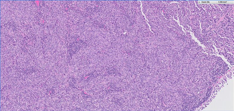
100 µm 100 µm
Figure 3 Sarcomatoid carcinoma. (A) Haematoxylin and eosin slide showing a combination of malignant epithelial and mesenchymal cells; (B)
immunohistochemical staining for cytokeratin-7 (CK-7).
eccentrically placed nucleus and optically clear cytoplasmic tumours composed of an admixture of carcinomatous
vacuoles resembling lipoblasts or signet‐ring cells (7). and sarcomatous components with an abrupt or gradual
There is no presence of mucin detectable, which would transition from one to another (Figure 3). In most cases,
distinguish it from signet ring cell tumours. The contents of the epithelial component consists of high grade UC with
the vacuoles are unknown, although postulated to contain possible epidermoid and/or glandular differentiation while
lipids based on animal studies but not demonstrable in the heterologous component consists of chondrosarcoma,
human tissue (70). The lipid cell component is variable but malignant fibrous histiocytoma, osteosarcoma,
can occupy up to 50% of the tumour, and usually admixed leiomyosarcoma, fibrosarcoma or rhabdomyosarcoma (79).
with a component of conventional UC. In the largest case While smoking does not seem to be a risk factor for the
series of 27 cases, other variants were also found with LCV development of SC, prior exposure to cyclophosphamide
and UC, including MPU (2 cases), PCV (1 case), squamous and pelvic irradiation may be risk factors (80). While there
differentiation (2 cases) and concomitant carcinoma-in-situ is evidence for a monoclonal origin for the development of
(8 cases) (71). both the epithelial and mesenchymal components of SC,
The typical presentation is haematuria and the age there may be genetic divergence in the subsequent clonal
groups affected range from 42–90 years of age with a male evolution (81).
preponderance. The majority of patients present with As a relatively uncommon variant of urothelial cancers,
muscle invasive disease (more than 80%) and were treated these patients are rarely recruited into clinical trials and hence,
with RC, including a small proportion (15%) with adjuvant it is challenging to define optimal treatment for these patients.
chemotherapy or chemoradiation therapy. At a median The existing literature supports an aggressive biological
follow-up of 28 months, the OS rate was 30% in this small behaviour of these tumours, which have a high tendency
cohort. In another case series of 5 patients, a majority for muscle invasive disease at clinical presentation (82).
of patients had cancer specific mortality between 8 to SC was also shown to have a poorer survival compared to
76 months from diagnosis, although the modality of conventional urothelial cancers, and a higher T stage at
treatment was not specified (72). presentation (82).
Hence, in LCV, there is a preponderance to advanced In a study of 221 cases with outcomes derived from the
stage at presentation associated with an aggressive SEER database, the disease prognosis was largely dependent on
behaviour, thereby suggesting that this variant should be the tumour stage at presentation (79). Individuals with regional
treated radically and aggressively upon diagnosis. or distant metastasis were at a high risk for cancer specific
mortality, when compared to those with localized disease.
The 1-, 5- and 10-year survival was 53.9%, 28.4% and 25.8%
Sarcomatoid carcinoma (SC)
respectively, reflecting the aggressive biological behaviour of SC.
SC is defined as a biphasic tumour consisting of malignant There was no significant difference in OS between those treated
epithelial and mesenchymal elements (73) . These with TURBT with radical radiation versus RC. However, in
tumors are relatively uncommon and most literature light of the aggressive behaviour of SC, one should pursue
is limited to case reports or limited case series (74-78). radical treatment where possible, and RC should be considered
The microscopic features of SCs include that of biphasic where surgically resectable in fit individuals.
© Translational Andrology and Urology. All rights reserved. Transl Androl Urol 2020 | http://dx.doi.org/10.21037/tau.2020.01.028 Tiwari et al. The optimal management of variant histology for invasive bladder cancer
Conclusions Prognostic and Clinical Importance of Urothelial and
Nonurothelial Histological Variants of Bladder Cancer in
The tumour variants of UC are detailed based on the
Predicting Oncological Outcomes in Patients with Muscle-
WHO 2016 classification of tumors of the urinary system,
invasive and Metastatic Bladder Cancer? A European
which highlights the importance of distinct morphological
Association of Urology Muscle Invasive and Metastatic
description of histological variants. The treatment and
Bladder Cancer Guidelines Panel Systematic Review. Eur
prognosis have been described albeit with a variable level
Urol Oncol 2019;2:625-42.
of evidence due to limited cases numbers reported in
6. Humphrey PA, Moch H, Reuter VE et al. The 2016
literature.
WHO Classification of Tumours of the Urinary System
C u r r e n t l y, t w o c h a l l e n g e s e x i s t i n t h e c l i n i c a l
and Male Genital Organs—Part B: Prostate and Bladder
management of UC variants in the urinary bladder, Tumours. Eur Urol 2016;70:106-19.
namely an accurate histopathological diagnosis and specific 7. Eble JN, Sauter G, Sesterhenn IA, et al. World Health
treatment strategies. Given that most therapeutic strategies Organization Classification of Tumours. Pathology and
are extrapolated from treatment options of conventional Genetics Tumours of the Urinary System and Male
UC, and while applicable in a proportion of patients, there Genital Organs 2004;10.
is clearly a need for more defined therapeutics guided by 8. Monn MF, Kaimakliotis HZ, Cheng L, et al.
data from specifically designed clinical trials. Contemporary bladder cancer: Variant histology may be a
significant driver of disease. Urol Oncol 2015;33:18.e15-
Acknowledgments 18.e20.
9. Xylinas E, Rink M, Brisuda A, et al. Impact of histological
None. variants on oncological outcomes of patients with
urothelial carcinoma of the bladder treated with radical
Footnote cystectomy. Eur J Cancer 2013;49:1889-97.
10. Shah RB, Montgomery JS, Kunju LP, et al. Variant
Conflicts of Interest: The authors have no conflicts of interest (divergent) histologic differentiation in urothelial
to declare. carcinoma is under-recognized in community practice:
Impact of mandatory central pathology review at a large
Ethical Statement: The authors are accountable for all referral hospital. Urol Oncol 2013;31:1650-5.
aspects of the work in ensuring that questions related 11. Sangoi AR, Beck AH, Hansel DE, et al. Interobserver
to the accuracy or integrity of any part of the work are Reproducibility in the Diagnosis of Invasive Micropapillary
appropriately investigated and resolved. Carcinoma of the Urinary Tract Among Urologic
Pathologists. Am J Surg Pathol 2010;34:1367-76.
References 12. Maranchie JK, Bouyounes BT, Wolf WCD, et al.
Clinical and pathological characteristics of micropapillary
1. Bray F, Ferlay J, Soerjomataram I, Siegel RL, et al. transitional cell carcinoma: A highly aggressive variant. J
Global cancer statistics 2018: GLOBOCAN estimates of Urol 2000;163:748-51.
incidence and mortality worldwide for 36 cancers in 185 13. Sangoi AR, Higgins JP, McKenney JK, et al.
countries. CA Cancer J Clin 2018;68:394-424. Immunohistochemical comparison of MUC1, CA125,
2. Antoni S, Ferlay J, Soerjomataram I, et al. Bladder Cancer and Her2Neu in invasive micropapillary carcinoma of the
Incidence and Mortality: A Global Overview and Recent urinary tract and typical invasive urothelial carcinoma with
Trends. Eur Urol 2017;71:96-108. retraction artifact. Mod Pathol 2009;22:660-7.
3. Flaig TW, Spiess PE, Agarwal N, et al. NCCN Guidelines 14. Willis DL, Fernandez MI, Pisters LL, et al. Clinical
Insights: Bladder Cancer, Version 5.2018. J Natl Compr Outcomes of cT1 Micropapillary Bladder Cancer. J Urol
Canc Netw 2018;16:1041-53. 2015;193:1129-34.
4. Chou R, Selph SS, Buckley DI, et al. Treatment of muscle- 15. Kamat AM, Gee JR, Dinney CPN, et al. The Case
invasive bladder cancer: A systematic review. Cancer for Early Cystectomy in the Treatment of Nonmuscle
2016;122:842-51. Invasive Micropapillary Bladder Carcinoma. J Urol
5. Veskimäe E, Espinos EL, Kamat AM, et al. What Is the 2006;175:881-5.
© Translational Andrology and Urology. All rights reserved. Transl Androl Urol 2020 | http://dx.doi.org/10.21037/tau.2020.01.02Translational Andrology and Urology, 2020 9
16. Amin MB, Ro JY, Silva EG, et al. Micropapillary variant Hum Pathol 2009;40:1461-6.
of transitional cell carcinoma of the urinary bladder. 31. Baydar D, Amin MB, Epstein JI. Osteoclast-rich
Histologic pattern resembling ovarian papillary serous undifferentiated carcinomas of the urinary tract. Mod
carcinoma. Am J Surg Pathol 1994;18:1224-32. Pathol 2006;19:161-71.
17. Kwon GY, Ro JY. Micropapillary Variant of Urothelial 32. Behzatoğlu K, Durak H, Oznur M, et al. Giant cell tumor-
Carcinoma. Adv Urol 2011;2011:217153. like lesion of the urinary bladder: a report of two cases
18. Samaratunga H, Khoo K. Micropapillary variant and literature review; giant cell tumor or undifferentiated
of urothelial carcinoma of the urinary bladder; a carcinoma? Diagn Pathol 2009;4:48.
clinicopathological and immunohistochemical study. 33. Amir G, Rosenmann E. Osteoclast-like giant cell tumour
Histopathology 2004;45:55-64. of the urinary bladder. Histopathology 1990;17:413-8.
19. Kamat AM, Dinney CPN, Tamboli P, et al. Micropapillary 34. Clusp BG. Tumors of Urinary Bladder, 3rd Ed. USA:
bladder cancer. Cancer 2007;110:62-7. ScienceDirect Topics, 2014:1230.
20. Compérat E, Roupret M, Ouzaïd I, et al. Micropapillary 35. Wu PJ, Su CK, Chen CL. Osteoclast-like Giant Cell
urothelial carcinoma of the urinary bladder: a Carcinoma of the Urinary Bladder. J Chin Med Assoc
clinicopathological analysis of 72 cases. Pathology 2009;72:495-7.
2010;42:650-4. 36. O'Connor RC, Hollowell CM, Laven BA, et al. Recurrent
21. Spaliviero M, Dalbagni G, Al-Ahmadie HA, et al. Clinical giant cell carcinoma of the bladder. J Urol 2002;167:1784.
Outcome of Patients with T1 Micropapillary Urothelial 37. Alexiev BA, Papadimitriou JC, Drachenberg CB, et al.
Carcinoma of the Bladder. J Urol 2014;192:702-7. Polyomavirus (BK)-associated pleomorphic giant cell
22. Sui W, Matulay JT, James MB, et al. Micropapillary Bladder carcinoma of the urinary bladder: a case report. Pathol Res
Cancer: Insights from the National Cancer Database. Pract 2013;209:255-9.
Bladder Cancer 2016;2:415-23. 38. Lopez Beltran A, Montironi R, Cheng L. Microcystic
23. Jackson BL, Mohammed A, Griffiths TRL, et al. Is urothelial carcinoma: morphology, immunohistochemistry
Immediate Radical Cystectomy Necessary for All Patients and clinical behaviour. Histopathology 2014;64:872-9.
with Non-Muscle-Invasive Micropapillary Bladder 39. Venyo AKG. Microcystic Variant of Urothelial Carcinoma.
Cancer? Urol Int 2016;96:32-8. Adv Urol 2013;2013:654751.
24. Meeks JJ, Taylor JM, Bochner BH, et al. Pathological 40. Rugvedita P. P Rugvedita Bladder urothelial carcinoma-
response to neoadjuvant chemotherapy for muscle- invasive microcystic variant 2013 Pathol Outlines.
invasive micropapillary bladder cancer. BJU Int 41. Paz A, Rath-Wolfson L, Mukamel E, et al. The clinical
2013;111:E325-E330. and histological features of transitional cell carcinoma of
25. Abufaraj M, Foerster B, Hassler MR, et al. Micropapillary the bladder with microcysts: analysis of 12 cases. Br J Urol
Urothelial Carcinoma of the Bladder: A Systematic 1997;79:722-5.
Review and Meta-analysis of Disease Characteristics and 42. Cox R, Epstein JI. Large Nested Variant of Urothelial
Treatment Outcomes. Eur Urol 2019;75:649-58. Carcinoma: 23 Cases Mimicking von Brunn Nests and
26. Fernández MI, Williams SB, Parikh S, et al. Clinical Inverted Growth Pattern of Noninvasive Papillary
risk stratification in patients with surgically resectable Urothelial Carcinoma. Am J Surg Pathol 2011;35:1337-42.
micropapillary bladder cancer. BJU Int 2017;119:684-91. 43. Murphy WM, Deana DG. The nested variant of
27. Guo CC, Dadhania V, Sykulski M, et al. Gene Expression transitional cell carcinoma: a neoplasm resembling
Profile of the Clinically Aggressive Micropapillary Variant proliferation of Brunn's nests. Mod Pathol 1992;5:240-3.
of Bladder Cancer. Eur Urol 2016;70:611-20. 44. Holmäng S, Johansson SL. The Nested Variant of
28. Kitazawa M, Kobayashi H, Sekine S, et al. Giant Cell Transitional Cell Carcinoma - A Rare Neoplasm with Poor
Tumor of the Bladder Associated with Transitional Cell Prognosis. Scand J Urol Nephrol 2001;35:102-5.
Carcinoma. J Urol 1985;133:472-5. 45. Drew PA, Furman J, Murphy WM, et al. The nested
29. Zhai QJ, Black J, Ro JY, et al. Histologic variants of variant of transitional cell carcinoma: an aggressive
infiltrating urothelial carcinoma. Arch Pathol Lab Med neoplasm with innocuous histology. Mod Pathol
2007;131:1244-56. 1996;9:989-94.
30. Lopez-Beltran A, Blanca A, Regueiro JC, et al. 46. Lin O, Cardillo M, Reuter VE, et al. Nested Variant
Pleomorphic giant cell carcinoma of the urinary bladder. of Urothelial Carcinoma: A Clinicopathologic and
© Translational Andrology and Urology. All rights reserved. Transl Androl Urol 2020 | http://dx.doi.org/10.21037/tau.2020.01.0210 Tiwari et al. The optimal management of variant histology for invasive bladder cancer
Immunohistochemical Study of 12 Cases. Mod Pathol 60. Lopez‐Beltran A. Further commentary: tumours of the
2003;16:1289-98. urinary system. In: Eble JN, Epstein JI, Sesterhenn
47. Linder BJ, Frank I, Tarrell RF, et al. Outcomes Following IA. editors. Pathology and Genetics of Tumours of the
Radical Cystectomy for Nested Variant of Urothelial Urinary System and Male Genital Organs. Lyon: IARC
Carcinoma: A Matched Cohort Analysis. J Urol Press, 2004:90-134.
2013;189:1670-5. 61. Rotellini M, Fondi C, Raspollini MR, et al. Clear Cell
48. Wasco MJ, Daignault S, Shah RB, et al. Nested variant Carcinoma of the Bladder in a Patient With a Earlier
of urothelial carcinoma: a clinicopathologic and Clear Cell Renal Cell Carcinoma: A Case Report With
immunohistochemical study of 30 pure and mixed cases. Morphologic, Immunohistochemical, and Cytogenetical
Hum Pathol 2010;41:163-71. Analysis. Appl Immunohistochem Mol Morphol
49. Lopez-Beltran A, Requena MJ, Cheng L, et al. 2010;18:396-9.
Plasmacytoid urothelial carcinoma of the bladder. Hum 62. Braslis KG, Jones A, Murphy D. Clear cell transitional cell
Pathol 2009;40:1023-8. carcinoma. Aust N Z J Surg 1997;67:906-8.
50. Nigwekar P, Tamboli P, Amin MB, et al. Plasmacytoid 63. Izquierdo-García FM, Garcia-Diez F, Suarez-Vilela D,
Urothelial Carcinoma: Detailed Analysis of Morphology et al. Lymphoepithelioma-like carcinoma of the bladder:
With Clinicopathologic Correlation in 17 Cases. Am J three cases with clinicopathological and p53 protein
Surg Pathol 2009;33:417-24. expression study. Virchows Arch 2004;444:420-5.
51. Kaimakliotis HZ, Monn MF, Cary KC, et al. Plasmacytoid 64. Yoshino T, Ohara S, Moriyama H. Lymphoepithelioma-
variant urothelial bladder cancer: Is it time to update the like carcinoma of the urinary bladder: a case report and
treatment paradigm? Urol Oncol 2014;32:833-8. review of the literature. BMC Res Notes 2014;7:779.
52. Ro JY, Shen SS, Cho NH, et al. Plasmacytoid Transitional 65. Holmäng S, Borghede G, Johansson SL. Bladder
Cell Carcinoma of Urinary Bladder: A Clinicopathologic carcinoma with lymphoepithelioma-like differentiation: A
Study of 9 Cases. Am J Surg Pathol 2008;32:752-7. report of 9 cases. J Urol 1998;159:779-82.
53. Keck B, Stoehr R, Lehmann J, et al. The plasmacytoid 66. Lopez-Beltrán A, Luque RJ, Quintero A, et al.
carcinoma of the bladder—rare variant of aggressive Lymphoepithelioma-like carcinoma of the urinary bladder:
urothelial carcinoma. Int J Cancer 2011;129:346-54. a clinicopathologic study of 13 cases. Virchows Arch
54. Dayyani F, Czerniak BA, Dinney CP, et al. Plasmacytoid 2001;438:552-7.
Urothelial Carcinoma, a Chemosensitive Cancer with 67. Williamson SR, Zhang S, Tan P-H, et al.
Poor Prognosis, and Peritoneal Carcinomatosis. J Urol Lymphoepithelioma-like Carcinoma of the Urinary
2013;189:1656-61. Bladder: Clinicopathologic, Immunohistochemical, and
55. Ricardo-Gonzalez RR, Nguyen M, McKenney JK, et al. Molecular Features. Am J Surg Pathol 2011;35:474-83.
Plasmacytoid Carcinoma of the Bladder: A Urothelial 68. Porcaro AB, Gilioli E, Migliorini F, et al. Primary
Carcinoma Variant With a Predilection for Intraperitoneal lymphoepithelioma-like carcinoma of the urinary bladder:
Spread. J Urol 2012;187:852-5. Report of one case with review and update of the literature
56. Keck B, Wach S, Stoehr R, et al. Plasmacytoid variant after a pooled analysis of 43 patients. Int Urol Nephrol
of bladder cancer defines patients with poor prognosis 2003;35:99.
if treated with cystectomy and adjuvant cisplatin-based 69. Tamas EF, Nielsen ME, Epstein JI, et al.
chemotherapy. BMC Cancer 2013;13:71. Lymphoepithelioma-like carcinoma of the urinary tract: a
57. Kotliar SN, Wood CG, Oyasu R, et al. Transitional cell clinicopathological study of 30 pure and mixed cases. Mod
carcinoma exhibiting clear cell features. A differential Pathol 2007;20:828-34.
diagnosis for clear cell adenocarcinoma of the urinary 70. Borzacchiello G, Ambrosio V, Roperto F, et al. Rare
tract. Arch Pathol Lab Med 1995;119:79-81. tumours in domestic animals: a lipid cell variant of
58. Kramer MW, Abbas M, Pertschy S, et al. Clear-cell variant urothelial carcinoma of the urinary bladder in a cow and a
urothelial carcinoma of the bladder: a case report and case of vesical carcinosarcoma in a dog. Vet Res Commun
review of the literature. Rare Tumors 2012;4:e48. 2004;28 Suppl 1:273-4.
59. Yamashita R, Yamaguchi R, Tobisu K, et al. Urothelial 71. Lopez-Beltran A, Amin MB, McKenney JK, et al.
carcinoma (clear cell variant) diagnosed with useful Urothelial Carcinoma of the Bladder, Lipid Cell Variant:
immunohistochemistry stain. Int J Urol 2006;13:1448-50. Clinicopathologic Findings and LOH Analysis. Am J Surg
© Translational Andrology and Urology. All rights reserved. Transl Androl Urol 2020 | http://dx.doi.org/10.21037/tau.2020.01.02Translational Andrology and Urology, 2020 11
Pathol 2010;34:371-6. response by neoadjuvant chemoradiotherapy. Int J Urol
72. Leroy X, Gonzalez S, Aubert S, et al. Lipoid-cell Variant 2007;14:79-81.
of Urothelial Carcinoma: A Clinicopathologic and 78. Ogishima T, Kawachi Y, Tanaka T, et al. Sarcomatoid
Immunohistochemical Study of Five Cases. Am J Surg carcinoma and carcinosarcoma of the urinary bladder. Int J
Pathol 2007;31:770-3. Urol 2002;9:354-8.
73. Wick MR, Swanson PE. Carcinosarcomas: current 79. Wang J, Wang FW, Kessinger A, et al. Clinical features
perspectives and an historical review of nosological of sarcomatoid carcinoma (carcinosarcoma) of the urinary
concepts. Semin Diagn Pathol 1993;10:118-27. bladder: analysis of 221 cases. Sarcoma 2010;2010:454792.
74. Giannopoulos A, Alivizatos G, Dimopoulos MA. 80. Lopez-Beltran A, Luque RJ, Montironi R, et al. Changes
Carcinosarcoma of the Bladder. Br J Urol 1991;67:106-7. produced in the urothelium by traditional and newer
75. Lopez-Beltran A, Pacelli A, Blute ML, et al. therapeutic procedures for bladder cancer. J Clin Path
Carcinosarcoma and sarcomatoid carcinoma of the 2002;55:641-7.
bladder: Clinicopathological study of 41 cases. J Urol 81. Sung MT, Wang M, Lopez-Beltran A, et al. Histogenesis
1998;159:1497-503. of sarcomatoid urothelial carcinoma of the urinary bladder:
76. Cheng L, Zhang S, Harrison BT, et al. Sarcomatoid evidence for a common clonal origin with divergent
Carcinoma of the Urinary Bladder: The Final Common differentiation. J Pathol 2007;211:420-30.
Pathway of Urothelial Carcinoma Dedifferentiation. Am J 82. Wright JL, Black PC, Dinney CP, et al. Differences in
Surg Pathol 2011;35:e34-e46. survival among patients with sarcomatoid carcinoma,
77. Hoshi S, Sasaki M, Tukigi M, et al. Case of carcinosarcoma carcinosarcoma and urothelial carcinoma of the bladder. J
of urinary bladder obtained a pathologically complete Urol 2007;178:2302-6; discussion 2307.
Cite this article as: Tiwari RV, Ngo NT, Lee LS. The optimal
management of variant histology in muscle invasive bladder
cancer. Transl Androl Urol 2020. doi: 10.21037/tau.2020.01.02
© Translational Andrology and Urology. All rights reserved. Transl Androl Urol 2020 | http://dx.doi.org/10.21037/tau.2020.01.02You can also read