Targeting FGFR pathway in human hepatocellular carcinoma (HCC) expressing pFGFR and pMET for anti-tumor activity.
←
→
Page content transcription
If your browser does not render page correctly, please read the page content below
Author Manuscript Published OnlineFirst on September 8, 2015; DOI: 10.1158/1535-7163.MCT-14-0780
   Author manuscripts have been peer reviewed and accepted for publication but have not yet been edited.

      Targeting FGFR pathway in human hepatocellular carcinoma (HCC) expressing
                               pFGFR and pMET for anti-tumor activity.

  Jae-Cheol Jo1,2,#, Eun Kyoung Choi1,2,#, Jae-Sik Shin1,2,#, Jai-Hee Moon1,2, Seung-Woo Hong1,2, Ha-
  Reum Lee1,2,6, Seung-Mi Kim1,2, Soo-A Jung1,2, Dae-Hee Lee1,2, Seang Hwan Jung1,2, Sun-Hye Lee1,3,
  Jeong Eun Kim1,2, Kyu-pyo Kim1,2, Yong Sang Hong1,2, Young-Ah Suh1,3, Se Jin Jang1,3, Eun Kyung
  Choi1,4, Jung Shin Lee1,2, Dong-Hoon Jin1,2,5,*, and Tae Won Kim1,2,*

  1
   Innovative Cancer Research, ASAN Institute for Life Science, Asan Medical Center, Seoul, Republic
  of Korea
  2
   Department of Oncology, Asan Medical Center, Seoul, Republic of Korea
  3
   Department of Pathology, Asan Medical Center, Seoul, Republic of Korea
  4
   Department of Radiation Oncology, Asan Medical Center, Seoul, Republic of Korea
  5
   Department of Convergence Medicine, University of Ulsan College of Medicine, Asan Medical
  Center, Seoul, Republic of Korea.
  6
   Department of Life Sciences, Sookmyung Women’s University, Seoul, Republic of Korea
  #
   These authors contributed equally to this work.

  *
   Correspondence to: Dong-Hoon Jin and Tae Won Kim, Department of Oncology, Asan Medical
  Center, 88 Olympicro-43gil, Songpa-gu, Seoul, 138-736, Republic of Korea; Tel: + 82 2 3010 2643 or
  82 2 3010 3910; Fax: + 82 2 3010 6961; E-mail: inno183@amc.seoul.kr (DH Jin),
  twkimmd@amc.seoul.kr (TW Kim)

  Running title: AZD4547 can overcome the resistance to the MET inhibitor.
  Keywords: FGFR inhibitor, MET inhibitor, Human hepatocellular carcinoma
  Abbreviation list: FGFR: Fibroblast growth factor, HCC: Human hepatocellular carcinoma, HGF: hepatocyte
  growth factor, FRS2: fibroblast growth factor receptor substrate 2
  Grant support: This study was supported by grants from the Korea Health 21 R & D Project, Ministry of
  Health and Welfare and Family Affairs, Republic of Korea (HI06C0868) (TW Kim), a National Research
  Foundation of Korea (NRF) grant funded by the Korean government (MEST) (2013R1A2A2A01067394) (DH
  Jin), and the Asan Institute for Life Sciences, Seoul, Republic of Korea (2014-622) (TW Kim).

  Conflict of Interest Statement: The authors declare no conflicts of interest.

  Total number of figures and tables: Six figures, seven supplementary figures, and one supplementary table

                                                          1

Downloaded from mct.aacrjournals.org on March 14, 2021. © 2015 American Association for Cancer Research.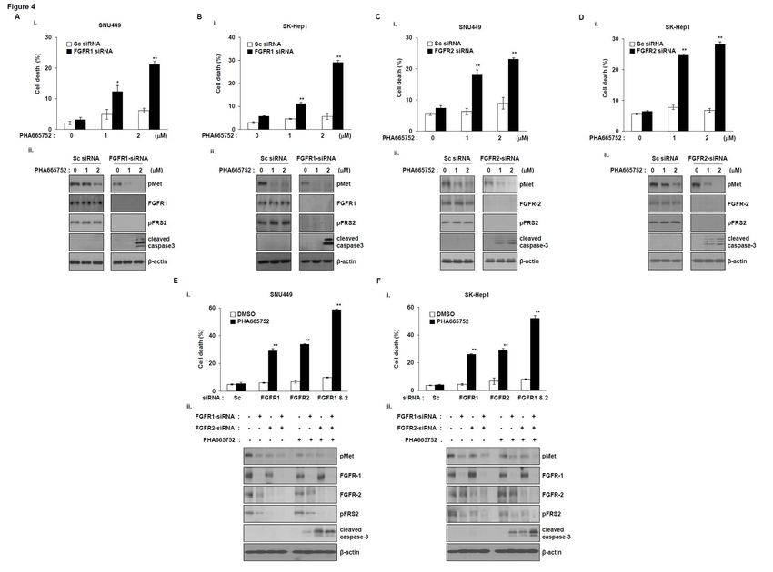
Author Manuscript Published OnlineFirst on September 8, 2015; DOI: 10.1158/1535-7163.MCT-14-0780
   Author manuscripts have been peer reviewed and accepted for publication but have not yet been edited.

  Abstract

       The MET receptor tyrosine kinase, the receptor for hepatocyte growth factor (HGF), has been

  implicated in cancer growth, invasion, migration, angiogenesis, and metastasis in a broad variety of

  human cancers, including human hepatocellular carcinoma (HCC). Recently, MET was suggested to

  be a potential target for the personalized treatment of HCC with an active HGF/MET signaling

  pathway. However, the mechanisms of resistance to MET inhibitors need to be elucidated in order to

  provide effective treatment. Here, we show that HCC cells exhibit different sensitivities to the MET

  inhibitor PHA665752, depending on the phosphorylation status of fibroblast growth factor receptor

  (FGFR). Treatment of cells expressing both phospho-FGFR and phospho-MET, with the inhibitor

  PHA665752, did not cause growth inhibition and cell death, whereas treatment with AZD4547, a pan-

  FGFR inhibitor, resulted in decreased colony formation and cleavage of caspase-3. Moreover,

  silencing of endogenous FGFR1 and FGFR2 by RNA interference of HCC cells expressing phospho-

  FGFR, phosph-FGFR2, and phospho-MET, overcame the resistance to PHA665752 treatment.

  Treatment of primary cancer cells from patients with HCC expressing both phospho-FGFR and

  phospho-MET with PHA665752 did not induce cell death, whereas AZD4547 treatment induced cell

  death through the cleavage of caspase-3. In addition, treatment of cells resistant to PHA665752 with

  AZD4547 abrogated the activation of downstream effectors of cell growth, proliferation, and survival.

  Based on these results, we conclude that the FGFR pathway is critical for HCC survival, and that

  targeting this pathway with AZD4547 may be beneficial for the treatment of patients with HCC

  expressing phospho-FGFR and phospho-MET.

                                                     2

Downloaded from mct.aacrjournals.org on March 14, 2021. © 2015 American Association for Cancer Research.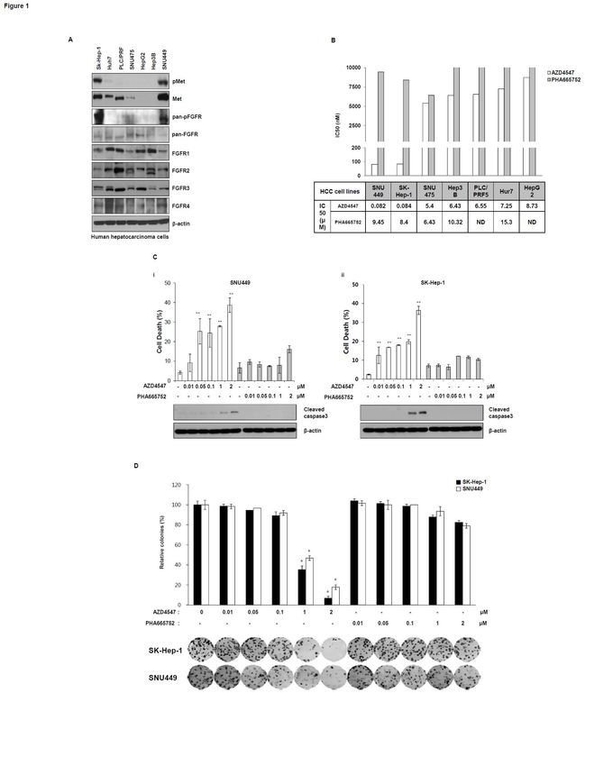
Author Manuscript Published OnlineFirst on September 8, 2015; DOI: 10.1158/1535-7163.MCT-14-0780
   Author manuscripts have been peer reviewed and accepted for publication but have not yet been edited.

  Introduction

       Human hepatocellular carcinoma (HCC) is the third leading cause of cancer-related deaths

  worldwide (1). The prognosis of patients with HCC remains poor, with a 1-year survival rate less than

  50%, despite recent improvements in overall survival. Treatment with a molecular targeted agent, the

  multikinase inhibitor sorafenib, has improved the overall survival in patients with HCC, and sorafenib

  is the only targeted agent shown to prolong the overall survival in randomized controlled trials (2, 3).

  Nevertheless, sorafenib has demonstrated modest efficacy in patients with HCC in clinical practice.

  Therefore, a better understanding of the cellular mechanisms in the development and proliferation of

  HCC is essential.

       Hepatocyte growth factor (HGF)-induced activation of the receptor tyrosine kinase, MET,

  triggers a variety of cellular responses, including proliferation, survival, cytoskeleton rearrangements,

  cell-cell dissociation, and motility (4). Although many studies have demonstrated that MET

  overexpression is associated with poor outcomes in HCC (5), definitive evidence that MET inhibition

  is an effective strategy for the treatment of HCC has not been established. However, You et al.

  suggested that MET is a potential target for the personalized treatment of HCC with active HGF/MET

  signaling, by demonstrating that inhibition of MET can significantly inhibit the growth of MET

  positive HCC tumors (6).

       Preclinical data have demonstrated that FGFR-mediated signaling may play a role in the cellular

  mechanisms underlying the pathogenesis of HCC (7). The main FGFRs expressed in liver tissues are

  FGFR3 and FGFR4, and several studies have suggested that these receptors may be involved in HCC

  tumorigenesis (8, 9). Moreover, the levels and activities of several FGFRs are known to be altered in

  HCC. For example, somatic mutations of FGFR1 have been found in several types of cancers,

  including gliomas and lung tumors (10, 11). In the case of FGFR2, its mutations have been found in

  endometrial carcinomas and gastric cancers (12, 13), whereas those of FGFR3 or FGFR4 have been

  found in bladder carcinomas, multiple myelomas (14-16), and primary rhabdomyosarcomas (17).

       Overexpression of the FGFR2 protein in tumors has been associated with advanced clinical
                                                     3

Downloaded from mct.aacrjournals.org on March 14, 2021. © 2015 American Association for Cancer Research.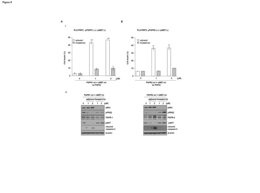
Author Manuscript Published OnlineFirst on September 8, 2015; DOI: 10.1158/1535-7163.MCT-14-0780
   Author manuscripts have been peer reviewed and accepted for publication but have not yet been edited.

  stages (18). Another study reported that FGFR2 overexpression in HCC tumors was associated with a

  high Ki-67 proliferation index and high levels of serum alpha-fetoprotein, and was linked to the

  development of multiple sites of metastasis (19). In addition, overexpression of FGFR3 has been

  associated with a higher grade and poorer differentiation status in HCC tumors, suggesting that

  increased FGFR3 expression may contribute to advanced HCC tumorigenesis (20). Thus, FGFRs may

  act as potent regulators in a variety of cancers. A study evaluating clinical HCC samples strongly

  suggested that FGFR4 was important for the proliferation and survival of HCC tumor cells, as well as

  for the secretion of alpha-fetoprotein (21).

       Although the biological roles of the inter-receptor tyrosine kinase signaling networks are still

  unclear, FGFR and MET have been recently reported to interact with each other to promote cell

  growth or acquired resistance to specific inhibitors in gastric cancer cell lines (22). In the present

  study, we therefore evaluated the response of HCC cell lines and primary HCC cells to a MET

  inhibitor, to determine whether inhibition of the FGFR pathway can overcome the resistance to MET

  inhibitors.

                                                     4

Downloaded from mct.aacrjournals.org on March 14, 2021. © 2015 American Association for Cancer Research.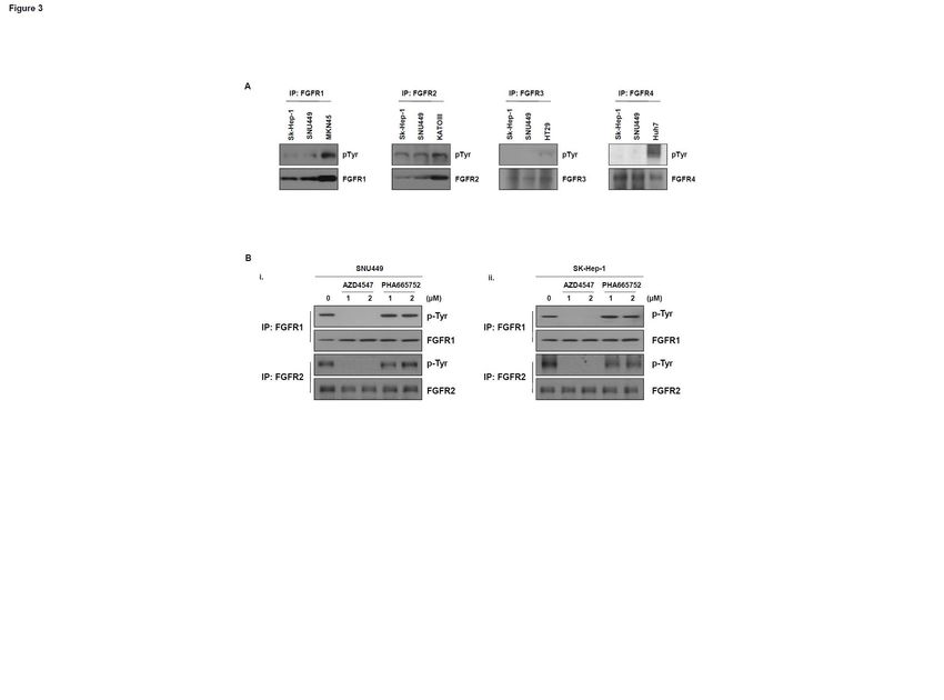
Author Manuscript Published OnlineFirst on September 8, 2015; DOI: 10.1158/1535-7163.MCT-14-0780
   Author manuscripts have been peer reviewed and accepted for publication but have not yet been edited.

  Materials and Methods

  Cell culture and reagents

       All human liver carcinoma cells were purchased from the ATCC (American Type Culture

  Collection, Manassas, VA, USA) who authenticated the identity of the cell lines, except for SNU449

  and SNU475, which were obtained from the KCLB (Korean Cell Line Bank, Seoul, Republic of

  Korea). All human liver carcinoma cell lines were maintained in RPMI-1640 medium or DMEM

  medium (GIBCO BRL, Grand Island, NY, USA) containing 10% fetal bovine serum (FBS; GIBCO

  BRL), 100 units penicillin, and 100 μL/mL streptomycin in a 5% CO2 incubator at 37°C. PHA665752,

  a specific MET inhibitor, was purchased from Selleck Chemicals LLC (Houston, TX, USA), and

  AZD4547, a pan-FGFR inhibitor, was kindly provided by AstraZeneca. Co., Ltd. (Macclesfield,

  Cheshire, UK).

  Human primary hepatocarcinoma cell culture

       Specimens of human primary hepatocarcinoma cancer tissues were obtained from patients after

  written informed consent and approval by the Research Ethics Board at Asan Medical Center. The

  study protocol followed the tenets of the Declaration of Helsinki. Tumor specimens were minced with

  scissors, and digested by incubation in Minimum Essential Medium (MEM) (GIBCO BRL)

  containing 1 mg/mL collagenase (Sigma-Aldrich, St. Louis, MO, USA) for 2 h at 37°C. Cells were

  washed with medium containing 10% FBS, followed by washing with phosphate-buffered saline (PBS,

  pH 7.4) to remove FBS. Next, the cells were plated in Human Hepatocyte Basal Medium (HBM)

  (Lonza, Walkersville, MD, USA) and cultured in a humidified incubator under 5% CO2 at 37 °C.

  Cell viability

       Cells (3 × 10⁵ cells per plate) were seeded in 60 mm plates and treated with various amounts of

  PHA665752 or AZD4547 for 48 h. Cell viability was determined using the trypan blue dye exclusion

  method.
                                                     5

Downloaded from mct.aacrjournals.org on March 14, 2021. © 2015 American Association for Cancer Research.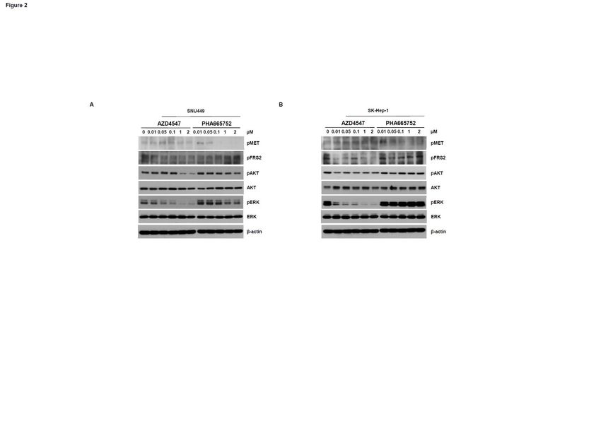
Author Manuscript Published OnlineFirst on September 8, 2015; DOI: 10.1158/1535-7163.MCT-14-0780
   Author manuscripts have been peer reviewed and accepted for publication but have not yet been edited.

  Clonogenic assay

       SNU449 and SK-Hep-1 cells, seeded at 3 × 10² cells per well in 6-well plates, were treated with

  AZD4547 or PHA665752 at a concentration of 1 µM for 24 h. The cells were cultured for 14 days.

  Colonies were fixed with 10% formalin and stained with 0.01% crystal violet dye solution. The

  numbers of colony-forming cells with a diameter ≥1 mm were counted.

  DNA, siRNA, and transfection

       MET, FGFR-1, and FGFR-2 cDNAs were purchased from Origene (Rockville, MD, USA). The

  mutated MET T1191I DNA was generated using a polymerase chain reaction (PCR)-based quick

  change site-directed mutagenesis kit (Intron Biotechnology, Seoul, Republic of Korea) and wild type

  MET. All DNA transfections were performed using Lipofectamine® 2000 reagent (Invitrogen,

  Carlsbad, CA, USA) according to the manufacturer’s instructions. For siRNA transfection, cells were

  transfected using Lipofectamine® RNAi MAX reagent (Invitrogen) according to the manufacturer’s

  protocol. The siRNA sequences for the transfection study were the following: FGFR1-siRNA, 5'-GAA

  GUG CAU ACA CCG AGA C-3'; FGFR2-siRNA, 5'-AAG UGC UGG CUC UGU UCA AUG-3';

  FGFR3-siRNA, 5'-AGA CGA TGC CAC TGA CAA G-3'; and FGFR4-siRNA, 5'-AUA CGG ACA

  UCA UCC UGU A-3'.

  Immunoblot and immunoprecipitation

       Total cellular proteins (20 μg) were subjected to sodium dodecylsulfate-polyacrylamide gel

  electrophoresis (SDS-PAGE) and transferred to polyvinylidene difluoride (PVDF) membranes. The

  membranes were blocked with 5% non-fat dry milk in TBS-T buffer (20 mM Tris-HCl, pH 7.4, 150

  mM NaCl, and 0.1% Tween 20) and incubated with the following antibodies: anti-Met, anti-FGFR2,

  and anti-β-actin antibodies (Santa Cruz Biotechnology, Santa Cruz, CA, USA); anti-phospho-Met

  (Y1234/1235), anti-phospho-FGFR (pan-pFGFR), anti-phospho-FRS2 (pFRS2), anti-phospho-AKT,
                                                     6

Downloaded from mct.aacrjournals.org on March 14, 2021. © 2015 American Association for Cancer Research.
Author Manuscript Published OnlineFirst on September 8, 2015; DOI: 10.1158/1535-7163.MCT-14-0780
Author manuscripts have been peer reviewed and accepted for publication but have not yet been edited.
anti-phospho-ERK, anti-phospho-tyrosine, anti-AKT, anti-ERK, and anti-caspase-3 antibodies (Cell
Signaling, Beverly, CA, USA); anti-FGFR1 and anti-AFP antibodies (Abcam, Cambridge, MA,
USA); and anti-FGFR3 and anti-FGFR4 (Abnova Corporation, Taipei, Taiwan) antibodies. Primary
antibodies were detected with horseradish peroxidase-conjugated goat anti-mouse or goat anti-rabbit
secondary antibody (Cell Signaling), as appropriate, and immunoreactive proteins were visualized
using an enhanced ECL detection reagent (GE Healthcare Bio-Sciences, Uppsala, Sweden).
For immunoprecipitation analyses, cell lysates were immunoprecipitated with anti-FGFR-1, anti-
FGFR2, anti-FGFR3, or anti-FGFR4 antibodies for 12 h at 4°C. Protein A/G Plus-Sepharose beads
(20 μL) (Santa Cruz Biotechnology) were then added, and the mixtures were incubated for an
additional 2 h at 4°C. The immunoprecipitates were washed with Nonidet P-40 lysis buffer, boiled in
2× SDS sample buffer, and analyzed by western blot analysis using anti-phospho-tyrosine antibody.
Immunofluorescence analysis
Human primary hepatocarcinoma cells were seeded on glass coverslips. The cells were fixed in
4% paraformaldehyde, and permeabilized-blocked, then incubated with primary antibodies. Following
washing, the cells were incubated with Alexa488- or Alexa594-labeled secondary antibodies, and the
cells were stained briefly with DAPI. After washing, the coverslips were mounted on glass slides
using mounting solution. The slides were analyzed by confocal microscopy (Carl Zeiss, Jena,
Germany).
Immunohistochemistry
The pMET and pFGFR in tumor tissues and tissue microarrays (TMAs) were detected using
immunohistochemistry (IHC). The HCC TMAs were purchased from US Biomax, Inc. (Rockville,
MD, USA). Immunohistochemistry was performed according to the manufacturer’s protocol. Antigen
retrieval was performed by heating the samples in citrate buffer (pH 6.0) in a microwave. The reaction
products were developed with diaminobenzidine (Dako, Hamburg, Germany), followed by
7
Downloaded from mct.aacrjournals.org on March 14, 2021. © 2015 American Association for Cancer Research.Author Manuscript Published OnlineFirst on September 8, 2015; DOI: 10.1158/1535-7163.MCT-14-0780
Author manuscripts have been peer reviewed and accepted for publication but have not yet been edited.
counterstaining with hematoxylin.
Statistical analysis
All data were analyzed using two-tailed Student’s t-tests. The level of statistical significance was
set at PAuthor Manuscript Published OnlineFirst on September 8, 2015; DOI: 10.1158/1535-7163.MCT-14-0780
Author manuscripts have been peer reviewed and accepted for publication but have not yet been edited.
Results
Human hepatoma cancer cells express resistance to a MET inhibitor dependent on the
phosphorylation status of FGFR.
It was recently reported that FGFR-expressing gastric cancer cells are sensitive to a MET
inhibitor (22), implying that some association exists between MET and FGFR against the sensitivity
to the MET inhibitor in a variety of cancer cells. We therefore investigated the dependence of the
inhibitory effects of a MET inhibitor on the status of FGFR in human hepatoma cell lines. First, we
examined the phosphorylation status of MET and FGFR, including the expression levels of both
proteins in multiple human hepatoma cell lines. Two of them, SK-Hep-1 and SNU449 cells, expressed
both phospho-MET and phospho-FGFR (Fig. 1A). We examined the inhibitory effects of treatment
with a MET inhibitor (PHA665752) or a pan-FGFR inhibitor (AZD4547) in various human hepatoma
cell lines. The results demonstrated that SNU449 and SK-Hep-1 cells were sensitive to AZD4547,
exhibiting IC50 values of 84 nM and 92 nM, respectively. By contrast, these cell lines were resistant to
PHA665752. Other cell lines were resistant to the two compounds (Fig. 1B). Based on the IC50 data,
we determined the concentration of AZD4547, and in parallel, determined the concentration of
PHA665752. These human hepatoma cell lines were selected to determine whether the differences in
sensitivities to MET inhibitor were dependent on the presence of phospho-FGFR, and were therefore
treated with various doses of each inhibitor that targets MET or FGFR. The results showed that cell
growth was inhibited by AZD4547 at lower concentrations, and cell death was induced at higher
concentrations (>1 µM) (Fig. 1C and Supplementary Fig 1). Notably, there was almost no change in
the population of dead cells in response to PHA665752, a selective MET inhibitor, whereas the
population of dead cells after the treatment with AZD4547, a pan-FGFR inhibitor, dramatically
increased (Fig. 1C, i). AZD4547 also induced the cleavage of pro-caspase-3 to yield active caspase-3,
but not in cells treated with PHA665752 (Fig. 1C, ii). To analyze the effects of AZD4547 or
PHA665752 on caspase-3 cleavage, we repeated the western blot analysis. AZD4547 treatment at 1 or
2 μM led to caspase-3 cleavage (Fig. 1C, lower panel). To further characterize the inhibitory effects of
9
Downloaded from mct.aacrjournals.org on March 14, 2021. © 2015 American Association for Cancer Research.Author Manuscript Published OnlineFirst on September 8, 2015; DOI: 10.1158/1535-7163.MCT-14-0780
Author manuscripts have been peer reviewed and accepted for publication but have not yet been edited.
each inhibitor, AZD4547 or PHA665752, on cells that express both phospho-MET and phospho-
FGFR, we performed colony-forming assays. Colony formation in SNU449 and SK-Hep1 cells was
significantly reduced by AZD4547, whereas a decrease in colony formation was not observed with
PHA665752 in both cell lines (Fig. 1D). Taken together, the results indicate that differences in
sensitivity to MET inhibitor are dependent on the phosphorylation status of FGFR in human
hepatocarcinoma cells.
MET inhibitor does not inhibit downstream signaling in hepatocarcinoma cells expressing
pMET and pFGFR.
As shown in Fig. 1, SNU449 and SK-Hep1 cells were insensitive to PHA665752, a selective
MET inhibitor, whereas both cell lines were sensitive to AZD4547, a pan-FGFR inhibitor. Based upon
these results, we investigated the effects of each inhibitor on their downstream signaling pathways.
Treatment with AZD4547 caused a decrease of FRS2 (fibroblast growth factor receptor substrate 2),
AKT, and ERK phosphorylation in SNU449 cells. However, phosphorylation of these proteins did not
decrease after exposure to PHA665752 (Fig. 2A). AZD4547 also blocked the phosphorylation of
FRS2, AKT, and ERK in SK-Hep1 cells, whereas PHA665752 did not (Fig. 2B). These results
indicated that inhibition of FGFR inhibits the phosphorylation of the downstream signaling molecules
FRS2, AKT, and ERK in hepatoma cells expressing pMET and pFGFR.
FGFR inhibitor, but not MET inhibitor, causes inhibition of FGFR1 or FGFR2 phosphorylation
in human hepatocarcinoma cells
We examined the phosphorylation status of FGFR1, FGFR2, FGFR3, and FGFR4 in SNU449
and SK-Hep1 cells that express both pMET and pFGFR. Notably, phosphorylation of FGFR1 and
FGFR2 was observed in both cell types, whereas FGFR3 and FGFR4 phosphorylation was not
detected (Fig. 3A), implying that phospho-FGFR1 and FGFR2 may affect sensitivity to the MET
inhibitor. We next examined the inhibition of FGFR1 and FGFR2 phosphorylation after treatment of
10
Downloaded from mct.aacrjournals.org on March 14, 2021. © 2015 American Association for Cancer Research.Author Manuscript Published OnlineFirst on September 8, 2015; DOI: 10.1158/1535-7163.MCT-14-0780
Author manuscripts have been peer reviewed and accepted for publication but have not yet been edited.
these cells with AZD4547 or PHA665752 using an immunoprecipitation assay. AZD4547 treatment
led to decreases in both p-FGFR1 and FGFR2 in the two selected cell lines, but PHA665752 treatment
did not induce a decrease (Fig. 3B).
Silencing of FGFR1 or FGFR2 overcomes the resistance to the MET inhibitor.
We next investigated the effects of members of the FGFR family on the sensitivity to MET
inhibitor. We analyzed the dependence of FGFR1 or FGFR2 on the sensitivity to MET inhibitor using
constructs expressing FGFR1 or FGFR2 small interfering RNA (siRNA). Initially, SNU449 cells
were transfected with FGFR1-siRNA or scrambled siRNA, followed by PHA665752 treatment. The
dead cell population was significantly increased in FGFR1-siRNA-treated cells following
PHA665752 treatment, but not in scrambled siRNA-treated cells (Fig. 4A, i). Additionally, the
cleavage of caspase-3 was significantly increased in FGFR1-siRNA-treated cells following
PHA665752 treatment, and the phosphorylation level of FRS2 was decreased after exposure to
PHA665752 (Fig. 4A, ii). The results showed that silencing of FGFR1 in SK-Hep1 cells increases cell
death (Fig. 4B), indicating that inhibition of FGFR1 can overcome the resistance to MET inhibitor.
As shown in Fig. 4A and B, SNU449 and SK-Hep1 cells expressed phospho-FGFR2 as well as
phospho-FGFR1. Thus, we investigated the effect of depletion of FGFR2 by siRNA on the sensitivity
to the MET inhibitor. Knockdown of FGFR2 led to increased cell death in SNU449 (Fig. 4C, i) and
SK-Hep1 cells (Fig. 4D, i) in response to PHA665752, but not in scrambled siRNA-treated cells, as
seen with FGFR1-siRNA. The cleavage of caspase-3 also increased in FGFR2-siRNA-treated cells
(Fig. 4C, ii and D, ii), indicating that inhibition of FGFR2 can overcome the resistance to MET
inhibitor. To further confirm these conclusions, we co-transfected cells that coexpressed pMET and
pFGFR with FGFR1- and 2-siRNA. Cell death was significantly higher in cells co-transfected with
FGFR1- and 2-siRNA than in cells transfected with FGFR1- or 2-siRNA (Figs. 4E, i and F, i). Double
knockdown also significantly increased the cleaved form of caspase-3 compared with single
knockdown (Figs. 4E, ii and F, ii). We next examined the effects of FGFR3 and FGFR4 knockdown
11
Downloaded from mct.aacrjournals.org on March 14, 2021. © 2015 American Association for Cancer Research.Author Manuscript Published OnlineFirst on September 8, 2015; DOI: 10.1158/1535-7163.MCT-14-0780
Author manuscripts have been peer reviewed and accepted for publication but have not yet been edited.
on the sensitivity to a MET inhibitor. Silencing FGFR3 and FGFR4 did not increase cell death in the
absence of PHA665752 (Supplementary Fig. 2), indicating that FGFR3 and FGFR4 do not affect
sensitivity to a MET inhibitor. In addition, MET inhibition induced cell death in the absence of
AZD4547 (Supplementary Fig. 3). These results indicate that FGFR1 and 2 silencing enhances MET
inhibitor sensitivity in human hepatocarcinoma cells.
FGFR1 or FGFR 2 confers resistance to the MET inhibitor.
To further characterize the dependence of FGFR1 or FGFR2 on the sensitivity to the MET
inhibitor, we co-transfected PLC/PRF5 cells that did not express pMET and pFGFR, with a construct
expressing FGFR1 cDNA and a MET CA (constitutively active) mutant or with FGFR2 cDNA and a
MET CA mutant, followed by treatment with PHA665752 or AZD4547. There was almost no
induction of cell death induced by PHA665752 in cells expressing FGFR1 cDNA and a MET CA
mutant, whereas treatment of AZD4547 led to increased cell death (Fig. 5A, i). Similarly, the dead cell
population in cells expressing FGFR2 cDNA and a MET CA mutant was significantly increased by
AZD4547, but not by PHA665752 (Fig. 5B, ii). Additionally, caspase-3 cleavage was significantly
increased in cells expressing FGFR1 or FGFR2 cDNA and a MET CA mutant following AZD4547,
but not PHA665752, treatment (Fig. 5A, i and B, ii). Moreover, c-MET altered the growth of these
cells (Supplementary Fig. 4). Together, these results indicate that FGFR1 and FGFR2 activities are
associated with resistance to the MET inhibitor in human heparocellular carcinoma cells expressing
pMET and pFGFR.
AZD4547 induces cell death in human primary hepatocarcinoma cells expressing pMET and
pFGFR.
Based on the above results, we examined the effect of each inhibitor, PHA665752 or AZD4547,
on primary cancer cells from patients with hepatocarcinomas. We first analyzed the phosphorylation
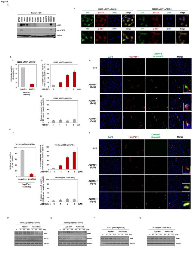
status of MET or FGFR. Two out of 14 primary HCC cells, 78019A and 3226B (patient numbers),
12
Downloaded from mct.aacrjournals.org on March 14, 2021. © 2015 American Association for Cancer Research.Author Manuscript Published OnlineFirst on September 8, 2015; DOI: 10.1158/1535-7163.MCT-14-0780
Author manuscripts have been peer reviewed and accepted for publication but have not yet been edited.
coexpressed pMET and pFGFR (Fig. 6A, i). We then performed immunofluorescence analysis on
primary HCC cells. The 78019A and 3226B cells expressed pMET, pFGFR, and AFP (Fig. 6A, ii).
Human primary HCC cells, which include stromal cells and tumor cells, were obtained from HCC
patient tissues. We analyzed the drug sensitivity of cells in the absence or presence of Hep-Par-1 as a
HCC marker. Approximately 18% of human primary HCC cells were found to be Hep-Par-1 positive.
AZD4547 treatment led to a dose-dependent caspase-3 cleavage in Hep-Par-1 positive cells, but not in
Hep-Par-1 negative cells. The Hep-Par-1 negative cells were almost always stromal cells, with no
induction of cell death by treatment with AZD4547. These results were confirmed by
immunofluorescence analysis, showing that Hep-Par-1 positive cells displayed sensitivity to
AZD4547 at 1, 2, and 5 μM (Fig. 6B and C). In addition, AZD4547 induced a decrease of pFRS2, but
not pMET, in 3226B and 78019A cells (Fig. 6D and E), indicating that AZD4547 can induce cell
death in human primary hepatocarcinoma cells expressing pMET and pFGFR.
Next, we examined the inhibitory effect of each inhibitor on human primary hepatocarcinoma
cells expressing pMET alone. PHA665752 induced a decrease of pMET, indicating that the MET
inhibitor can act on human primary hepatocarcinoma cells expressing pMET alone (Fig. 6F and G).
However, the concentration at which significant induction of caspase-3 cleavage was observed may
not be clinically achievable with AZD4547.
13
Downloaded from mct.aacrjournals.org on March 14, 2021. © 2015 American Association for Cancer Research.Author Manuscript Published OnlineFirst on September 8, 2015; DOI: 10.1158/1535-7163.MCT-14-0780
Author manuscripts have been peer reviewed and accepted for publication but have not yet been edited.
Discussion
The receptor tyrosine kinase, MET, that controls cell growth, proliferation, survival, and motility,
is constitutively activated and amplified in several cancer types, including non-small cell lung cancers,
hepatocarcinomas, and gastric cancers (23). Thus, MET is considered a clinically important target for
therapies against these types of cancers. Agents targeting MET have been evaluated and have recently
entered clinical trials. Major problems of therapies targeting tyrosine kinases involve the lack of
response to treatment of many tumors, and/or the drug resistance that eventually develops. In the
present study, we show that AZD4547, a pan-FGFR inhibitor, can overcome the resistance to MET
inhibitor in human hepatocarcinoma cells.
It was recently reported that activation of RTK (receptor tyrosine kinase) in gastric cancer cells
mediates resistance to MET inhibition, implicating that some association exists between RTK and
MET during the induction of drug resistance. Specifically, Kataoka et al. reported that TKI appears to
be more effective against human gastric cancer, by blocking inter-RTK signaling networks that
depend on MET or FGFR2 (22). Foretinib (GSK1363089), a multi-kinase inhibitor of MET and
VEGFRs, shows different sensitivities that are dependent on FGFR expression (22). The FGFR
inhibitor that has been most extensively studied in patients with HCC is brivanib, an ATP-competitive
dual inhibitor of VEGFR and FGFR1-3 (24). Despite strong preclinical and phase II data of HCC (25,
26), brivanib failed in large randomized phase III trials in both the first- and second-line settings for
patients with advanced HCC (27, 28). Nonetheless, FGFR inhibition remains an attractive therapeutic
target for HCC, and requires further investigation of its possible clinical applications. Efforts continue
to explore the use of multikinase inhibitors that also target FGFR in patients with advanced HCC, and
ongoing first-line trials include a randomized phase III study of lenvatinib compared with sorafenib, a
randomized phase II trial of dovitinib compared with sorafenib in the Asia-Pacific region, and a phase
I/II trial of nintedanib in combination with sorafenib. Conversely, our findings have shown that a pan-
FGFR inhibitor, AZD4547, appears to be more effective than a selective MET inhibitor, PHA665752,
against HCC expressing phospho-MET and phospho-FGFR. Thus, the inhibition of MET in
14
Downloaded from mct.aacrjournals.org on March 14, 2021. © 2015 American Association for Cancer Research.Author Manuscript Published OnlineFirst on September 8, 2015; DOI: 10.1158/1535-7163.MCT-14-0780
Author manuscripts have been peer reviewed and accepted for publication but have not yet been edited.
hepatocarcinoma cells expressing both pMET and pFGFR has not been effective, unlike that in human
gastric cancer cells. Although the most robust effects on inducing cell death in HCC cell lines were
seen at concentrations at or above 1 µM, which might be above clinically achievable concentrations of
AZD4547, significant effects were also seen at lower concentrations. Similarly, the expression of
downstream signaling molecules decreased after exposure to AZD4547 at concentrations above 50
nM.
Based upon these results, we confirmed that an association exists between pMET and pFGFR,
using primary cancer cells from patients with hepatocarcinoma. Interestingly, two out of 14 primary
cancer cells, 78019A and 3226B, expressed both pMET and pFGFR. According to some reports, MET
activation is relevant to hepatitis B virus infection in hepatocarcinoma. Hepatitis B virus X (HBx)
protein increases invasiveness via the promoter activity of MET (29). Additionally, hepatitis B and C
virus-infected mouse models highly express HGF (30). However, the infectious status of these viruses
might not be related to FGFR and MET signaling activation in the 14 primary HCCs (Supplementary
Table. 1). Consistent with the results from cancer cell lines, treatment of primary cancer cells that
express pMET and pFGFR with PHA665752 as a MET inhibitor was not effective, whereas AZD4547
was more effective against both primary cancer cells, implying that FGFR kinase is more important
than MET kinase for hepatocarcinoma cell survival (Fig. 6). Thus, FGFR kinase may serve as a potent
target of several therapeutic strategies for human heparocellular carcinoma. We therefore investigated
the expressions of pFGFR and pMET in other primary HCC cell lines.
One limitation of the current study is that it was based on a small sample size of primary HCC
samples. The subset of HCC samples with both FGFR and Met signaling activation likely comprises a
small proportion of the total HCC population. We analyzed the expression of pFGFR and pMET in a
tissue microarray (TMA) by immunohistochemistry. TMA slides were purchased from US Biomax
(the donors were from China). Nine of 53 TMA cores (16%) were double positive for the expression
of pFGFR and pMET (Supplementary Fig. 5). In addition, other studies reported that MET
15
Downloaded from mct.aacrjournals.org on March 14, 2021. © 2015 American Association for Cancer Research.Author Manuscript Published OnlineFirst on September 8, 2015; DOI: 10.1158/1535-7163.MCT-14-0780
Author manuscripts have been peer reviewed and accepted for publication but have not yet been edited.
amplification is 27% (31) and FGFR amplification is 31% (32) in HCC. Given the ethnic background
of the donors, we expect similar results in Korean patients, but it would be helpful to confirm this
possibility using a larger cohort. Nonetheless, our results provide the first evidence regarding the
relationship of pMET and pFGFR in HCC.
In conclusion, we have evaluated the effects of AZD4547, a pan-FGFR inhibitor dependent on
MET activity/phosphorylation, in human hepatocarcinoma cells. Given the increased effects on
inducing HCC cell death at higher concentrations, it is also possible that other FGFR inhibitors, which
can be used at higher tolerable doses, even for a shorter time than used with AZD4547, might be more
efficacious in such patients. The success of personalized or precision medicine using targeted
therapies, based on the particular genotype of cancer, depends on the ability to identify patients who
will benefit from specific drugs. Thus, our findings may provide the rationale for clinical development
of FGFR inhibitors, such as AZD4547, for the treatment of selected patient populations with
hepatocarcinoma that express phospho-MET and phospho-FGFR.
16
Downloaded from mct.aacrjournals.org on March 14, 2021. © 2015 American Association for Cancer Research.Author Manuscript Published OnlineFirst on September 8, 2015; DOI: 10.1158/1535-7163.MCT-14-0780
Author manuscripts have been peer reviewed and accepted for publication but have not yet been edited.
Acknowledgements
This study was supported by the grants from the Korea Health 21 R&D Project, Ministry of Health
and Welfare and Family Affairs, Republic of Korea (HI06C0868), (TW Kim) a National Research
Foundation of Korea (NRF) grant funded by the Korean government (MEST) (No.
2013R1A2A2A01067394) (DH Jin), and the Asan Institute for Life Sciences, Seoul, Republic of
Korea (2014-622) (TW Kim). The use of AZD4547, a pan-FGFR inhibitor, was supported by
AstraZeneca through a research material transfer agreement between AstraZeneca and the ASAN
Institute for Life Sciences.
17
Downloaded from mct.aacrjournals.org on March 14, 2021. © 2015 American Association for Cancer Research.Author Manuscript Published OnlineFirst on September 8, 2015; DOI: 10.1158/1535-7163.MCT-14-0780
Author manuscripts have been peer reviewed and accepted for publication but have not yet been edited.
References
1. El-Serag HB, Rudolph KL. Hepatocellular carcinoma: epidemiology and molecular carcinogenesis.
Gastroenterology. 2007;132:2557-76.
2. Zhang T, Ding X, Wei D, Cheng P, Su X, Liu H, et al. Sorafenib improves the survival of patients
with advanced hepatocellular carcinoma: a meta-analysis of randomized trials. Anticancer Drugs.
2010;21:326-32.
3. Villeneuve JP, Richer G, Cote J, Guevin R, Marleau D, Joly JG, et al. Chronic carriers of hepatitis B
antigen (HBsAg). Histological, biochemical, and immunological findings in 31 voluntary blood
donors. Am J Dig Dis. 1976;21:18-25.
4. Ma PC, Maulik G, Christensen J, Salgia R. c-Met: structure, functions and potential for therapeutic
inhibition. Cancer Metastasis Rev. 2003;22:309-25.
5. Kaposi-Novak P, Lee JS, Gomez-Quiroz L, Coulouarn C, Factor VM, Thorgeirsson SS. Met-
regulated expression signature defines a subset of human hepatocellular carcinomas with poor
prognosis and aggressive phenotype. J Clin Invest. 2006;116:1582-95.
6. You H, Ding W, Dang H, Jiang Y, Rountree CB. c-Met represents a potential therapeutic target for
personalized treatment in hepatocellular carcinoma. Hepatology. 2011;54:879-89.
7. Ahmad I, Iwata T, Leung HY. Mechanisms of FGFR-mediated carcinogenesis. Biochim Biophys
Acta. 2012;1823:850-60.
8. Qiu WH, Zhou BS, Chu PG, Chen WG, Chung C, Shih J, et al. Over-expression of fibroblast
growth factor receptor 3 in human hepatocellular carcinoma. World J Gastroenterol. 2005;11:5266-72.
9. French DM, Lin BC, Wang M, Adams C, Shek T, Hotzel K, et al. Targeting FGFR4 inhibits
hepatocellular carcinoma in preclinical mouse models. PLoS One. 2012;7:e36713.
10. Rand V, Huang J, Stockwell T, Ferriera S, Buzko O, Levy S, et al. Sequence survey of receptor
tyrosine kinases reveals mutations in glioblastomas. Proc Natl Acad Sci U S A. 2005;102:14344-9.
11. Grose R, Dickson C. Fibroblast growth factor signaling in tumorigenesis. Cytokine Growth Factor
Rev. 2005;16:179-86.
18
Downloaded from mct.aacrjournals.org on March 14, 2021. © 2015 American Association for Cancer Research.Author Manuscript Published OnlineFirst on September 8, 2015; DOI: 10.1158/1535-7163.MCT-14-0780
Author manuscripts have been peer reviewed and accepted for publication but have not yet been edited.
12. Dutt A, Salvesen HB, Chen TH, Ramos AH, Onofrio RC, Hatton C, et al. Drug-sensitive FGFR2
mutations in endometrial carcinoma. Proc Natl Acad Sci U S A. 2008;105:8713-7.
13. Jang JH, Shin KH, Park JG. Mutations in fibroblast growth factor receptor 2 and fibroblast growth
factor receptor 3 genes associated with human gastric and colorectal cancers. Cancer Res.
2001;61:3541-3.
14. Gomez-Roman JJ, Saenz P, Molina M, Cuevas Gonzalez J, Escuredo K, Santa Cruz S, et al.
Fibroblast growth factor receptor 3 is overexpressed in urinary tract carcinomas and modulates the
neoplastic cell growth. Clin Cancer Res. 2005;11:459-65.
15. Chesi M, Nardini E, Lim RS, Smith KD, Kuehl WM, Bergsagel PL. The t(4;14) translocation in
myeloma dysregulates both FGFR3 and a novel gene, MMSET, resulting in IgH/MMSET hybrid
transcripts. Blood. 1998;92:3025-34.
16. Tomlinson DC, Baldo O, Harnden P, Knowles MA. FGFR3 protein expression and its relationship
to mutation status and prognostic variables in bladder cancer. J Pathol. 2007;213:91-8.
17. Taylor JGt, Cheuk AT, Tsang PS, Chung JY, Song YK, Desai K, et al. Identification of FGFR4-
activating mutations in human rhabdomyosarcomas that promote metastasis in xenotransplanted
models. J Clin Invest. 2009;119:3395-407.
18. Gatius S, Velasco A, Azueta A, Santacana M, Pallares J, Valls J, et al. FGFR2 alterations in
endometrial carcinoma. Mod Pathol. 2011;24:1500-10.
19. Harimoto N, Taguchi K, Shirabe K, Adachi E, Sakaguchi Y, Toh Y, et al. The significance of
fibroblast growth factor receptor 2 expression in differentiation of hepatocellular carcinoma.
Oncology. 2010;78:361-8.
20. Cheng AL, Shen YC, Zhu AX. Targeting fibroblast growth factor receptor signaling in
hepatocellular carcinoma. Oncology. 2011;81:372-80.
21. Ho HK, Pok S, Streit S, Ruhe JE, Hart S, Lim KS, et al. Fibroblast growth factor receptor 4
regulates proliferation, anti-apoptosis and alpha-fetoprotein secretion during hepatocellular carcinoma
progression and represents a potential target for therapeutic intervention. J Hepatol. 2009;50:118-27.
19
Downloaded from mct.aacrjournals.org on March 14, 2021. © 2015 American Association for Cancer Research.Author Manuscript Published OnlineFirst on September 8, 2015; DOI: 10.1158/1535-7163.MCT-14-0780
Author manuscripts have been peer reviewed and accepted for publication but have not yet been edited.
22. Kataoka Y, Mukohara T, Tomioka H, Funakoshi Y, Kiyota N, Fujiwara Y, et al. Foretinib
(GSK1363089), a multi-kinase inhibitor of MET and VEGFRs, inhibits growth of gastric cancer cell
lines by blocking inter-receptor tyrosine kinase networks. Invest New Drugs. 2012;30:1352-60.
23. Trusolino L, Bertotti A, Comoglio PM. MET signalling: principles and functions in development,
organ regeneration and cancer. Nat Rev Mol Cell Biol. 2010;11:834-48.
24. Huynh H, Ngo VC, Fargnoli J, Ayers M, Soo KC, Koong HN, et al. Brivanib alaninate, a dual
inhibitor of vascular endothelial growth factor receptor and fibroblast growth factor receptor tyrosine
kinases, induces growth inhibition in mouse models of human hepatocellular carcinoma. Clin Cancer
Res. 2008;14:6146-53.
25. Finn RS, Kang YK, Mulcahy M, Polite BN, Lim HY, Walters I, et al. Phase II, open-label study of
brivanib as second-line therapy in patients with advanced hepatocellular carcinoma. Clin Cancer Res.
2012;18:2090-8.
26. Park JW, Finn RS, Kim JS, Karwal M, Li RK, Ismail F, et al. Phase II, open-label study of
brivanib as first-line therapy in patients with advanced hepatocellular carcinoma. Clin Cancer Res.
2011;17:1973-83.
27. Johnson PJ, Qin S, Park JW, Poon RT, Raoul JL, Philip PA, et al. Brivanib versus sorafenib as
first-line therapy in patients with unresectable, advanced hepatocellular carcinoma: results from the
randomized phase III BRISK-FL study. J Clin Oncol. 2013;31:3517-24.
28. Llovet JM, Decaens T, Raoul JL, Boucher E, Kudo M, Chang C, et al. Brivanib in patients with
advanced hepatocellular carcinoma who were intolerant to sorafenib or for whom sorafenib failed:
results from the randomized phase III BRISK-PS study. J Clin Oncol. 2013;31:3509-16.
29. Xie B, Tang C, Chen P, Gou YB, Xiao J, Du H. [The effect of hepatitis B virus X protein on the c-
met promoter activity in HepG2 cells]. Zhonghua Gan Zang Bing Za Zhi. 2009;17:292-6.
30. Xie Q, Su Y, Dykema K, Johnson J, Koeman J, De Giorgi V, et al. Overexpression of HGF
Promotes HBV-Induced Hepatocellular Carcinoma Progression and Is an Effective Indicator for Met-
Targeting Therapy. Genes Cancer. 2013;4:247-60.
20
Downloaded from mct.aacrjournals.org on March 14, 2021. © 2015 American Association for Cancer Research.Author Manuscript Published OnlineFirst on September 8, 2015; DOI: 10.1158/1535-7163.MCT-14-0780
Author manuscripts have been peer reviewed and accepted for publication but have not yet been edited.
31. Korhan P, Erdal E, Kandemis E, Cokakli M, Nart D, Yilmaz F, et al. Reciprocal activating
crosstalk between c-Met and caveolin 1 promotes invasive phenotype in hepatocellular carcinoma.
PLoS One. 2014;9:e105278.
32. Riehle KJ, Yeh MM, Yu JJ, Kenerson HL, Harris WP, Park JO, et al. mTORC1 and FGFR1
signaling in fibrolamellar hepatocellular carcinoma. Mod Pathol. 2015;28:103-10.
21
Downloaded from mct.aacrjournals.org on March 14, 2021. © 2015 American Association for Cancer Research.Author Manuscript Published OnlineFirst on September 8, 2015; DOI: 10.1158/1535-7163.MCT-14-0780 Author manuscripts have been peer reviewed and accepted for publication but have not yet been edited. Figure Legends Figure 1. Effects of MET or pan-FGFR inhibitor on hepatocellular carcinoma (HCC) cell lines. A. Expression levels of phospho-MET and phospho-FGFR were evaluated by western blotting of extracts of HCC cell lines with anti-pMET or pFGFR antibodies. β-actin was used as a loading control. B. Various HCC cell lines were seeded in 96-well plates and treated with a MET inhibitor (PHA665752) or a pan-FGFR inhibitor (AZD4547). After incubation for 72 h, MTS assays were performed. C. Differential effects of the MET inhibitor (PHA665752) or pan-FGFR inhibitor (AZD4547) on SNU449 and SK-HEP-1 cells. SNU449 (i) and SK-HEP-1 (ii) cells were treated with different concentrations of PHA665752 or AZD4547 for 48 h. Cell death was determined using the trypan blue dye exclusion assay. Cells were harvested and analyzed by western blotting using antibody against cleaved caspase-3. β-actin was used as a loading control. The graphs present the mean ± s.d. of three separate experiments performed in triplicate. *P
Author Manuscript Published OnlineFirst on September 8, 2015; DOI: 10.1158/1535-7163.MCT-14-0780
   Author manuscripts have been peer reviewed and accepted for publication but have not yet been edited.

  lines were used as positive controls for each antibody. B. SNU449 (i) and SK-HEP-1 (ii) cells were

  treated with MET or pan-FGFR inhibitor and immunoprecipitated with antibodies directed against

  FGFR1 or FGFR2. The immunoprecipitates were subjected to western blot analysis using a phospho-

  tyrosine antibody.

  Figure 4. Silencing of FGFR1 or FGFR2 overcomes the resistance to the MET inhibitor. (A and

  B). SNU449 (A) and SK-HEP-1 (B) cells were transfected with siRNA directed against FGFR1 or

  control siRNA (sc) for 48 h. The transfected cells were treated with the indicated concentrations of

  PHA665752. Cell death was evaluated using the trypan blue dye exclusion assay. Cells were

  harvested and analyzed by western blotting using antibodies directed against phospho-MET, phospho-

  FRS2, and cleaved caspase-3; β-actin was used as loading control. SNU449 (C) and SK-HEP-1 (D)

  cells were transfected with scrambled siRNA or FGFR2-specific siRNA for 24 h, and then treated

  with PHA665752 at the indicated concentrations for an additional 48 h. Cell death was determined

  using the trypan blue dye exclusion assay. Cells were harvested, and immunoblot analyses were

  performed to determine the expression of phospho-MET, phospho-FRS2, and cleaved caspase-3; β-

  actin was used as loading control. Error bars indicate the s.d. of three individual experiments. *P
Author Manuscript Published OnlineFirst on September 8, 2015; DOI: 10.1158/1535-7163.MCT-14-0780
   Author manuscripts have been peer reviewed and accepted for publication but have not yet been edited.

  MET inhibitor, at the indicated concentrations for another 48 h. Cell death was determined using the
                                                                                              **
  trypan blue dye exclusion method. The column and error bars represent the mean ± s.d.         P
Downloaded from mct.aacrjournals.org on March 14, 2021. © 2015 American Association
Downloaded from mct.aacrjournals.org on March 14, 2021. © 2015 American Association for Cancer Research.
Downloaded from mct.aacrjournals.org on March 14, 2021. © 2015 American Association for Cancer Research.
Downloaded from mct.aacrjournals.org on March 14, 2021. © 2015 American Association for Cancer Research.
Downloaded from mct.aacrjournals.org on March 14, 2021. © 2015 American Association for Cancer Research.
Downloaded from mct.aacrjournals.org on March 14, 2021. © 2015 American Association
Author Manuscript Published OnlineFirst on September 8, 2015; DOI: 10.1158/1535-7163.MCT-14-0780
              Author manuscripts have been peer reviewed and accepted for publication but have not yet been edited.

Targeting FGFR pathway in human hepatocellular carcinoma
(HCC) expressing pFGFR and pMET for anti-tumor activity.
Jae-Cheol Jo, Eun-Kyoung Choi, Jae-Sik Shin, et al.

Mol Cancer Ther Published OnlineFirst September 8, 2015.

 Updated version        Access the most recent version of this article at:
                        doi:10.1158/1535-7163.MCT-14-0780

   Supplementary        Access the most recent supplemental material at:
         Material       http://mct.aacrjournals.org/content/suppl/2015/09/05/1535-7163.MCT-14-0780.DC1

          Author        Author manuscripts have been peer reviewed and accepted for publication but have not yet been
       Manuscript       edited.

      E-mail alerts     Sign up to receive free email-alerts related to this article or journal.

     Reprints and       To order reprints of this article or to subscribe to the journal, contact the AACR Publications
    Subscriptions       Department at pubs@aacr.org.

      Permissions       To request permission to re-use all or part of this article, use this link
                        http://mct.aacrjournals.org/content/early/2015/09/05/1535-7163.MCT-14-0780.
                        Click on "Request Permissions" which will take you to the Copyright Clearance Center's (CCC)
                        Rightslink site.

           Downloaded from mct.aacrjournals.org on March 14, 2021. © 2015 American Association for Cancer Research.
You can also read