EPSTEIN-BARR VIRUS NUCLEAR ANTIGEN 3C RECRUITS HISTONE DEACETYLASE ACTIVITY AND ASSOCIATES WITH THE COREPRESSORS MSIN3A AND NCOR IN HUMAN B-CELL LINES
←
→
Page content transcription
If your browser does not render page correctly, please read the page content below
JOURNAL OF VIROLOGY, Apr. 2003, p. 4261–4272 Vol. 77, No. 7
0022-538X/03/$08.00⫹0 DOI: 10.1128/JVI.77.7.4261–4272.2003
Copyright © 2003, American Society for Microbiology. All Rights Reserved.
Epstein-Barr Virus Nuclear Antigen 3C Recruits Histone Deacetylase
Activity and Associates with the Corepressors mSin3A and
NCoR in Human B-Cell Lines
Jason S. Knight, Ke Lan, Chitra Subramanian, and Erle S. Robertson*
Department of Microbiology and Abramson Comprehensive Cancer Center, University of Pennsylvania
Medical School, Philadelphia, Pennsylvania 19104
Received 1 October 2002/Accepted 6 January 2003
Downloaded from http://jvi.asm.org/ on February 14, 2021 by guest
Epstein-Barr virus (EBV) nuclear antigen 3C (EBNA3C) is a known regulatory transcription factor that has
been shown to interact with histone deacetylase 1 (HDAC1) when cotransfected in human cell lines and by in
vitro binding experiments. Previous studies have shown that EBNA3C interacts with p300 and prothymosin
alpha (ProT␣) in EBV-infected cells and may be involved in recruiting acetyltransferases to the chromatin for
acetylation of histones and transcriptional activation. EBNA3C has also been shown to function as a repressor
of transcription when directed to promoters. In this report, we show that EBNA3C complexed with ProT␣ can
also recruit deacetylase activity and associates in a complex that includes HDAC1 and HDAC2 in human B
cells. A complex of EBNA3C and ProT␣ coimmunoprecipitated with HDAC1 and HDAC2 in cell lines stably
expressing EBNA3C. Additionally, this complex associated with the mSin3A and NCoR corepressors in
EBNA3C-expressing cell lines and may function in a complex with additional transcription factors known to
be repressors of transcription. EBNA3C in complex with ProT␣ recruited deacetylase activity in cell lines
stably expressing EBNA3C, and this activity was shown to be partially sensitive to trichostatin A (TSA). This
suggests an association with other deacetylases that are insensitive to the general inhibitory effects of TSA, as
the entire activity was not abolished in multiple assays. The association between EBNA3C and the corepressors
as well as HDACs is likely to depend on the presence of ProT␣ in the complex. Immunoprecipitation with
anti-ProT␣ antibody immunoprecipitated EBNA3C and the other repressors, whereas immunoprecipitation
with anti-EBNA3C antibody resulted in little or no association with these molecules associated with transcrip-
tion repression. Clearly, EBNA3C functions as a component of a number of dynamic complexes which function
in repression and activation of transcription.
Regulators of cellular pathways are common targets usurped well as repression of transcription and are capable of activating
by specific proteins encoded by DNA tumor viruses (12, 18, 20, or repressing transcription when fused to the GAL4 DNA
25). Epstein-Barr virus (EBV) is a known human DNA tumor binding domain, which targets the fusion protein to GAL4-
virus which targets B lymphocytes and epithelial cells and is responsive elements (5, 35). The activation domain is rich in
tightly associated with a number of human cancers (19, 20, 25, glutamines and prolines and is similar to the c-Jun/c-Fos family
33). The initial discovery of EBV was linked to its association of transcription factors (5, 21). Other studies have shown that
with Burkitt’s lymphoma in the early 1960s, and the intense EBNA3C associates with the retinoblastoma protein in vitro,
studies which followed led to identification of the viral genes although this has not been demonstrated in EBV-infected cells
expressed during latent infection and those that are essential or when cotransfected with EBNA3C (3). Additional studies
for EBV-mediated transformation of primary B lymphocytes have shown that EBNA3C also associates with the transcrip-
(6, 25). Of the EBV nuclear antigen 3 (EBNA3) family of tional repressor RBP-J, also targeted by EBNA2, the known
proteins, EBNA3C was shown to be critical for the immortal- EBV activator of transcription (21, 27, 28). EBNA2 activates
ization process and is expressed from the major latent Cp transcription of the major EBV latent promoters through its
promoter, located approximately 110 kbp upstream of the interaction with RBP-J, one of the critical components dere-
open reading frame (31, 37). pressed at these major latent promoters (14, 15, 41).
EBNA3C is an EBV-encoded transcription regulatory factor Other factors known to associate with EBNA3C include the
992 amino acids in size, based on the sequence, and is localized acetyltransferase p300, the DEAD box protein, the nuclear
in the nucleus, as shown with its punctate signals in immuno-
protein prothymosin alpha (ProT␣), and the suppressor of
fluorescence assays (13, 31). It is associated with a number of
metastasis Nm23-H1 (9, 35, 40). These interacting molecules
cellular factors involved in transcription regulation (Fig. 1) (9).
are all involved in transcriptional regulation as activators or
Specific domains of the protein are involved in activation as
repressors of transcription and have been shown to have a
direct effect on regulating the activity of EBNA3C on the
major EBV latent promoters. Moreover, EBNA3C modulates
* Corresponding author. Mailing address: Department of Microbi- the acetyltransferase activity of p300 when cotransfected in
ology and the Abramson Comprehensive Cancer Center, University of
cells as well as in EBV-transformed B lymphocytes (9). These
Pennsylvania Medical School, 201E Johnson Pavilion, 3610 Hamilton
Walk, Philadelphia, PA 19104. Phone: (215) 746-0114. Fax: (215) 898- studies demonstrate that EBNA3C is also associated with co-
9557. E-mail: erle@mail.med.upenn.edu. activator complexes and that these activities are regulated
4261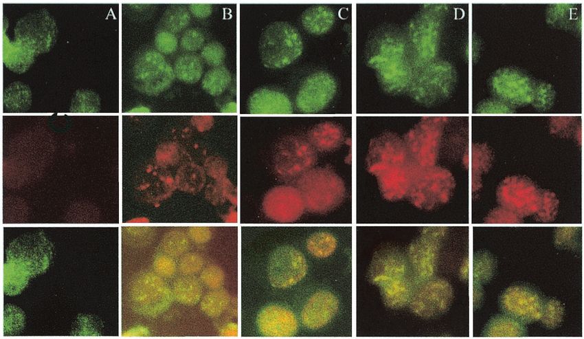
4262 KNIGHT ET AL. J. VIROL.
FIG. 1. Scheme showing the EBNA3C protein and the various domains. The homology domain is the region of highest homology to the other
EBNA3 family members (26). The repression domains and activation domains are indicated. Regions that bind p300 at the amino terminus and
carboxy terminus of EBNA3C are shown, and the interaction domains for ProT␣ and HDAC1 are shown within the amino-terminal 400 amino
acids (aa) (9, 24). The RBP-J binding site has also been mapped within the homology domain (27). LZ, leucine zipper; NLS, nuclear localization
signal; AD, acidic domain.
Downloaded from http://jvi.asm.org/ on February 14, 2021 by guest
through association with other factors that interact with the transferred to a 0.4-cm cuvette and electroporated at 975 F and 220 V. The
glutamine-rich domain known to be involved in transcriptional electroporated cells were then transferred to 10 ml of complete medium, fol-
lowed by incubation at 37°C and 5% CO2. Transfections were harvested after
activation. 24 h and assayed for activity.
EBNA3C binds histone deacetylase 1 (HDAC1) in vitro in a Deacetylase assays. 293 cells were transfected with CsCl-purified DNA with
region of the amino-terminal 200 amino acids known to asso- the appropriate constructs as described above. Cells were transfected with equiv-
ciate with the transcription repressor RBP-J (24). This inter- alent amounts of DNA for all transfections by normalizing with vector DNA.
Transfection efficiencies were determined by cotransfecting green fluorescent
action suggests that the association of EBNA3C with RBP-J
protein (GFP) expression vector in each transfection and counting GFP-positive
may also include complexing with HDACs and other molecules cells. Deacetylase assays were based on instructions obtained from the assay kit,
containing deacetylase activities in EBV-transformed cells. purchased from Upstate Biotechnology Inc., and performed essentially as per the
In this report we show that EBNA3C complexed with ProT␣ manufacturer’s instructions. Briefly, transfected cells were incubated for 24 h
associates with HDAC1 and HDAC2 and the corepressors posttransfection, harvested, and lysed in radioimmunoprecipitation assay
(RIPA) buffer (0.5% NP-40, 10 mM Tris [pH 7.5], 2 mM EDTA, 150 mM NaCl,
mSin3A and NCoR. These complexes recruited histone
supplemented with protease inhibitors). Cell debris was removed by centrifuga-
deacetylase activity in EBNA3C-expressing cell lines. Addi- tion, and the soluble lysate was transferred to a fresh tube. Rabbit polyclonal
tionally, the majority of the deacetylase activity was associated antibody against ProT␣ was used to immunoprecipitate complexes of ProT␣ and
with EBNA3C and ProT␣. However, the activity seen here may deacetylases for assays.
not be due only to the association with HDAC1, as EBNA3C Briefly, bound complexes were incubated with tritium-labeled acetylated pep-
tides corresponding to bovine histone H4 (amino acids 1 to 24). The reaction mix
and ProT␣ may be associated with other deacetylases yet to be was incubated at 37°C for 2 h with gentle agitation. The reaction was stopped by
characterized which are sensitive to trichostatin A (TSA) in- addition of incubation buffer containing 50 mM Tris, 150 mM NaCl, 5 mM
hibition. The deacetylase activity was coimmunoprecipitated EDTA, 0.5% NP-40, and 2 mM phenylmethylsulfonyl fluoride supplemented
with complexes associated with ProT␣. Moreover, in the pres- with 1 M HCl and 0.16 M acetic acid. To extract free acetate, 500 l of ethyl
ence of EBNA3C, this deacetylase activity immunoprecipi- acetate was added; the sample was vortexed, and the organic phase was sepa-
rated from the aqueous phase by centrifugation. The organic phase was removed
tated by antibodies to ProT␣ was enhanced by at least 50% and added to 2 ml of scintillation cocktail, the sample was vortexed, and the
over the initial activity. Therefore, EBNA3C can influence the amount of tritium released was determined by liquid scintillation counting.
cellular deacetylase activity through interactions with ProT␣ in GST binding assays, Western blots, and immunofluorescence assays. Gluta-
association with deacetylases and corepressors. thione S-transferase (GST) fusion proteins were purified from bulk Escherichia
coli cultures following induction with isopropylthiogalactopyranoside (IPTG).
Cells were lysed by sonication in NETN buffer (0.5% NP-40, 20 mM Tris [pH
MATERIALS AND METHODS 8.0], 1 mM EDTA, and 100 mM NaCl) supplemented with Sarkosyl and dithio-
Antibodies and cell lines. EBNA3C rabbit polyclonal antibody and RBP-J threitol. Cell debris was removed by centrifugation, and Sarkosyl was neutralized
antibodies were provided by Elliott Kieff, Harvard Medical School, Boston, with Triton X-100. GST fusion proteins were then purified from the supernatants
Mass. The A10 monoclonal antibody that recognizes EBNA3C was a gift from by incubation with glutathione-Sepharose beads. Protein concentrations were
Martin Rowe (22). HDAC1, HDAC2, mSin3A, and NCoR antibodies were determined by sodium dodecyl sulfate-polyacrylamide gel electrophoresis (SDS-
purchased from Santa Cruz Biotechnology, Inc. PAGE) with subsequent Coomassie brilliant blue staining.
BJAB cells are EBV-negative B cells isolated from a Burkitt’s lymphoma For GST pulldown assays, 5 ⫻ 107 cells were collected and washed in phos-
patient and were provided by Elliott Kieff. Human embryonic kidney fibroblast phate-buffered saline. Cells were lysed in RIPA buffer with 1% NP-40 and then
293 and 293T cells, transformed with E1A and T antigens, respectively, were cleared by centrifugation and transferred to a cold, sterile microcentrifuge tube.
obtained from Jon Aster, Brigham and Women’s Hospital, Boston, Mass. Lym- GST protein was added to the lysates for 30 min to preclear them, and then the
phoblastoid cell lines (LCLs) are EBV-transformed primary B lymphocytes in- GST fusion protein was added for 3 h with rotation at 4°C. Bound proteins were
fected with recombinant P3HR-1 rescued for EBNA2 and EBNALP (1, 8). collected by centrifugation and then washed four times in RIPA buffer with
All B-cell lines were grown in RPMI 1640 containing 10% fetal bovine serum protease inhibitors. Protein lysates were solubilized in SDS-lysis buffer and
supplemented with 10 mM glutamine, 25 U of penicillin per ml, and 25 g of fractionated on SDS-polyacrylamide gels. Fractionated proteins were then trans-
streptomycin per ml (Invitrogen-Gibco, Gaithersburg, Md.). Adherent cells were ferred to 0.4-m-pore nitrocellulose membranes, blocked in 5% nonfat milk, and
grown in Dulbecco’s modified Eagle’s medium containing 10% fetal bovine incubated in primary antibody overnight at 4°C with moderate rocking. Primary
serum supplemented with streptomycin and penicillin as above. All cell lines antibodies were used typically at a dilution of 1:100 in phosphate-buffered saline.
were grown at 37°C with 5% CO2 and passaged every 3 to 4 days. Protein-antibody complexes were detected with secondary antibody against
Transfections. BJAB and 293 cells were transfected by electroporation with a mouse, rabbit, or goat immunoglobulin or protein A conjugated to horseradish
Bio-Rad Gene Pulser II electroporator; 10 million cells harvested in exponential peroxidase at the dilutions recommended by the manufacturer (Amersham Inc.).
phase were collected and washed in phosphate-buffered saline and then resus- Immunofluorescence analyses were performed essentially as described previ-
pended in 400 l of RPMI with DNA for transfection. Resuspended cells were ously. Briefly, fixed cells were blocked in the appropriate serum and then incu-VOL. 77, 2003 EBNA3C RECRUITS HDAC ACTIVITY IN HUMAN B CELLS 4263
Downloaded from http://jvi.asm.org/ on February 14, 2021 by guest
FIG. 2. EBNA3C and ProT␣ recruit deacetylase activity in transient-transfection assays. (A) 293T cells were transfected with constructs
containing ProT␣ or ProT␣ and EBNA3C with vector alone as the DNA control. Harvested lysates were immunoprecipitated with antibody against
ProT␣ and assayed for deacetylase activity in the presence and absence of inhibitory concentrations of TSA. A positive-control HeLa lysate for
deacetylase activity was provided by Upstate Biotechnology Inc. as part of the assay kit and was used to validate our results for deacetylase activity
(panels A and B, right sample). ProT␣ immunoprecipitated deacetylase activity in this assay and was enhanced by the coexpression of EBNA3C
(E3C). (B) Truncated regions of EBNA3C coexpressed with ProT␣ showed that the amino-terminal 368 amino acids enhanced deacetylase activity
almost to the extent of wild-type EBNA3C. The carboxy terminus of EBNA3C showed increased deacetylase activity but not to the extent seen
with the amino-terminal region when compared to the positive control used in our assays. Protein lysates were analyzed by Western blot for levels
of expression of the transfected protein with the Myc monoclonal antibody to detect ProT␣ and A10 monoclonal antibody for detection of the
EBNA3C protein. Protein lysate was used as a loading control and was stained with Ponceau S.
bated with the specific primary antibody for HDAC1, HDAC2, mSin3A, and used as a positive control. Note that these control extracts were
NCoR (Santa Cruz Inc.) diluted at 1:200 or mouse anti-EBNA3C ascites diluted only used to determine activity and to validate our assay (Fig.
at 1:1,000 in phosphate-buffered saline for 1 h. Cells were washed and then
further incubated with the appropriate secondary antibody conjugated to fluo- 2A).
rescein isothiocyanate or Texas Red at 1:1,000 dilutions in phosphate-buffered The level of deacetylase activity was about twofold over that
saline for 1 h. Slides were washed and visualized with an Olympus XI70 inverted with the vector alone when ProT␣ was expressed from the
fluorescence microscope (Olympus Inc.) with an attached digital PixelFly camera
heterologous cytomegalovirus immediate-early promoter (Fig.
and software (Cooke, Inc.).
2A). The ProT␣ expression vector was monitored by Western
blotting for the Myc epitope fused at the carboxy terminus.
RESULTS Western blots with the 9E10 Myc monoclonal antibody showed
EBNA3C enhances the deacetylase activity associated with that similar levels of Myc-tagged ProT␣ protein were ex-
ProT␣ when coexpressed in transient-transfection assays. Pre- pressed in all transfected cells. EBNA3C levels were also con-
vious studies indicated that EBNA3C is associated with firmed with the A10 monoclonal antibody, which is reactive to
deacetylase activity in transient assays (24). To investigate this EBNA3C (22).
further, we decided to determine if EBNA3C and ProT␣ were In an effort to determine the effects of specific EBNA3C
associated with deacetylase activity by evaluating the level of domains on the recruitment of deacetylase activity, we com-
deacetylation when ProT␣ was immunoprecipitated alone or pared the domain shown to be involved in binding HDAC1,
with EBNA3C. Therefore, we transfected a ProT␣-expressing located within the amino-terminal 207-amino-acid region, with
vector either alone or with EBNA3C-expressing constructs in the amino-terminal region that includes the leucine zipper and
293 cells. Our results show that in the presence of EBNA3C, the entire homology region for the EBNA3 family of proteins
the level of deacetylation associated with ProT␣ increased to (26, 30, 31). In this deacetylase assay, we showed that ProT␣
three times over that with the vector alone. This was similar to and full-length EBNA3C recruited equivalent amounts of the
the levels seen with deacetylase activity from the HeLa control deacetylase activity and that together the activity was doubled
extract provided by Upstate Biotechnologies Inc., which was (Fig. 2B). When the amino-terminal 207 amino acids of4264 KNIGHT ET AL. J. VIROL.
Downloaded from http://jvi.asm.org/ on February 14, 2021 by guest
FIG. 3. GST pulldown assays show that HDAC1 and HDAC2 associate with ProT␣ and EBNA3C in EBNA3C-expressing cell lines. BJAB
E3C.7 and E3C.10 are cell lines that stably express EBNA3C under the control of a simian virus 40 promoter. GST protein was used as a control,
and GST-ProT␣ fusion protein was used in the experimental lanes. All binding assays were done at 4°C for 3 h and then washed four times. Bound
proteins were then solubilized and fractionated on SDS–8% PAGE. Proteins were transferred to nitrocellulose and blotted for EBNA3C with
monoclonal antibody A10. The blot was then stripped and reprobed consecutively with specific antibodies against HDAC1 and HDAC2 (Santa
Cruz Inc.).
EBNA3C were expressed along with ProT␣, a limited amount gels. The results of Western blots for HDAC1 and HDAC2
of activity, approximately 60% of that seen with either ProT␣ indicated that EBNA3C and ProT␣ associated with both these
or EBNA3C alone, was seen. However, when the region ex- deacetylases in EBNA3C-expressing cell lines. Interestingly,
pressing the amino-terminal 368 amino acids was introduced in HDAC1 showed similar levels of association on visual inspec-
the system, the activity obtained was almost equivalent to that tion with GST-ProT␣ in EBNA3C and control cell lines. How-
with the combination of ProT␣ and full-length EBNA3C (Fig. ever, HDAC2 had little or no signal with ProT␣ in BJAB
2B). Assays evaluating the carboxy-terminal 630 amino acids control cells but was clearly visible above background levels of
also showed deacetylase activity but less than that seen with the association in BJAB cell lines stably expressing EBNA3C (Fig.
368-amino-acid amino-terminal region. Based on these results, 3, compare lanes 3, 6, and 9).
we suggest that the amino-terminal and carboxy-terminal re- Complexes involved in repression of gene expression include
gions of EBNA3C may both contribute to the recruitment of known corepressor molecules such as NCoR and mSin3A. We
deacetylase activity. This may include recruitment and binding wanted to determine if these complexes containing HDAC1
to deacetylases HDAC1 and HDAC2 as well as other core- and HDAC2 may also contain these large corepressor proteins.
pressors known to be associated with deacetylase activity. In pulldown assays performed with the GST fusion of ProT␣,
HDAC1, HDAC2, mSin3A, and NCoR associate with we showed that mSin3A as well as NCoR associated with
EBNA3C and ProT␣ in EBNA3C-expressing Burkitt’s lym- EBNA3C and ProT␣ in EBNA3C-expressing cell lines (Fig. 4).
phoma cell lines. Previous studies showed that EBNA3C binds Additionally, we showed that these proteins also associated
to HDAC1 in vitro and that the region of EBNA3C bound to with ProT␣ in the absence of EBNA3C, suggesting that ProT␣
HDAC1 is similar to that seen with RBP-J, a known cellular may be recruiting these large molecules to complexes and
transcription repressor associated with EBNA3C (24). How- acting as a stabilizing component of these large transcriptional
ever, there has been no direct evidence showing that HDAC1 megacomplexes (Fig. 4). NCoR as well as mSin3A precipitated
associates with EBNA3C in EBNA3C-expressing cells or in with the ProT␣ fusion protein, as seen for EBNA3C, suggest-
EBV-transformed LCLs. Therefore, we wanted to determine if ing that these molecules may exist in a complex that includes
the association with the cellular deacetylases requires other EBNA3C in EBNA3C-expressing cells. A faint background
factors that may function as stabilizers for active complexes band in the GST control lanes was seen with this EBNA3C
which contain these deacetylases. Based on our previous data, Western blot but was not typically seen in subsequent blots. In
we hypothesized that ProT␣ may be a critical component of contrast to HDAC2, NCoR and mSin3A were pulled down
this complex, as was previously demonstrated for acetylase with ProT␣ in the presence and in the absence of EBNA3C in
complexes containing EBNA3C (9). the cell lines analyzed (Fig. 4).
We utilized the GST-ProT␣ fusion protein to pull down We next wanted to determine if these complexes seen with
complexes containing human deacetylases 1 and 2, existing as the GST fusion protein with ProT␣ were also seen with anti-
subunits of larger corepressor complexes containing EBNA3C. bodies against ProT␣. We used polyclonal rabbit antibody
In cells stably expressing EBNA3C, we showed that EBNA3C against ProT␣ and performed immunoprecipitation assays
as well as HDAC1 and HDAC2 were brought down in complex with Western blot analysis to determine if these complexes
with the GST-ProT␣ fusion protein. Lysates from two isogenic were stable to immunoprecipitation as well as GST pulldown.
EBNA3C-expressing BJAB cell lines were harvested from 50 In these assays, we showed again that HDAC1 and HDAC2
million cells, lysed in RIPA buffer, and incubated with GST- coimmunoprecipitated with ProT␣ and EBNA3C (Fig. 5). The
ProT␣ bound to beads. Bound complexes were washed and blots were stripped and reprobed each time with a different
solubilized in lysis buffer and then fractionated on SDS-PAGE antibody to the HDAC and corepressor molecules. The dataVOL. 77, 2003 EBNA3C RECRUITS HDAC ACTIVITY IN HUMAN B CELLS 4265
Downloaded from http://jvi.asm.org/ on February 14, 2021 by guest
FIG. 4. GST pulldown assays show that corepressors NCoR and mSin3A associate with ProT␣ and EBNA3C in EBNA3C-expressing cell lines.
BJAB E3C.7 and E3C.10 are cell lines that stably express EBNA3C under the control of a simian virus 40 promoter. As in Fig. 3, cells expressing
EBNA3C were lysed and proteins were incubated with the GST-ProT␣ fusion. Bound proteins were solubilized and run on SDS–6% PAGE. The
proteins were transferred to nitrocellulose and probed with antibodies against NCoR and mSin3A (Santa Cruz, Inc.).
from these Western blots show that NCoR and mSin3A coim- Complexes associated with EBNA3C and ProT␣ recruit hi-
munoprecipitated in EBNA3C-expressing cell lines as well as stone deacetylase activity in B-cell lines stably expressing the
in the control cell line BJAB containing the vector alone (Fig. EBNA3C protein. Studies utilizing a transient system indicate
5). Similar levels of proteins were seen in the immunoprecipi- that EBNA3C is associated with deacetylase activity and that it
tation lane for the EBNA3C-expressing cell lines and the neg- can associate with human HDAC1 when overexpressed by
ative control (Fig. 5). cotransfection (24). We wanted to determine if this activity
FIG. 5. Corepressors NCoR and mSin3A and HDAC1 and HDAC2 associate with EBNA3C complexed with ProT␣ in cell lines stably
expressing EBNA3C. Immunoprecipitation analysis with ProT␣ rabbit antibody showed that the HDACs and corepressors were associated with
ProT␣ and EBNA3C in EBNA3C-expressing cell lines. The blot was stripped and reprobed with specific antibodies to the antigens indicated. L,
lysate; PC, preclear; IP, immunoprecipitate.4266 KNIGHT ET AL. J. VIROL.
may be associated with ProT␣ and EBNA3C, as EBNA3C has
been shown to have both activating and repressive functions.
ProT␣ was previously shown to associate with the acetyltrans-
ferase p300 and with EBNA3C (9). Therefore, we wanted to
determine if ProT␣ may also be associated with deacetylases
associated with transcription repression potentially regulated
by EBNA3C.
In this study, we coimmunoprecipitated complexes with
ProT␣ antibody and tested for deacetylase activity. Western
blotting with EBNA3C-specific monoclonal antibody A10 was
used to determine the expression levels for EBNA3C in two
cell lines stably expressing EBNA3C, E3C.7 and E3C.10, and
in two recently transformed LCLs, LCL1 and LCL2. BJAB was
used as a negative control. ProT␣ levels were monitored by
Downloaded from http://jvi.asm.org/ on February 14, 2021 by guest
Western blot analysis with the ProT␣ antibody. HeLa cell
extract from the manufacturer was used as a positive control
for deacetylase activity and was provided by Upstate Biotech-
nology Inc.
The results of these experiments showed that the cell lines
expressing greater amounts of EBNA3C resulted in a greater
association with deacetylase activity compared to LCLs ex-
pressing a lower amount of EBNA3C and other EBNA pro-
teins, including EBNA2 and EBNALP (2, 25, 39) (Fig. 6). In
fact, the amount of deacetylase activity was greater than that
seen with the extract supplied from HeLa cells used as a pos-
itive control. The amount of ProT␣ immunoprecipitated was FIG. 6. EBNA3C and ProT␣ recruit deacetylase activity in cell
similar in each case, as demonstrated by Western blot and the lines stably expressing EBNA3C. HeLa cell extract was used as a
control for deacetylase activity and was based on the deacetylase kit
protein loading control (Fig. 6, compare middle and lower from Upstate Biotechnology Inc. LCLs showed deacetylase activity
panels). equivalent to that seen with the EBV-negative BJAB cell control.
EBNA3C-associated deacetylase activity in stable BJAB cell Immunoprecipitation of ProT␣ with rabbit polyclonal antibody to
lines is sensitive to TSA. The above results suggest that ProT␣ was followed by incubation with 14C-labeled acetylated pep-
tides. The labeled molecules removed by deacetylation were counted
EBNA3C recruits deacetylase activity when complexed with in a scintillation counter and plotted. All experiments were done in
ProT␣ and that this activity is enhanced as EBNA3C levels triplicate, and the means of the experiments are shown. Western blots
increase in the cell. The level of deacetylase activity in LCLs for detection of EBNA3C (E3C) and ProT␣ are shown below, as is a
are similar to that in the EBV-negative control. This may be a Ponceau S-stained gel showing the protein loading control.
consequence of the expression of other EBNA proteins in
EBV-transformed LCLs or simply indicate that a smaller
amount of EBNA3C is present in LCLs. In order to determine HDAC1 binds to a similar region at the amino terminus of
if the deacetylase activity was inhibited by a general deacety- EBNA3C adjacent to the RBP-J binding site, as determined
lase inhibitor, we used TSA at concentrations of 10 mM and 50 by a series of in vitro binding studies (24). Here we found that,
mM. In this assay, we showed that TSA inhibited the deacety- in contrast to cell lines stably expressing large amounts of
lase activity of EBNA3C-expressing cells; 10 mM TSA reduced EBNA3C, ProT␣ had little or no association with HDAC1,
the activity approximately 30%, and 50 mM reduced the levels HDAC2, and the corepressors mSin3A and NCoR in EBV-
to 75 to 90% of the initial amounts in EBNA3C-overexpressing transformed LCLs. Immunoprecipitation experiments from
lines (Fig. 7). The fact that the deacetylase activity associated two independent transformed cell lines showed that there was
with EBNA3C can be efficiently inhibited by TSA at low con- no detectable signal above the background for the preimmune
centrations without affecting cell viability indicates that control lane with rabbit polyclonal ProT␣ antibody (Fig. 8).
EBNA3C and ProT␣ may exist in repressor complexes with These results suggest that other competing molecules may
multiple partners to mediate repression of specific cellular and sequester EBNA3C and ProT␣ away from these repressor
viral promoters. Based on these data, we decided to look for complexes or that the limited number of molecules available
possible association of corepressors in the complex with for targeting deacetylases in LCLs is below the detection limit
EBNA3C in cell lines stably expressing EBNA3C as well as in of our immunoprecipitation analysis. Other EBV nuclear an-
EBV-transformed LCLs. tigens may also play a role in disrupting these complexes while
Association of complexes of HDACs and corepressors with functioning as regulators of gene expression from viral and
EBNA3C is limited in EBV-transformed LCLs. The associa- cellular promoters required for maintenance of the trans-
tion of EBNA3C with corepressors and HDACs in EBNA3C- formed state. Additionally, it is possible that this specific an-
expressing cell lines suggests that these molecules may function tibody used to immunoprecipitate the ProT␣ complex may also
in regulating cellular and viral promoters in EBV-immortal- destabilize some components of the complexes.
ized cells. It was previously shown that RBP-J, a known cel- One interesting observation made in these experiments was
lular repressor, interacts in vitro with EBNA3C (28) and that that in EBV-transformed LCLs, no NCoR signal was detectedVOL. 77, 2003 EBNA3C RECRUITS HDAC ACTIVITY IN HUMAN B CELLS 4267
Downloaded from http://jvi.asm.org/ on February 14, 2021 by guest
FIG. 7. Deacetylation activity associated with EBNA3C and ProT␣ is inhibited by TSA. Deacetylation assays were performed as described for
Fig. 6, and samples were incubated with increasing concentrations of TSA. Western blots show levels of ProT␣ and EBNA3C (E3C) as well as the
protein loading control.
by Western blot repeated in multiple assays (Fig. 8). However, EBNA3C may be localized in compartments similar to those
the signal was clearly seen in the lysate lane in EBV-negative of these cellular molecules. However, the association may be
BJAB cells. The signal was not seen in the lysate of LCL1 and temporally regulated, based on the specific phase of the cell
LCL2 cells even after longer exposures (data not shown). cycle. Therefore, we performed immunofluorescence analysis
These studies suggest that the expression of NCoR may be with HDAC1, HDAC2, mSin3A, and NCoR to determine if
downregulated in primary B lymphocytes either directly or EBNA3C was in the same nuclear compartment. EBNA3C-
indirectly by an EBV-expressed latent antigen. However, it is expressing cells were fixed and probed with antibodies for the
also possible that NCoR is not expressed in primary B lym- HDACs, mSin3A, and NCoR, followed by incubation with
phocytes. This needs to be explored further, as the levels of EBNA3C monoclonal antibody A10. Fluorescein isothiocya-
NCoR in uninfected primary B cells have to be compared to nate and Texas Red conjugated to the appropriate secondary
those seen in LCLs and other Burkitt’s lymphoma cell lines. antibody were used for detection. The results of these studies
To determine if complexes might be differentially detected showed that EBNA3C was localized in the same nuclear com-
when immunoprecipitating with antibody reactive to EBNA3C partments in B cells as HDAC1, HDAC2, mSin3A, and NCoR
rather than ProT␣, we chose to use a rabbit polyclonal anti- molecules (Fig. 9B).
body specific for EBNA3C, which recognizes an epitope in the
carboxy terminus. In these experiments, we again showed that DISCUSSION
little or no association detectable above background was seen
(Fig. 9A). However, we did show that RBP-J could be coim- The association of EBNA3C with deacetylases and corepres-
munoprecipitated with the same antibody against EBNA3C in sors has not been previously shown in cell lines or EBV-trans-
EBNA3C-expressing cell lines and LCLs (Fig. 9A). No RBP-J formed LCLs. EBNA3C is known to have both activation and
signal was seen in the EBNA3C-negative BJAB cells but was repression functions, and specific domains have been mapped
seen in the EBNA3C-expressing cell lines and EBV-trans- for the specific functions of transcription repression and acti-
formed LCL1 (Fig. 9A). vation (21, 24, 26). We have shown that EBNA3C binds to the4268 KNIGHT ET AL. J. VIROL.
Downloaded from http://jvi.asm.org/ on February 14, 2021 by guest
FIG. 8. Immunoprecipitation analysis with anti-ProT␣ antibody in lysates from LCLs showed that there was little or no detectable association
with HDAC1 and HDAC2 as well as mSin3A and NCoR. No NCoR signal was present in the LCLs even after prolonged exposure to film. Some
HDAC1 and HDAC2 were detected, but the levels were barely above the background levels of control preimmune serum. The signal in the
immune experimental lane was lower that that seen in the EBNA3C-expressing cell lines. L, lysate; PC, preclear; IP, immunoprecipitate.
p300 coactivator and is associated with histone acetyltrans- ProT␣ that is yet to be determined in EBV-infected cells and
ferase activity in EBV-transformed LCLs (9). These studies is expected to be important for the association with numerous
indicate that ProT␣ may function as a displacement factor for other cellular factors.
histone H1, similar to the HMG1 factor that allows access to Deacetylase assays to determine the regions of EBNA3C
the promoter regions (7, 10, 11, 16, 23). Here we show that involved in recruiting deacetylase activity showed that the do-
EBNA3C associated with HDAC1 and HDAC2 and also the mains at both the amino and carboxy terminus contain activity
corepressors mSin3A and NCoR in EBNA3C-expressing cells with approximately equivalent potency. This indicates that the
but had little association in EBV-transformed LCLs, as deter- regions of the molecules previously mapped to have repressive
mined by our immunoprecipitation analyses. functions may independently recruit deacetylases. The fact that
Previous in vitro studies showed that HDAC1 interacts with EBNA3C synergizes the activity seen with ProT␣ suggests that
EBNA3C at the amino-terminal region and that the deacety- these molecules may be associated with a larger complex con-
lase activity is associated with its amino-terminal region (24). taining deacetylase activity, as no deacetylase activity has pre-
However, to date no association has been demonstrated in viously been assigned to either ProT␣ or EBNA3C. Large
EBV-transformed LCLs or in EBNA3C-expressing cells. In complexes containing deacetylases may require association
this report we show that HDAC1 and HDAC2 associate with with specific factors for triggering activity. Some molecules
EBNA3C in a complex with ProT␣ and that this is important may have partial activity but becomes fully active in the pres-
for the in vivo association of EBNA3C and HDACs as well as ence of the required molecules recruited to the complex, which
other corepressor molecules, including mSin3A and NCoR. may include EBNA3C, ProT␣, and other corepressors, includ-
One interesting observation in our studies was that little asso- ing NCoR and mSin3A (Fig. 10).
ciation was seen when we immunoprecipitated directly with The association of HDAC1 with ProT␣ as well as EBNA3C
antibodies to the EBNA3C carboxy terminus. However, we indicates that these molecules may be part of a larger repressor
were able to show association when we immunoprecipitated complex stabilized by ProT␣ (Fig. 10). HDAC2 seems to be
with anti-ProT␣ antibodies. These results suggest that ProT␣ is more abundant in its association with ProT␣ and EBNA3C, as
required to hold the complex together and that ProT␣ may be shown in the GST-ProT␣ pulldown experiments. Therefore,
functioning to stabilize complexes with EBNA3C, the deacety- HDAC2 may be the more predominant deacetylase associated
lases HDAC1 and HDAC2, and the corepressors mSin3A and in complex with EBNA3C and ProT␣, as HDAC2 had little or
NCoR. Additionally, there is likely to be a level of regulation no association with ProT␣ in the absence of EBNA3C, sug-
that is dependent on the posttranslational modification of gesting a requirement for EBNA3C to recruit enhanced activ-VOL. 77, 2003 EBNA3C RECRUITS HDAC ACTIVITY IN HUMAN B CELLS 4269
Downloaded from http://jvi.asm.org/ on February 14, 2021 by guest
FIG. 9. Immunoprecipitation analysis with rabbit anti-EBNA3C antibody showed that HDAC1, HDAC2, mSin3A, and NCoR were not directly
immunoprecipitated with EBNA3C. However, RBP-J was immunoprecipitated with anti-EBNA3C, as expected in EBNA3C-expressing lines and
LCL1 (panel A). Immunofluorescence analysis showed that EBNA3C (E3C) was localized to the same nuclear compartment as HDAC1, HDAC2,
mSin3A, and NCoR (panel B, A to E) in EBNA3C-expressing cells. L, lysate; PC, preclear; IP, immunoprecipitate.
ity. In comparison, the corepressors mSin3A and NCoR asso- transformed LCLs, suggesting a lack of expression of NCoR.
ciated to similar levels compared to that seen with EBNA3C. This lack of expression may be dependent on the presence of
It was clear from the immunoprecipitation data that EBV latent antigens, possibly the EBNAs. However, further
EBNA3C was brought down in the same manner as NCoR, studies will elucidate the possible regulation of NCoR expres-
mSin3A, HDAC1, and HDAC2 in stable cell lines. Surpris- sion by an EBV antigen. It is possible that EBNA2 and
ingly, NCoR was not detected even in the lysates of EBV- EBNALP or even EBNA3C itself may have a direct regulatory4270 KNIGHT ET AL. J. VIROL.
Downloaded from http://jvi.asm.org/ on February 14, 2021 by guest
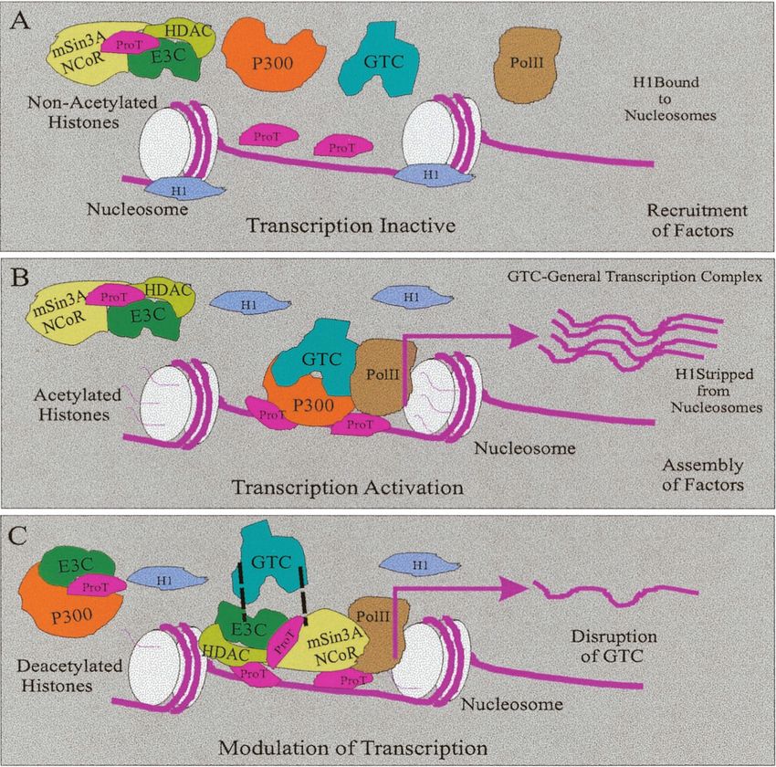
FIG. 10. Schematic showing the putative role of EBNA3C in regulating chromatin acetylation and deacetylation, resulting in transcriptional
control. EBNA3C associates with the acetylase p300, the deacetylases HDAC1 and HDAC2, and the corepressors mSin3A and NCoR. These
complexes are stabilized by ProT␣, which also acts in inhibiting histone H1 from binding to the nucleosome, allowing access of the large activation
and repressor complexes. Therefore, EBNA3C is involved in regulating viral and cellular promoters, and this intrinsically requires the cellular
molecule ProT␣ to modulate the associated activity.
role on the NCoR promoter, and we will continue to address proteins. It should be noted that to date, no direct association
these studies with further experimentation. Clearly, in LCLs between RBP-J and HDACs has been demonstrated in hu-
the deacetylase activity is reduced compared to that seen with man cells or EBV-transformed LCLs. The interaction has only
the cell lines stably expressing EBNA3C. This may be a reflec- been shown by in vitro binding studies. It is possible that
tion of the levels of EBNA3C expressed in these LCLs com- RBP-J is outcompeted by other repressor molecules with a
pared to the stable cell lines or the result of expression of other higher affinity for HDAC1 as well as other HDACs and that
latent genes, including EBNA2 and EBNALP, the coactivator those associations are temporally regulated. The interaction of
of EBNA2, which may counter the overall effects at specific the essential EBV antigens with regulators of gene expression
promoters recruiting deacetylases and corepressors. is expected to be tightly regulated and dependent on the pres-
The putative involvement of RBP-J in these complexes is a ence of specific cellular factors required for maintaining the
reasonable question to address, as RBP-J was previously transformed phenotype of the infected cells.
shown to be a major player in EBV-mediated events and Interestingly, in LCLs the association of HDAC1, HDAC2,
shown to be a repressor of both viral and cellular promoters (4, and corepressors mSin3A and NCoR with EBNA3C and
14, 15). This is functionally reversed by EBNA2 and further ProT␣ was negligible compared to the control background
regulated by EBNA3C (27). To address this directly, we per- lanes. This was surprising, as we expected that the levels would
formed immunoprecipitation and Western blotting for RBP-J be comparable to that in the EBNA3C cell lines and may be a
and, as expected, were able to show association with EBNA3C. function of the additional EBNAs expressed in EBV-trans-
This may indicate that RBP-J is only one of the major regu- formed LCLs. Immunofluorescence analysis with antibodies
lators associated with the essential EBV latent antigens and against HDAC1 and HDAC2 as well as mSin3A and NCoR
that it may associate with other complexes with the EBNA indicated that they were localized in the same nuclear com-VOL. 77, 2003 EBNA3C RECRUITS HDAC ACTIVITY IN HUMAN B CELLS 4271
partments as EBNA3C. Additionally, the association requires 9. Cotter, M. A., 2nd, and E. S. Robertson. 2000. Modulation of histone acetyl-
transferase activity through interaction of Epstein-Barr nuclear antigen 3C
ProT␣ and most likely other factors yet to be identified to with prothymosin alpha. Mol. Cell. Biol. 20:5722–5735.
stabilize the complex. As these factors localize in the same 10. Diaz-Jullien, C., A. Perez-Estevez, G. Covelo, and M. Freire. 1996. Prothy-
compartment but have different affinities for direct association, mosin alpha binds histones in vitro and shows activity in nucleosome assem-
bly assay. Biochim. Biophys. Acta 1296:219–227.
it is possible that their association with EBNA3C is dependent 11. Ding, H. F., M. Bustin, and U. Hansen. 1997. Alleviation of histone H1-
on the cell cycle, as it was previously shown that the expression mediated transcriptional repression and chromatin compaction by the acidic
of ProT␣ is upregulated during the S/G2 phase of the cell cycle activation region in chromosomal protein HMG-14. Mol. Cell. Biol. 17:
5843–5855.
(17, 36). ProT␣ is induced as the cell proliferates and at the 12. Farrell, P. J., I. Cludts, and A. Stuhler. 1997. Epstein-Barr virus genes and
S/G2 transition, as it may be required for stabilizing repression cancer cells. Biomed. Pharmacother. 51:258–267.
13. Fennewald, S., V. Van Santen, and E. Kieff. 1984. The nucleotide sequence
and activation complexes. The absence of ProT␣ results in a of a messenger RNA transcribed in latent growth transforming virus infec-
block to cell division and proliferation, suggesting that cellular tion indicates that it may encode a membrane protein. J. Virol. 51:411–419.
molecules required for driving these cellular processes are 14. Grossman, S. R., E. Johannsen, X. Tong, R. Yalamanchili, and E. Kieff.
1994. The Epstein-Barr virus nuclear antigen 2 transactivator is directed to
intrinsically linked with ProT␣ (29, 32, 34, 38). The fact that response elements by the J kappa recombination signal binding protein.
EBNA3C binds ProT␣ clearly supports the hypothesis that Proc. Natl. Acad. Sci. USA 91:7568–7572.
Downloaded from http://jvi.asm.org/ on February 14, 2021 by guest
EBNA3C is involved in regulating gene expression. 15. Henkel, T., P. D. Ling, S. D. Hayward, and M. G. Peterson. 1994. Mediation
of Epstein-Barr virus EBNA2 transactivation by recombination signal-bind-
The association with p300 and acetylase activity as well as ing protein J kappa. Science 265:92–95.
HDACs, corepressors, and deacetylase activity in B cells 16. Juan, L. J., R. T. Utley, M. Vignali, L. Bohm, and J. L. Workman. 1997.
H1-mediated repression of transcription factor binding to a stably positioned
strengthens the argument for a role of EBNA3C in both acti- nucleosome. J. Biol. Chem. 272:3635–3640.
vation and repression of transcription. In our model, EBNA3C 17. Karetsou, Z., R. Sandaltzopoulos, M. Frangou-Lazaridis, C. Y. Lai, O.
is associated with corepressor complexes that include deacety- Tsolas, P. B. Becker, and T. Papamarcaki. 1998. Prothymosin alpha modu-
lates the interaction of histone H1 with chromatin. Nucleic Acids Res.
lases (HDACs). This complex is recruited to promoters, result- 26:3111–3118.
ing in downregulation of specific viral and cellular genes. 18. Kieff, E. 1998. Current perspectives on the molecular pathogenesis of virus-
ProT␣ stabilizes these complexes containing the acetylases and induced cancers in human immunodeficiency virus infection and acquired
immunodeficiency syndrome. J. Natl. Cancer Inst. Monogr. 23:7–14.
deacetylases and is critical for these complexes to be fully 19. Kieff, E. 1996. Epstein-Barr Virus and its replication, 3rd ed., vol. 2. Lippin-
active (Fig. 10). EBNA3C seems to be in separate complexes cott-Raven, Philadelphia, Pa.
with acetylases and deacetylases. The associated functions may 20. Klein, E. 1998. The complexity of the Epstein-Barr virus infection in humans.
Pathol. Oncol. Res. 4:3–7.
then be dependent on cell cycle events as well as contributions 21. Marshall, D., and C. Sample. 1995. Epstein-Barr virus nuclear antigen 3C is
from other cellular and viral antigens involved in regulating a transcriptional regulator. J. Virol. 69:3624–3630.
22. Maunders, M. J., L. Petti, and M. Rowe. 1994. Precipitation of the Epstein-
cell growth and proliferation. More studies dissecting these Barr virus protein EBNA 2 by an EBNA 3c-specific monoclonal antibody.
cellular processes and their regulation should shed light on the J. Gen. Virol. 75:769–778.
roles of ProT␣ and EBNA3C and their abilities to regulate 23. Nightingale, K., S. Dimitrov, R. Reeves, and A. P. Wolffe. 1996. Evidence for
a shared structural role for HMG1 and linker histones B4 and H1 in orga-
these associated cellular events. nizing chromatin. EMBO J. 15:548–561.
24. Radkov, S. A., R. Touitou, A. Brehm, M. Rowe, M. West, T. Kouzarides, and
ACKNOWLEDGMENTS M. J. Allday. 1999. Epstein-Barr virus nuclear antigen 3C interacts with
histone deacetylase to repress transcription. J. Virol. 73:5688–5697.
We thank Fernando Dominguez for the ProT␣ construct and Elliott 25. Rickinson, A., and E. Kieff. 1996. Epstein-Barr virus, 3rd ed., vol. 2. Lippin-
Kieff for the EBNA3C expression construct. We thank Rama Orre for cott-Raven, Philadelphia, Pa.
initial help with the deacetylase assays. 26. Robertson, E. S. 1997. The Epstein-Barr virus EBNA3 protein family as
This work was supported by grants from the Leukemia and Lym- regulators of transcription. Epstein-Barr Virus Rep. 4:143–150.
phoma Society of America and the National Cancer Institute 27. Robertson, E. S., S. Grossman, E. Johannsen, C. Miller, J. Lin, B. Tomkin-
son, and E. Kieff. 1995. Epstein-Barr virus nuclear protein 3C modulates
(CA072150 to E.S.R.). E.S.R. is a Leukemia and Lymphoma Society of transcription through interaction with the sequence-specific DNA-binding
America Scholar. J.S.K. is supported by NIH graduate training grant protein J. J. Virol. 69:3108–3116.
T32-AI-07632-02 at the University of Pennsylvania Medical School. 28. Robertson, E. S., J. Lin, and E. Kieff. 1996. The amino-terminal domains of
Epstein-Barr virus nuclear proteins 3A, 3B, and 3C interact with RBPJ.
REFERENCES J. Virol. 70:3068–3074.
1. Adams, A., G. Bjursell, C. Kaschka-Dierich, and T. Lindahl. 1977. Circular 29. Roson, E., R. Gallego, T. Garcia-Caballero, E. P. Heimer, A. M. Felix, and
Epstein-Barr virus genomes of reduced size in a human lymphoid cell line of F. Dominguez. 1990. Prothymosin alpha expression is associated to cell
infectious mononucleosis origin. J. Virol. 22:373–380. division in rat testis. Histochemistry 94:597–599.
2. Alfieri, C., M. Birkenbach, and E. Kieff. 1991. Early events in Epstein-Barr 30. Sample, C., and B. Parker. 1994. Biochemical characterization of Epstein-
virus infection of human B lymphocytes. Virology 181:595–608. (Erratum, Barr virus nuclear antigen 3A and 3C proteins. Virology 205:534–539.
Virology 185:946.) 31. Sample, J., L. Young, B. Martin, T. Chatman, E. Kieff, and A. Rickinson.
3. Allday, M. J., and P. J. Farrell. 1994. Epstein-Barr virus nuclear antigen 1990. Epstein-Barr virus types 1 and 2 differ in their EBNA-3A, EBNA-3B,
EBNA3C/6 expression maintains the level of latent membrane protein 1 in and EBNA-3C genes. J. Virol. 64:4084–4092.
G1-arrested cells. J. Virol. 68:3491–3498. 32. Sburlati, A. R., R. E. Manrow, and S. L. Berger. 1991. Prothymosin alpha
4. Aster, J. C., E. S. Robertson, R. P. Hasserjian, J. R. Turner, E. Kieff, and J. antisense oligomers inhibit myeloma cell division. Proc. Natl. Acad. Sci. USA
Sklar. 1997. Oncogenic forms of NOTCH1 lacking either the primary bind- 88:253–257.
ing site for RBP-J or nuclear localization sequences retain the ability to 33. Sixbey, J. W., J. G. Nedrud, N. Raab-Traub, R. A. Hanes, and J. S. Pagano.
associate with RBP-J and activate transcription. J. Biol. Chem. 272:11336– 1984. Epstein-Barr virus replication in oropharyngeal epithelial cells.
11343. N. Engl. J. Med. 310:1225–1230.
5. Bain, M., R. J. Watson, P. J. Farrell, and M. J. Allday. 1996. Epstein-Barr 34. Smith, M. R., A. al-Katib, R. Mohammad, A. Silverman, P. Szabo, S. Khil-
virus nuclear antigen 3C is a powerful repressor of transcription when teth- nani, W. Kohler, R. Nath, and M. G. Mutchnick. 1993. Prothymosin alpha
ered to DNA. J. Virol. 70:2481–2489. gene expression correlates with proliferation, not differentiation, of HL-60
6. Burkitt, D. W. D. 1966. Geographical and tribal distribution of the African cells. Blood 82:1127–1132.
lymphoma in Uganda. Br. Med. J. 5487:569–573. 35. Subramanian, C., M. A. Cotter 2nd, and E. S. Robertson. 2001. Epstein-Barr
7. Bustin, M. 2001. Chromatin unfolding and activation by HMGN(*) chro- virus nuclear protein EBNA-3C interacts with the human metastatic sup-
mosomal proteins. Trends Biochem. Sci. 26:431–437. pressor Nm23-H1: a molecular link to cancer metastasis. Nat. Med. 7:350–355.
8. Cohen, J. I., F. Wang, J. Mannick, and E. Kieff. 1989. Epstein-Barr virus 36. Szabo, P., D. Ehleiter, E. Whittington, and M. E. Weksler. 1992. Prothymo-
nuclear protein 2 is a key determinant of lymphocyte transformation. Proc. sin alpha expression occurs during G1 in proliferating B or T lymphocytes.
Natl. Acad. Sci. USA 86:9558–9562. Biochem. Biophys. Res. Commun. 185:953–959.4272 KNIGHT ET AL. J. VIROL.
37. Tomkinson, B., E. Robertson, and E. Kieff. 1993. Epstein-Barr virus nuclear 40. Zhao, B., and C. E. Sample. 2000. Epstein-Barr virus nuclear antigen 3C
proteins EBNA-3A and EBNA-3C are essential for B-lymphocyte growth activates the latent membrane protein 1 promoter in the presence of Ep-
transformation. J. Virol. 67:2014–2025. stein-Barr virus nuclear antigen 2 through sequences encompassing an Spi-
38. Wu, C. L., A. L. Shiau, and C. S. Lin. 1997. Prothymosin alpha promotes cell 1/Spi-B binding site. J. Virol. 74:5151–5160.
proliferation in NIH 3T3 cells. Life Sci. 61:2091–2101. 41. Zimber-Strobl, U., L. J. Strobl, C. Meitinger, R. Hinrichs, T. Sakai, T.
39. Young, L., C. Alfieri, K. Hennessy, H. Evans, C. O’Hara, K. C. Anderson, J. Furukawa, T. Honjo, and G. W. Bornkamm. 1994. Epstein-Barr virus nuclear
Ritz, R. S. Shapiro, A. Rickinson, E. Kieff, et al. 1989. Expression of Epstein- antigen 2 exerts its transactivating function through interaction with recom-
Barr virus transformation-associated genes in tissues of patients with EBV bination signal binding protein RBP-J kappa, the homologue of Drosophila
lymphoproliferative disease. N. Engl. J. Med. 321:1080–1085. Suppressor of Hairless. EMBO J. 13:4973–4982.
Downloaded from http://jvi.asm.org/ on February 14, 2021 by guestYou can also read


