P63 and p53: Collaborative Partners or Dueling Rivals? - Frontiers
←
→
Page content transcription
If your browser does not render page correctly, please read the page content below
MINI REVIEW
                                                                                                                                                published: 05 July 2021
                                                                                                                                        doi: 10.3389/fcell.2021.701986

                                            p63 and p53: Collaborative Partners
                                            or Dueling Rivals?
                                            Dana L. Woodstock 1 , Morgan A. Sammons 1* and Martin Fischer 2*
                                            1
                                              Department of Biological Sciences, The State University of New York at Albany, Albany, NY, United States, 2 Computational
                                            Biology Group, Leibniz Institute on Aging – Fritz Lipmann Institute (FLI), Jena, Germany

                                            The tumor suppressor p53 and its oncogenic sibling p63 (1Np63) direct opposing fates
                                            in tumor development. These paralog proteins are transcription factors that elicit their
                                            tumor suppressive and oncogenic capacity through the regulation of both shared and
                                            unique target genes. Both proteins predominantly function as activators of transcription,
                                            leading to a paradigm shift away from 1Np63 as a dominant negative to p53 activity. The
                                            discovery of p53 and p63 as pioneer transcription factors regulating chromatin structure
                                            revealed new insights into how these paralogs can both positively and negatively
                                            influence each other to direct cell fate. The previous view of a strict rivalry between
                             Edited by:     the siblings needs to be revisited, as p53 and p63 can also work together toward a
                     Brigitte M. Pützer,
University Hospital Rostock, Germany
                                            common goal.
                     Reviewed by:           Keywords: p53, p63, tumor suppressor, oncogene, transcription factor, pioneer factor
                        Matt Fisher,
      Cold Spring Harbor Laboratory,
                      United States        INTRODUCTION
                 Adone Mohd-Sarip,
          Queen’s University Belfast,      The p53 transcription factor family comprises the three members p53, p63, and p73. Although
                    United Kingdom
                                           it is evolutionarily the youngest, p53 is the eponymous member of the family. The transcription
                 *Correspondence:          factors evolved from an ancestral p63/p73 gene that can be found in most invertebrates (Belyi
                Morgan A. Sammons          et al., 2010; Rutkowski et al., 2010). While the ancestral p63/p73 gene protects organismal integrity
           masammons@albany.edu
                                           and the germ line by inducing cell death upon DNA damage, higher vertebrates possess all three
                       Martin Fischer
          Martin.Fischer@leibniz-fli.de
                                           p53 family members with diversified functions. In particular the intricate relationship between
                                           the family members and their overlapping and opposing functions have been subject to intense
                    Specialty section:     research and debate.
         This article was submitted to         The family matters are complicated by the fact that p53, p63, and p73 comprise multiple
                   Molecular Medicine,     isoforms. The TP53, TP63, and TP73 genes encode for major isoform groups that are controlled by
                a section of the journal   distinct promoters leading to transcripts that differ in their N-terminus (Murray-Zmijewski et al.,
  Frontiers in Cell and Developmental      2006). In the case of TP63, these isoforms are highly cell type-dependent. The long isoform TAp63 is
                                Biology
                                           mainly expressed in germ cells and the shorter 1Np63 isoform is predominantly expressed in basal
             Received: 28 April 2021       and stratifying epithelia. In contrast, full-length p53 is expressed across essentially all tissues. In
            Accepted: 14 June 2021
                                           addition to different N-termini generated through alternative promoter usage, alternative splicing
            Published: 05 July 2021
                                           leads to additional isoforms for each p53 family member that differ in their C-termini, such as α, β,
                              Citation:    and γ isoforms (Murray-Zmijewski et al., 2006).
       Woodstock DL, Sammons MA
                                               The full-length isoforms p53α, TAp63α, and TAp73α function as haplo-insufficient tumor
  and Fischer M (2021) p63 and p53:
    Collaborative Partners or Dueling
                                           suppressors (Venkatachalam et al., 1998; Flores et al., 2005). In addition, p73 functions in
                                Rivals?    neuronal development, multi-ciliated cell differentiation, and metabolism (Nemajerova et al.,
      Front. Cell Dev. Biol. 9:701986.     2018). In contrast, 1Np63 governs epidermis development (Mills et al., 1999; Yang et al., 1999)
     doi: 10.3389/fcell.2021.701986        and is an oncogenic driver that is overexpressed or amplified in squamous cell carcinoma

Frontiers in Cell and Developmental Biology | www.frontiersin.org                  1                                              July 2021 | Volume 9 | Article 701986
Woodstock et al.                                                                                                              p53/p63 Sibling Rivalry

(Campbell et al., 2018; Gatti et al., 2019). The opposing directions       analyses of chromatin immunoprecipitation (ChIP-seq) (Yang
in tumor development driven by the tumor suppressor p53α                   et al., 2006; Kouwenhoven et al., 2010; McDade et al., 2012) led to
(p53 hereafter) and the proto-oncoprotein 1Np63 involve the                the identification of p63 recognition motifs with high similarity
potential for a serious sibling rivalry. During the past two               to p53 motifs that sill showed some uniqe characteristics. These
decades, the relationship between p53 and p63 has been the basis           unique characteristics, however, differed substantially between
for several hypotheses and debates. Here, we provide an updated            the studies. A recent study addressed the question of p53 and
view on this relationship with an emphasis on recent genome-               p63-specific DNA recognition motifs using a meta-analysis of
wide studies and we discuss whether these siblings might get               ChIP-seq datasets combined with an iterative de novo motif
along as much as they fight.                                               search approach (Riege et al., 2020). The results imply that p53
                                                                           relies on one CWWG core motif with flanking regions, while
                                                                           p63 relies on two CNNG (N = A/C/G/T) core motifs with little
HISTORY FUELED A SIBLING RIVALRY                                           importance of flanking regions. These findings support and
                                                                           expand one of the models established earlier (McDade et al.,
In 1998, the discovery of p63 laid the foundation for a history            2014) and explain a substantial number of genomic regions that
of sibling rivalry with its more famous sibling p53. The first             are bound by only p53 or p63 (Riege et al., 2020; Figure 1B). DNA
study of p63 found that both its full-length isoform TAp63 and             recognition motifs alone, however, cannot explain productive
the shorter 1Np63 can bind to DNA motifs that are similar                  binding of p53 or p63, which occurs when p53 or p63 bind
to those of p53, but only TAp63 harbored a transactivation                 to a genomic region that functions as a cis-regulatory region
domain (TAD). In contrast to TAp63, 1Np63 lacked a canonical               to transcriptionally regulate a proximal or distal gene that is
TAD and was found to confer negative effects over other p53                linked to it. In fact, p53 and p63 regulate largely non-overlapping
family members and its own isoforms (Yang et al., 1998). The               gene sets (Gallant-Behm et al., 2012; Riege et al., 2020), which
function of 1Np63 as dominant negative regulator of its family             indicates that additional layers of regulation play an important
members was fueled by the initial and additional reporter assays           role in p53 and p63-mediated gene regulation.
using exogenous expression of different isoforms of p53 family
members (Yang et al., 1998; Westfall et al., 2003). While Yang
et al. (1998) used a minimal promoter containing multiple p53              ENGAGING CHROMATIN – THE
binding sites to drive a β-galactosidase reporter, Westfall et al.         PIONEER ROLE OF THE p53 FAMILY
(2003) employed promoter regions of CDKN1A (p21) containing
known p53 binding sites to drive a luciferase reporter. Both               Generally, transcription factors bind to nucleosome-free DNA
studies showed that high expression of 1Np63 was associated                and are inhibited by nucleosomes. This rule is broken by
with a reduced ability of p53 to trans-activate the reporter genes,        pioneer transcription factors, which can bind to nucleosomal
highlighting a potential sibling rivalry (Figure 1A).                      DNA either sequence-dependent or independent (Zaret and
                                                                           Mango, 2016). Numerous recent studies suggest that the p53
                                                                           family of transcription factors are pioneer transcription factors
DNA RECOGNITION SEQUENCE                                                   (Sammons et al., 2015; Yu and Buck, 2019; Yu et al., 2021;
SPECIFICITY                                                                Figure 2A). Indeed, differential pioneer activity of p53 and p63
                                                                           has the potential to explain some of the observed gene regulatory
The p53 family shares a highly conserved DNA binding domain                differences between these two siblings.
(DBD) through which its members bind to very similar DNA                       p53 can bind to nucleosomal DNA, although this strongly
motifs. Consequently, p53, p63, and p73 share many binding                 depends on the specific location of the response element relative
sites, but they also bind to many unique sites (Lin et al., 2009;          to the nucleosome dyad (Sahu et al., 2010; Yu and Buck, 2019).
McDade et al., 2014; Riege et al., 2020). In agreement with                ChIP-seq studies suggest that about 50% of p53 binding sites
their ability to also bind to unique genomic sites, differences in         are nucleosome-occupied, but to date, the role of nucleosome-
their DBDs have been reported with regard to thermostability,              bound p53 in transcription has only been studied at a single
hydrophobic potentials (Enthart et al., 2016), zinc-coordination           gene level (Espinosa and Emerson, 2001; Laptenko et al., 2011).
(Lokshin et al., 2007), and redox sensitivity (Tichý et al., 2013).        A subset of p53 binding sites display de novo DNA accessibility
In addition to small differences in their DBD, the different               and potential enhancer activity upon p53 binding, suggesting a
C-terminal domains of the p53 family may affect their DNA                  requirement for p53 pioneer activity (Younger and Rinn, 2017),
binding specificity (Sauer et al., 2008; Laptenko et al., 2015).           although p53 is not required for accessibility at the majority of
For example, p53 is well-established to bind to two decameric              its binding sites (Karsli Uzunbas et al., 2019). The full activity of
half sites that both harbor the sequence RRRCWWGYYY                        p53-bound regulatory regions depends on other factors, such as
(R = A/G; W = A/T; Y = C/T). The substantial number                        SP1 and the AP-1 family (Daino et al., 2006; Catizone et al., 2020),
of unique genomic sites bound by p53 and p63 motivated                     but whether they facilitate DNA accessibility or other aspects of
a series of studies that investigated potential differences                transcriptional activation is not fully understood.
in their DNA recognition motifs. Using either systematic                       Given the high sequence and functional homology to p53,
evolution of ligands by exponential enrichment (SELEX)                     perhaps it is unsurprising that p63 has also emerged as a
(Ortt and Sinha, 2006; Perez et al., 2007) or high-throughput              pioneer transcription factor with similar nucleosomal constraints

Frontiers in Cell and Developmental Biology | www.frontiersin.org      2                                        July 2021 | Volume 9 | Article 701986
Woodstock et al. p53/p63 Sibling Rivalry
FIGURE 1 | (A) Simplified model of transcriptional repression by 1Np63 and its dominant negative effect on p53, largely based on Westfall et al. (2003). (B) DNA
motifs recognized either by both p53 and 1Np63 (common) or solely by p53 or 1Np63. Major motifs taken from Riege et al. (2020).
(Yu et al., 2021). Control of cell type-specific chromatin p63 and p53 regulate largely non-overlapping gene sets, and
accessibility and gene expression is a feature shared by many argue that 1Np63 is more likely to activate than to repress
other pioneer transcription factors (Iwafuchi-Doi and Zaret, its target genes (Riege et al., 2020). While genome-wide data
2014, 2016; Zaret and Mango, 2016). Unlike p53, p63 has a revealed an essentially exclusive trans-activator function for p53
widespread role in establishing and maintaining accessible DNA (Allen et al., 2014; Fischer et al., 2014, 2016a; Sullivan et al.,
at transcriptional regulatory regions associated with epithelial 2018; Sammons et al., 2020), a trans-repressor function could
cell maturation (Bao et al., 2015; Kouwenhoven et al., 2015; not be ruled out for 1Np63 (Yang et al., 2006; Riege et al.,
Qu et al., 2018; Li et al., 2019; Lin-Shiao et al., 2019). The 2020). Interestingly, during epithelial cell maturation, 1Np63
identification of p63 as a pioneer factor for epithelial-specific represses early surface ectoderm gene promoters presumably
regulatory elements provides a direct molecular connection to through chromatin looping-mediated recruitment of repressive
the long-known genetic requirement for p63 in epithelial biology. histone modifications (Pattison et al., 2018), providing a novel
We are just beginning to understand the specific contexts in mechanism for p63-mediated repression. The specific impact of
which p53 and p63 use their pioneer factor activity, or don’t this, and the ability of 1Np63 to recruit negative transcriptional
(Figures 2A,B). Nucleosomes, and chromatin structure in regulators like histone deacetylases and histone variants (LeBoeuf
general, remain a potent regulator of transcription factor activity, et al., 2010; Ramsey et al., 2011; Gallant-Behm et al., 2012), on p53
including the pioneers p53 and p63. activity remains to be fully explored.
The predominant function of 1Np63 as trans-activator and
the small overlap between p63 and p53-regulated genes suggest
p53 AND p63 – DUELING RIVALS? any model of 1Np63 functioning strictly as a dominant negative
regulator of p53 cannot be upheld. p53 and p63 have many
The initial model of 1Np63 functioning as repressor and opportunities to regulate each other, given the high overlap
dominant negative regulator of its siblings was based on its in genomic binding locations (McDade et al., 2014; Riege
missing TAD (Yang et al., 1998), and was fueled by experiments et al., 2020). Importantly though, the 180 high-confidence direct
using reporter gene assays and exogenous expression of p63 or 1Np63 targets and 343 high-confidence direct p53 targets
its siblings (Yang et al., 1998; Westfall et al., 2003; Figure 1A). identified by meta-analyses of ChIP-seq and transcriptomic data
Wild-type p53 and p63 are unlikely to form oligomers in vivo (Fischer, 2017; Riege et al., 2020) contain an overlap of only 23
(Davison et al., 1999; Gaiddon et al., 2001), ruling out the genes, 19 (>82%) of which are commonly up-regulated by p53
formation of p53:p63 heterotetramers as a mechanism for any and 1 Np63.
dominant negative activity. Notably, 1Np63 was found to Having this much organized data at hand, what do we know
harbor alternative TADs that enable it to trans-activate genes
we about CDKN1A and SFN, the genes that initially fueled
(King et al., 2003; Helton et al., 2006). Results from the first
p53 and p630 s history of sibling rivalry (Westfall et al., 2003)?
integration of 1Np63 ChIP-seq and transcriptomic data revealed
CDKN1A has been identified in essentially all p53 ChIP-seq and
1Np63 genome binding to be associated largely with trans-
activation (Yang et al., 2006). Transcriptome analyses revealed transcriptome profiling datasets as a direct p53 target gene. While
that p63 and p53-regulated genes show only very little overlap 1Np63 can bind to the CDKN1A promoter, only 4 and 3 out of 16
(Gallant-Behm et al., 2012), which was inconsistent with 1Np63 datasets on 1Np63-dependent gene regulation identified 1Np63
functioning as a negative regulator of its siblings. A broad meta- to significantly up and down-regulate CDKN1A, respectively.
analysis of ChIP-seq and transcriptomic data corroborated that SFN was identified as a direct p53 and 1Np63 target that is
Frontiers in Cell and Developmental Biology | www.frontiersin.org 3 July 2021 | Volume 9 | Article 701986Woodstock et al. p53/p63 Sibling Rivalry
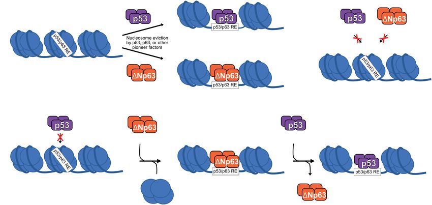
FIGURE 2 | (A) p53 and 1Np63 can act as pioneer factors to evict/remodel nucleosomes and facilitate DNA accessibility, although other pioneer factors are likely
responsible for nucleosome eviction at the majority of p53/1Np63 binding sites. (B) Certain nucleosome contexts, such as when the p53/p63 RE motif is near the
nucleosome dyad, are recalcitrant to p53 and/or 1Np63 pioneer factor activity and remain unbound (Yu and Buck, 2019; Yu et al., 2021). (C) In epithelial cell types,
p53 DNA binding can be facilitated by 1Np630 s pioneer factor activity, although specific biochemical mechanisms are still being fully elucidated.
typically up-regulated by both siblings (Fischer, 2017; Riege sites and diminishes the ability of p53 to activate nearby genes
et al., 2020). Together, these data suggest that the view of (Karsli Uzunbas et al., 2019). These sites are nucleosome rich with
1Np63 functioning as a potent negative regulator of p53 can little to no DNA accessibility in the absence of 1Np63 (Thurman
be rejected and replaced with a context-dependent model where et al., 2012), and p53 does not bind these sites in non-epithelial
1Np63 functions as either a trans-activator or a repressor cell types (Nguyen et al., 2018; Karsli Uzunbas et al., 2019).
depending on cell type and binding location. Future work will Presumably, this is due to the ability of 1Np63 to mediate local
undoubtedly be focused on better defining the context for these chromatin accessibility with its pioneer factor activity (Sammons
opposing activities. et al., 2015; Karsli Uzunbas et al., 2019). Modulation of local and
distal chromatin states, be it to facilitate transcriptional activation
(Fessing et al., 2011; Bao et al., 2015; Li et al., 2019; Catizone
p53 AND p63 – COLLABORATIVE et al., 2020) or repression (Gallant-Behm et al., 2012; Pattison
PARTNERS? et al., 2018) appears to be a key function of 1Np63 and paves the
way for the field to resolve many of the incongruent observations
Although they appear to regulate a mostly unique set of target regarding 1Np630 s influence on p53.
genes and have non-overlapping cellular roles, genetic evidence
suggests that p53 and p63 cooperate to regulate DNA damage-
induced apoptosis in mouse embryonic fibroblasts (Flores et al., OPPOSING DIRECTIONS IN TUMOR
2002). p53 binds to cell cycle arrest target genes like CDKN1A DEVELOPMENT
in the absence of p63, but was unable to interact with promoters
of the pro-apoptotic genes NOXA and BAX (Flores et al., 2002). While current data suggest that p53 and 1Np63 are more likely
The specific molecular mechanisms regulating this apparent to cooperate than to compete at DNA, they remain functionally
collaborative effort for p53 and p63 are still unknown, but quite different. Perhaps most importantly, 1Np63 promotes
the recent identification of 1Np63 as a pioneer transcription while p53 restricts cellular growth. As a consequence, 1Np63 is
factor provides one possibility. p53 genomic binding and gene a key oncogenic driver in squamous cell carcinoma (Campbell
regulatory activity is expanded in epithelial cell types (McDade et al., 2018; Gatti et al., 2019) while p53 is the best-known tumor
et al., 2014; Sammons et al., 2015; Nguyen et al., 2018; suppressor. The context-dependent tumor suppressor role of p63
Karsli Uzunbas et al., 2019). These novel p53 binding sites (Flores et al., 2005; Keyes et al., 2006) appears to be largely
have epithelial cell-specific DNA accessibility, have chromatin reflected by the tumor suppressive function of the TAp63 isoform
modifications associated with active enhancers, and, importantly, that induces apoptosis and senescence (Gressner et al., 2005; Suh
are strongly bound by 1Np63. Inhibition of 1Np63 leads to et al., 2006; Guo et al., 2009). The contrary direction driven by p53
depletion of active transcription-associated hallmarks at these and 1Np63 in tumor development can be explained on the one
Frontiers in Cell and Developmental Biology | www.frontiersin.org 4 July 2021 | Volume 9 | Article 701986Woodstock et al. p53/p63 Sibling Rivalry
hand by their unique target genes. While unique direct 1Np63 The other factors and precise context required for p53 and
target genes encode for several proteins that promote squamous 1Np63 to elicit productive binding to DNA and to regulate
cell cancer growth, inflammation, and invasion (Somerville et al., distinct target genes remain unclear. Although 1Np63 occupies
2018, 2020; Riege et al., 2020), unique p53 target genes encode most sites that can be bound by p53, it appears to affect only
inducers of cell cycle arrest and apoptosis (Fischer, 2017). On the a very small subset of the associated genes. It is unknown how
other hand, there is the large set of cell cycle genes differentially 1Np63 distinguishes between the many sites it activates, the
regulated by p53 and 1Np63 (Riege et al., 2020). p53 employs smaller number of sites it represses, and the majority of sites it
its direct target gene CDKN1A, encoding the cyclin-dependent appears to not affect transcriptionally. Along those lines, when
kinase inhibitor p21, to reactivate the cell cycle trans-repressor and where are p53 and 1Np63 pioneer factors? The context and
complexes DREAM and RB:E2F (Fischer et al., 2016a,b; Schade the extent to which pioneer activity is required for p53 family
et al., 2019; Uxa et al., 2019). While it is not completely function remains an important and active area of investigation.
understood how 1Np63 up-regulates cell cycle genes, it was And how collaborative is their oft-forgotten sibling p73?
suggested to inhibit the p21–p130 (DREAM) axis (Truong et al., Despite beginning their relationship as rivals, p53 and
2006; McDade et al., 2011) and to trans-activate multiple cell cycle 1Np63 appear to cooperate with each other when mutually
genes directly (Riege et al., 2020). We have a detailed picture of beneficial. Identifying the situations when these two transcription
how p53 down-regulates cell cycle genes and sustains cell cycle factors are collaborators and when they are competitors may
arrest (Schade et al., 2019; Uxa et al., 2019). It remains unresolved, provide a blueprint to better understand mechanisms of how
however, whether the regulation of cell cycle genes is cause or transcription factor families that share binding sites and target
consequence of the growth-promoting function of 1Np63, as genes coordinate their efforts.
it is well established that high expression of cell cycle genes is
associated with cancer and worse prognosis (Whitfield et al.,
2006). Together, the unique direct p53 and 1Np63 target genes AUTHOR CONTRIBUTIONS
as well as the differential regulation of cell cycle genes elicited by
p53 and 1Np63 offer a partial, but direct, explanation for their MS and MF conceptualized the manuscript and prepared the
opposing functions in tumor development. figures. DW, MS, and MF performed the literature review,
provided an outline, wrote and edited the manuscript, and
DISCUSSION approved the submitted version. All authors contributed to the
article and approved the submitted version.
Sibling rivalry can happen in any family and it is no different
for the p53 transcription factor family. p63 was within p530 s
considerably large shadow from the beginning, but p63 has FUNDING
started to step into the light with the discoveries of its clear
genetic requirement during development, regulation of a pro- This work was supported by grants from the National Institutes of
epithelial gene network, and pioneer activity. Now, what are Health (R35 GM138120 to MS and T32 GM132066 to DW). The
the key questions that need to be addressed regarding the publication of this manuscript was funded by the Open Access
collaboration and competition between p53 and p63? Fund of the Leibniz Association.
REFERENCES Daino, K., Ichimura, S., and Nenoi, M. (2006). Both the basal transcriptional
activity of the GADD45A gene and its enhancement after ionizing irradiation
Allen, M. A., Andrysik, Z., Dengler, V. L., Mellert, H. S., Guarnieri, A., Freeman, are mediated by AP-1 element. Biochim. Biophys. Acta 1759, 458–469. doi:
J. A., et al. (2014). Global analysis of p53-regulated transcription identifies 10.1016/j.bbaexp.2006.09.005
its direct targets and unexpected regulatory mechanisms. Elife 3:e02200. doi: Davison, T. S., Vagner, C., Kaghad, M., Ayed, A., Caput, D., and Arrowsmith,
10.7554/eLife.02200 C. H. (1999). p73 and p63 Are Homotetramers Capable of Weak Heterotypic
Bao, X., Rubin, A. J., Qu, K., Zhang, J., Giresi, P. G., Chang, H. Y., et al. (2015). A Interactions with Each Other but Not with p53. J. Biol. Chem. 274, 18709–
novel ATAC-seq approach reveals lineage-specific reinforcement of the open 18714. doi: 10.1074/jbc.274.26.18709
chromatin landscape via cooperation between BAF and p63. Genome Biol. Enthart, A., Klein, C., Dehner, A., Coles, M., Gemmecker, G., Kessler, H., et al.
16:284. doi: 10.1186/s13059-015-0840-9 (2016). Solution structure and binding specificity of the p63 DNA binding
Belyi, V. A., Ak, P., Markert, E., Wang, H., Hu, W., Puzio-Kuter, A., et al. (2010). domain. Sci. Rep. 6:26707. doi: 10.1038/srep26707
The origins and evolution of the p53 family of genes. Cold Spring Harb. Perspect. Espinosa, J. M., and Emerson, B. M. (2001). Transcriptional Regulation by
Biol. 2:a001198. doi: 10.1101/cshperspect.a001198 p53 through Intrinsic DNA/Chromatin Binding and Site-Directed Cofactor
Campbell, J. D., Yau, C., Bowlby, R., Liu, Y., Brennan, K., Fan, H., et al. Recruitment. Mol. Cell 8, 57–69. doi: 10.1016/S1097-2765(01)00283-0
(2018). Genomic, Pathway Network, and Immunologic Features Distinguishing Fessing, M. Y., Mardaryev, A. N., Gdula, M. R., Sharov, A. A.,
Squamous Carcinomas. Cell Rep. 23, 194–212.e6. doi: 10.1016/j.celrep.2018.03. Sharova, T. Y., Rapisarda, V., et al. (2011). p63 regulates Satb1 to
063 control tissue-specific chromatin remodeling during development
Catizone, A. N., Uzunbas, G. K., Celadova, P., Kuang, S., Bose, D., and Sammons, of the epidermis. J. Cell Biol. 194, 825–839. doi: 10.1083/jcb.2011
M. A. (2020). Locally acting transcription factors regulate p53-dependent cis- 01148
regulatory element activity. Nucleic Acids Res. 48, 4195–4213. doi: 10.1093/nar/ Fischer, M. (2017). Census and evaluation of p53 target genes. Oncogene 36,
gkaa147 3943–3956. doi: 10.1038/onc.2016.502
Frontiers in Cell and Developmental Biology | www.frontiersin.org 5 July 2021 | Volume 9 | Article 701986Woodstock et al. p53/p63 Sibling Rivalry Fischer, M., Grossmann, P., Padi, M., and DeCaprio, J. A. (2016a). Integration Laptenko, O., Shiff, I., Freed-Pastor, W., Zupnick, A., Mattia, M., Freulich, E., et al. of TP53, DREAM, MMB-FOXM1 and RB-E2F target gene analyses identifies (2015). The p53 C Terminus Controls Site-Specific DNA Binding and Promotes cell cycle gene regulatory networks. Nucleic Acids Res. 44, 6070–6086. doi: Structural Changes within the Central DNA Binding Domain. Mol. Cell 57, 10.1093/nar/gkw523 1034–1046. doi: 10.1016/j.molcel.2015.02.015 Fischer, M., Quaas, M., Steiner, L., and Engeland, K. (2016b). The p53-p21- LeBoeuf, M., Terrell, A., Trivedi, S., Sinha, S., Epstein, J. A., Olson, E. N., et al. DREAM-CDE/CHR pathway regulates G2/M cell cycle genes. Nucleic Acids Res. (2010). Hdac1 and Hdac2 act redundantly to control p63 and p53 functions in 44, 164–174. doi: 10.1093/nar/gkv927 epidermal progenitor cells. Dev. Cell 19, 807–818. doi: 10.1016/j.devcel.2010.10. Fischer, M., Steiner, L., and Engeland, K. (2014). The transcription factor p53: not a 015 repressor, solely an activator. Cell Cycle 13, 3037–3058. doi: 10.4161/15384101. Li, L., Wang, Y., Torkelson, J. L., Shankar, G., Pattison, J. M., Zhen, H. H., 2014.949083 et al. (2019). TFAP2C- and p63-Dependent Networks Sequentially Rearrange Flores, E. R., Sengupta, S., Miller, J. B., Newman, J. J., Bronson, R., Crowley, D., et al. Chromatin Landscapes to Drive Human Epidermal Lineage Commitment. Cell (2005). Tumor predisposition in mice mutant for p63 and p73: evidence for Stem Cell 24, 271–284.e8. doi: 10.1016/j.stem.2018.12.012 broader tumor suppressor functions for the p53 family. Cancer Cell 7, 363–373. Lin, Y.-L., Sengupta, S., Gurdziel, K., Bell, G. W., Jacks, T., and Flores, E. R. (2009). doi: 10.1016/j.ccr.2005.02.019 p63 and p73 transcriptionally regulate genes involved in DNA repair. PLoS Flores, E. R., Tsai, K. Y., Crowley, D., Sengupta, S., Yang, A., McKeon, F., et al. Genet. 5:e1000680. doi: 10.1371/journal.pgen.1000680 (2002). p63 and p73 are required for p53-dependent apoptosis in response to Lin-Shiao, E., Lan, Y., Welzenbach, J., Alexander, K. A., Zhang, Z., Knapp, M., et al. DNA damage. Nature 416, 560–564. doi: 10.1038/416560a (2019). p63 establishes epithelial enhancers at critical craniofacial development Gaiddon, C., Lokshin, M., Ahn, J., Zhang, T., and Prives, C. (2001). A Subset of genes. Sci. Adv. 5:eaaw0946. doi: 10.1126/sciadv.aaw0946 Tumor-Derived Mutant Forms of p53 Down-Regulate p63 and p73 through a Lokshin, M., Li, Y., Gaiddon, C., and Prives, C. (2007). p53 and p73 display Direct Interaction with the p53 Core Domain. Mol. Cell. Biol. 21, 1874–1887. common and distinct requirements for sequence specific binding to DNA. doi: 10.1128/MCB.21.5.1874-1887.2001 Nucleic Acids Res. 35, 340–352. doi: 10.1093/nar/gkl1047 Gallant-Behm, C. L., Ramsey, M. R., Bensard, C. L., Nojek, I., Tran, J., Liu, M., et al. McDade, S. S., Henry, A. E., Pivato, G. P., Kozarewa, I., Mitsopoulos, C., Fenwick, (2012). 1Np63α represses anti-proliferative genes via H2A.Z deposition. Genes K., et al. (2012). Genome-wide analysis of p63 binding sites identifies AP- Dev. 26, 2325–2336. doi: 10.1101/gad.198069.112 2 factors as co-regulators of epidermal differentiation. Nucleic Acids Res. 40, Gatti, V., Fierro, C., Annicchiarico-Petruzzelli, M., Melino, G., and Peschiaroli, 7190–7206. doi: 10.1093/nar/gks389 A. (2019). 1Np63 in squamous cell carcinoma: defining the oncogenic routes McDade, S. S., Patel, D., and McCance, D. J. (2011). p63 maintains keratinocyte affecting epigenetic landscape and tumour microenvironment. Mol. Oncol. 13, proliferative capacity through regulation of Skp2-p130 levels. J. Cell Sci. 124, 981–1001. doi: 10.1002/1878-0261.12473 1635–1643. doi: 10.1242/jcs.084723 Gressner, O., Schilling, T., Lorenz, K., Schulze Schleithoff, E., Koch, A., Schulze- McDade, S. S., Patel, D., Moran, M., Campbell, J., Fenwick, K., Kozarewa, I., et al. Bergkamen, H., et al. (2005). TAp63α induces apoptosis by activating signaling (2014). Genome-wide characterization reveals complex interplay between TP53 via death receptors and mitochondria. EMBO J. 24, 2458–2471. doi: 10.1038/sj. and TP63 in response to genotoxic stress. Nucleic Acids Res. 42, 6270–6285. emboj.7600708 doi: 10.1093/nar/gku299 Guo, X., Keyes, W. M., Papazoglu, C., Zuber, J., Li, W., Lowe, S. W., et al. (2009). Mills, A. A., Zheng, B., Wang, X. J., Vogel, H., Roop, D. R., and Bradley, A. (1999). TAp63 induces senescence and suppresses tumorigenesis in vivo. Nat. Cell Biol. P63 Is a P53 Homologue Required for Limb and Epidermal Morphogenesis. 11, 1451–1457. doi: 10.1038/ncb1988 Nature 398, 708–713. doi: 10.1038/19531 Helton, E. S., Zhu, J., and Chen, X. (2006). The unique NH2-terminally deleted Murray-Zmijewski, F., Lane, D. P., and Bourdon, J.-C. (2006). P53/P63/P73 (DeltaN) residues, the PXXP motif, and the PPXY motif are required for Isoforms: an Orchestra of Isoforms To Harmonise Cell Differentiation and the transcriptional activity of the DeltaN variant of p63. J. Biol. Chem. 281, Response To Stress. Cell Death Differ. 13, 962–972. doi: 10.1038/sj.cdd.440 2533–2542. doi: 10.1074/jbc.M507964200 1914 Iwafuchi-Doi, M., and Zaret, K. S. (2014). Pioneer transcription factors in cell Nemajerova, A., Amelio, I., Gebel, J., Dötsch, V., Melino, G., and Moll, U. M. reprogramming. Genes Dev. 28, 2679–2692. doi: 10.1101/gad.253443.114 (2018). Non-oncogenic roles of TAp73: from multiciliogenesis to metabolism. Iwafuchi-Doi, M., and Zaret, K. S. (2016). Cell fate control by pioneer transcription Cell Death Differ. 25:144. doi: 10.1038/CDD.2017.178 factors. Development 143, 1833–1837. doi: 10.1242/dev.133900 Nguyen, T.-A. T., Grimm, S. A., Bushel, P. R., Li, J., Li, Y., Bennett, B. D., et al. Karsli Uzunbas, G., Ahmed, F., and Sammons, M. A. (2019). Control of p53- (2018). Revealing a human p53 universe. Nucleic Acids Res. 46, 8153–8167. dependent transcription and enhancer activity by the p53 family member p63. doi: 10.1093/nar/gky720 J. Biol. Chem. 294, 10720–10736. doi: 10.1074/jbc.RA119.007965 Ortt, K., and Sinha, S. (2006). Derivation of the consensus DNA-binding sequence Keyes, W. M., Vogel, H., Koster, M. I., Guo, X., Qi, Y., Petherbridge, K. M., for p63 reveals unique requirements that are distinct from p53. FEBS Lett. 580, et al. (2006). p63 heterozygous mutant mice are not prone to spontaneous 4544–4550. doi: 10.1016/j.febslet.2006.07.004 or chemically induced tumors. Proc. Natl. Acad. Sci. U. S. A. 103, 8435–8440. Pattison, J. M., Melo, S. P., Piekos, S. N., Torkelson, J. L., Bashkirova, E., doi: 10.1073/pnas.0602477103 Mumbach, M. R., et al. (2018). Retinoic acid and BMP4 cooperate with p63 King, K. E., Ponnamperuma, R. M., Yamashita, T., Tokino, T., Lee, L. A., to alter chromatin dynamics during surface epithelial commitment. Nat. Genet. Young, M. F., et al. (2003). deltaNp63alpha functions as both a positive and a 50:1658. doi: 10.1038/s41588-018-0263-0 negative transcriptional regulator and blocks in vitro differentiation of murine Perez, C. A., Ott, J., Mays, D. J., and Pietenpol, J. A. (2007). p63 consensus DNA- keratinocytes. Oncogene 22, 3635–3644. doi: 10.1038/sj.onc.1206536 binding site: identification, analysis and application into a p63MH algorithm. Kouwenhoven, E. N., Oti, M., Niehues, H., van Heeringen, S. J., Schalkwijk, J., Oncogene 26, 7363–7370. doi: 10.1038/sj.onc.1210561 Stunnenberg, H. G., et al. (2015). Transcription factor p63 bookmarks and Qu, J., Tanis, S. E. J., Smits, J. P. H., Kouwenhoven, E. N., Oti, M., van den regulates dynamic enhancers during epidermal differentiation. EMBO Rep. 16, Bogaard, E. H., et al. (2018). Mutant p63 Affects Epidermal Cell Identity 863–878. doi: 10.15252/embr.201439941 through Rewiring the Enhancer Landscape. Cell Rep. 25, 3490–3503.e4. doi: Kouwenhoven, E. N., van Heeringen, S. J., Tena, J. J., Oti, M., Dutilh, B. E., 10.1016/j.celrep.2018.11.039 Alonso, M. E., et al. (2010). Genome-Wide Profiling of p63 DNA–Binding Ramsey, M. R., He, L., Forster, N., Ory, B., and Ellisen, L. W. (2011). Sites Identifies an Element that Regulates Gene Expression during Limb Physical association of HDAC1 and HDAC2 with p63 mediates transcriptional Development in the 7q21 SHFM1 Locus. PLoS Genet. 6:e1001065. doi: 10.1371/ repression and tumor maintenance in squamous cell carcinoma. Cancer Res. 71, journal.pgen.1001065 4373–4379. doi: 10.1158/0008-5472.CAN-11-0046 Laptenko, O., Beckerman, R., Freulich, E., and Prives, C. (2011). p53 binding to Riege, K., Kretzmer, H., Sahm, A., McDade, S. S., Hoffmann, S., and nucleosomes within the p21 promoter in vivo leads to nucleosome loss and Fischer, M. (2020). Dissecting the DNA binding landscape and gene transcriptional activation. Proc. Natl. Acad. Sci. U. S. A. 108, 10385–10390. regulatory network of p63 and p53. Elife 9:e63266. doi: 10.7554/eLife. doi: 10.1073/pnas.1105680108 63266 Frontiers in Cell and Developmental Biology | www.frontiersin.org 6 July 2021 | Volume 9 | Article 701986
Woodstock et al.                                                                                                                                            p53/p63 Sibling Rivalry

Rutkowski, R., Hofmann, K., and Gartner, A. (2010). Phylogeny and Function of                 Venkatachalam, S., Shi, Y. P., Jones, S. N., Vogel, H., Bradley, A.,
   the Invertebrate p53 Superfamily. Cold Spring Harb. Perspect. Biol. 2:a001131.                Pinkel, D., et al. (1998). Retention of wild-type p53 in tumors
   doi: 10.1101/cshperspect.a001131                                                              from p53 heterozygous mice: reduction of p53 dosage can promote
Sahu, G., Wang, D., Chen, C. B., Zhurkin, V. B., Harrington, R. E., Appella, E., et al.          cancer formation. EMBO J. 17, 4657–4667. doi: 10.1093/emboj/17.
   (2010). p53 binding to nucleosomal DNA depends on the rotational positioning                  16.4657
   of DNA response element. J. Biol. Chem. 285, 1321–1332. doi: 10.1074/jbc.                  Westfall, M. D., Mays, D. J., Sniezek, J. C., and Pietenpol, J. A. (2003). The
   M109.081182                                                                                   Np63 Phosphoprotein Binds the p21 and 14-3-3 Promoters In Vivo and Has
Sammons, M. A., Nguyen, T.-A. T., McDade, S. S., and Fischer, M. (2020). Tumor                   Transcriptional Repressor Activity That Is Reduced by Hay-Wells Syndrome-
   suppressor p53: from engaging DNA to target gene regulation. Nucleic Acids                    Derived Mutations. Mol. Cell. Biol. 23, 2264–2276. doi: 10.1128/MCB.23.7.
   Res. 48, 8848–8869. doi: 10.1093/nar/gkaa666                                                  2264-2276.2003
Sammons, M. A., Zhu, J., Drake, A. M., and Berger, S. L. (2015). TP53 engagement              Whitfield, M. L., George, L. K., Grant, G. D., and Perou, C. M. (2006). Common
   with the genome occurs in distinct local chromatin environments via pioneer                   markers of proliferation. Nat. Rev. Cancer 6, 99–106.
   factor activity. Genome Res. 25, 179–188. doi: 10.1101/gr.181883.114                       Yang, A., Kaghad, M., Wang, Y., Gillett, E., Fleming, M. D., Dötsch, V., et al.
Sauer, M., Bretz, A. C., Beinoraviciute-Kellner, R., Beitzinger, M., Burek, C.,                  (1998). P63, a P53 Homolog At 3Q27–29, Encodes Multiple Products With
   Rosenwald, A., et al. (2008). C-terminal diversity within the p53 family accounts             Transactivating, Death-Inducing, and Dominant-Negative Activities. Mol. Cell
   for differences in DNA binding and transcriptional activity. Nucleic Acids Res.               2, 305–316. doi: 10.1016/S1097-2765(00)80275-0
   36, 1900–1912. doi: 10.1093/nar/gkn044                                                     Yang, A., Schweitzer, R., Sun, D., Kaghad, M., Walker, N., Bronson, R. T., et al.
Schade, A. E., Fischer, M., and DeCaprio, J. A. (2019). RB, p130 and p107                        (1999). p63 is essential for regenerative proliferation in limb, craniofacial and
   differentially repress G1/S and G2/M genes after p53 activation. Nucleic Acids                epithelial development. Nature 398, 714–718. doi: 10.1038/19539
   Res. 47, 11197–11208. doi: 10.1093/nar/gkz961                                              Yang, A., Zhu, Z., Kapranov, P., McKeon, F., Church, G. M., Gingeras, T. R.,
Somerville, T. D., Biffi, G., Daßler-Plenker, J., Hur, S. K., He, X.-Y., Vance, K. E.,           et al. (2006). Relationships between p63 Binding, DNA Sequence, Transcription
   et al. (2020). Squamous trans-differentiation of pancreatic cancer cells promotes             Activity, and Biological Function in Human Cells. Mol. Cell 24, 593–602. doi:
   stromal inflammation. Elife 9:e53381. doi: 10.7554/eLife.53381                                10.1016/j.molcel.2006.10.018
Somerville, T. D. D., Xu, Y., Miyabayashi, K., Tiriac, H., Cleary, C. R., Maia-               Younger, S. T., and Rinn, J. L. (2017). p53 regulates enhancer accessibility and
   Silva, D., et al. (2018). TP63-Mediated Enhancer Reprogramming Drives the                     activity in response to DNA damage. Nucleic Acids Res. 45, 9889–9900. doi:
   Squamous Subtype of Pancreatic Ductal Adenocarcinoma. Cell Rep. 25, 1741–                     10.1093/nar/gkx577
   1755.e7. doi: 10.1016/j.celrep.2018.10.051                                                 Yu, X., and Buck, M. J. (2019). Defining TP53 pioneering capabilities with
Suh, E.-K., Yang, A., Kettenbach, A., Bamberger, C., Michaelis, A. H., Zhu, Z., et al.           competitive nucleosome binding assays. Genome Res. 29, 107–115. doi: 10.1101/
   (2006). p63 protects the female germ line during meiotic arrest. Nature 444,                  gr.234104.117
   624–628. doi: 10.1038/nature05337                                                          Yu, X., Singh, P. K., Tabrejee, S., Sinha, S., and Buck, M. J. (2021). 1Np63
Sullivan, K. D., Galbraith, M. D., Andrysik, Z., and Espinosa, J. M. (2018).                     is a pioneer factor that binds inaccessible chromatin and elicits chromatin
   Mechanisms of transcriptional regulation by p53. Cell Death Differ. 25, 133–143.              remodeling. Epigenetics Chromatin 14:20. doi: 10.1186/s13072-021-00394-8
   doi: 10.1038/cdd.2017.174                                                                  Zaret, K. S., and Mango, S. E. (2016). Pioneer transcription factors, chromatin
Thurman, R. E., Rynes, E., Humbert, R., Vierstra, J., Maurano, M. T., Haugen, E.,                dynamics, and cell fate control. Curr. Opin. Genet. Dev. 37, 76–81. doi: 10.1016/
   et al. (2012). The accessible chromatin landscape of the human genome. Nature                 j.gde.2015.12.003
   489, 75–82. doi: 10.1038/nature11232
Tichý, V., Navrátilová, L., Adámik, M., Fojta, M., and Brázdová, M. (2013). Redox             Conflict of Interest: The authors declare that the research was conducted in the
   state of p63 and p73 core domains regulates sequence-specific DNA binding.                 absence of any commercial or financial relationships that could be construed as a
   Biochem. Biophys. Res. Commun. 433, 445–449. doi: 10.1016/j.bbrc.2013.02.097               potential conflict of interest.
Truong, A. B., Kretz, M., Ridky, T. W., Kimmel, R., and Khavari, P. A. (2006).
   p63 regulates proliferation and differentiation of developmentally mature                  Copyright © 2021 Woodstock, Sammons and Fischer. This is an open-access article
   keratinocytes. Genes Dev. 20, 3185–3197. doi: 10.1101/gad.1463206                          distributed under the terms of the Creative Commons Attribution License (CC BY).
Uxa, S., Bernhart, S. H., Mages, C. F. S., Fischer, M., Kohler, R., Hoffmann, S.,             The use, distribution or reproduction in other forums is permitted, provided the
   et al. (2019). DREAM and RB cooperate to induce gene repression and cell-                  original author(s) and the copyright owner(s) are credited and that the original
   cycle arrest in response to p53 activation. Nucleic Acids Res. 47, 9087–9103.              publication in this journal is cited, in accordance with accepted academic practice. No
   doi: 10.1093/nar/gkz635                                                                    use, distribution or reproduction is permitted which does not comply with these terms.

Frontiers in Cell and Developmental Biology | www.frontiersin.org                         7                                                  July 2021 | Volume 9 | Article 701986
You can also read