Circulating Vitamin D Levels and DNA Repair Capacity in Four Molecular Subtypes of Women with Breast Cancer - MDPI
←
→
Page content transcription
If your browser does not render page correctly, please read the page content below
International Journal of
            Molecular Sciences

Article
Circulating Vitamin D Levels and DNA Repair
Capacity in Four Molecular Subtypes of Women with
Breast Cancer
Carmen Ortiz-Sánchez 1, * , Jarline Encarnación-Medina 1 , Ralphdy Vergne 2 , Luis Padilla 3
and Jaime Matta 1
 1    Department of Basic Sciences, Ponce Research Institute, Ponce Health Sciences University-School of
      Medicine, Ponce, PR 00716, USA; jencarnacion@psm.edu (J.E.-M.); jmatta@psm.edu (J.M.)
 2    Biomedical Sciences Department, Interamerican University of Puerto Rico, Ponce, PR 00715, USA;
      ralphdy.vergne@upr.edu
 3    Biology Department, University of Puerto Rico at Ponce, Ponce, PR 00716, USA; luis.padilla7@upr.edu
 *    Correspondence: carmenortiz@psm.edu; Tel.: +1-787-840-2575 (ext. 2197)

 Received: 19 August 2020; Accepted: 18 September 2020; Published: 19 September 2020

 Abstract: Vitamin D regulates estrogen synthesis among other mechanisms involved in breast cancer
 (BC) development; however, no evidence has been found regarding its relationship with DNA
 repair capacity (DRC). Therefore, the objective of this study was to elucidate whether DRC levels
 are linked with plasma 25(OH)D levels. BC cases and controls were selected from our BC cohort.
 DRC levels were assessed in lymphocytes through the host-cell reactivation assay. 25(OH)D levels
 were measured using the UniCel DxI 600 Access Immunoassay System. BC cases (n = 91) showed
 higher 25(OH)D levels than the controls (n = 92) (p = 0.001). When stratifying BC cases and controls
 into low and high DRC categories, BC cases with low DRC (n = 74) had the highest 25(OH)D levels
 (p = 0.0001). A positive correlation between 25(OH)D and DRC levels was found for the controls
 (r = 0.215, p = 0.043) while a negative correlation was found for BC cases (r = −0.236, p = 0.026).
 Significant differences in 25(OH)D levels were observed when stratifying by molecular subtypes
 (p = 0.0025). Our study provides evidence of a link between 25(OH)D and DRC in BC along with
 a description of to how 25(OH)D levels vary across subtypes. The positive correlation observed in
 the control group suggests that 25(OH)D contributes differently to DRC levels once the malignancy
 is developed.

 Keywords: plasma 25-hydroxyvitamin D (25(OH)D); vitamin D; breast cancer; DNA repair capacity;
 host-cell reaction assay; nucleotide excision repair; molecular subtypes

1. Introduction
      Worldwide, breast cancer (BC) accounts for nearly a quarter of all cancers in women [1]. About 1
in 8 women are expected to be diagnosed with BC. This accounts for 268,600 new cases of invasive
BC in women in the U.S. only, including Hispanics [2,3]. For the last three decades, the use of
vitamins, multivitamins, and supplements (i.e., calcium) has been attractive for BC prevention or
as adjuvant treatment for this disease [4,5]. Vitamin D has different roles in cancer and healthy
states. Previous studies have shown that vitamin D plays an important role in preventing the tumor
initiation through anti-inflammatory and antioxidant defense mechanisms and DNA damage repair
processes [6]. This highlights the role of vitamin D in cancer prevention. However, inconsistencies in
study results hinder a clear understanding about whether vitamin supplementation could be beneficial
once BC is developed [7,8]. A case–control study by Vergne et al., 2013 suggests that multivitamin
supplementation could be an independent protective factor for BC. However, when other individual

Int. J. Mol. Sci. 2020, 21, 6880; doi:10.3390/ijms21186880                          www.mdpi.com/journal/ijms
Int. J. Mol. Sci. 2020, 21, 6880 2 of 13
vitamins were taken into consideration (i.e., vitamins A, E, and C), no positive results were found.
Interestingly, this study also found an association between calcium intake and having high overall
DNA repair capacity (DRC) levels measured through the nucleotide excision repair (NER) pathway [9].
This pathway is one of the various DNA repair mechanisms responsible for maintaining the genomic
stability and preventing alterations to the DNA that could potentially lead to cancer [10]. Therefore,
it is not surprising that defective DNA repair, measured in lymphocytes, has been identified as a
risk factor for different types of cancer [11–13], including BC [14]. Variability on DRC levels has been
reported among the four principal molecular BC subtypes, where the triple-negative subtype showed
the lowest levels [15].
Breast tumors may (+) or may not (−) have three hormonal receptors: estrogen (ER), progesterone
(PR), and HER2 (human epidermal growth factor receptor 2). Based on their hormonal receptor status,
four principal molecular BC subtypes have been identified: luminal A (ER+, PR+, HER2−), luminal B
(ER+, PR+, HER2+), HER2+ (ER−, PR−, HER2+), and triple-negative (TN) (ER−, PR−, HER2−).
Various studies have aimed at elucidating the role of vitamin D (25-hydroxyvitamin D (25(OH)D))
in BC [16–18] and more specifically among molecular subtypes. Low serum 25(OH)D levels have been
associated with aggressive phenotypes and worse prognosis in several molecular BC subtypes [19].
A substantial amount of studies have shown discrepancies regarding vitamin D supplementation
and its role in BC based on in vitro [20,21], in vivo [22,23], and clinical studies [18,24]. Other studies
have reported an association between higher 25(OH)D levels and a lower BC risk but these findings have
been inconsistent [8,25]. As part of its many physiological functions, vitamin D signaling influences
estrogen synthesis. Since BC is known to be a hormonal cancer, due to its dependence on estrogen to
promote cell proliferation [26], there is a need to understand the effects of vitamin D in women with
BC [27,28]. Since previous studies from our laboratory established an association between DRC levels
and ER positivity in women with BC, the main aim of this study was to elucidate whether DRC is
linked with plasma 25(OH)D levels. A secondary aim was to understand whether this effect can be
observed across the four principal BC subtypes. In general, we expected to observe differences in
vitamin D levels between women with and without BC. We hypothesize that 25(OH)D levels will vary
among women with different molecular BC subtypes and that women with ER+ breast tumors will
present distinct plasma vitamin D levels when compared to women with ER− tumors.
2. Results
2.1. Plasma 25(OH)D Levels
Overall, BC cases included women in the age range from 41 to 60 years of age with high DRC
levels, and a BMI over 25 kg/m2 who were not undergoing menopause. Controls were similar to
BC cases regarding age and BMI (Table 1). In terms of DRC levels, most of the BC cases had low
DRC levels using the previously established cut-off of low (Int. J. Mol. Sci. 2020, 21, 6880 3 of 13
Pairwise comparisons were performed to evaluate the mean differences among groups (Table 3).
Controls with high DRC levels had 5.54 ng/mL above the mean 25(OH)D levels when compared
to controls with low DRC levels (p < 0.05). The mean 25(OH)D levels for BC cases with high DRC
levels was 7.77 ng/mL less than BC cases with low DRC levels (p < 0.05). Cases with high DRC levels
had lower 25(OH)D levels when compared to the controls independently of DRC levels. However,
significant differences were found when comparing BC cases high DRC with controls also with high
DRC levels (p < 0.05).
Table 1. Description of the study population including women with and without breast cancer.
Controls BC Cases
Variables p-Value 1
n = 92 (50.27%) n = 91 (49.73%)
DRCInt. J. Mol. Sci. 2020, 21, 6880 4 of 13
Table 2. Mean 25(OH)D level comparisons among women with and without breast cancer with low
and high DNA repair capacity.
25(OH)D (ng/mL) 25(OH)D (ng/mL) 95% Confidence Interval
Stratifications p-Value
(Mean ± SD) a (Mean ± SD) * Lower Bound Upper Bound
Controls 38.10 (9.95) 38.55 (1.03) 36.51 40.59
0.123 a , 0.001 *
BC Cases 41.12 (10.25) 40.82 (1.03) 38.79 42.85
Controls LDRC 35.41 (10.46) 35.74(1.40) 32.98 38.43
0.0001 a , 0.0001*
Controls HDRC 41.18 (8.43) 41.27 (1.41) 38.49 44.05
BC Cases
42.55 (10.25) 42.36 (1.10) 40.18 44.53
LDRC
BC Cases
34.90 (7.78) 34.59 (2.30) 30.06 39.13
HDRC
aCrude mean comparisons; * adjusted by age and BMI. BC: breast cancer; LDRC: low DNA repair capacity; HDRC:
high DNA repair capacity.
Table 3. Differences in mean 25(OH)D levels among women with and without breast cancer with low
and high DNA repair capacity.
Mean 25(OH)D 95% Confidence Interval for
Stratifications Difference Significance b Difference b
(ng/mL) Lower Bound Upper Bound
Controls HDRC −5.54 * 0.006 −9.45 −1.63
Controls LDRC BC cases LDRC −6.62 * 0.0001 −10.14 −3.10
BC cases HDRC 1.14 0.671 −4.17 6.45
Controls LDRC 5.54 * 0.006 1.63 9.45
Controls HDRC BC cases LDRC −1.09 0.545 −4.62 2.45
BC cases HDRC 6.68 * 0.014 1.36 12.00
Controls LDRC 6.62 * 0.0001 3.10 10.14
BC cases LDRC Controls HDRC 1.09 0.545 −2.45 4.62
BC cases HDRC 7.77 * 0.003 2.74 12.80
Controls LDRC −1.14 0.617 −6.45 4.17
BC cases HDRC Controls HDRC −6.68 * 0.014 −12.00 −1.36
BC cases LDRC −7.77 * 0.003 −12.80 −2.74
Based on estimated marginal means. * 25(OH)D mean difference is significant at the 0.05 level; b adjustment
for multiple comparisons: Least Significant Difference (equivalent to no adjustments). BC: breast cancer; LDRC:
low DNA repair capacity; HDRC: high DNA repair capacity.
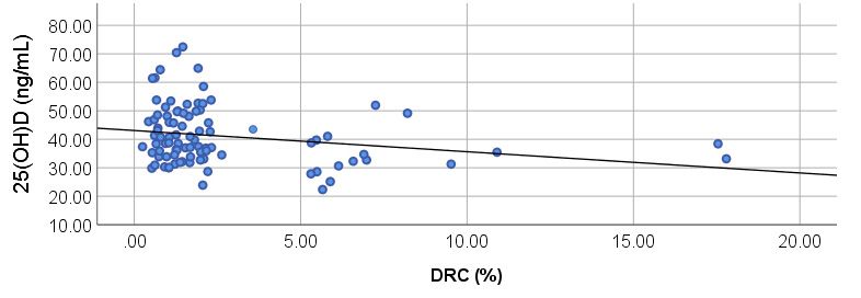
2.2. Correlation between 25(OH)D and DRC Levels
To study the relationship between 25(OH)D levels and DRC, crude and adjusted partial lineal
correlation analyses were performed (Table 4). The partial correlation Model 1 shows a positive
correlation of 0.215 for the controls (Figure 1A) (p = 0.040). For the BC cases group (model 2), a negative
correlation was found with a coefficient of −0.236 (p = 0.030) (Figure 1B). After adjusting for confounders
in both models, a statistical significance was still detected, showing a relationship between DRC and
25(OH)D levels (Table 4).
Table 4. Correlation analyses between plasma 25(OH)D and DNA repair capacity levels in women
with and without breast cancer.
R2 Testing Significance
Stratifications
Model 1 Model 2 p-Value a p-Value *
Controls 0.215 - 0.040 0.043
BC Cases - −0.236 0.030 0.026
a Crude mean comparisons; * adjusted by age and BMI. BC: breast cancer.R2 Testing Significance
Stratifications
Model 1 Model 2 p-value a p-value *
Controls 0.215 - 0.040 0.043
Int. J. Mol. Sci. 2020, 21, 6880 BC Cases - −0.236 0.030 0.026 5 of 13
a Crude mean comparisons; * adjusted by age and BMI. BC: breast cancer
(a)
(b)
Figure 1. Correlations between DNA repair capacity (DRC) and 25(OH)D levels in breast cancer cases
and controls.
Figure Partial correlation
1. Correlations between DNA analyses were
repair performed
capacity (DRC)to test25(OH)D
and any correlations between
levels in breast DRCcases
cancer (%)
and
andplasma vitamin
controls. PartialDcorrelation
levels in (a)analyses = 0.0215,
controls (rwere p = 0.043)
performed andany
to test (b) BC cases (r = −0.236;
correlations = 0.026).
betweenp DRC (%)
and plasma vitamin
2.3. Relationship betweenDMolecular
levels in (a) controls
Breast (r = 0.0215,
Cancer Subtypesp =and
0.043) and (b) BC
25(OH)D cases (r = −0.236; p = 0.026).
Levels
To further elucidate the relevance of our results, additional analyses were performed to evaluate
whether plasma 25(OH)D levels vary depending on the molecular subtype of the tumor. Since previous
studies from our research team showed that the DRC level distribution is positively skewed in BC
cases [15], we focused our analysis on BC cases with low DRC levels only. Mean 25(OH)D levels were
compared among women with breast tumors that were classified as luminal A, luminal B, HER2+,
and TN. Controls were also included on these analyses.
Crude analysis showed significant differences in 25(OH)D levels when stratifying by molecular
subtypes: Luminal A (n = 17), Luminal B (n = 11), HER2+ (n = 9), and TN (n = 17); and comparing
with low DRC controls (n =46) (p = 0.0025) (Table 5). Statistical significance increased after adjustment
by age and BMI (p = 0.001), BC cases with HER2+ tumors showed the highest 25(OH)D levels
(47.70 ± 3.14 ng/mL) followed by BC cases with TN tumors (45.08 ± 2.24 ng/mL). Luminal B BC
cases had a mean 25(OH)D concentration of 40.50 ± 2.78 ng/mL while luminal A BC cases had
38.73 ± 2.26 ng/mL. Controls showed the lowest 25(OH)D levels with 35.51 ± 1.36 ng/mL.
When exploring the mean differences in 25(OH)D levels among groups, significant differences
were observed between controls and cases with HER2+ and TN BC. Controls had 12.19 ng/mL and
9.57 ng/mL less than HER2+ (p = 0.001) and TN (p = 0.0001) BC cases, respectively (Table 6). BC cases
with luminal A tumors had 8.98 ng/mL less 25(OH)D levels than HER2+ BC cases (p = 0.025). Therefore,
our results show that vitamin D levels vary among molecular BC subtypes. In addition, our results
show that ER negative subtypes have the highest 25(OH)D levels.Int. J. Mol. Sci. 2020, 21, 6880 6 of 13
Table 5. Mean 25(OH)D level comparisons among women with low DNA repair capacity levels
stratified by molecular subtypes.
25(OH)D (ng/mL) 25(OH)D (ng/mL) 95% Confidence Interval
Stratifications p-Value
(Mean ± SD) a (Mean ± SD) * Lower Bound Upper Bound
Controls 38.41 (10.46) 35.51 (1.36) 32.81 38.211
0.0025 a , 0.001 *
Luminal A 38.46 (6.79) 38.73 (2.26) 34.23 43.22
Luminal B 40.99 (8.22) 40.50(2.78) 34.98 46.03
HER2+ 48.83 (11.34) 47.70 (3.14) 41.46 53.94
Triple-negative 44.71 (10.26) 45.08 (2.24) 40.62 49.53
a Crude mean comparisons; * adjusted by age and BMI.
Table 6. Differences in mean 25(OH)D levels among women with BC with low DRC with different
molecular subtypes.
95% Confidence Interval for
Stratifications Mean 25(OH)D Difference Significance b Difference b
Lower Bound Upper Bound
Luminal A 3.21 0.225 −8.45 2.01
Luminal B −4.99 0.110 −11.14 1.16
Controls
HER2+ −12.19 * 0.001 −19.01 −5.38
TN −9.57 * 0.0001 −14.78 −4.36
Controls 3.21 0.225 −2.02 8.45
Luminal B −1.78 0.621 −8.90 5.34
Luminal A
HER2+ −8.98 * 0.025 −16.78 −1.18
TN −6.35 0.050 −12.71 0.002
Controls 4.99 0.110 −1.16 11.14
Luminal A 1.78 0.621 −5.34 8.90
Luminal B
HER2+ −7.20 0.089 −15.52 1.13
TN −4.58 0.204 −11.68 2.53
Controls 12.19 * 0.001 5.38 19.05
Luminal A 8.98 * 0.025 1.18 16.78
HER2+
Luminal B 7.20 0.089 −1.13 15.53
TN 2.63 0.497 −5.01 10.26
Controls 9.57 * 0.0001 4.36 14.78
Luminal A 6.35 0.050 −0.002 12.71
TN
Luminal B 4.58 0.204 −2.53 11.68
HER2+ −2.63 0.497 −10.26 5.01
Based on estimated marginal means. * 25(OH)D mean difference is significant at the 0.05 level; b adjustment for
multiple comparisons: Least Significant Difference (equivalent to no adjustments).
3. Discussion
Vitamin D is a molecule with pleiotropic functions. This study adds a new functional dimension
to 25(OH)D, the major circulating form of vitamin D, namely, the link between its plasma levels and
DRC levels in women with BC. This study also presents the differences in 25(OH)D levels across the
four principal molecular BC subtypes in Puerto Rican women. Our study is the first, to our knowledge,
in aiming to elucidate the relationship between 25(OH)D and DRC using lymphocytes as surrogate
markers for the participants’ DRC specifically through the NER pathway. Interestingly, our results
show plasma 25(OH)D levels vary significantly in women with and without BC. Moreover, BC cases
with low DRC levels had higher levels of 25(OH)D when compared to BC cases with high DRC
levels. This finding is further confirmed by the negative partial correlation found between DRC and
25(OH)D levels in BC cases. Of the relationship between plasma 25(OH)D levels and DNA repair,
little is known. A study by Gonzalez-Suarez et al. (2011) provided the first evidence of a link between
vitamin D and double-strand break DNA repair [31], showing that calcitriol (biologically-active form
of vitamin D) could stabilize 53BP1 (p53 binding protein 1) levels in human cells. 53BP1 has beenInt. J. Mol. Sci. 2020, 21, 6880 7 of 13
recently highlighted as a novel target for BRCA1-deficient TN BC [32]. Moreover, Gonzalo (2014)
presented a novel sensitization strategy for TN BC by taking advantage of the calcitriol-mediated
53BP1 stabilization [32].
As for DNA damage, Wang et al. (2016) evaluated the connection between oxidative damage
and vitamin D levels in a group of non-obese, non-smoking young adults; however, no association
was found [33]. Interestingly, our results show a positive correlation between 25(OH)D levels and
DRC levels for the controls. This could support our hypothesis that 25(OH)D levels change once the
malignancy is developed. Moreover, adding to the benefits of maintaining optimal levels of vitamin D.
As of today, no studies linking single-strand break repair and 25(OH)D have been performed.
Our results show a significant difference in vitamin D levels between treatment-naïve BC cases
and controls at the time of diagnosis. Previous studies report different outcomes for this comparison
such as the study by Abbas et al. (2009) in a German BC case–control cohort, in which plasma 25(OH)D
levels were 18.2 ng/mL and 20.5 ng/mL for cases and controls, respectively. Although the BC cases were
described as recently diagnosed, the difference between time of diagnosis and blood collection was
189 days [34]. Similar trends have been reported by several groups [16,19,35,36]; however, we have
identified several factors that could potentially explain our results. First, most of the studies regarding
25(OH)D and BC have been performed in White women with none (or a limited number) considering
Hispanics [16,19,24,37]. This is important since previous studies have documented that ethnicity or
race may influence the association between vitamin D levels and BC risk [38]. Since UV radiation is
the main source for vitamin D synthesis, the geographical location of the study population is crucial
in order to establish comparisons among studies. Although most of the vitamin D studies in BC
have been performed in the US, other studies have been performed in Korean [38], German [34],
Japanese [39], and French [35] women cohorts. As of today, few studies have been performed in
Hispanic populations [40,41] and none including Puerto Rican women with BC. Few studies had been
performed to measure the plasma vitamin D levels in the Puerto Rican population [42–44]; however,
the largest study was performed by Suárez-Martínez et al. (2013). In this study, the laboratory test
results of 4090 individuals who had vitamin D levels measured at a reference laboratory located in
Puerto Rico’s metropolitan area were analyzed. This group reported that around 32% of the studied
population was vitamin D sufficient (>30 ng/mL) [42]. Of this study group 3414 of the participants were
women with a mean vitamin D concentration of 26.9 ± 12.3 ng/mL. Although the Suárez-Martinez’s
study provides the first report of plasma vitamin D levels in Puerto Rico, the lack of relevant participants’
information does not allow for a clear comparison with our study. First, no exclusion criteria were
established. No medical information was collected; therefore, there is no assurance of the health status
of the study participants. In addition, no epidemiological information was collected and only six
municipalities (out of 78) of are represented by their findings whereas our study includes participants
from almost 36 municipalities.
Another important consideration is the timing of blood sample collection in relation to BC
diagnosis [37]. In most of the studies, the time between blood collection and diagnosis ranges between
2 months and years [16,37,45,46]. In our study, the blood collection was performed at the time of
diagnosis which allowed us to avoid any confounding effects due to BC treatment. As previously
reported, BC therapy can alter plasma vitamin D levels [47,48]. It has been reported that after 6 months
of chemotherapy, vitamin D levels can be reduced to almost 5.52 ng/mL (p = 0.003). In contrast, patients
receiving anti-hormone therapy showed an increase in vitamin D levels at 6 months and 12 months
(+3.00 ng/mL and +6.47 ng/mL, respectively) [47].
Our results show that plasma 25(OH)D levels are significantly different across the four
principal molecular BC subtypes, with ER negative subtypes having the highest levels. The work
of Peppone et al. (2012) is one of the few studies that aimed to elucidate the relationship of plasma
vitamin D among molecular BC subtypes. This group found that women with basal-like tumors
had lower vitamin D levels than women with luminal A tumors with 24.2 ng/mL and 32.8 ng/mL,
respectively (p = 0.04) [19]. In contrast with our findings, this group found that women with ER+Int. J. Mol. Sci. 2020, 21, 6880 8 of 13
tumors had higher vitamin D levels than women with ER− tumors. This group also found significant
differences in plasma vitamin D levels among the four molecular subtypes; however, in contrast with
our findings, they found that ER− subtypes had the lowest vitamin D concentrations [19].
This study provides the first evidence of a link between plasma 25(OH)D and DRC levels in
Hispanic women with and without BC. This study also shows the variation in 25(OH)D levels across
the four principal molecular BC subtypes in this population. Moreover, the positive correlation
observed in the control group suggests that circulating 25(OH)D contributes differently to the DRC
levels once the malignancy is developed. This finding adds to the known benefits of maintaining
optimal vitamin D levels in women without BC. Since having a low DRC level has been associated
with an increased BC risk, maintaining optimal vitamin D levels could be viewed as a potential tool for
chemoprevention. On the other hand, once the malignancy is developed, our results show that vitamin
D levels become altered and that there are variations among molecular subtypes. Various studies
suggest that a different approach regarding vitamin use is recommended for cancer patients. A recent
study by Ambrose et al. (2020), highlights that the use of multivitamin and antioxidants before and
during chemotherapy could have a negative impact on recurrence and overall survival in women with
BC [49]. Therefore, our study adds to the overall knowledge regarding vitamin D levels in recently
diagnosed treatment-naïve women with BC. Our results also provide new insights on the role of
25(OH)D in DRC levels in women with BC while opening new avenues for mechanistic studies to
study this effect.
4. Materials and Methods
4.1. Patient Recruitment
This study was approved by the Ponce Health Sciences University Institutional Review Board
(IRB #120207-JM renewed on 24 October 2019). Informed consent and blood samples were collected by
the study nurse along with epidemiological data through a questionnaire. Participants were selected
from our BC cohort of Puerto Rican women recruited from 2006 to 2013 [50]. Women without BC
(controls) were required to have a normal breast examination performed by a primary care physician
and a normal mammography 6 months prior to study enrolment. The inclusion criteria for women
with BC (cases) were recently diagnosed, treatment-naïve (had not received blood transfusions,
chemotherapy, or radiotherapy) patients with primary breast tumors. Pathology reports from BC
cases were obtained to confirm the diagnosis, and collect clinicopathological variables such as: tumor
grade, tumor size, and other clinically relevant information. For this study, we established an exclusion
criteria based on previous studies including: intake of vitamin D, vitamin E, vitamin C, calcium,
steroids, cortisone, hormone replacement therapy [51], insulin, or any immune repressors [52].
4.2. Hormone Receptor Status
Medical records from women with BC included in this study were reviewed to collect receptor
status data (ER, PR, and HER2). Receptor status was assessed on the participants’ formalin-fixed
tumors using immunohistochemistry (IHC) according to ASCO (American Society of Clinical Oncology)
and CAP (College of American Pathologists) guidelines [53,54] in 10 private laboratories in Puerto
Rico. Information regarding ER and PR status included: percentage of positive-staining cells, intensity
of staining (weak, moderate, or strong), and a result interpretation based on the percentage of invasive
tumor cells that were positively stained for ER/PR (where ≥1% meant “receptor positive” andInt. J. Mol. Sci. 2020, 21, 6880 9 of 13
4.3. DNA Repair Capacity (DRC) Measurements
DRC was assessed in the participants’ lymphocytes through the host-cell reactivation (HCR) assay
with a luciferase reporter gene, as previously published [50]. Lymphocytes were used as surrogate
markers of the participants’ DRC [55,56]. At the moment of recruitment, peripheral blood samples
(30 mL) were collected in heparinized tubes from each participant to obtain lymphocytes that were
assayed in batches. Lymphocytes with >95% viability were incubated for 72 h with phytohemagglutinin
and then transfected with undamaged or UVC light damaged plasmid DNA. After transfection,
repair-transcription-blocking damage was introduced exogenously on foreign DNA; then DRC was
measured via HCR [57]. The HCR allows for a direct measurement of in vivo DRC. This approach
measured the unaffected phenotype, which reflects the cells’ inherent DRC, measured primarily in
terms of their NER activity [57]. Cells isolated from Xeroderma pigmentosum patients corresponding
to complementation groups C and D were used as internal controls (GM02246D and GM02253F,
respectively; Coriell Institute Medical Research; Camden, NJ, USA).
4.4. Calculation of DRC
Gene expression of luciferase activity was measured using a luminometer (Turner Designs,
model TD-20/20, Sunnyvale, CA, USA). DRC was calculated as the percentage of luciferase activity
present after damaged plasmid DNA repair, compared to the undamaged plasmid DNA repair (100%).
Results were expressed as percentage of residual luciferase reporter gene expression (% luciferase
activity in luminescence units).
4.5. Plasma 25-Hydroxyvitamin D Levels
Plasma samples obtained from peripheral blood were stored in aliquots (−80 ◦ C) until vitamin D
levels were measured in a single batch in October 2017. Vitamin D levels were measured using the
UniCel DxI 600 Access Immunoassay System (Beckman Coulter, CA, USA) through the Access 25(OH)
Vitamin D Total Assay which detects both 25(OH)vitamin D2 and 25(OH)vitamin D3. For this study,
we used the 25(OH)D as an indicator of vitamin D levels in plasma. The analysis was performed at
Laboratorios Ramírez, a CLIA-certified laboratory located in Ponce, Puerto Rico.
4.6. Statistical Analyses
Demographic and clinicopathological variables were analyzed using contingency tables and
chi-square tests. Yates’ correction was applied when the number of observations was less than 5.
Mean 25(OH)D comparisons between and among groups were performed using an independent t
test and analysis of covariance (ANCOVA) adjusting for potential cofounders including age and BMI.
Pairwise comparisons were performed to detect differences in 25(OH)D after adjustment for age and
BMI. To assess the relationship of the DRC on the 25(OH)D levels, partial lineal correlation analyses
were performed also adjusting by age and BMI. Significance levels were established using a p-value
cutoff of 0.05 based on a two-tail test. The data were analyzed using SPSS 25.0 software (Chicago,
IL, USA).
Author Contributions: Conceptualization, C.O.-S. and J.E.-M.; data curation, C.O.-S., J.E.-M., R.V. and L.P.;
formal analysis, C.O.-S. and J.E.-M.; funding acquisition, J.M.; investigation, C.O.-S. and J.E.-M.; methodology,
C.O.-S. and J.E.-M.; project administration, J.M.; writing—original draft, C.O.-S. and J.E.-M.; writing—review
and editing, C.O.-S., J.E.-M., R.V., L.P. and J.M. All authors have read and agreed to the published version of
the manuscript.
Funding: This study was supported by grants from the NCI Center to Reduce Health Disparities and NIH-MBRS
Program grants #S06GM008239-20 (J.M., principal investigator), 9SC1CA182846-04 (C.O.-S., corresponding author;
J.E.-M., co-investigator; J.M., principal investigator), and 5SC1CA157250-02 to Ponce Health Sciences University
(PHSU) and PHSU-Moffitt Cancer Center Partnership grant #5U54CA163071-04 (J.M., principal investigator).
Additional support was provided by the 5R25GM096955-08 (R.V., co-investigator; L.P., co-investigator) grant from
NIH-NIGMS. Additional support was provided by the Ponce Research Institute (C.O.-S., corresponding author).Int. J. Mol. Sci. 2020, 21, 6880 10 of 13
Acknowledgments: Special thanks go to Luisa Morales for performing the host-cell reactivation assay
measurements. Additionally, the authors acknowledge the support provided by Laboratorios Ramírez at Ponce,
Puerto Rico for performing the plasma 25(OH)D analyses.
Conflicts of Interest: The authors declare no conflict of interest.
Abbreviations
BC Breast cancer
DRC DNA repair capacity
LDRC Low DRC
HDRC High DRC
NER Nucleotide excision repair
ER Estrogen
PR Progesterone
HER2 Human epidermal growth factor receptor 2
TN Triple-negative
25(OH)D 25-hydroxyvitamin D
BMI Body mass index
UV Ultraviolet
IRB Institutional Review Board
IHC Immunohistochemistry
ASCO American Society of Clinical Oncology
CAP College of American Pathologists
FDA Food and Drug Administration
FISH Fluorescence In Situ Hybridization
HCR Host-cell reactivation
CLIA Clinical Laboratory Improvement Amendments
ANCOVA Analysis of covariance
References
1. Ferlay, J.; Soerjomataram, I.; Ervik, M.; Dikshit, R.; Eser, S.; Mathers, C.; Rebelo, M.; Parkin, D.M.;
Forman, D.; Bray, F. GLOBOCAN 2012 v1.0, Cancer Incidence and Mortality Worldwide: IARC CancerBase No. 11;
International Agency for Research on Cancer: Lyon, France, 2013.
2. American Cancer Society. Breast Cancer Facts & Figures 2019–2020; American Cancer Society, Inc.: Atlanta,
GA, USA, 2019.
3. Martinez Tyson, D.; Medina-Ramirez, P.; Flores, A.M.; Siegel, R.; Aguado Loi, C. Unpacking Hispanic
Ethnicity—Cancer Mortality Differentials among Hispanic Subgroups in the United States, 2004–2014.
Front. Public Health 2018, 6, 219. [CrossRef] [PubMed]
4. Yasueda, A.; Urushima, H.; Ito, T. Efficacy and Interaction of Antioxidant Supplements as Adjuvant Therapy
in Cancer Treatment: A Systematic Review. Integr. Cancer Ther. 2016, 15, 17–39. [CrossRef] [PubMed]
5. Norman, H.A.; Butrum, R.R.; Feldman, E.; Heber, D.; Nixon, D.; Picciano, M.F.; Rivlin, R.; Simopoulos, A.;
Wargovich, M.J.; Weisburger, E.K.; et al. The Role of Dietary Supplements during Cancer Therapy. J. Nutr.
2003, 133, 3794S–3799S. [CrossRef]
6. Jeon, S.-M.; Shin, E.-A. Exploring vitamin D metabolism and function in cancer. Exp. Mol. Med. 2018, 50, 20.
[CrossRef]
7. Kennel, K.A.; Drake, M.T. Vitamin D in the cancer patient. Curr. Opin. Support. Palliat. Care 2013, 7, 272–277.
[CrossRef]
8. Crew, K.D. Vitamin D: Are we ready to supplement for breast cancer prevention and treatment? ISRN Oncol.
2013, 2013, 483687. [CrossRef]
9. Vergne, Y.; Matta, J.; Morales, L.; Vargas, W.; Alvarez-Garriga, C.; Bayona, M. Breast Cancer and DNA Repair
Capacity: Association with Use of Multivitamin and Calcium Supplements. Integr. Med. 2013, 12, 38–46.
10. Marteijn, J.A.; Lans, H.; Vermeulen, W.; Hoeijmakers, J.H. Understanding nucleotide excision repair and its
roles in cancer and ageing. Nat. Rev. Mol. Cell Biol. 2014, 15, 465–481. [CrossRef]Int. J. Mol. Sci. 2020, 21, 6880 11 of 13
11. Wei, Q.; Cheng, L.; Hong, W.K.; Spitz, M.R. Reduced DNA repair capacity in lung cancer patients. Cancer Res.
1996, 56, 4103–4107.
12. Wei, Q.; Matanoski, G.M.; Farmer, E.R.; Hedayati, M.A.; Grossman, L. DNA repair and aging in basal cell
carcinoma: A molecular epidemiology study. Proc. Natl. Acad. Sci. USA 1993, 90, 1614–1618. [CrossRef]
13. Hu, J.J.; Hall, M.C.; Grossman, L.; Hedayati, M.; McCullough, D.L.; Lohman, K.; Case, L.D.
Deficient Nucleotide Excision Repair Capacity Enhances Human Prostate Cancer Risk. Cancer Res. 2004, 64,
1197–1201. [CrossRef] [PubMed]
14. Ramos, J.M.; Ruiz, A.; Colen, R.; Lopez, I.D.; Grossman, L.; Matta, J.L. DNA repair and breast carcinoma
susceptibility in women. Cancer 2004, 100, 1352–1357. [CrossRef] [PubMed]
15. Matta, J.; Ortiz, C.; Encarnacion, J.; Dutil, J.; Suarez, E. Variability in DNA Repair Capacity Levels among
Molecular Breast Cancer Subtypes: Triple Negative Breast Cancer Shows Lowest Repair. Int. J. Mol. Sci.
2017, 18, 1505. [CrossRef]
16. McCullough, M.L.; Stevens, V.L.; Patel, R.; Jacobs, E.J.; Bain, E.B.; Horst, R.L.; Gapstur, S.M.; Thun, M.J.;
Calle, E.E. Serum 25-hydroxyvitamin D concentrations and postmenopausal breast cancer risk: A nested case
control study in the Cancer Prevention Study-II Nutrition Cohort. Breast Cancer Res. 2009, 11, 28. [CrossRef]
[PubMed]
17. Deeb, K.K.; Trump, D.L.; Johnson, C.S. Vitamin D signalling pathways in cancer: Potential for anticancer
therapeutics. Nat. Rev. Cancer 2007, 7, 684–700. [CrossRef]
18. Palmieri, C.; MacGregor, T.; Girgis, S.; Vigushin, D. Serum 25-hydroxyvitamin D levels in early and advanced
breast cancer. J. Clin. Pathol. 2006, 59, 1334–1336. [CrossRef] [PubMed]
19. Peppone, L.J.; Rickles, A.S.; Janelsins, M.C.; Insalaco, M.R.; Skinner, K.S. The Association between Breast
Cancer Prognostic Indicators and Serum 25-OH Vitamin D Levels. Ann. Surg. Oncol. 2012, 19, 2590–2599.
[CrossRef]
20. Swami, S.; Raghavachari, N.; Muller, U.R.; Bao, Y.P.; Feldman, D. Vitamin D Growth Inhibition of Breast
Cancer Cells: Gene Expression Patterns Assessed by cDNA Microarray. Breast Cancer Res. Treat. 2003, 80,
49–62. [CrossRef]
21. James, S.Y.; Mackay, A.G.; Colston, K.W. Effects of 1,25 dihydroxyvitamin D3 and its analogues on induction
of apoptosis in breast cancer cells. J. Steroid Biochem. Mol. Biol. 1996, 58, 395–401. [CrossRef]
22. Matthews, D.; LaPorta, E.; Zinser, G.M.; Narvaez, C.J.; Welsh, J. Genomic Vitamin D signaling in Breast
Cancer: Insights from Animal Models and Human Cells. J. Steroid Biochem. Mol. Biol. 2010, 121, 362–367.
[CrossRef]
23. Welsh, J. Vitamin D and breast cancer: Insights from animal models. Am. J. Clin. Nutr. 2004, 80 (Suppl. 6),
1721S–1724S. [CrossRef] [PubMed]
24. Crew, K.D.; Gammon, M.D.; Steck, S.E.; Hershman, D.L.; Cremers, S.; Dworakowski, E.; Shane, E.; Terry, M.B.;
Desai, M.; Teitelbaum, S.L.; et al. Association between Plasma 25-Hydroxyvitamin D and Breast Cancer Risk.
Cancer Prev. Res. 2009, 2, 598–604. [CrossRef] [PubMed]
25. Goodwin, P.J.; Ennis, M.; Pritchard, K.I.; Koo, J.; Hood, N. Prognostic effects of 25-hydroxyvitamin D levels
in early breast cancer. J. Clin. Oncol. 2009, 27, 3757–3763. [CrossRef] [PubMed]
26. Lumachi, F.; Santeufemia, D.A.; Basso, S.M. Current medical treatment of estrogen receptor-positive breast
cancer. World J. Biol. Chem. 2015, 6, 231–239. [CrossRef] [PubMed]
27. Lisse, T.S.; Hewison, M.; Adams, J.S. Hormone response element binding proteins: Novel regulators of
vitamin D and estrogen signaling. Steroids 2011, 76, 331–339. [CrossRef] [PubMed]
28. Santos-Martinez, N.; Diaz, L.; Ordaz-Rosado, D.; Garcia-Quiroz, J.; Barrera, D.; Avila, E.; Halhali, A.;
Medina-Franco, H.; Ibarra-Sanchez, M.J.; Esparza-Lopez, J.; et al. Calcitriol restores antiestrogen
responsiveness in estrogen receptor negative breast cancer cells: A potential new therapeutic approach.
BMC Cancer 2014, 14, 230. [CrossRef]
29. Encarnacion, J.; Ortiz, C.; Vergne, R.; Vargas, W.; Coppola, D.; Matta, J.L. High DRC Levels Are Associated
with Let-7b Overexpression in Women with Breast Cancer. Int. J. Mol. Sci. 2016, 17, 865. [CrossRef] [PubMed]
30. Han, J.; Guo, X.; Yu, X.; Liu, S.; Cui, X.; Zhang, B.; Liang, H. 25-Hydroxyvitamin D and Total Cancer Incidence
and Mortality: A Meta-Analysis of Prospective Cohort Studies. Nutrients 2019, 11, 2295. [CrossRef]
31. Gonzalez-Suarez, I.; Redwood, A.B.; Grotsky, D.A.; Neumann, M.A.; Cheng, E.H.; Stewart, C.L.; Dusso, A.;
Gonzalo, S. A new pathway that regulates 53BP1 stability implicates cathepsin L and vitamin D in DNA
repair. EMBO J. 2011, 30, 3383–3396. [CrossRef]Int. J. Mol. Sci. 2020, 21, 6880 12 of 13
32. Gonzalo, S. Novel roles of 1alpha,25(OH)2D3 on DNA repair provide new strategies for breast cancer
treatment. J. Steroid Biochem. Mol. Biol. 2014, 144, 59–64. [CrossRef]
33. Wang, E.W.; Collins, A.R.; Pang, M.Y.C.; Siu, P.P.M.; Lai, C.K.Y.; Woo, J.; Benzie, I.F.F. Vitamin D and
oxidation-induced DNA damage: Is there a connection? Mutagenesis 2016, 31, 655–659. [CrossRef] [PubMed]
34. Abbas, S.; Chang-Claude, J.; Linseisen, J. Plasma 25-hydroxyvitamin D and premenopausal breast cancer
risk in a German case-control study. Int. J. Cancer 2009, 124, 250–255. [CrossRef] [PubMed]
35. Engel, P.; Fagherazzi, G.; Boutten, A.; Dupré, T.; Mesrine, S.; Boutron-Ruault, M.-C.; Clavel-Chapelon, F.
Serum 25(OH) Vitamin D and Risk of Breast Cancer: A Nested Case-Control Study from the French E3N
Cohort. Cancer Epidemiol. Biomark. Prev. 2010, 19, 2341–2350. [CrossRef]
36. Rejnmark, L.; Tietze, A.; Vestergaard, P.; Buhl, L.; Lehbrink, M.; Heickendorff, L.; Mosekilde, L.
Reduced Prediagnostic 25-Hydroxyvitamin D Levels in Women with Breast Cancer: A Nested Case-Control
Study. Cancer Epidemiol. Biomark. Prev. 2009, 18, 2655–2660. [CrossRef] [PubMed]
37. Bertone-Johnson, E.R.; Chen, W.Y.; Holick, M.F.; Hollis, B.W.; Colditz, G.A.; Willett, W.C.; Hankinson, S.E.
Plasma 25-Hydroxyvitamin D and 1,25-Dihydroxyvitamin D and Risk of Breast Cancer. Cancer Epidemiol.
Biomark. Prev. 2005, 14, 1991–1997. [CrossRef] [PubMed]
38. Kim, H.J.; Lee, Y.M.; Ko, B.S.; Lee, J.W.; Yu, J.H.; Son, B.H.; Gong, G.-Y.; Kim, S.B.; Ahn, S.H. Vitamin D
Deficiency is Correlated with Poor Outcomes in Patients with Luminal-type Breast Cancer. Ann. Surg. Oncol.
2011, 18, 1830–1836. [CrossRef]
39. Kawase, T.; Matsuo, K.; Suzuki, T.; Hirose, K.; Hosono, S.; Watanabe, M.; Inagaki, M.; Iwata, H.; Tanaka, H.;
Tajima, K. Association between vitamin D and calcium intake and breast cancer risk according to menopausal
status and receptor status in Japan. Cancer Sci. 2010, 101, 1234–1240. [CrossRef]
40. Farrag, S.E.; Dwivedi, A.K.; Otoukesh, S.; Badri, N.J.; Sanchez, L.A.; Nahleh, Z.A. Prevalence of Low Vitamin
D in Patients with Breast Cancer in a Predominantly Hispanic Population at the American-Mexican Border.
Nutr. Cancer 2017, 69, 819–824. [CrossRef]
41. Wu, Y.; Sarkissyan, M.; Clayton, S.; Chlebowski, R.; Vadgama, J.V. Association of Vitamin D3 Level with
Breast Cancer Risk and Prognosis in African-American and Hispanic Women. Cancers 2017, 9, 144. [CrossRef]
42. Suarez-Martinez, E.B.; Perez, C.M.; Cruz, S.K.; Khorsandi, S.; Chardon, C.; Ferder, L. Importance of vitamin
D and vitamin D levels status in Puerto Ricans. J. Health Care Poor Underserved 2013, 24 (Suppl. 4), 38–47.
[CrossRef]
43. Palacios, C.; Gil, K.; Pérez, C.M.; Joshipura, K. Determinants of Vitamin D Status among Overweight and
Obese Puerto Rican Adults. Ann. Nutr. Metab. 2012, 60, 35–43. [CrossRef] [PubMed]
44. Dávila, L.H.; Rivera, N.R.; Valentin, M.L.; Haddock, L.; Martínez, R.R.; Bossolo, A.G.; Vick, M.R. Prevalence
of vitamin D insufficiency and deficiency among medical residents of the University Hospital in San Juan,
Puerto Rico. Puerto Rico Health Sci. J. 2015, 34, 83–88.
45. Janowsky, E.C.; Lester, G.E.; Weinberg, C.R.; Millikan, R.C.; Schildkraut, J.M.; Garrett, P.A.; Hulka, B.S.
Association between low levels of 1,25-dihydroxyvitamin D and breast cancer risk. Public Health Nutr. 1999,
2, 283–291. [CrossRef] [PubMed]
46. Yao, S.; Kwan, M.L.; Ergas, I.J.; Roh, J.M.; Cheng, T.D.; Hong, C.C.; McCann, S.E.; Tang, L.; Davis, W.;
Liu, S.; et al. Association of Serum Level of Vitamin D at Diagnosis with Breast Cancer Survival: A Case-Cohort
Analysis in the Pathways Study. JAMA Oncol. 2017, 3, 351–357. [CrossRef] [PubMed]
47. Kim, H.J.; Koh, B.S.; Yu, J.H.; Lee, J.W.; Son, B.H.; Kim, S.B.; Ahn, S.H. Changes in serum hydroxyvitamin D
levels of breast cancer patients during tamoxifen treatment or chemotherapy in premenopausal breast cancer
patients. Eur. J. Cancer 2014, 50, 1403–1411. [CrossRef]
48. Kok, D.E.; van den Berg, M.M.G.A.; Posthuma, L.; van ’t Erve, I.; van Duijnhoven, F.J.B.; de Roos, W.K.;
Grosfeld, S.; Los, M.; Sommeijer, D.W.; van Laarhoven, H.W.M.; et al. Changes in Circulating Levels of
25-hydroxyvitamin D3 in Breast Cancer Patients Receiving Chemotherapy. Nutr. Cancer 2019, 71, 756–766.
[CrossRef]
49. Ambrosone, C.B.; Zirpoli, G.R.; Hutson, A.D.; McCann, W.E.; McCann, S.E.; Barlow, W.E.; Kelly, K.M.;
Cannioto, R.; Sucheston-Campbell, L.E.; Hershman, D.L.; et al. Dietary Supplement Use During
Chemotherapy and Survival Outcomes of Patients with Breast Cancer Enrolled in a Cooperative Group
Clinical Trial (SWOG S0221). J. Clin. Oncol. 2020, 38, 804–814. [CrossRef]Int. J. Mol. Sci. 2020, 21, 6880 13 of 13
50. Matta, J.; Echenique, M.; Negron, E.; Morales, L.; Vargas, W.; Gaetan, F.S.; Lizardi, E.R.; Torres, A.; Rosado, J.O.;
Bolanos, G.; et al. The association of DNA Repair with breast cancer risk in women. A comparative
observational study. BMC Cancer 2012, 12, 490. [CrossRef]
51. Deng, H.W.; Li, J.; Li, J.L.; Johnson, M.; Gong, G.; Davis, K.M.; Recker, R.R. Change of bone mass in
postmenopausal Caucasian women with and without hormone replacement therapy is associated with
vitamin D receptor and estrogen receptor genotypes. Hum. Genet. 1998, 103, 576–585. [CrossRef]
52. Yetley, E.A. Assessing the vitamin D status of the US population. Am. J. Clin. Nutr. 2008, 88, 558S–564S.
[CrossRef]
53. Wolff, A.C.; Hammond, M.E.; Hicks, D.G.; Dowsett, M.; McShane, L.M.; Allison, K.H.; Allred, D.C.;
Bartlett, J.M.; Bilous, M.; Fitzgibbons, P.; et al. Recommendations for human epidermal growth factor
receptor 2 testing in breast cancer: American Society of Clinical Oncology/College of American Pathologists
clinical practice guideline update. Arch. Pathol. Lab. Med. 2014, 138, 241–256. [CrossRef] [PubMed]
54. Hammond, M.E.; Hayes, D.F.; Wolff, A.C.; Mangu, P.B.; Temin, S. American Society of Clinical
Oncology/College of American Pathologists Guideline Recommendations for Immunohistochemical Testing
of Estrogen and Progesterone Receptors in Breast Cancer. J. Oncol. Pract. Am. Soc. Clin. Oncol. 2010, 6,
195–197. [CrossRef] [PubMed]
55. Mendez, P.; Taron, M.; Moran, T.; Fernandez, M.A.; Requena, G.; Rosell, R. A modified host-cell reactivation
assay to quantify DNA repair capacity in cryopreserved peripheral lymphocytes. DNA Repair 2011, 10,
603–610. [CrossRef] [PubMed]
56. Athas, W.F.; Hedayati, M.A.; Matanoski, G.M.; Farmer, E.R.; Grossman, L. Development and field-test
validation of an assay for DNA repair in circulating human lymphocytes. Cancer Res. 1991, 51, 5786–5793.
[PubMed]
57. Wang, L.; Wei, Q.; Shi, Q.; Guo, Z.; Qiao, Y.; Spitz, M.R. A modified host-cell reactivation assay to measure
repair of alkylating DNA damage for assessing risk of lung adenocarcinoma. Carcinogenesis 2007, 28,
1430–1436. [CrossRef] [PubMed]
© 2020 by the authors. Licensee MDPI, Basel, Switzerland. This article is an open access
article distributed under the terms and conditions of the Creative Commons Attribution
(CC BY) license (http://creativecommons.org/licenses/by/4.0/).You can also read