Deep Learning and Medical Diagnosis: A Review of Literature - MDPI
←
→
Page content transcription
If your browser does not render page correctly, please read the page content below
Multimodal Technologies
           and Interaction

Review
Deep Learning and Medical Diagnosis: A Review
of Literature
Mihalj Bakator *       ID
                            and Dragica Radosav
 Technical Faculty “Mihajlo Pupin” in Zrenjanin, University of Novi Sad, Djure Djakovica bb,
 23000 Zrenjanin, Serbia; radosav@tfzr.uns.ac.rs
 * Correspondence: mihaljbakator@gmail.com; Tel.: +381-61-180-6169

 Received: 20 June 2018; Accepted: 14 August 2018; Published: 17 August 2018

 Abstract: In this review the application of deep learning for medical diagnosis is addressed.
 A thorough analysis of various scientific articles in the domain of deep neural networks application in
 the medical field has been conducted. More than 300 research articles were obtained, and after several
 selection steps, 46 articles were presented in more detail. The results indicate that convolutional neural
 networks (CNN) are the most widely represented when it comes to deep learning and medical image
 analysis. Furthermore, based on the findings of this article, it can be noted that the application of deep
 learning technology is widespread, but the majority of applications are focused on bioinformatics,
 medical diagnosis and other similar fields.

 Keywords: deep learning; medical diagnosis; segmentation; CNN

1. Introduction
      Neural networks have advanced at a remarkable rate, and they have found practical applications
in various industries [1]. Deep neural networks define inputs to outputs through a complex
composition of layers which present building blocks including transformations and nonlinear
functions [2]. Now, deep learning can solve problems which are hardly solvable with traditional
artificial intelligence [3]. Deep learning can utilize unlabeled information during training; it is
thus well-suited to addressing heterogeneous information and data, in order to learn and acquire
knowledge [4]. The applications of deep learning may lead to malicious actions, however the positive
use of this technology is much broader. Back in 2015, it was noted that deep learning has a clear
path towards operating with large data sets, and thus, the applications of deep learning are likely to
be broader in the future [3]. A large number of newer studies have highlighted the capabilities of
advanced deep learning technologies, including learning from complex data [5,6], image recognition [7],
text categorization [8] and others. One of the main applications of deep learning is for medical
diagnosis [9,10]. This includes but is not limited to health informatics [11], biomedicine [12], and
magnetic resonance image MRI analysis [13]. More specific uses of deep learning in the medical field
are segmentation, diagnosis, classification, prediction, and detection of various anatomical regions of
interest (ROI). Compared to traditional machine learning, deep learning is far superior as it can learn
from raw data, and has multiple hidden layers which allow it to learn abstractions based on inputs [5].
The key to deep learning capabilities lies in the capability of the neural networks to learn from data
through general purpose learning procedure [5].
      The main goal of this review is to address the applications of deep learning in medical diagnosis
in a concise and simple manner. Why is this important? It was noticed that a large number of scientific
papers define various applications of deep learning in great detail. However, the number of papers
that actually provide a concise review of deep learning application in medical diagnosis are scarce.
Scientific terminology in the domain of deep learning can be confusing for researchers outside of this

Multimodal Technologies and Interact. 2018, 2, 47; doi:10.3390/mti2030047          www.mdpi.com/journal/mti
Multimodal
MultimodalTechnologies and Interact.
Technol. Interact. 2018,
2018, 2, 2, 47
x FOR PEER REVIEW 2 2ofof
1213
outside of this topic. This review paper provides a concise and simple approach to deep learning
topic. This review paper provides a concise and simple approach to deep learning applications in
applications in medical diagnosis, and it can moderately contribute to the existing body of literature.
medical diagnosis, and it can moderately contribute to the existing body of literature. The following
The following research questions are used as guidelines for this article:
research questions are used as guidelines for this article:
How diverse is the application of deep learning in the field of medical diagnosis?
• How diverse is the application of deep learning in the field of medical diagnosis?
Can deep learning substitute the role of doctors in the future?
• Can deep learning substitute the role of doctors in the future?
Does deep learning have a future or will it become obsolete?
• Does deep learning have a future or will it become obsolete?
This paper includes three main sections. In the first section the research methodology is
This paper
described. includes three
Afterwards, main sections.
the review of deepIn the firstapplication
learning section the research methodology
in medical is described.
diagnosis is addressed.
Afterwards, the review of deep learning application in medical diagnosis is addressed.
Finally, the results are discussed, conclusions are drawn, and future research is suggested. Finally, the
results are discussed, conclusions are drawn, and future research is suggested.
2. Method
2. Method
2.1.Flow
2.1. FlowDiagram
Diagramofofthe
theResearch
Research
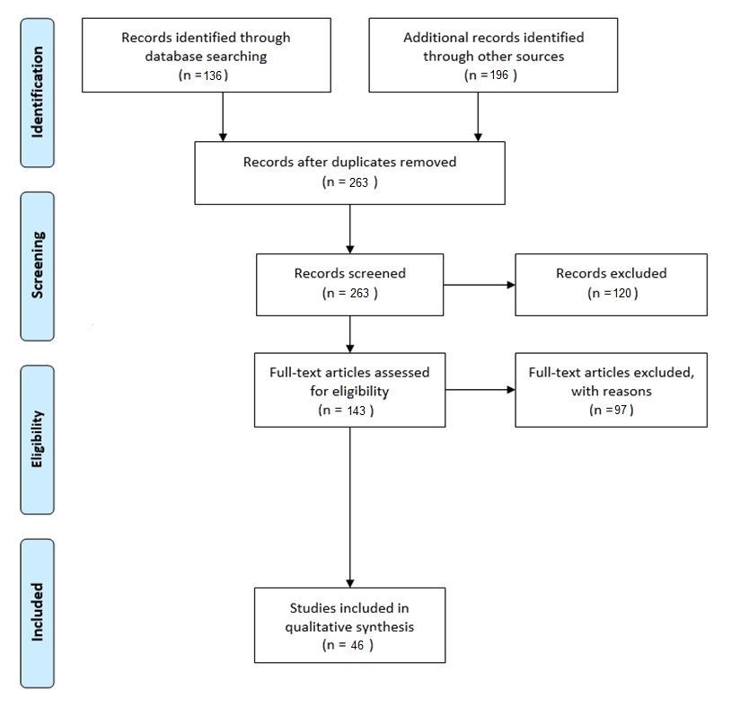
Theresearch
The researchprocess
processisisininaccordance
accordancewith
withthe
thePRISMA
PRISMAflowflowdiagram
diagramand andprotocol
protocol[14],
[14],and
and
depicts the conducted steps from identifying articles to eligible articles for further analysis.
depicts the conducted steps from identifying articles to eligible articles for further analysis. The The
mentionedflow
mentioned flowdiagram
diagramisisshown
shownininFigure
Figure1.1.
Figure 1. Flow diagram of the review process.
Figure 1. Flow diagram of the review process.
There
Thereare
arefour
fourmain
mainsections
sectionsininthe
theflow
flowdiagram.
diagram.Firstly,
Firstly,article
articleidentification
identificationisisconducted.
conducted.
This
This includes acquiring articles from various sources. The next section of the diagram includesthe
includes acquiring articles from various sources. The next section of the diagram includes the
screening
screeningprocess.
process.Article
Articleduplicates
duplicateswerewereexcluded.
excluded.Furthermore,
Furthermore,the thearticles
articlesare
arescreened
screenedonce
once
more and inadequate articles are removed. In the third section, full-articles were analyzed in orderMultimodal Technologies and Interact. 2018, 2, 47 3 of 12
more and inadequate articles are removed. In the third section, full-articles were analyzed in order
to determine the eligibility of the articles for further review. Ineligible articles were excluded from
further review. The fourth and final section includes studies/articles that were thoroughly analyzed.
2.2. Literature Sources
In order to investigate the applications of deep learning in medical diagnosis, 263 articles
published in the domain were analyzed. The main sources of these articles are presented in Table 1.
Table 1. Literature sources.
Literature Source ISSN
Briefings in Bioinformatics 1477-4054
Expert Systems with Application 0957-4174
IEEE Transactions Medical Imaging 1558-254X
Medical Image Analysis 1361-8423
Molecular Pharmaceutics 1543-8392
Nature 1476-4687
Neural Computing and Applications 0941-0643
Neurocomputing 0925-2312
These journals were chosen so that the credibility of this review paper is not compromised.
However, there is a wide variety of other literature sources that are also adequate for this review.
2.3. Data Collection Process
The data collection process included extensive research of articles that addressed the applications
of deep learning in the medical field. These articles were downloaded and analyzed in order to acquire
sufficient theoretical information on the subject. The results in this paper are qualitative in nature, and
the main focus is to review the applications of deep learning, and to answer the research questions
which were outlined in the introduction section of this paper. In sum, the data collection process was
conducted in four main phases:
• Phase 1: Searching articles in credible journals. This included the use of keywords presented
under the Section 2.4 of this paper. At this point the articles were thoroughly analyzed.
• Phase 2: Analyzing the literature and excluding articles that do not fit the eligibility criteria.
As there was no special screening during the search process, at this point the articles were
analyzed and selected for further analysis.
• Phase 3: Thorough analysis of eligible articles conducted and the qualitative data classified in
accordance with the aim of the review. At this stage there was a possibility of bias towards clearly
written and conducted research articles.
• Phase 4: Qualitative data obtained and notes taken in order to concisely present the data in the
results section of this paper. Data was collected in the form remarks and notes of what type of
data and methods were used, and on what applications.
2.4. Obtained Literature and Eligibility Criteria
When the necessary literature for this systematic review was gathered, it was important to include
various fields where deep learning is practically used. Therefore, the following keywords were used in
the search engine:
• deep learning practical applications
• deep learning and medical diagnosis
• deep learning and MRI
• deep learning CTMultimodal Technologies and Interact. 2018, 2, 47 4 of 12
• deep learning segmentation in medicine
• deep learning classification in medicine
• deep learning diagnosis medicine
• deep learning application medicine
This way it was ensured that a wide variety of articles will be included in the review. The year of
article publication was also considered; the earliest article dates from 2014, while the majority of other
reviewed articles are from 2016, 2017 and 2018. However, for the introduction section of this review,
earlier articles were also addressed.
2.5. Risk of Bias in Individual Studies
There was no major bias during the data analysis. However, if an article was not about the
application of deep learning in the field of medical diagnosis or medicine in general, it was then
excluded from further analysis. This type of review paper allows the inclusion of articles, regardless
of sample size, location, and data. There may seem to be a minor bias towards articles that address
deep learning applications in medicine, particularly in cancer detection. However, this is due to the
sheer number of articles that is much higher in this specific domain, as compared to other diseases.
Therefore, this minor bias does not have a major impact, or indeed, any impact, on the obtained results.
3. Results
When it comes to deep learning and its application for medical diagnosis, there are two main
approaches. The first approach is classification that includes reducing potential outcomes (diagnosis)
by mapping data to specific outcomes. The second approach is physiological data which includes
medical images and data from other sources are used to identify and diagnose tumors, or other
diseases [15]. In addition, deep learning can be used for dietary assessment support [16]. For a
certainty, deep learning is applied in various ways when it comes to medical diagnosis.
Brief reviews of individual articles in the domain of deep learning and medical diagnosis are
given in Table 2.
Table 2. Results of individual articles in the domain of deep learning and medical diagnosis.
Reference Method Data Source Application/Remarks
Computed Anatomical localization; the results indicate that 3D localization of
[17] CNN
tomography (CT) anatomical regions is possible with 2D images.
Automated segmentation; liver, heart and great vessels segmentation; it
[18] CNN MRI was concluded that this approach has great potential for
clinical applications.
Brain tumor grading; a 3-layered CNN has a 18% performance
[19] CNN MRI
improvement over to the baseline neural network.
Glaucoma detection; the experiments were performed on SCES and ORIGA
[20] CNN Fundus images datasets; further, it was noted that this approach may be great for
glaucoma detection.
Alzheimer’s disease prediction; the accuracy of this approach is far
[21] CNN MRI
superior compared to 2D methods
Automatic breast tissue classification; the pectoral muscles were detected
[22] CNN Mammography
with high accuracy (0.83) while nipple detection had lower accuracy (0.56).
Automatic detection of myocardial infarction; average accuracy was 93.53%
[23] CNN ECG
with noise and 95.22% without noise.
[24] CNN CT Automated segmentation of human brain structures.
Automatic diagnosis for detecting breast cancer; the accuracy of the overall
[25] DBN-NN Mammography neural network was 99.68%, the sensitivity was 100%, and the specificity
was 99.47%.
Breast lesion and pulmonary nodule detection/diagnosis; the results
Ultrasound of indicated that there is a significant increase in performance. In addition, it
[26] SDAE
breasts, and lung CT was noted that deep learning techniques have the potential to change CAD
systems, with ease, and without the need for structural redesign.Multimodal Technologies and Interact. 2018, 2, 47 5 of 12
Table 2. Cont.
Reference Method Data Source Application/Remarks
Classification of skin cancer; the results of the study were satisfactory, as
the deep convolutional neural networks achieve performance similar to the
[27] CNN Clinical images expertise of 21 board-certified dermatologist. This can lead to mobile
dermatology diagnosis, providing millions of people with universal
diagnostic care.
Drug-induced liver injury (DILI) prediction. The model had an accuracy of
86.9%, sensitivity of 82.5%, and specificity of 92.9%. Overall, deep learning
Various medical
[28] UGRNN gave significantly better results in opposite to other DILI prediction models.
data sets
In sum, deep learning can lower the health risk for humans when it comes
to DILI.
Automatic grading system for nuclear cataracts; this method improved the
[29] CRNN Fundus images clinical management of this cataract disease, and it has a potential for other
eye disease diagnosis.
Chest pathology identification; the area under the curve for heart detection
was 0.89, for right pleural effusion detection 0.93, and for the classification
[30] CNN X-ray between a healthy and an abnormal chest X-ray was 0.79. It was concluded
that it is possible for non-medical images, and datasets to be sufficient for
recognition of medical images.
Skin cancer diagnosis; the results were promising, and there is a possibility
[31] SA-SVM Dermoscopic images to use this type of self -advised SVM method in cases where is limited
labeled data.
Multi-classification of breast cancer; a great performance of 93.2% accuracy
[32] CSDCNN Mammography
was achieved on large-scale datasets
Brain tumor segmentation; with this method the whole brain can be
[33] DNN MRI
segmented in 25 s to 3 min, thus making it a great segmentation tool.
[34] CNN MRI Brain lesion segmentation; this approach produced great results.
Breast density classification; Radiologist have a problem to differentiate
between the two types of density. A learning model was developed that
[35] CNN Mammography
helped radiologist in the diagnosis process. Deep learning was found to be
useful when it comes to realistic diagnosis, and overall clinical needs.
Breast tumor classification; the results indicated that the deep learning
Shear-wave
[36] RBM model achieved a remarkable accuracy rate of 93.4%, with 88.6% sensitivity,
elastography SWE
and 97.1% specificity.
Urinary bladder segmentation; this method can overcome strong
[37] DL-CNN CT urography
boundaries between two regions that have large differences of gray levels.
Glioblastoma segmentation; this approach allowed a large U-Net training
with small datasets, without significant overfitting. Taken into
[38] U-Net MRI
consideration that patients move during the segmentation process, there
may be performance increase switching to 3D convolutions.
Disease staging and prognosis of smokers; this type of chronic lung illness
[39] CNN CT
prognosis is powerful for risk assessment.
Cancer detection from gene data; the study was successful as this method
Various medical
[40] SDAE managed to extract genes that are helpful when it comes to
images
cancer prediction.
Ultrasound segmentation of first trimester placenta; it was noted that this
The 3D volumetric
[41] CNN approach had similar performance compared to results that were acquired
ultrasound data
through MRI data.
Segmentation of the striatum; two serial CNN architectures were used; the
[42] CNN MRI speed and accuracy of this approach makes it adequate for application in
neuroscience and other clinical fields.
Images produced Lymph node metastases detection in breast cancer; the best performing
[43] CNN
with a digital slider algorithm achieved performance comparable to a pathologist.
Brain segmentation; the results are comparable of other
[44] CNN MRI
state-of-the-art performance.
CAD classifier with Lung nodule classification; this approach resulted an accuracy of 75.01%
[45] deep features from CT and sensitivity of 83.35%; false positive was 0.39 per patient over 10
autoencoder cross validations.
[46] CNN, DBN, SDAE CT Lung cancer diagnosis; highest accuracy was achieved with DBN (0.8119).
Breast cancer diagnosis; with this approach the achieved accuracy was
[47] CNN MRI
96.39%, the sensitivity was 97.73%, and the specificity was 94.87%.
Lung nodule detection; the network was trained with weak label
[48] CNN CT information; 3D segmentation could exclude air tracts in the lungs, thus
reducing false positives.
Planar projection Microcalcifications detection in digital breast tomosynthesis; the best
[49] DLCNN
(PPJ) image obtained AUC was 0.933.Multimodal Technologies and Interact. 2018, 2, 47 6 of 12
Table 2. Cont.
Reference Method Data Source Application/Remarks
[50] CNN CT Pancreas segmentation; average dice scores were from 46.6% to 68.8%.
Diagnosis of diabetic retinopathy; on a dataset of 80,000 images the
[51] CNN Fundus images
accuracy was 75% and the sensitivity is 95%.
Breast and fibroglandular tissue segmentation; average Dice Similarity
[52] U-Net MRI Coefficients (DSC) were 0.850 for 3C U-net, 0.811 for 2C U-net, and 0.671
for atlas-based method.
Breast cancer histopathology classification; average recognition rate was
Histopathology
[53] CNN 82.13% for classification tasks, and 80.10% accuracy when it comes to
images
magnification estimation.
Retina vessel segmentation; this method reduces the number of
[54] CNN Fundus images
false-positives.
Brain segmentation; the performance is dependent of several factors such
[55] CNN MRI
as initialization, preprocessing, and post-processing.
Automatic brain segmentation; this approach can be used for accurate
[56] CNN MRI
brain segmentation results.
Identifying and classifying tumor-associated stroma from breast biopsies;
[57] CNN Digital images the study revealed that this deep learning approach was able to define
stromal features of ductal carcinoma in situ grade.
[58] CNN Gastric images Gastric cancer identification; the accuracy of classification was 97.93%.
Lytic and sclerotic metastatic spinal lesion detection, segmentation and
classification; the obtained results were quantitatively compared to other
[59] CNN CT methods and it was concluded that this approach can provide better
accuracy even for small lesions (greater than 1.4 mm3 and diameter greater
than 0.7 mm).
In this study the predictive test was conducted both with deep learning
and non-deep-learning. The deep learning approach had an accuracy of
[60] DCNN MRI
84%, sensitivity of 69.6% and specificity of 83.9%. The non-deep-learning
approach had 70% accuracy, 49.4% sensitivity, and 81.7% specificity.
Lung cancer nodule malignancy classification; with this approach a high
[61] CNN CT level of accuracy was achieved 99%. This is proportionate to the accuracy
of an experienced radiologist.
Digital pathology Nuclei detection of breast cancer; the stacked sparse autoencoder (SSAE)
[62] SSAE
images approach can outperform state of the art nuclear detection strategies.
Furthermore, the synthesis of the results is presented in Table 3.
Table 3. Synthesis of articles by type of deep learning method, data source and application.
Type of Deep Learning Method Number of Articles
CNN 32
RBM 1
SA-SVM 1
CRNN 1
Other 3
DBN 1
SDAE 2
UGRNN 1
Multiple 1
U-Net 2
CSDCNN 1
Type of Data Source Number of Articles
X-ray 1
Ultrasound 2
CT 10
MRI 13
Fundus photography 4
Mammography 4
Other data 12
Application Type Number of Articles
Localization 1
Segmentation 14
Grading 2
Detection 8
Prediction 4
Classification 8
Diagnosis 6
Identification 2Multimodal Technologies and Interact. 2018, 2, 47 7 of 12
In the next section the results are discussed.
4. Discussion
Discussing the Results
The main goal of this paper was to review various articles in the domain of deep learning
application in medical diagnosis. After analyzing more than 300 articles, 46 were further examined,
and the individual results of each article were presented. There was no need for quantitative data
analysis, as the nature of this review was to present the variety of deep learning uses in the medical
field. The synthesis of data was conducted in a simple way. Some of the methods used for synthesis
were in accordance with other similar studies [63–65]. According to the gathered data, the most
widely used deep learning method is convolutional neural networks (CNNs). In addition, MRI was
most frequently used as training data. When it comes to the specific use, segmentation is the most
represented. It is important to note, that the article review and analysis was biased towards newer
(published 2015 and later) articles, and articles that included “deep learning” in the title. It can be seen
that there is a large variety in the type of data that is used to train and apply deep neural networks.
CT scan images, MRIs, fundus photography and other types of data can be used for expert-level
diagnosis. However, as noted in other studies, neural networks use energy to activate neurons. With
the human brain, during the thought process only a small number of neurons are active, while the
neighboring neurons are shut down until needed. Communication “costs” are reduced through
single-task allocation for neighboring neurons [65]. It is expected that artificial neural networks will
further develop in the future, thus managing to complete more complex tasks.
The concise nature of this review can moderately contribute to the existing body of literature.
The aim was to provide an objective, simple and a concise article. The individual research results
provide sufficient information and insight into the applications of deep learning for detecting,
classifying, segmenting and diagnosing various diseases and abnormalities in specific anatomical
regions of interest (ROI). Without a doubt deep learning application in the medical field will further
develop as it has already achieved remarkable results in medical image analysis [66], and more
precisely, in image-based cancer detection and diagnosis [67]. This may increase the efficiency and
quality of healthcare in the long-run, thus reducing the risk of late-diagnosis of serious diseases.
However, as mentioned before, there is still a long way to go before general purpose neural networks
will be commercially relevant. Finally, it is expected that artificial intelligence will “rise” through the
combination of representation learning and complex reasoning [3].
5. Conclusions
5.1. Research Questions
In the introduction section of this review, three main research questions were investigated:
• How diverse is the application of deep learning in the field of medical diagnosis?
Deep learning methods have a wide application in the medical field. In this case, medical
diagnosis is conducted through use-cases of deep learning networks. As mentioned before, these
include detection, segmentation, classification, prediction and other. The results of the reviewed
studies indicate that deep learning methods can be far superior in comparison to other high-performing
algorithms. Therefore, it is safe to assume that deep learning is and will continue to diversify its uses.
• Can deep learning substitute the role of doctors in the future?
The future development of deep learning promises more applications in various fields of medicine,
particularly in the domain of medical diagnosis. However, in the current state, it is not evident that
deep learning can substitute the role of doctors/clinicians in medical diagnosis. So far, deep learning
can provide good support for experts in the medical field.Multimodal Technologies and Interact. 2018, 2, 47 8 of 12
• Does deep learning have a future or will it become obsolete?
All indicators point towards an even wider use of deep learning in various fields. Deep learning
has already found its application in transportation and greenhouse-gas emission control [68], traffic
control [69], text classification [8,70], object detection [71], speech detection [72,73], translation [74]
and in other fields. These applications were not so represented in the past. Traditional approaches
to various similarity measures are ineffective when compared to deep learning [63]. Based on these
findings, it can be suggested that deep learning and deep neural networks will prevail, and that they
will find many other uses in the near future.
5.2. Limitations and Future Research
The main limitation of this paper is the absence of meta-analysis of quantitative data. However,
considering the main goal of this paper, this limitation does not devalue the contribution of the review.
For future research, a more categorized review should be conducted. In addition, the development
and application of deep learning through defined periods of time could be added. A theoretical
introduction to future reviews is also recommended. In this case, the theoretical background did not
contain a detailed explanation of how deep neural networks function. However, given the nature
of the review, and the target audience (researchers whose domain of expertise is not deep learning
focused), such a theoretical approach was not deemed necessary.
Author Contributions: M.B. conducted the investigation, data curation, and writing of the original draft. D.R.
contributed in the form of supervision, conceptualization, and methodology.
Funding: This research received no external funding.
Conflicts of Interest: The authors declare no conflict of interest.
References
1. Szegedy, C.; Wei, L.; Yang, J.; Sermanet, P.; Reed, S.; Anguelov, D.; Erhan, D.; Vanhoucke, V.; Rabinovich, A.
Going deeper with convolutions. In Proceedings of the IEEE Conference on Computer Vision and Pattern
Recognition, Boston, MA, USA, 7–12 June 2015; pp. 1–9.
2. Abadi, M.; Chu, A.; Goodfellow, I.; McMahan, H.B.; Mironov, I.; Talwar, K.; Zhang, L. Deep Learning with
Differential Privacy. In Proceedings of the 2016 ACM SIGSAC Conference on Computer and Communications
Security, Vienna, Austria, 24–28 October 2016; pp. 308–318.
3. LeCun, Y.; Bengio, Y.; Hinton, G. Deep learning. Nature 2015, 436–444. [CrossRef] [PubMed]
4. Chen, X.-W.; Lin, X. Big data deep learning: Challenges and perspectives. IEEE Access 2014, 2, 514–525.
[CrossRef]
5. Miotto, R.; Wang, F.; Wang, S.; Jiang, X.; Dudley, J.T. Deep learning for healthcare: Review, opportunities and
challenges. Brief Bioinform. 2017. [CrossRef] [PubMed]
6. Wei, J.; He, J.; Chen, K.; Zhou, Y.; Tang, Z. Collaborative filtering and deep learning based recommendation
system for cold start items. Expert Syst. Appl. 2017, 69, 29–39. [CrossRef]
7. Shin, H.C.; Roth, H.R.; Gao, M.; Lu, L.; Xu, Z.; Nogues, I.; Summers, R.M. Deep Convolutional Neural
Networks for Computer-Aided Detection: CNN Architectures, Dataset Characteristics and Transfer Learning.
IEEE Trans. Med. Imaging 2016, 35, 1285–1298. [CrossRef] [PubMed]
8. Song, J.; Qin, S.; Zhang, P. Chinese text categorization based on deep belief networks. In Proceedings of
the 2016 IEEE/ACIS 15th International Conference on Computer and Information Science Computer and
Information Science (ICIS), Okayama, Japan, 26–29 June 2016; pp. 1–5.
9. Lee, J.G.; Jun, S.; Cho, Y.W.; Lee, H.; Kim, G.B.; Seo, J.B.; Kim, N. Deep Learning in Medical Imaging: General
Overview. Korean J. Radiol. 2017, 18, 570–584. [CrossRef] [PubMed]
10. Suzuki, K. Overview of deep learning in medical imaging. Radiol. Phys. Technol. 2017, 10, 257–273. [CrossRef]
[PubMed]
11. Ravì, D.; Wong, C.; Deligianni, F.; Berthelot, M.; Andreu-Perez, J.; Lo, B.; Yang, G.-Z. Deep learning for health
informatics. IEEE J. Biomed. Health Inf. 2017, 21, 4–21. [CrossRef] [PubMed]Multimodal Technologies and Interact. 2018, 2, 47 9 of 12
12. Mamoshina, P.; Vieira, A.; Putin, E.; Zhavoronkov, A. Applications of Deep Learning in Biomedicine.
Mol. Pharm. 2016, 13, 1445–1454. [CrossRef] [PubMed]
13. Liu, J.; Pan, Y.; Li, M.; Chen, Z.; Tang, L.; Lu, C.; Wang, J. Applications of deep learning to MRI images:
A survey. Big Data Mining Anal. 2018, 1, 1–18. [CrossRef]
14. Moher, D.; Liberati, A.; Tetzlaff, J.; Altman, D.G.; Prisma Group. Preferred reporting items for systematic
reviews and meta-analyses: The PRISMA statement. Int. J. Surg. 2010, 8, 336–341. [CrossRef] [PubMed]
15. Greenspan, H.; Van Ginneken, B.; Summers, R.M. Guest editorial deep learning in medical imaging:
Overview and future promise of an exciting new technique. IEEE Trans. Med. Imaging 2016, 35, 1153–1159.
[CrossRef]
16. Mezgec, S.; Koroušić Seljak, B. NutriNet: A Deep Learning Food and Drink Image Recognition System for
Dietary Assessment. Nutrients 2017, 9, 657. [CrossRef] [PubMed]
17. De Vos, B.D.; Wolterink, J.M.; de Jong, P.A.; Viergever, M.A.; Išgum, I. 2D image classification for 3D anatomy
localization: Employing deep convolutional neural networks. In Proceedings of the Medical Imaging 2016:
Image Processing, San Diego, CA, USA, 1–3 March 2016; Volume 9784.
18. Dou, Q.; Yu, L.; Chen, H.; Jin, Y.; Yang, X.; Qin, J.; Heng, P.A. 3D deeply supervised network for automated
segmentation of volumetric medical images. Med Image Anal. 2017, 41, 40–54. [CrossRef] [PubMed]
19. Pan, Y.; Huang, W.; Lin, Z.; Zhu, W.; Zhou, J.; Wong, J.; Ding, Z. Brain tumor grading based on neural
networks and convolutional neural networks. In Proceedings of the 2015 37th Annual International
Conference of the IEEE Engineering in Medicine and Biology Society (EMBC), Milan, Italy, 25–29 August
2015; pp. 699–702.
20. Chen, X.; Xu, Y.; Wong, D.W.K.; Wong, T.Y.; Liu, J. Glaucoma detection based on deep convolutional neural
network. In Proceedings of the 2015 37th Annual International Conference of the IEEE, Engineering in
Medicine and Biology Society (EMBC), Milan, Italy, 25–29 August 2015; pp. 715–718.
21. Payan, A.; Montana, G. Predicting Alzheimer’s disease: A neuroimaging study with 3D convolutional neural
networks. arXiv 2015, arXiv:1502.02506.
22. Dubrovina, A.; Kisilev, P.; Ginsburg, B.; Hashoul, S.; Kimmel, R. Computational mammography using deep
neural networks. Comput. Methods Biomech. Biomed. Eng. Imaging Vis. 2016, 6, 243–247. [CrossRef]
23. Acharya, U.R.; Fujita, H.; Oh, S.L.; Hagiwara, Y.; Tan, J.H.; Adam, M. Application of deep convolutional
neural network for automated detection of myocardial infarction using ECG signals. Inf. Sci. 2017, 415,
190–198. [CrossRef]
24. Mehta, R.; Majumdar, A.; Sivaswamy, J. BrainSegNet: A convolutional neural network architecture for
automated segmentation of human brain structures. J. Med. Imaging (Bellingham) 2017, 4, 024003. [CrossRef]
[PubMed]
25. Abdel-Zaher, A.M.; Eldeib, A.M. Breast cancer classification using deep belief networks. Expert Syst. Appl.
2016, 46, 139–144. [CrossRef]
26. Cheng, J.Z.; Ni, D.; Chou, Y.H.; Qin, J.; Tiu, C.M.; Chang, Y.C.; Huang, C.S.; Shen, D.; Chen, C.M. Computer-
Aided Diagnosis with Deep Learning Architecture: Applications to Breast Lesions in US Images and
Pulmonary Nodules in CT Scans. Sci. Rep. 2016, 6, 24454. [CrossRef] [PubMed]
27. Esteva, A.; Kuprel, B.; Novoa, R.A.; Ko, J.; Swetter, S.M.; Blau, H.M.; Thrun, S. Dermatologist-level
classification of skin cancer with deep neural networks. Nature 2017, 542, 115–118. [CrossRef] [PubMed]
28. Xu, Y.; Dai, Z.; Chen, F.; Gao, S.; Pei, J.; Lai, L. Deep Learning for Drug-Induced Liver Injury. J. Chem.
Inf. Model. 2015, 55, 2085–2093. [CrossRef] [PubMed]
29. Gao, X.; Lin, S.; Wong, T.Y. Automatic Feature Learning to Grade Nuclear Cataracts Based on Deep Learning.
IEEE Trans. Biomed. Eng. 2015, 62, 2693–2701. [CrossRef] [PubMed]
30. Bar, Y.; Diamant, I.; Wolf, L.; Greenspan, H. Deep learning with non-medical training used for chest pathology
identification. In Proceedings of the Medical Imaging 2015: Computer-Aided Diagnosis, Orlando, FL, USA,
3–7 November 2015; Volume 9414.
31. Masood, A.; Al-Jumaily, A.; Anam, K. Self-supervised learning model for skin cancer diagnosis. In
Proceedings of the 2015 7th International IEEE/EMBS Conference Neural Engineering (NER), Montpellier,
France, 22–24 April 2015; pp. 1012–1015.
32. Han, Z.; Wei, B.; Zheng, Y.; Yin, Y.; Li, K.; Li, S. Breast Cancer Multi-classification from Histopathological
Images with Structured Deep Learning Model. Sci. Rep. 2017, 7, 4172. [CrossRef] [PubMed]Multimodal Technologies and Interact. 2018, 2, 47 10 of 12
33. Havaei, M.; Davy, A.; Warde-Farley, D.; Biard, A.; Courville, A.; Bengio, Y.; Larochelle, H. Brain tumor
segmentation with Deep Neural Networks. Med. Image Anal. 2017, 35, 18–31. [CrossRef] [PubMed]
34. Kamnitsas, K.; Ledig, C.; Newcombe, V.F.J.; Simpson, J.P.; Kane, A.D.; Menon, D.K.; Glocker, B. Efficient
multi-scale 3D CNN with fully connected CRF for accurate brain lesion segmentation. Med. Image Anal.
2017, 36, 61–78. [CrossRef] [PubMed]
35. Mohamed, A.A.; Berg, W.A; Peng, H.; Luo, Y.; Jankowitz, R.C.; Wu, S. A deep learning method for classifying
mammographic breast density categories. Med. Phys. 2018, 45, 314–321. [CrossRef] [PubMed]
36. Zhang, Q.; Xiao, Y.; Dai, W.; Suo, J.; Wang, C.; Shi, J.; Zheng, H. Deep learning based classification of breast
tumors with shear-wave elastography. Ultrasonics 2016, 72, 150–157. [CrossRef] [PubMed]
37. Cha, K.H.; Hadjiiski, L.; Samala, R.K.; Chan, H.P.; Caoili, E.M.; Cohan, R.H. Urinary bladder segmentation in
CT urography using deep-learning convolutional neural network and level sets. Med. Phys. 2016, 43, 1882.
[CrossRef] [PubMed]
38. Isensee, F.; Kickingereder, P.; Bonekamp, D.; Bendszus, M.; Wick, W.; Schlemmer, H.P.; Maier-Hein, K. Brain
Tumor Segmentation Using Large Receptive Field Deep Convolutional Neural Networks. Bildverarb. Med.
2017, 86–91. [CrossRef]
39. González, G.; Ash, S.Y.; Vegas-Sánchez-Ferrero, G.; Onieva Onieva, J.; Rahaghi, F.N.; Ross, J.C.; Washko, G.R.
Disease staging and prognosis in smokers using deep learning in chest computed tomography. Am. J. Respir.
Crit. Care Med. 2018, 197, 193–203. [CrossRef] [PubMed]
40. Danaee, P.; Ghaeini, R.; Hendrix, D.A. A deep learning approach for cancer detection and relevant gene
identification. Pac. Symp. Biocomput. 2017, 2017, 219–229.
41. Looney, P.; Stevenson, G.N.; Nicolaides, K.H.; Plasencia, W.; Molloholli, M.; Natsis, S.; Collins, S.L. Automatic
3D ultrasound segmentation of the first trimester placenta using deep learning. In Proceedings of the 2017
IEEE 14th International Symposium on Biomedical Imaging, Biomedical Imaging (ISBI 2017), Melbourne,
Australia, 18–21 April 2017; pp. 279–282.
42. Choi, H.; Jin, K.H. Fast and robust segmentation of the striatum using deep convolutional neural networks.
J. Neurosci. Methods 2016, 274, 146–153. [CrossRef] [PubMed]
43. Ehteshami Bejnordi, B.; Veta, M.; Johannes van Diest, P.; van Ginneken, B.; Karssemeijer, N.; Litjens, G.;
Venancio, R. Diagnostic Assessment of Deep Learning Algorithms for Detection of Lymph Node Metastases
in Women With Breast Cancer. JAMA 2017, 318, 2199–2210. [CrossRef] [PubMed]
44. Kleesiek, J.; Urban, G.; Hubert, A.; Schwarz, D.; Maier-Hein, K.; Bendszus, M.; Biller, A. Deep MRI brain
extraction: A 3D convolutional neural network for skull stripping. Neuroimage 2016, 129, 460–469. [CrossRef]
[PubMed]
45. Kumar, D.; Wong, A.; Clausi, D.A. Lung nodule classification using deep features in CT images.
In Proceedings of the 2015 12th Conference on Robot VisionComputer and Robot Vision (CRV), Halifax, NS,
Canada, 3–5 June 2015; pp. 133–138.
46. Sun, W.; Zheng, B.; Qian, W. Computer aided lung cancer diagnosis with deep learning algorithms. In
Proceedings of the Medical Imaging 2016: Computer-Aided Diagnosis, San Diego, CA, USA, 27 February–3
March 2016; Volime 9785.
47. Rasti, R.; Teshnehlab, M.; Phung, S.L. Breast cancer diagnosis in DCE-MRI using mixture ensemble of
convolutional neural networks. Pattern Recognit. 2017, 72, 381–390. [CrossRef]
48. Anirudh, R.; Thiagarajan, J.J.; Bremer, T.; Kim, H. Lung nodule detection using 3D convolutional neural
networks trained on weakly labeled data. In Proceedings of the Medical Imaging 2016: Computer-Aided
Diagnosis, San Diego, CA, USA, 27 February–3 March 2016; Volume 9785.
49. Samala, R.K.; Chan, H.P.; Hadjiiski, L.M.; Cha, K.; Helvie, M.A. Deep-learning convolution neural network
for computer-aided detection of microcalcifications in digital breast tomosynthesis. In Proceedings of
the Medical Imaging 2016: Computer-Aided Diagnosis, San Diego, CA, USA, 27 February–3 March 2016;
Volume 9785.
50. Roth, H.R.; Farag, A.; Lu, L.; Turkbey, E.B.; Summers, R.M. Deep convolutional networks for pancreas
segmentation in CT imaging. Med. Imaging Image Process. 2015, 9413, 94131G.
51. Pratt, H.; Coenen, F.; Broadbent, D.M.; Harding, S.P.; Zheng, Y. Convolutional Neural Networks for Diabetic
Retinopathy. Proced. Comput. Sci. 2016, 90, 200–205. [CrossRef]Multimodal Technologies and Interact. 2018, 2, 47 11 of 12
52. Dalmis, M.U.; Litjens, G.; Holland, K.; Setio, A.; Mann, R.; Karssemeijer, N.; Gubern-Merida, A. Using
deep learning to segment breast and fibroglandular tissue in MRI volumes. Med. Phys. 2017, 44, 533–546.
[CrossRef] [PubMed]
53. Bayramoglu, N.; Kannala, J.; Heikkilä, J. Deep learning for magnification independent breast cancer
histopathology image classification. In Proceedings of the 2016 23rd International Conference on Pattern
Recognition (ICPR), Cancún, Mexico, 4–8 December 2016; pp. 2440–2445.
54. Fu, H.; Xu, Y.; Wong, D.W.K.; Liu, J. Retinal vessel segmentation via deep learning network and
fully-connected conditional random fields. In Proceedings of the Biomedical Imaging (ISBI), 2016 IEEE 13th
International Symposium on Biomedical Imaging, Prague, Czech, 13–16 April 2016; pp. 698–701.
55. Akkus, Z.; Galimzianova, A.; Hoogi, A.; Rubin, D.L.; Erickson, B.J. Deep Learning for Brain MRI
Segmentation: State of the Art and Future Directions. J. Digit. Imaging 2017, 30, 449–459. [CrossRef]
[PubMed]
56. Moeskops, P.; Viergever, M.A.; Mendrik, A.M.; de Vries, L.S.; Benders, M.J.; Isgum, I. Automatic Segmentation
of MR Brain Images With a Convolutional Neural Network. IEEE Trans. Med. Imaging 2016, 35, 1252–1261.
[CrossRef] [PubMed]
57. Ehteshami Bejnordi, B.; Mullooly, M.; Pfeiffer, R.M.; Fan, S.; Vacek, P.M.; Weaver, D.L.; Sherman, M.E. Using
deep convolutional neural networks to identify and classify tumor-associated stroma in diagnostic breast
biopsies. Mod. Pathol. 2018. [CrossRef] [PubMed]
58. Li, Y.; Li, X.; Xie, X.; Shen, L. Deep learning based gastric cancer identification. In Proceedings of the 2018
IEEE 15th International Symposium on Biomedical Imaging, Biomedical Imaging (ISBI 2018), Washington,
DC, USA, 4–7 April 2018; pp. 182–185.
59. Chmelik, J.; Jakubicek, R.; Walek, P.; Jan, J.; Ourednicek, P.; Lambert, L.; Amadori, E.; Gavelli, G. Deep
convolutional neural network-based segmentation and classification of difficult to define metastatic spinal
lesions in 3D CT data. Med. Image Anal. 2018. [CrossRef] [PubMed]
60. Wang, X.; Yang, W.; Weinreb, J.; Han, J.; Li, Q.; Kong, X.; Yan, Y.; Ke, Z.; Luo, B.; Liu, T.; et al. Searching
for prostate cancer by fully automated magnetic resonance imaging classification: Deep learning versus
non-deep learning. Sci. Rep. 2017, 7, 15415. [CrossRef] [PubMed]
61. Causey, J.L.; Zhang, J.; Ma, S.; Jiang, B.; Qualls, J.A.; Politte, D.G.; Prior, F.; Zhang, S.; Huang, X. Highly
accurate model for prediction of lung nodule malignancy with CT scans. Sci. Rep. 2018, 8, 9286. [CrossRef]
[PubMed]
62. Xu, J.; Xiang, L.; Liu, Q.; Gilmore, H.; Wu, J.; Tang, J.; Madabhushi, A. Stacked sparse autoencoder (SSAE)
for nuclei detection on breast cancer histopathology images. IEEE Trans. Med. Imaging 2016, 35, 119–130.
[CrossRef] [PubMed]
63. Faust, O.; Hagiwara, Y.; Hong, T.J.; Lih, O.S.; Acharya, U.R. Deep learning for healthcare applications based
on physiological signals: A review. Comput. Methods Programs Biomed. 2018. [CrossRef] [PubMed]
64. Litjens, G.; Kooi, T.; Bejnordi, B.E.; Setio, A.A.A.; Ciompi, F.; Ghafoorian, M.; Sánchez, C.I. A survey on deep
learning in medical image analysis. Med. Image Anal. 2017, 42, 60–88. [CrossRef] [PubMed]
65. Schmidhuber, J. Deep learning in neural networks: An overview. Neural Netw. 2015, 61, 85–117. [CrossRef]
[PubMed]
66. Shen, D.; Wu, G.; Suk, H.I. Deep learning in medical image analysis. Annu. Rev. Biomed. Eng. 2017, 19,
221–248. [CrossRef] [PubMed]
67. Hu, Z.; Tang, J.; Wang, Z.; Zhang, K.; Zhang, L.; Sun, Q. Deep Learning for Image-based Cancer Detection
and Diagnosis—A Survey. Pattern Recognit. 2018. [CrossRef]
68. Madu, C.N.; Kuei, C.-H.; Lee, P. Urban sustainability management: A deep learning perspective. Sustain.
Cities Soc. 2017, 30, 1–17. [CrossRef]
69. Fadlullah, Z.M.; Tang, F.; Mao, B.; Kato, N.; Akashi, O.; Inoue, T.; Mizutani, K. State-of-the-Art Deep
Learning: Evolving Machine Intelligence Toward Tomorrow’s Intelligent Network Traffic Control Systems.
IEEE Commun. Surv. Tutor. 2017, 19, 2432–2455. [CrossRef]
70. Yousefi-Azar, M.; Hamey, L. Text summarization using unsupervised deep learning. Expert Syst. Appl. 2017,
68, 93–105. [CrossRef]
71. Zhou, X.; Gong, W.; Fu, W.; Du, F. Application of deep learning in object detection. In Proceedings of the
2017 IEEE/ACIS 16th International Conference on Computer and Information Science (ICIS), Wuhan, China,
24–26 May 2017; pp. 631–634.Multimodal Technologies and Interact. 2018, 2, 47 12 of 12
72. Badjatiya, P.; Gupta, S.; Gupta, M.; Varma, V. Deep learning for hate speech detection in tweets.
In Proceedings of the 26th International Conference on World Wide Web Companion, Perth, Australia,
3–7 April 2017; pp. 759–760.
73. Deng, L. Deep learning: From speech recognition to language and multimodal processing. APSIPA Trans.
Signal Inf. Process. 2016, 5. [CrossRef]
74. Singh, S.P.; Kumar, A.; Darbari, H.; Singh, L.; Rastogi, A.; Jain, S. Machine translation using deep learning:
An overview. In Proceedings of the 2017 International Conference on Computer, Communications and
Electronics (Comptelix), Jaipur, India, 1–2 July 2017; pp. 162–167.
© 2018 by the authors. Licensee MDPI, Basel, Switzerland. This article is an open access
article distributed under the terms and conditions of the Creative Commons Attribution
(CC BY) license (http://creativecommons.org/licenses/by/4.0/).You can also read