The Effect of pH and Viscosity on Magnetophoretic Separation of Iron Oxide Nanoparticles - MDPI
←
→
Page content transcription
If your browser does not render page correctly, please read the page content below
magnetochemistry

Article
The Effect of pH and Viscosity on Magnetophoretic Separation
of Iron Oxide Nanoparticles
Leonie Wittmann               , Chiara Turrina        and Sebastian P. Schwaminger *

                                          Bioseparation Engineering Group, Department of Mechanical Engineering, Technical University of Munich,
                                          Boltzmanstr. 15, 85748 Garching bei München, Germany; l.wittmann@tum.de (L.W.); c.turrina@tum.de (C.T.)
                                          * Correspondence: s.schwaminger@tum.de

                                          Abstract: Magnetic nanoparticles (MNPs) are used for magnetophoresis-based separation processes
                                          in various biomedical and engineering applications. Essential requirements are the colloidal stability
                                          of the MNPs and the ability to be separated even in low magnetic field gradients. Bare iron oxide
                                          nanoparticles (BIONs) with a diameter of 9.2 nm are synthesized via coprecipitation, exhibiting a
                                          high saturation magnetization of 70.84 Am2 kg−1 and no remanence. In our study, zeta potential,
                                          dynamic light scattering (DLS), and sedimentation analysis show that the aggregation behavior
                                          of BIONs is influenced by pH and viscosity. Small aggregate clusters are formed with either low
                                          or high pH values or increased viscosity. Regarding magnetophoresis-based separation, a higher
                                          viscosity leads to lower magnetophoretic velocities, similar to how small aggregates do. Additionally,
                                          cooperative magnetophoresis, the joint motion of strongly interacting particles, affects the separation
                                          of the BIONs, too. Our study emphasizes the effect of pH and viscosity on the physicochemical
                                          characteristics of MNPs, resulting in different aggregation behavior. Particularly, for high viscous
                                working media in downstream processing and medicine, respectively, the viscosity should be taken

                                          into account, as it will affect particle migration.
Citation: Wittmann, L.; Turrina, C.;
Schwaminger, S.P. The Effect of pH
                                          Keywords: magnetophoresis; magnetic separation process; iron oxide nanoparticles; aggregation;
and Viscosity on Magnetophoretic
                                          pH; viscosity; sedimentation analyzer; zeta potential; hydrodynamic diameter
Separation of Iron Oxide
Nanoparticles. Magnetochemistry 2021,
7, 80. https://doi.org/10.3390/
magnetochemistry7060080
                                          1. Introduction
Academic Editor: Marie Frenea-Robin             Magnetic nanoparticles (MNPs) have become an important nanomaterial in biotechnol-
                                          ogy, in chemistry, and in medicine [1–6]. They entail characteristics such as biocompatibility,
Received: 29 April 2021                   high binding capacities, and cost-efficient production via coprecipitation. Moreover, their
Accepted: 30 May 2021                     superparamagnetic properties are advantageous during the separation process, as the
Published: 3 June 2021                    MNPs show no remanence at room temperature. However, when applying a magnetic
                                          field, they possess a high magnetic susceptibility and can be easily separated [7,8]. For
Publisher’s Note: MDPI stays neutral      biological samples, it is challenging to separate a target entity from a complex mixture
with regard to jurisdictional claims in   with different components. Here, magnetophoresis-based processes provide a simple and
published maps and institutional affil-   efficient method, where the desired entity (magnetic or magnetically labeled with MNPs)
iations.                                  is isolated by applying an external magnetic field [9–11]. For the method of High Gradient
                                          Magnetic Separation (HGMS), a suspension containing the magnetic material is pumped
                                          through a separation chamber, and it is trapped by a magnetically susceptible matrix due
                                          to high magnetic field gradients (102 –104 T m−1 ) [9,11,12]. In contrast, low magnetic field
Copyright: © 2021 by the authors.         gradients (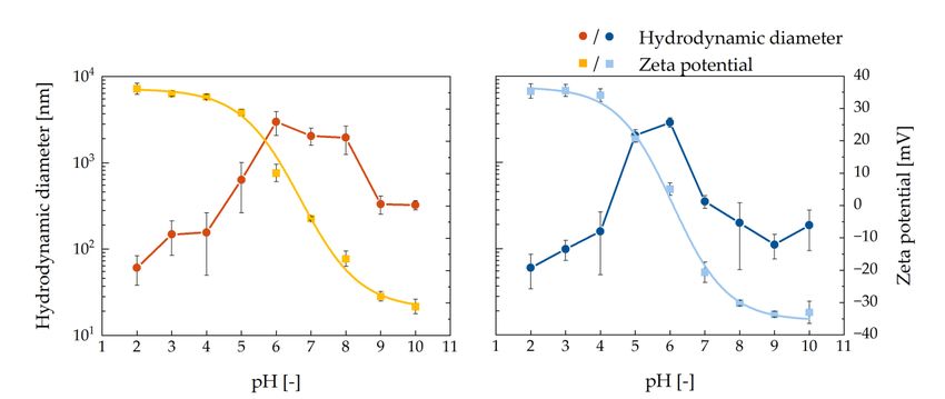
Magnetochemistry 2021, 7, 80                                                                                           2 of 13

                               of a permanent magnet with a high magnetic field gradient is more advantageous, as
                               Alnaimat et al. summarize [15].
                                      For LGMS or HGMS applications, such as in microfluidics [16] or in medicine [17–22],
                               it is important to have a defined and stable particle size. Hence, MNPs need to meet the
                               following requirements. On the one hand, they should be colloidal stable and not form
                               aggregates so that they remain their defined size during fractionation or separation [5].
                               Particularly, for medical purposes, such as hyperthermia treatment or magnetic resonance
                               imaging, the particle diameter should be smaller than 100 nm without forming large
                               aggregates [17,19,23,24]. On the other hand, it should be possible to isolate the colloidal
                               stable MNPs with the respective magnetic field gradient [25].
                                      Regarding downstream processing, the handled liquids, e.g., cell lysates or hy-
                               drolysates coming out of fermentation, pose another challenge. Viscosity becomes an
                               essential factor, influencing the separation performance [26–28]. Furthermore, in medical
                               applications, the medium blood exhibits a higher viscosity, being fivefold higher than
                               the one of water [29]. In addition to the higher viscosity, serum proteins or other blood
                               components might influence the aggregation behavior [20,21]. However, the so-called
                               biocorona, which is determined by the liquid components such as biomolecules, bacterial
                               debris, proteins, or lipids, could also prevent the aggregation of MNPs [30]. Socoliuc
                               et al. recently emphasized the importance of characterizing the aggregation behavior
                               of MNPs in the respective medium, e.g., cell culture media, and not only in water [18].
                               Besides the intrinsic characteristics of the working liquid, an increase in viscosity could
                               be advantageous for the microfluidic processing of magnetic particles [31]. In order to
                               avoid sedimentation effects during the process, Solsona et al. developed a microfluidic chip
                               for the magnetophoretic sorting of single-catalyst particles composed of iron [31]. Hence,
                               working with different viscosities might facilitate process handling, or a specific medium
                               might give another viscosity. We would like to point out that besides other parameters,
                               e.g., pH, the viscosity might influence the particle aggregation behavior as well.
                                      Concerning an LGMS process, the control of aggregation is important, as it has
                               been shown that, despite low magnetic field gradients, the particles can be separated,
                               mainly due to cooperative magnetophoresis [32–34]. By extending the DLVO theory with
                               the magnetic interaction, this phenomenon can be explained [35]. Here, particles form
                               aggregates because of the magnetic field gradient, as their magnetic dipoles align, and
                               the cooperative effect speeds up the magnetophoretic separation. According to Faraudo
                               et al., these aggregates are reversible for high zeta potentials. Electrostatic repulsion and
                               magnetic attraction form a secondary minimum besides the primary one, separated by a
                               potential barrier [35]. The magnetic Bjerrum length poses a parameter, which enables the
                               evaluation of aggregation throughout a magnetophoretic process. It describes the distance
                               of two parallel dipoles, where the magnetic force equals the thermal energy. Due to this
                               parameter, it can be estimated if the magnetic dipoles are interacting with each other or not.
                                                                                     1
                                                                           µ 0 · m2 3
                                                                     λB =                                                 (1)
                                                                          2·π·kB ·T

                               The Bjerrum length is dependent on the permeability of free space µ0 = 4π × 10−7 H m−1 ,
                               the Boltzmann’s constant kB = 1.381 × 10⁻²³ J K−1 , the temperature T, and the magnetic
                               dipole, which is written as
                                                                              4
                                                                  m = MS ·ρp · ·π·r3 ,                                 (2)
                                                                              3
                               where the saturation magnetization per unit mass of colloid is MS , the particle density is
                               ρP , and the particle radius is r.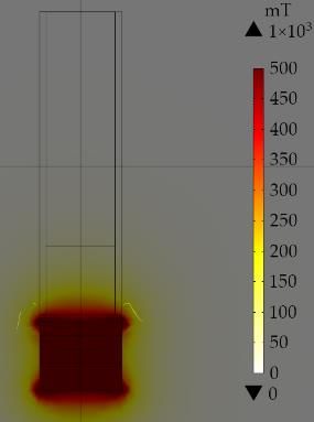
Magnetochemistry 2021, 7, x FOR PEER REVIEW 3 of 13
Magnetochemistry 2021, 7, 80 3 of 13
This study emphasizes the importance of understanding the aggregation behavior of
bare iron oxide nanoparticles (BIONs) in different viscosities. The influence on a separa-
This study
tion process emphasizes
in a low magneticthe importance
field gradient isofinvestigated
understanding themagnetophoretic
by the aggregation behavior
sed-
of bare iron oxide nanoparticles (BIONs) in different viscosities.
imentation velocity with a sedimentation analyzer. The following hypotheses The influence are
on apro-
sep-
aration
posed: process in a low magnetic field gradient is investigated by the magnetophoretic
sedimentation velocity with a sedimentation analyzer. The following hypotheses are
1.proposed:
The aggregation behavior of BIONs in the gravity field is dependent on pH and vis-
cosity, respectively. Therefore, the colloidal stability can be selectively controlled.
1. The aggregation behavior of BIONs in the gravity field is dependent on pH and
2. During magnetophoresis, these effects directly influence the separation process. The
viscosity, respectively. Therefore, the colloidal stability can be selectively controlled.
aggregate size, as well as viscosity, result in different magnetophoretic velocities.
2. During magnetophoresis, these effects directly influence the separation process. The
Four different
aggregate pHasvalues
size, well asbetween 4 and
viscosity, 9, in
result which clearly
different show the pH’s influence
magnetophoretic velocities.on
aggregate size and therefore on magnetophoretic sedimentation velocity, are chosen. The
Four different pH values between 4 and 9, which clearly show the pH’s influence on
viscosity of water (η = 0.888 mPa s) is compared with the 2.5-fold viscosity. A higher vis-
aggregate size and therefore on magnetophoretic sedimentation velocity, are chosen. The
cosity can be beneficial for magnetophoretic sorting processes, as field-induced aggrega-
viscosity of water (η = 0.888 mPa s) is compared with the 2.5-fold viscosity. A higher viscos-
tion and convection only play a minor role at elevated viscosities [31,33]. The higher vis-
ity can be beneficial for magnetophoretic sorting processes, as field-induced aggregation
cosity is obtained by adding sucrose (η = 2.227 mPa s).
and convection only play a minor role at elevated viscosities [31,33]. The higher viscosity
is obtained by adding sucrose (η = 2.227 mPa s).
2. Results and Discussion
2. Results
2.1. and Discussion
Particle Characterization
2.1. BIONs
Particleare
Characterization
synthesized via coprecipitation. They exhibit a high saturation magneti-
zationBIONs
of 70.84 Am
are 2 kg−1 (Figure
synthesized via 1a), no remanence
coprecipitation. andexhibit
They no hysteresis at 0 Oe [36–38].
a high saturation The
magnetiza-
chemical Am2 kg−and
composition
tion of 70.84 1 (Figure 1a), no remanence and no hysteresis at 0 Oe [36–38]. The
the crystalline spinel structure were previously described by
chemical composition
Schwaminger et al. [39].and
With theTransmission
crystalline spinel structure
Electron were previously
Microscopy (TEM) (Figuredescribed by
1b), the
Schwaminger
optical diameteretofal.
the[39]. With
BIONs is Transmission Electron
examined, resulting Microscopy
in an (TEM)
average single (Figure
particle 1b), the
diameter
ofoptical diameter
9.92 nm, whichof
is the BIONs
similar is examined,
to previous resulting in of
measurements anBIONs
average single particle diameter
[38–40].
of 9.92 nm, which is similar to previous measurements of BIONs [38–40].
(a) (b)
Figure
Figure1.1.(a)
(a)Superconducting
Superconductingquantum
quantum interference
interference (SQUID) measurement for
(SQUID) measurement for the
thebare
bareiron
ironoxide
oxidenanoparticles
nanoparticles(BIONs)
(BIONs)at
at300
300KKwith
witha aLangevinMod
LangevinModfit.fit.(b)
(b)Transmission
Transmission Electron Microscopy (TEM) image of the BIONs.
Electron Microscopy (TEM) image of the BIONs.
2.2. Influencing the Colloidal Stability of the BIONs due to Viscosity and pH
2.2. Zeta
Influencing the is
potential Colloidal Stability ofpotential
the electrostatic the BIONsat due to Viscosityslipping
the particle’s and pH plane, which pre-
Zeta
sents the potential
interface is the the
between electrostatic potential
moving fluid and theat fluid
the particle’s slipping
attached to plane,
the particle which
surface.
presents the interface between the moving fluid and the fluid attached to the
Therefore, the zeta potential is used as a relative measurand for the surface potential and, particle
surface.
thus, Therefore,
for the magnitude theofzeta potentialcharge.
a particle’s is usedDueas to
a relative
the extentmeasurand
of electricfor the surface
repulsion be-
potential
tween and, thus,infor
the particles thethe magnitude
solution, of a particle’s
the colloidal charge.
stability can beDue to the extent
evaluated. of electric
The repulsion
repulsion
energy between
is thereby the particles
dependent on in
thethe solution,radius
particles’ the colloidal
and thestability
extent ofcan be evaluated.
shielding, whichTheis
repulsion energy is thereby dependent on the particles’ radius and the extent of shielding,
which is affected by the ionic strength of the solvent [41]. Figure 2 illustrates the course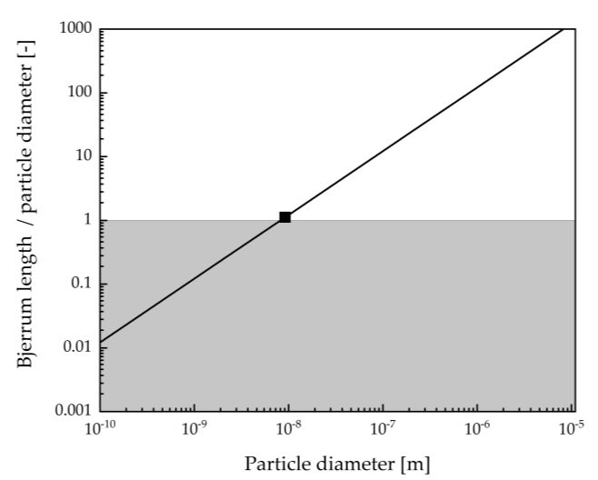
Magnetochemistry 2021, 7, x FOR PEER REVIEW 4 of 13
Magnetochemistry 2021, 7, 80 affected by the ionic strength of the solvent [41]. Figure 2 illustrates the course 4of zeta
of 13
potential versus pH and presents the amphiphilic character of the BIONs’ surface. At very
low and high pH, it ranges between 20 and 35 mV or −20 and −35 mV, meaning the BIONs’
net charge is positive or negative, respectively. Thus, the particles’ repulsion is high, and
of zeta
they potential versus
are colloidally stable,pH and presents
leading to less the amphiphilic
aggregation. The character of the BIONs’
hydrodynamic diameter surface.
meas-
At very low and high pH, it ranges between 20 and 35 mV or −20 and −35 mV, meaning
urement goes in line with this assumption, as it shows low diameters for low and high pH
the BIONs’ net charge is positive or negative, respectively. Thus, the particles’ repulsion
values. While the isoelectric point (IEP), where the potential is 0, is 6.69 for the low viscous
is high, and they are colloidally stable, leading to less aggregation. The hydrodynamic
solution (η = 0.888 mPa s) (Figure 2a), it is 6.07 for the high viscous solution (η = 2.227 mPa
diameter measurement goes in line with this assumption, as it shows low diameters for low
s) and
(Figure
high2b)
pH[42]. Here,
values. Whilethetheparticles formpoint
isoelectric aggregates up tothe
(IEP), where 3000 nm in is
potential both
0, isliquids
6.69 forbe-
cause of their
the low low
viscous superficial
solution charge.
(η = 0.888 mPaComparing
s) (Figure 2a), the particle
it is 6.07 fordistributions
the high viscous of both solu-
solution
tions,
(η = 2.227 mPa s) (Figure 2b) [42]. Here, the particles form aggregates up to 3000 nm in both In
the one with the low viscosity (Figure 2a) exhibits a wide distribution over pH.
contrast,
liquids for the other
because onelow
of their (Figure 2b), large
superficial aggregates
charge. Comparing can only be observed
the particle at pH 5of
distributions and
6. both
Thus, in the higher
solutions, the one viscous
with the solution, the colloidal
low viscosity (Figure stability
2a) exhibits of the BIONs
a wide is given over
distribution over a
broader pH range
pH. In contrast, forfrom pH 2one
the other to 4(Figure
and pH 2b),7large
to 10.aggregates
In addition, can the
onlypolydispersity
be observed at pH index
5 and 6. Thus, in the higher viscous solution, the colloidal stability
(PDI) of the measured samples (Figure S1) confirms these results, as it is between 0.18 ± of the BIONs is given
over
0.09 a broader
and pHfor
0.24 ± 0.04 range from viscous
the high pH 2 to solution,
4 and pHwhereas
7 to 10. In addition,
it is betweenthe polydispersity
0.22 ± 0.03 and 0.54
indexfor
± 0.20 (PDI) of the
the low measured
viscous samples
solution. (Figure S1)
Compared confirms
to the thesearesults,
literature, PDI belowas it 0.7
is between
is consid-
0.18 ± 0.09 and 0.24 ± 0.04 for the high viscous solution, whereas
ered as nearly monodisperse [43]. For pH 2, 3, 9, and 10, the PDI for both solutions it is between 0.22 ± 0.03 is
and 0.54 ± 0.20 for the low viscous solution. Compared to the literature,
similar; however, for the low viscous samples, the PDI at pH 5, 6, 7, and 8 is higher. a PDI below 0.7 This
is
considered as nearly monodisperse [43]. For pH 2, 3, 9, and 10, the PDI for both solutions
indicates that the BIONs around the IEP possess a higher heterogeneity in aggregate size.
is similar; however, for the low viscous samples, the PDI at pH 5, 6, 7, and 8 is higher. This
Additionally, this effect can be seen in the intensity distributions of all samples in Figures
indicates that the BIONs around the IEP possess a higher heterogeneity in aggregate size.
S2 and S3. For the water solution, heterogeneous aggregates are detected between pH 4
Additionally, this effect can be seen in the intensity distributions of all samples in Figures
andS2 8,
andwhile thethe
S3. For particle
water distributions for the higher
solution, heterogeneous viscosity
aggregates areare uniform.
detected Particularly,
between pH 4
forand
low8, pH values,
while the particles
the particle might for
distributions reactthewith the viscosity
higher acid andare therefore
uniform. possess a higher
Particularly,
stability.
for low pH values, the particles might react with the acid and therefore possess a higher
stability.
(a) (b)
Figure 2. Hydrodynamic
Figure 2. Hydrodynamic diameter
diameterand
andzeta
zetapotential
potentialmeasurements
measurements of BION suspension
of BION suspensionininpH
pH2 2toto1010
(a)(a)
in in deionized
deionized
water (η =(η0.888
water mPa
= 0.888 s) s)
mPa (b)(b)
and sucrose
and sucrosesolution
solution(η
(η==2.227
2.227 mPa
mPa s).
s).
Theanalysis
The analysisofofBIONs
BIONsinin other
other sugar solutions,
solutions,such
suchasasfructose
fructoseand and glucose, at at
glucose, pHpH 7 7
resultedininsimilar
resulted similarparticle
particledistributions
distributions asas sucrose
sucrose(Figure
(FigureS4).
S4).InInthe
theliterature, thethe
literature, effect
effect
onon thecolloidal
the colloidalstability
stabilityof
ofparticles
particles due
due to
to higher
higherviscosity
viscositybybysugars,
sugars,suchsuchasas
sucrose,
sucrose,is is
confirmed. Previously, Szalai et al. synthesized ex situ coated magnetite nanoparticles
confirmed. Previously, Szalai et al. synthesized ex situ coated magnetite nanoparticles with
gelatin
with and and
gelatin sucrose. They They
sucrose. reported higherhigher
reported colloidal stabilitystability
colloidal of their particles
of their when adding
particles when
sucrose [44]. Sun et al. propose that the multiple hydroxyl groups, as they are present in
adding sucrose [44]. Sun et al. propose that the multiple hydroxyl groups, as they are
sucrose, can adsorb or chelate onto the magnetite surface, resulting in steric hindrance, as it
present in sucrose, can adsorb or chelate onto the magnetite surface, resulting in steric
is known to conventional surfactants [45,46]. Benítez et al. describe another effect of sugars,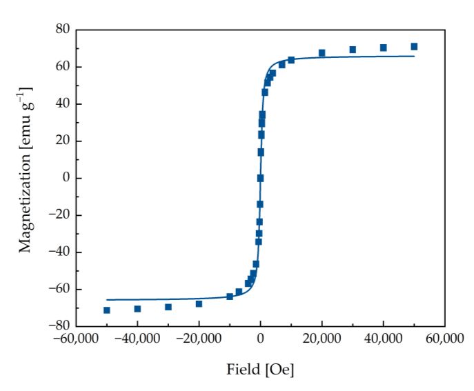
hindrance, as it is known to conventional surfactants [45,46]. Benítez et al. describe an-
Magnetochemistry 2021, 7, 80 other effect of sugars, where they claim that sucrose might influence particles’5 ofcolloidal13
stability because of the hydration capacity. They extend the DLVO theory by the hydra-
tion repulsive energy, which occurs as water molecules around the particle surface restrict
them in their
where motion,
they claim thatknown
sucroseas hydration
might pressure
influence [47,48].
particles’ Westability
colloidal assumebecause
that stabilization
of the
due to the higher
hydration viscosity
capacity. withthe
They extend sucrose
DLVOistheory
reached because
by the of arepulsive
hydration synergyenergy,
of the which
mentioned
occurs as water molecules around the particle surface restrict them in their motion, known
effects.
as hydration pressure [47,48]. We assume that stabilization due to the higher viscosity with
2.3.sucrose is reached
Dependence of thebecause of a synergy
Magnetophoretic of the mentioned
Velocity effects.
on Aggregate Size and Viscosity
2.3.The effect ofofpH
Dependence and viscosity on
the Magnetophoretic the aggregation
Velocity behavior
on Aggregate Size of our BIONs indicates
and Viscosity
that it is essential to evaluate the influence of different aggregate
The effect of pH and viscosity on the aggregation behavior of sizes
our regarding
BIONs indi-a mag-
netophoresis-based separation
cates that it is essential process.
to evaluate The Space-and-Time-resolved
the influence of different aggregate sizesExtinction
regardingProfiles
a
(STEP) technology is based
magnetophoresis-based on the process.
separation sedimentation of particles due to magnetophoresis.
The Space-and-Time-resolved Extinction Pro- It
files (STEP) technology is based on the sedimentation of particles due to magnetophoresis.
describes the particles’ movement towards the magnetic field direction if the buoyancy
andIt friction
describesforce
the particles’
is overcomemovement
(Figuretowards
3). the magnetic field direction if the buoyancy
and friction force is overcome (Figure 3).
(a) (b) (c)
Figure 3. (a)3.Schematic
Figure illustration
(a) Schematic illustrationof ofthe sedimentation
the sedimentation analyzer (LUMiReader)
analyzer (LUMiReader) with
with thethe involved
involved forces
forces (magnetic
(magnetic force force
Fmag, the
Fmaggravitational forceforce
, the gravitational Fgrav,Fand
grav , the
anddrag
the force
drag F
force
drag).
F (b)
drag Experimental
). (b) set-up.
Experimental The
set-up. cuvette
The filled
cuvette with
filled sample
with samplefluid is
placed on a magnet, and parallel light with a wavelength of 870 nm transmits the sample. A light
fluid is placed on a magnet, and parallel light with a wavelength of 870 nm transmits the sample. A light sensor records sensor records transmis-
sion profiles,
transmissionand profiles,
the magnetophoretic velocity of velocity
and the magnetophoretic the particles
of theisparticles
calculated. The magnetic
is calculated. flux density
The magnetic in the direction
flux density in the of
the particle
directionmovement is 40movement
of the particle mT at theissample top
40 mT at theand 350 top
sample mT andat the
350sample bottom.
mT at the sample(c) Calculated
bottom. magnetic
(c) Calculated flux density
magnetic
alongflux
thedensity
y-axis. along
Calculations
the y-axis.were performed
Calculations with
were COMSOL
performed withMultiphysics 5.6.
COMSOL Multiphysics 5.6.
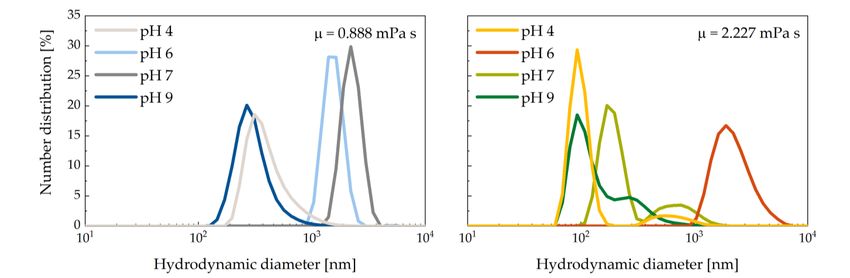
Figure
Figure 4 4presents
presentsthe the magnetophoretic
magnetophoretic velocityvelocityderived
derivedfromfromSTEPs overover
STEPs pH 4,pH6, 7,
4, 6, 7,
and 9 at η = 0.888 and η = 2.227 mPa s. For pH 4 and 9, the magnetophoretic
and 9 at η = 0.888 and η = 2.227 mPa s. For pH 4 and 9, the magnetophoretic velocity velocity is is
noticeably lower than for pH 6 and 7, which goes in line with the measured particle size
noticeably lower than for pH 6 and 7, which goes in line with the measured particle size
distributions via dynamic light scattering (DLS) measurement (Figure 5). The latter pH
distributions
values showvia dynamic
around lightofscattering
one power (DLS) measurement
ten higher diameters than pH 4 and(Figure
9 because 5).the
The latter pH
surface
values
of theshow
BIONs around one positively
is charged power of or ten higher diameters
negatively, than
respectively. pH 4the
Hence, and 9 because
attractive the sur-
forces
face of the BIONs is charged positively or negatively, respectively.
major the repulsive ones so that the particles do not resist aggregation. The same pattern Hence, the attractive
forces major
can be the repulsive
observed for the highones so that
viscous the particles
samples. Here, atdopHnot resist isaggregation.
6, which almost the IEP Theof same
the BIONs,
pattern can beaggregates
observedwith a hydrodynamic
for the high viscousdiameter 2764 ±
samples.ofHere, at 446
pH nm show aishigher
6, which almost the
IEPmagnetophoretic
of the BIONs,velocity in contrast
aggregates with toa other pH values where
hydrodynamic the hydrodynamic
diameter of 2764 ± 446 diameter
nm show a
is smaller (Figure 5b). Here, for pH 4, 7, and 9, a second mode with
higher magnetophoretic velocity in contrast to other pH values where the hydrodynamic larger aggregates is
detected, which is not visible in the intensity distributions (Figure S2). Even if the amount
diameter is smaller (Figure 5b). Here, for pH 4, 7, and 9, a second mode with larger aggre-
of these larger aggregates is lower compared to the first mode, they might influence the
gates is detected,
measured which is not
magnetophoretic visibleThe
velocity. in the intensity
velocity distributions
differences are lower (Figure
comparedS2). to
Even
the if the
amount of these
less viscous largerFollowing
samples. aggregates is lower
Equation compared
(7), the particle toradius
the first mode, proportional
is directly they might influ-
ence themagnetophoretic
to the measured magnetophoretic
force during thevelocity. The velocity
sedimentation differences
process. Therefore, theare lower com-
measured
pared to theare
velocities less viscous
in line with samples. Following
the theoretical Equation (7), the particle radius is directly
assumptions.
proportional to the magnetophoretic force during the sedimentation process. Therefore,
the measured velocities are in line with the theoretical assumptions.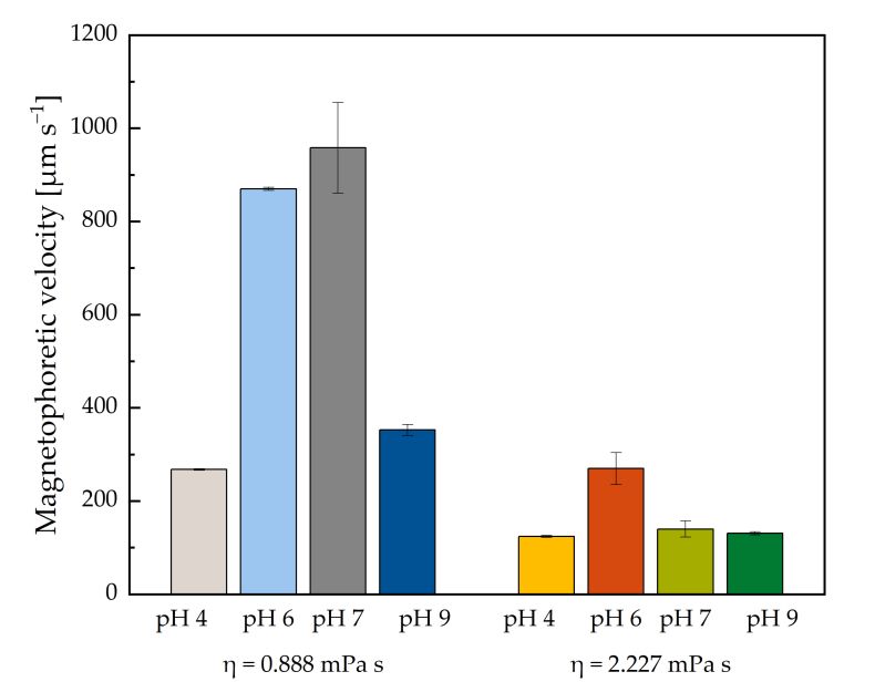
Magnetochemistry 2021, 7, x FOR PEER REVIEW 6 of 13
Magnetochemistry 2021, 7, 80 6 of 13
Magnetochemistry 2021, 7, x FOR PEER REVIEW 6 of 13
Figure
Figure
Figure4. Magnetophoretic
4.4.
Magnetophoretic
Magnetophoreticvelocity over
velocity
velocity pHpH
over
over 4, 6,
pH 7,
4,4, and
6,6,
7, 7,
and9and
of9 BION
9 of
of suspension
BION
BION in deionized
suspension
suspension water
in deionized
in deionized waterwater
(η(η
(η == 0.888
0.888 mPa
= 0.888mPas)s)and
mPa sucrose
s)and
and solution
sucrose
sucrose (η =(η
solution
solution 2.227
(η mPamPa
== 2.227
2.227 s) with
mPa awith
corresponding
s)s)with meanmean
a acorresponding
corresponding diameter
mean of of of
diameter
diameter
DLS
DLSDLSmeasurement.
measurement.
measurement.
(a) (b)
Figure Figure
5. Particle size distribution
5. Particle at pH at
size distribution 4, pH
6, 7,4,and
6, 7, 9and
of BION suspension
9 of BION (a) in
suspension (a)deionized water
in deionized (η =(η0.888
water mPa s) (b) and
sucrosesucrose
solutionsolution
(η = 2.227 mPa
(a)
s).
(b)= 0.888 mPa s) (b) and
(η = 2.227 mPa s).
Figure 5. Particle size distribution at pH 4, 6, 7, and 9 of BION suspension (a) in deionized water (η = 0.888 mPa s) (b) and
sucrose solution (η = 2.227 mPa s). Besides
Besidesthe theaggregate
aggregatesize,size,viscosity
viscosity is is another parameter
parameterthat thatinfluences
influencesthe theseparation
separa-
tion process.
process. TheThe magnetophoretic
magnetophoretic velocities
velocities of allofmeasured
all measured pH values
pH values are lower
are lower for the forhigher
the
higher viscosity
viscosity (η = (η
2.227 = 2.227
mPa mPa
s) s)
compared compared
to the to the
lower lower
one (η
Besides the aggregate size, viscosity is another parameter that influences theone
= (η
0.888 = 0.888
mPa s),mPa
as thes), as the separa-
viscosity
is indirectly
viscosity proportional
is indirectly to the velocity,
proportional as written
to the velocity, in Equation
as written (10). Due
in Equation (10).toDuetheto higher
the
tion process. The magnetophoretic velocities of all measured pH values are lower for the
viscosity,
higher the drag
viscosity, theforce
dragincreases and slows
force increases anddownslowsthe downparticle motion along
the particle motionthe magnetic
along the
higher
field
viscosity The
gradient.
(η =gravitational
2.227 mPa s) compared
force does not
toinfluence
the lower this
one (η =as0.888
effect, values
mPafor
s), as the
the
magnetic field gradient. The gravitational force does not influence this effect, as values for
viscosity is
sedimentation
the indirectly
sedimentationvelocity proportional
velocityshowshowvery to
verylow the
lowvalues velocity,
values for as written
for both viscosities in Equation
viscosities (Table
(Table 1). (10).
1). Here,
Here,theDue
the to the
higher viscosity,
sedimentation
sedimentation the drag
velocity
velocity is is force
similar
similar increases
when
when having
having and the
the slows
same
same down
size
size ofof the particle
aggregates
aggregates atat motionpH
different
different along
pH the
magnetic
valuesonly
values field
only inina gradient.
agravitationalThefield.
gravitational gravitational
field. force does not influence this effect, as values for
the sedimentation
Takingthese
Taking velocity
thesefindings
findings show very
together,
together, low
theviscosity
the values
viscosity for both
influences
influences viscosities
the
the (Tableof1).
colloidalstability
colloidal stability Here,
ofthe
the the
particles
sedimentationand the
particles and thevelocity magnetophoresis process
is similar process
magnetophoresis when havingitself. These effects
the same
itself. These should
size
effects be taken
of aggregates
should into
be taken at account
intodifferent
ac- pH
whenwhen
count
values working
only in awith
working high viscous
with high viscous
gravitational liquids,
field. such as
liquids, cellas
such lysates or blood,
cell lysates in order
or blood, into obtain
order to a
distinct
obtain separation
a distinct process.process.
separation
Taking these findings together, the viscosity influences the colloidal stability of the
particles and the magnetophoresis process itself. These effects should be taken into ac-
count when working with high viscous liquids, such as cell lysates or blood, in order to
obtain a distinct separation process.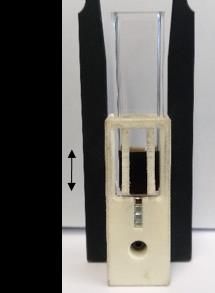
Magnetochemistry 2021, 7, x FOR PEER REVIEW 7 of 13
Table 1. Measurement of the sedimentation velocity and the corresponding hydrodynamic diameter of the samples in the
Magnetochemistry
sedimentation 2021, 7, 80
analyzer. 7 of 13
η = 0.888 mPa s η = 2.227 mPa s
Sedimentation Velocity
Table 1. Measurement Hydrodynamic
of the sedimentation Diameter
velocity Sedimentation
and the corresponding Velocity
hydrodynamic diameter ofHydrodynamic
the samples in the Diameter
pH
(µm s−1)
sedimentation analyzer. (nm) (µm s−1) (nm)
4 1.57 ± 0.09 156 ± 106 0.88 ± 0.70 158 ± 108
η = 0.888 mPa s η = 2.227 mPa s
6 3.82 ± 0.85 2986 ± 920 2.30 ± 0.01 2764 ± 446
Sedimentation Velocity Hydrodynamic Diameter Sedimentation Velocity Hydrodynamic Diameter
7 pH 3.69 ± 0.14(µm s−1 ) 2047 ± 461
(nm)
0.33 ± 0.28
(µm s−1 ) (nm)
351 ± 58
9 1.43 ± 0.37 331 ± 79 0.31 ± 0.06 111 ± 35
4 1.57 ± 0.09 156 ± 106 0.88 ± 0.70 158 ± 108
6 3.82 ± 0.85 2986 ± 920 2.30 ± 0.01 2764 ± 446
7 3.69 ± 0.14 Regarding 2047
a LGMS
± 461process, the particle
0.33 ±size
0.28 poses an essential
351factor
± 58 in the kinetics
9 1.43 ± 0.37of the separation and the resulting process efficiency. As mentioned±above,
331 ± 79 0.31 ± 0.06 111 35 mainly the
cooperative effect of magnetic aggregation speeds up the magnetophoretic motion of the
Regarding
particles a LGMS
[49]. Here, theprocess,
Bjerrumthe particle
length cansize
beposes
used anin essential
order to factor
evaluatein the kinetics
the formation of
of the separation and the resulting process efficiency. As mentioned
aggregates in a magnetic field [9,50]. Therefore, we calculate the ratio of the above, mainly theBjerrum
cooperative effect of magnetic aggregation speeds up the magnetophoretic motion of the
length λB and the particle diameter d (Figure 6). As seen in Figure 6, particles do not form
particles [49]. Here, the Bjerrum length can be used in order to evaluate the formation
aggregates in a magnetic field if λB d−1 < 1 with a saturation magnetization of 70.84 Am2
of aggregates in a magnetic field [9,50]. Therefore, we calculate the ratio of the Bjerrum
kg −1 [49]. For our BIONs with a particle diameter of 9.92 nm, the ratio is 1.12, which implies
length λB and the particle diameter d (Figure 6). As seen in Figure 6, particles do not form
magnetic
aggregatesaggregate formation.
in a magnetic field if λBThis
d−1 value,
< 1 withmeasured bymagnetization
a saturation TEM, is close of to one;
70.84 Am however,
2
kg−is
this the minimal
1 [49]. limit,with
For our BIONs as the particle
a particle diameter
diameter in nm,
of 9.92 suspension
the ratio isis1.12,
>100which
nm, depending
implies on
the bufferaggregate
magnetic conditions. Hence,This
formation. thevalue,
magnetophoretic
measured by TEM,velocity might
is close behowever,
to one; additionally
this influ-
is the minimal
enced by this limit, as but
effect, the particle
from adiameter
practicalin suspension
process view,is >100 nm, depending
cooperative on the
magnetophoresis
bufferbe
might conditions.
beneficialHence, the magnetophoretic
to implement such an LGMS velocity mightbecause
process be additionally influenced
the separation efficiency
by this
increases. effect, but from a practical process view, cooperative magnetophoresis might be
beneficial to implement such an LGMS process because the separation efficiency increases.
Figure 6.
Figure Log–log plot
6. Log–log plotofofthe
theratio of of
ratio thethe
Bjerrum
Bjerrumlength and and
length the particle diameter
the particle over the
diameter particle
over the particle
diameter. The shaded region corresponds to particle diameters where no
diameter. The shaded region corresponds to particle diameters where no aggregation dueaggregation due to ato a
magnetic field occurs, and the solid line describes the Bjerrum length for particles with
magnetic field occurs, and the solid line describes the Bjerrum length for particles with the same the same
magnetization. The
magnetization. Thesymbol
symbol represents
representsour our
particle size and
particle sizeindicates that magnetic
and indicates aggregation
that magnetic takes
aggregation
place (transparent region).
takes place (transparent region).
Besides the cooperative magnetophoresis, Leong et al. recently discussed the effect of
Besides the cooperative magnetophoresis, Leong et al. recently discussed the effect
convective magnetophoresis [33,41]. They introduce the dimensionless Grashof number
ofGrconvective magnetophoresis [33,41]. They introduce the dimensionless Grashof num-
m (Equation (3)), which is dependent on the magnetic field gradient ∇B, the volumetric
ber Grm (Equation (3)), which is dependent on the magnetic field gradient ∇B, the volu-
metric magnetization M, the particle concentration difference between the bulk solution,
the collection plane cs − c∞ and the length of the analyzed system Lc.Magnetochemistry 2021, 7, 80 8 of 13
magnetization M, the particle concentration difference between the bulk solution, the
collection plane cs − c∞ and the length of the analyzed system Lc .
3
ρ·∇B· ∂M∂c H ·(cs − c∞ )·Lc
Grm = (3)
η2
It describes the convection induced by an external magnetic field, which happens due
to the rapid acceleration of the particles. Leong et al. state that the Grashof number is mostly
dependent on the concentration, resulting in a higher magnetophoretic velocity for higher
particle concentrations. However, in our study, all parameters are kept constant besides
the viscosity. Its square is indirectly proportional to the dimensionless number, so one can
say that an increase in viscosity lowers the convection and therefore, the magnetophoretic
velocity as well.
In addition to both discussed effects, the cooperative and the convective magne-
tophoresis, the diffusion, described by the Stokes–Einstein Equation, is influenced by
viscosity as shown in Equation (4) [51]:
kB ·T
Do = (4)
6·π·r·η
According to Equation (4), the diffusion coefficient is decreasing with a higher viscosity
and thus, Brownian motion is lower as well. Hence, it evolves that viscosity noticeably
influences various parameters during a magnetophoresis-based process and should be
always considered.
To sum up, the colloidal stability of BIONs is controllable due to a change in pH
value, but also a higher viscosity leads to stabilization. The former is based on electrostatic
stabilization effects, whereas the various latter effects, such as a steric hindrance and/or
hydration repulsion due to sucrose, might be involved. The different aggregate sizes and
the viscosity influences the magnetophoresis process (Equations (5)–(10)). Particularly,
for a separation process with high viscous liquids, the efficiency is lower [26]. Moreover,
depending on the aggregate size, MNP–MNP interactions, as cooperative or convective
magnetophoresis and Brownian motion, have to be considered, as these effects influence
magnetophoretic processes, e.g., a microfluidic fractionation.
3. Materials and Methods
Coprecipitation of Fe2+ /3+ ions was used to synthesize the BIONs [38]. For this,
28.9 g of sodium hydroxide (722 mmol, 4.10 equivalents (eq.), Carl Roth GmbH + Co.
KG, Karlsruhe, Germany) was dissolved in 400 mL of degassed water under a nitrogen
atmosphere. A solution, containing 86.6 g of FeCl3 ·6H2 O (320 mmol, 1.82 eq., Sigma
Aldrich Merck KGaA, Darmstadt, Germany) and 35.0 g of FeCl2 ·4H2 O (176 mmol, 1.0 eq.,
Sigma Aldrich Merck KGaA, Darmstadt, Germany) in 160 mL of degassed water, was
added slowly under continuous stirring. The temperature was kept constant at 27 ◦ C via
a water bath. A black precipitate built up immediately, and the reaction was continued
under stable conditions for a further 30 min. Then, the precipitate was washed ~15 times
with deionized water via magnetic decantation with a neodymium iron boron magnet in a
glass bottle until the conductivity was below 200 µS cm−1 . The BIONs were stored under
a nitrogen atmosphere at 4 ◦ C. The magnetic susceptibility was analyzed by the SQUID
device Quantum Design MPMS XL-7 (Quantum Design GmbH, Darmstadt, Germany).
Therefore, the particles were lyophilized, and then they were glued into a small tube. TEM
was performed with the JEM 1400 Plus microscope (JEOL GmbH, Freising, Germany), and
the recorded images were subsequently evaluated by using ImageJ software.
All experiments were performed either in water (η = 0.888 mPa s, 25 ◦ C) or in a 24%
(w/w) sucrose solution (η = 2.227 mPa s, 25 ◦ C, Carl Roth GmbH + Co. KG, Karlsruhe,
Germany), fructose solution (η = 1.9858 mPa s, Carl Roth GmbH + Co. KG), or glucose
solution (η = 2.1201 mPa s, AppliChem GmbH, Darmstadt, Germany). The pH wasMagnetochemistry 2021, 7, 80 9 of 13
adjusted by adding 0.1 M or 1 M sodium hydroxide (Carl Roth GmbH + Co. KG, Karlsruhe,
Germany) or hydrochloric acid (VWR International GmbH, Darmstadt, Germany). A
solution of 1 g L−1 BIONs was used, dispersed via an ultrasonication probe (5 min, 10 s on,
15 s off, 20%, Branson Ultrasonics Corporation, Danbury, United States of America). For
zeta potential and hydrodynamic diameter by DLS evaluation, a ZetaSizer XS (Malvern
Panalytical GmbH, Kassel, Germany) was used. Both measurements were performed
at 25 ◦ C in 1 mL of a 1 g L−1 solution in five and three measuring cycles in duplicates,
respectively. The IEP was determined by applying a Boltzmann fit.
STEPs were recorded in duplicates at an optical wavelength of 870 nm (LUMiReader,
LUM GmbH, Berlin, Germany) to calculate the magnetophoretic velocity. According to
Newton’s second law of motion, magnetophoresis is composed of the gravitational force
Fgrav , the magnetic force Fmag and the viscous drag force Fdrag , pointing in the opposite
direction of the former two forces.
mp du
= Fgrav + Fmag + Fdrag (5)
dt
Here, mp and u are the mass and the velocity of the particle, respectively. For simplicity,
the inertial term can be neglected [49,52].
0 = Fgrav + Fmag + Fdrag (6)
The magnetic force is determined by the magnetic field gradient ∇H, the permeabilities µ0
and µr , the relative permittivity K, and the particle radius r:
Fmag = 3·µ0 ·µr ·K·∇H2 ·2·π·r3 (7)
According to Stokes’ law, the movement of a spherical particle in an incompressible fluid
can be described as follows, where η corresponds to the fluid viscosity:
Fdrag = −6·u·π·η·r (8)
The gravitational force is based on Newton’s law of gravitation,
4
Fgrav = ρp – ρf ·g·π·r3 , (9)
3
which is dependent on the density difference of particle and fluid ρp − ρf and the free-fall
acceleration g. The force balance, as written in Equations (6)–(9), can be solved for the
velocity as written in Equation (10):
2· ρp –ρf ·g·r2 µ ·µr ·K·∇H2 ·r2
v= + 0 (10)
9·η 2·η
For the measurement, a disposable cuvette (1 × 1 × 4.4 cm) filled with 1 mL of solution
was placed onto a stack of five cylindrical neodymium boron ferrite (NdFeB) magnets
(diameter = 12 mm, height = 2 mm, N45, Webcraft GmbH, Gottmadingen, Germany).
The built-in temperature control assured a constant temperature of 25 ◦ C. The magnetic
flux density was measured with a Hall detector PCE-MF M 3000 (PCE Instruments UK
Limited, Southampton Hampshire, United Kingdom). The obtained transmittance profiles
were integrated over the sample height, leading to an integral transmittance for each
measurement time. For data evaluation, it was converted into an integral extinction
value, which is directly proportional to the particle concentration after Lambert–Beer’s
law. Particle movement due to the magnetic and gravitational force induces the change in
particle concentration. Therefore, the time-dependent variation of the extinction enablesMagnetochemistry 2021, 7, 80 10 of 13
calculating the magnetophoretic velocity vmag , where L is the mean length defined by half
of the sample height.
v= (11)
t
A cumulative distribution function φ (vmag ) is obtained by plotting the relative extinction
over the magnetophoretic velocity [53]. The relative extinction Erel is determined by
the initial extinction of the sample E0 and the minimal extinction Emin at the end of
magnetophoresis measurement.
E − Emin
Erel (t) = , (12)
E0 − Emin
For better comparability, the value at t0.5 was used, which is the distribution function’s
median. It describes the velocity of 50% of the particles at the time t0.5 , where the extinction
has fallen by half. The sedimentation analysis experiments were performed with a magnetic
flux density between 40 and 350 mT, which corresponds to a magnetization between 29.31
and 54.40 Am2 kg−1 .
4. Conclusions
BIONs with an average diameter of 9.2 nm were synthesized via coprecipitation,
showing a high saturation magnetization [38]. Zeta potential and hydrodynamic diameter
measurements showed that pH and viscosity influence the colloidal stability of the particles.
pH values close to the IEP resulted in large particle agglomerates; however, electrostatic
stabilization was observed for high and low pH values. The particle distribution over
different pHs narrowed due to the increase in viscosity with sucrose. This could be
explained by the hydration repulsion and the steric stabilization effect of sucrose. The
aggregate size and viscosity directly influence a magnetophoretic process. The particle
diameter is proportional to the magnetophoretic velocity, which results in high velocities
for large aggregates and lower velocities for small particle diameters. By increasing
the viscosity, the drag force counteracts the magnetophoretic force and decreases the
magnetophoretic velocity of all aggregate sizes. This study emphasizes the underestimated
effect of viscosity by using the simple method of sedimentation analysis, in addition to
zeta potential and DLS measurement. In downstream processing or medical applications,
liquids such as cell suspension or blood will affect particle migration, resulting in a lower
separation. Particularly, the stabilizing effect of sucrose should be further examined, as
the formation of aggregates is one of the main reasons for using different coatings for the
steric stabilization of the BIONs [54]. In further studies, these new findings can be used to
control the aggregation behavior for applications in medicine or biotechnology.
Supplementary Materials: The following are available online at https://www.mdpi.com/article/
10.3390/magnetochemistry7060080/s1, Figure S1: Polydispersity Index of BION suspension from
pH 2 to 10 in deionized water (η = 0.888 mPa s) and sucrose solution (η = 2.227 mPa s), Figure S2:
Intensity distribution data of pH 2 to 10 for high viscous BION suspension. Technical triplicates are
shown for each duplicate, Figure S3: Intensity distribution data of pH 2 to 10 for low viscous BION
suspension. Technical triplicates are shown for each duplicate, Figure S4: Number distribution of
BIONs in fructose, glucose, and sucrose solution at pH 7.
Author Contributions: Conceptualization, L.W. and S.P.S.; methodology, L.W. and C.T.; software,
L.W.; validation, L.W.; formal analysis, L.W., C.T.; investigation, L.W.; resources, S.P.S.; data curation,
L.W.; writing—original draft preparation, L.W.; writing—review and editing, L.W. and S.P.S.; visual-
ization, L.W.; supervision, S.P.S., project administration, S.P.S.; funding acquisition, S.P.S. All authors
have read and agreed to the published version of the manuscript.
Funding: This research was funded by the German Research Foundation (DFG) (Project Number:
441672360). We appreciate the support from the German Research Foundation (DFG) and the
Technical University of Munich (TUM) in the framework of the Open-Access Publishing Program
and the support by TUM International Graduate School of Science and Engineering (IGSSE).Magnetochemistry 2021, 7, 80 11 of 13
Institutional Review Board Statement: Not applicable.
Informed Consent Statement: Not applicable.
Data Availability Statement: The original data related to this research can be asked any time to the
corresponding author’s email: l.wittmann@tum.de.
Acknowledgments: We would like to thank Sonja Berensmeier for laboratory resources and valuable
discussions. In addition, we acknowledge Matthias Opel for the SQUID measurement and Carsten
Peters for support with TEM imaging.
Conflicts of Interest: The authors declare no conflict of interest. The funders had no role in the design
of the study; in the collection, analyses, or interpretation of data; in the writing of the manuscript, or
in the decision to publish the results.
References
1. Fraga-García, P.; Kubbutat, P.; Brammen, M.; Schwaminger, S.; Berensmeier, S. Bare Iron Oxide Nanoparticles for Magnetic
Harvesting of Microalgae: From Interaction Behavior to Process Realization. Nanomaterials 2018, 8, 292. [CrossRef]
2. Schnell, F.; Kube, M.; Berensmeier, S.; Schwaminger, S.P. Magnetic Recovery of Cellulase from Cellulose Substrates with Bare Iron
Oxide Nanoparticles. ChemNanoMat 2019, 5, 422–426. [CrossRef]
3. Zhang, H.; Ding, W.; Li, S.; Ya, S.; Li, F.; Qiu, B. On-chip analysis of magnetically labeled cells with integrated cell sorting and
counting techniques. Talanta 2020, 220, 121351. [CrossRef]
4. Puddu, M.; Paunescu, D.; Stark, W.J.; Grass, R.N. Magnetically Recoverable, Thermostable, Hydrophobic DNA/Silica Encapsu-
lates and Their Application as Invisible Oil Tags. ACS Nano 2014, 8, 2677–2685. [CrossRef]
5. Ling, W.; Wang, M.; Xiong, C.; Xie, D.; Chen, Q.; Chu, X.; Qiu, X.; Li, Y.; Xiao, X. Synthesis, surface modification, and applications
of magnetic iron oxide nanoparticles. J. Mater. Res. 2019, 34, 1828–1844. [CrossRef]
6. Liu, S.; Yu, B.; Wang, S.; Shen, Y.; Cong, H. Preparation, surface functionalization and application of Fe3O4 magnetic nanoparticles.
Adv. Colloid Interface Sci. 2020, 281, 102165. [CrossRef]
7. Kodama, R. Magnetic nanoparticles. Magnetochemistry 2020, 6. [CrossRef]
8. Massart, R. Preparation of aqueous magnetic liquids in alkaline and acidic media. IEEE Trans. Magn. 1981, 17, 1247–1248.
[CrossRef]
9. Leong, S.S.; Yeap, S.P.; Lim, J. Working principle and application of magnetic separation for biomedical diagnostic at high- and
low-field gradients. Interface Focus 2016, 6, 20160048. [CrossRef]
10. Lozar, T.; Jesenko, T.; Prevodnik, V.K.; Cemazar, M.; Hosta, V.; Jericevic, A.; Nolde, N.; Kuhar, C.G. Preclinical and Clinical
Evaluation of Magnetic-Activated Cell Separation Technology for CTC Isolation in Breast Cancer. Front. Oncol. 2020, 10, 1–10.
[CrossRef]
11. Miltenyi, S.; Müller, W.; Weichel, W.; Radbruch, A. High gradient magnetic cell separation with MACS. Cytometry 1990, 11,
231–238. [CrossRef]
12. Fratzl, M.; Delshadi, S.; Devillers, T.; Bruckert, F.; Cugat, O.; Dempsey, N.M.; Blaire, G. Magnetophoretic induced convective
capture of highly diffusive superparamagnetic nanoparticles. Soft Matter 2018, 14, 2671–2681. [CrossRef]
13. Corchero, J.L.; Villaverde, A. Biomedical applications of distally controlled magnetic nanoparticles. Trends Biotechnol. 2009, 27,
468–476. [CrossRef] [PubMed]
14. Toh, P.Y.; Yeap, S.P.; Kong, L.P.; Ng, B.W.; Chan, D.J.C.; Ahmad, A.L.; Lim, J.K. Magnetophoretic removal of microalgae from
fishpond water: Feasibility of high gradient and low gradient magnetic separation. Chem. Eng. J. 2012, 211-212, 22–30. [CrossRef]
15. Alnaimat, F.; Karam, S.; Mathew, B.; Mathew, B. Magnetophoresis and Microfluidics: A Great Union. IEEE Nanotechnol. Mag.
2020, 14, 24–41. [CrossRef]
16. Robert, D.; Pamme, N.; Conjeaud, H.; Gazeau, F.; Iles, A.; Wilhelm, C. Cell sorting by endocytotic capacity in a microfluidic
magnetophoresis device. Lab. Chip 2011, 11, 1902–1910. [CrossRef] [PubMed]
17. Kievit, F.M.; Zhang, M. Surface Engineering of Iron Oxide Nanoparticles for Targeted Cancer Therapy. Acc. Chem. Res. 2011, 44,
853–862. [CrossRef]
18. Socoliuc, V.; Peddis, D.; Petrenko, V.I.; Avdeev, M.V.; Susan-Resiga, D.; Szabó, T.; Turcu, R.; Tombácz, E.; Vékás, L. Magnetic
Nanoparticle Systems for Nanomedicine—A Materials Science Perspective. Magnetochemistry 2020, 6, 2. [CrossRef]
19. Dulińska-Litewka, J.; Łazarczyk, A.; Hałubiec, P.; Szafrański, O.; Karnas, K.; Karewicz, A. Superparamagnetic Iron Oxide
Nanoparticles—Current and Prospective Medical Applications. Materials 2019, 12, 617. [CrossRef]
20. Ansari, S.A.M.K.; Ficiarà, E.; Ruffinatti, F.A.; Stura, I.; Argenziano, M.; Abollino, O.; Cavalli, R.; Guiot, C.; D’Agata, F. Magnetic
Iron Oxide Nanoparticles: Synthesis, Characterization and Functionalization for Biomedical Applications in the Central Nervous
System. Materials 2019, 12, 465. [CrossRef]
21. Vangijzegem, T.; Stanicki, D.; Laurent, S. Magnetic iron oxide nanoparticles for drug delivery: Applications and characteristics.
Expert Opin. Drug Deliv. 2019, 16, 69–78. [CrossRef]
22. Soetaert, F.; Korangath, P.; Serantes, D.; Fiering, S.; Ivkov, R. Cancer therapy with iron oxide nanoparticles: Agents of thermal and
immune therapies. Adv. Drug Deliv. Rev. 2020, 163-164, 65–83. [CrossRef] [PubMed]Magnetochemistry 2021, 7, 80 12 of 13
23. Belanova, A.A.; Gavalas, N.; Makarenko, Y.M.; Belousova, M.M.; Soldatov, A.V.; Zolotukhin, P.V. Physicochemical Properties
of Magnetic Nanoparticles: Implications for Biomedical Applications In Vitro and In Vivo. Oncol. Res. Treat. 2018, 41, 139–143.
[CrossRef]
24. Nam, J.; Huang, H.; Lim, H.; Lim, C.; Shin, S. Magnetic Separation of Malaria-Infected Red Blood Cells in Various Developmental
Stages. Anal. Chem. 2013, 85, 7316–7323. [CrossRef] [PubMed]
25. Pamme, N.; Wilhelm, C. Continuous sorting of magnetic cells via on-chip free-flow magnetophoresis. Lab. Chip 2006, 6, 974–980.
[CrossRef] [PubMed]
26. Roth, H.-C.; Prams, A.; Lutz, M.; Ritscher, J.; Raab, M.; Berensmeier, S. A high-gradient magnetic separator for highly viscous
process liquors in industrial biotechnology. Chem. Eng. Technol. 2016, 39, 469–476. [CrossRef]
27. Mishima, F.; Hayashi, S.; Akiyama, Y.; Nishijima, S. Development of a Superconducting High Gradient Magnetic Separator for a
Highly Viscous Fluid. IEEE Trans. Appl. Supercond. 2011, 22, 3700204. [CrossRef]
28. Gupta, A.K.; Gupta, M. Synthesis and surface engineering of iron oxide nanoparticles for biomedical applications. Biomaterials
2005, 26, 3995–4021. [CrossRef]
29. Coppola, L.; Caserta, F.; De Lucia, D.; Guastafierro, S.; Grassia, A.; Coppola, A.; Marfella, R.; Varricchio, M. Blood viscosity and
aging. Arch. Gerontol. Geriatr. 2000, 31, 35–42. [CrossRef]
30. Alphandéry, E. Bio-synthesized iron oxide nanoparticles for cancer treatment. Int. J. Pharm. 2020, 586, 119472. [CrossRef]
31. Solsona, M.; Nieuwelink, A.-E.; Meirer, F.; Abelmann, L.; Odijk, M.; Olthuis, W.; Weckhuysen, B.M.; Berg, A.V.D. Magnetophoretic
Sorting of Single Catalyst Particles. Angew. Chem. Int. Ed. 2018, 57, 10589–10594. [CrossRef] [PubMed]
32. Faraudo, J.; Andreu, J.S.; Calero, C.; Camacho, J. Predicting the Self-Assembly of Superparamagnetic Colloids under Magnetic
Fields. Adv. Funct. Mater. 2016, 26, 3837–3858. [CrossRef]
33. Leong, S.S.; Ahmad, Z.; Low, S.C.; Camacho, J.; Faraudo, J.; Lim, J. Unified View of Magnetic Nanoparticle Separation under
Magnetophoresis. Langmuir 2020, 36, 8033–8055. [CrossRef]
34. Yavuz, C.T.; Mayo, J.T.; Yu, W.W.; Prakash, A.; Falkner, J.C.; Yean, S.; Cong, L.; Shipley, H.J.; Kan, A.; Tomson, M.; et al. Low-Field
Magnetic Separation of Monodisperse Fe3O4 Nanocrystals. Science 2006, 314, 964–967. [CrossRef]
35. Faraudo, J.; Camacho, J. Cooperative magnetophoresis of superparamagnetic colloids: Theoretical aspects. Colloid Polym. Sci.
2009, 288, 207–215. [CrossRef]
36. Schwaminger, S.P.; Fraga-García, P.; Selbach, F.; Hein, F.G.; Fuß, E.C.; Surya, R.; Roth, H.-C.; Blank-Shim, S.A.; Wagner, F.E.;
Heissler, S.; et al. Bio-nano interactions: Cellulase on iron oxide nanoparticle surfaces. Adsorption 2016, 23, 281–292. [CrossRef]
37. Bean, C.P.; Livingston, J.D. Superparamagnetism. J. Appl. Phys. 1959, 30, S120–S129. [CrossRef]
38. Turrina, C.; Berensmeier, S.; Schwaminger, S. Bare Iron Oxide Nanoparticles as Drug Delivery Carrier for the Short Cationic
Peptide Lasioglossin. Pharmaceuticals 2021, 14, 405. [CrossRef]
39. Schwaminger, S.; Syhr, C.; Berensmeier, S. Controlled Synthesis of Magnetic Iron Oxide Nanoparticles: Magnetite or Maghemite?
Crystals 2020, 10, 214. [CrossRef]
40. Schwaminger, S.P.; Bauer, D.; Fraga-García, P.; Wagner, F.E.; Berensmeier, S. Oxidation of magnetite nanoparticles: Impact on
surface and crystal properties. CrystEngComm 2016, 19, 246–255. [CrossRef]
41. Schwaminger, S.P.; Schwarzenberger, K.; Gatzemeier, J.; Lei, Z.; Eckert, K. Magnetically Induced Aggregation of Iron Oxide
Nanoparticles for Carrier Flotation Strategies. ACS Appl. Mater. Interfaces 2021, 13, 20830–20844. [CrossRef] [PubMed]
42. Schwaminger, S.P.; Blank-Shim, S.A.; Scheifele, I.; Fraga-García, P.; Berensmeier, S. Peptide binding to metal oxide nanoparticles.
Faraday Discuss. 2017, 204, 233–250. [CrossRef] [PubMed]
43. Stetefeld, J.; McKenna, S.; Patel, T.R. Dynamic light scattering: A practical guide and applications in biomedical sciences. Biophys.
Rev. 2016, 8, 409–427. [CrossRef] [PubMed]
44. Szalai, A.J.; Manivannan, N.; Kaptay, G. Super-paramagnetic magnetite nanoparticles obtained by different synthesis and
separation methods stabilized by biocompatible coatings. Colloids Surfaces A Physicochem. Eng. Asp. 2019, 568, 113–122. [CrossRef]
45. Sun, X.; Zheng, C.; Zhang, F.; Yang, Y.; Wu, G.; Yu, A.; Guan, N. Size-Controlled Synthesis of Magnetite (Fe3O4) Nanoparticles
Coated with Glucose and Gluconic Acid from a Single Fe(III) Precursor by a Sucrose Bifunctional Hydrothermal Method. J. Phys.
Chem. C 2009, 113, 16002–16008. [CrossRef]
46. Cushing, B.L.; Kolesnichenko, V.; O’Connor, C.J. Recent Advances in the Liquid-Phase Syntheses of Inorganic Nanoparticles.
Chem. Rev. 2004, 104, 3893–3946. [CrossRef] [PubMed]
47. Benítez, E.I.; Genovese, D.B.; Lozano, J.E. Effect of typical sugars on the viscosity and colloidal stability of apple juice. Food
Hydrocoll. 2009, 23, 519–525. [CrossRef]
48. Chalikian, T.V. Ultrasonic and Densimetric Characterizations of the Hydration Properties of Polar Groups in Monosaccharides. J.
Phys. Chem. B 1998, 102, 6921–6926. [CrossRef]
49. Leong, S.S.; Ahmad, Z.; Lim, J. Magnetophoresis of superparamagnetic nanoparticles at low field gradient: Hydrodynamic effect.
Soft Matter 2015, 11, 6968–6980. [CrossRef]
50. Cuevas, G.D.L.; Faraudo, J.; Camacho, J. Low-Gradient Magnetophoresis through Field-Induced Reversible Aggregation. J. Phys.
Chem. C 2008, 112, 945–950. [CrossRef]
51. Einstein, A. Über die von der molekularkinetischen Theorie der Wärme geforderte Bewegung von in ruhenden Flüssigkeiten
suspendierten Teilchen. Ann. Phys. 1905, 322, 549–560. [CrossRef]
52. Furlani, E.P. Analysis of particle transport in a magnetophoretic microsystem. J. Appl. Phys. 2006, 99, 024912. [CrossRef]You can also read


