Involvement of Lhcb6 and Lhcb5 in Photosynthesis Regulation in Physcomitrella patens Response to Abiotic Stress - MDPI
←
→
Page content transcription
If your browser does not render page correctly, please read the page content below
International Journal of
            Molecular Sciences

Article
Involvement of Lhcb6 and Lhcb5 in Photosynthesis
Regulation in Physcomitrella patens Response to
Abiotic Stress
Xingji Peng 1,† , Xingguang Deng 1,† , Xiaoya Tang 1 , Tinghong Tan 1 , Dawei Zhang 1 , Baohui Liu 2
and Honghui Lin 1, *
 1    Ministry of Education Key Laboratory for Bio-Resource and Eco-Environment, College of Life Science, State
      Key Laboratory of Hydraulics and Mountain River Engineering, Sichuan University, Chengdu 610064, China
 2    School of Life Sciences, Guangzhou University, Guangzhou 510006, China
 *    Correspondence: hhlin@scu.edu.cn
 †    These authors contributed equally to the work.

 Received: 25 June 2019; Accepted: 22 July 2019; Published: 26 July 2019

 Abstract: There are a number of highly conserved photosystem II light-harvesting antenna proteins
 in moss whose functions are unclear. Here, we investigated the involvement of chlorophyll-binding
 proteins, Lhcb6 and Lhcb5, in light-harvesting and photosynthesis regulation in Physcomitrella patens.
 Lhcb6 or Lhcb5 knock-out resulted in a disordered thylakoid arrangement, a decrease in the number of
 grana membranes, and an increase in the number of starch granule. The absence of Lhcb6 or Lhcb5
 did not noticeably alter the electron transport rates. However, the non-photochemical quenching
 activity in the lhcb5 mutant was dramatically reduced when compared to wild-type or lhcb6 plants
 under abiotic stress. Lhcb5 plants were more sensitive to photo-inhibition, while lhcb6 plants showed
 little difference compared to the wild-type plants under high-light stress. Moreover, both mutants
 showed a growth malformation phenotype with reduced chlorophyll content in the gametophyte.
 These results suggested that Lhcb6 or Lhcb5 played a unique role in plant development, thylakoid
 organization, and photoprotection of PSII in Physcomitrella, especially when exposed to high light or
 osmotic environments.

 Keywords: minor antenna protein; Physcomitrella patens; growth malformation; chloroplast
 ultrastructure; photoprotection

1. Introduction
     To harvest solar energy efficiently, photosynthetic organisms use groups of light-harvesting
antenna proteins that bind carotenoids and chlorophylls. In higher plants, these proteins are composed
of an inner and outer antenna. The photosystem (PS) II outer antenna consists of different heterotrimers
of Lhcb1, Lhcb2, and Lhcb3, which is encoded by Lhcb1, Lhcb2, and Lhcb3 genes, while minor antenna
complexes, such as chlorophyll protein 29 (CP29), CP26, and CP24 are encoded by Lhcb4, Lhcb5, and
Lhcb6, respectively [1]. A structural analysis of an Lhcb super-complex and PSII organization has
revealed that Lhcb5 (also named CP26) is located between the strongly bound LHCII trimer and
dimeric PSII core complex, whereas Lhcb4 (also named CP29) is situated on the opposite side of the
dimeric PSII core complex which is associated with moderately bound LHCII trimer and Lhcb6 (also
named CP24) [2]. Additional LHCII trimers surround the PSII structure by connecting with Lhcb6 to
the core of PSII in higher plants [3,4].
     There are two mechanisms that regulate the absorption of light energy from Lhc (light-harvesting
complex): (1) the light-harvesting antennas quench excess harvested light, which is known as
non-photochemical quenching (NPQ) [5,6]; (2) state transition, which is a process associated with PSII

Int. J. Mol. Sci. 2019, 20, 3665; doi:10.3390/ijms20153665                           www.mdpi.com/journal/ijms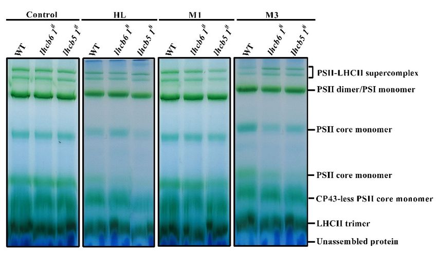
Int. J. Mol. Sci. 2019, 20, 3665                                                                  2 of 14

or PSI that can balance the electron transfer [7]. The largest NPQ is dependent on a low thylakoid
lumen pH and defined as energy quenching (qE) [8]. Among these minor antenna proteins, only Lhcb4
can be phosphorylated in monocotyledons, and this phosphorylation is involved in state transitions
and photo-inhibition recovery [9,10]. The photo-protective role of CP29 phosphorylation reduces
singlet oxygen production and enhances excess energy dissipation [11]. Other minor antennas, such as
Lhcb6 and Lhcb5, participate in the xanthophyll cycle, which can alleviate light damage [12–15].
     The moss, Physcomitrella patens, represents an excellent system to study gene function due to
its high homologous recombination frequency and complete sequence genome [16]. Research on
the organism has diverged from green lineages to vascular plants and enriches our understanding
of how photosynthetic organisms adapt to different environmental conditions [17]. The PSI protein
super-complex component Lhcb9 was reported to change the absorption properties of PSII by
harboring red-shifted chlorophyll [18,19]. Physcomitrella contains both LhcSR (light-harvesting complex
stress-related) and PsbS (PSII subunit S) [20]. LhcSR is an ancient light-harvesting protein that has
been reported to regulate excess light energy absorption and dissipation in green algae, while in higher
plants, LhcSR is functionally replaced by PsbS proteins [21]. However, little is known about the minor
antenna proteins in Physcomitrella.
     In this work, our results showed the function of minor antenna proteins Lhcb6 and Lhcb5 in
light-harvesting and regulation of photosynthesis when exposed to environmental stress conditions by
generating specific mutants that lacked one or both proteins. Under conditions of high light or osmotic
stress, NPQ was dramatically reduced in mutants. Moreover, mutants showed deformed leaves, a
lower content of chlorophyll content and PSII activity, which suggests that Lhcb6 and Lhcb5 played
important roles in the organization of photosynthetic complexes in grana partitions. Taken together,
our results indicated that minor antenna protein Lhcb5 and Lhcb6 of Physcomitrella played significant
roles in the function and structure of PSII, especially under abiotic stress.

2. Results

2.1. Knock-Out of Lhcb6 or Lhcb5 Altered Chloroplast Organization and Lhc Proteins Accumulation
      Although the mutations of Lhcb6 and Lhcb5 in higher plants inhibit the interaction of photosystem
II subunits and electron transport rate in grana membranes [22], little is known about the effects
of minor antenna proteins in P. patens. To investigate the physiological function of minor antenna
proteins, we generated Lhcb6 or Lhcb5 knock-out mutants in Physcomitrella. At least three independent
lines were isolated and retained for further characterization. The expression of the gene level and
accumulation of protein level were detected in mutants by qRT-PCR and Western blotting. As shown
Figure 1B,C, mRNA and protein levels were hardly detected in the corresponding mutants when
compared with wild-type. The alterations in Lhc stoichiometry in the thylakoid of mutants were
verified by immunoblotting and quantitative PCR analysis (Figure 2). In lhcb6 1# , the expression
levels of Lhc components Lhca and Lhcb4 were decreased sharply, while Lhcb5, PsbS and violaxanthin
de-epoxidase (vde) were increased compared with wild-type plants. Apart from induced Lhcb6, all these
genes in lhcb5 1# displayed the same expression pattern as lhcb6 1# . In addition, the LhcSR subunit
content did not show obvious changes in lhcb6 1# , but demonstrated an evident decline in lhcb5 1# . The
LhcSR subunit content was reduced in lhcb5 1# but only changed by a small amount in lhcb6 1# .
      To investigate the effects of Lhcb6 and Lhcb5 on chloroplast organization, ultrathin sections of
leaves were analyzed (Figure 3). Under normal growth conditions, wild-type (WT) plant chloroplasts
showed a characteristic organization of stromal membrane with interconnecting grana stacks and large
starch granules in most sections. The lhcb6 1# plants showed a lamella with a disordered arrangement
of thylakoids, reduced stacked grana, and increased plastoglobules and starch granules. Chloroplasts
from mutant lhcb5 1# accumulated more starch granules and plastoglobules but displayed a higher ratio
of stromal membrane to grana stacks when compared with WT. These results demonstrated that Lhcb6
and Lhcb5 influenced the ultrastructure of chloroplast and the accumulation of thylakoid proteins.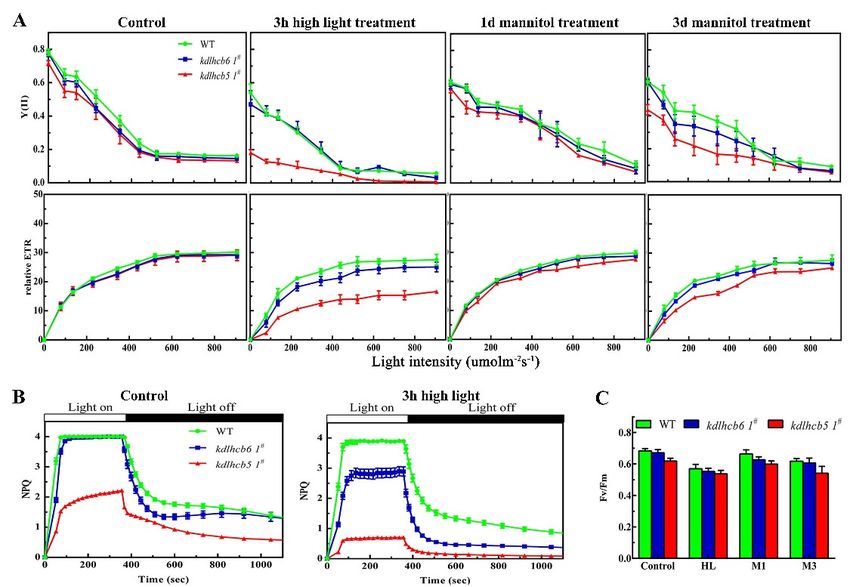
Int. J. Mol. Sci. 2019, 20, x FOR PEER REVIEW                                                                     3 of 14
Int. J. Mol. Sci. 2019, 20, x FOR PEER REVIEW                                                                     3 of 14

 demonstrated that Lhcb6 and Lhcb5 influenced the ultrastructure of chloroplast and the
demonstrated that Lhcb6 and Lhcb5 influenced the ultrastructure of chloroplast and the
 accumulation
Int.                 of thylakoid
     J. Mol. Sci. 2019,            proteins.
accumulation        of 20, 3665
                        thylakoid proteins.                                       3 of 14

      Figure 1. The identification of knock-out mutants. (A) The plasmid vector’s schematic maps of
     Figure 1. The
                The identification
                     identification  of
                                     of knock-out
                                        knock-out    mutants. (A)   The
                                                                (A) The   plasmid
                                                                           plasmid vector’s
                                                                     Theexpression    vector’s schematic
                                                                                               schematic   maps
                                                                                                           maps of
      pTN182-PpLhcb5/6    used for knock-out     a generation. (B)                     of PpLhcb6   and PpLhcb5,  in
     pTN182-PpLhcb5/6 usedused for
                                for knock-out
                                    knock-out  aageneration.
                                                   generation.(B)(B)
                                                                   The
                                                                     Theexpression of of  PpLhcb6
                                                                                            PpLhcb6andand
                                                                                                        PpLhcb5, in
                                                                                                           PpLhcb5,
      wild-type and knock-out    mutants, was analyzed    by RT-PCR.    (C)expression
                                                                             Immunoblotting     of Lhcb6  and Lhcb5
     wild-type
     in        andand
        wild-type   knock-out   mutants, was was
                        knock-out             analyzed by RT-PCR.      (C) Immunoblotting      of Lhcb6  and Lhcb5
      from  Arabidopsis  (Col) and mutants,
                                     Physcomitrella analyzed  by(WT)
                                                     (wild-type   RT-PCR.and (C)   Immunoblotting
                                                                               lines                  of Lhcb6
                                                                                      of knock-out mutants).   and
                                                                                                                The
     from
     Lhcb5 Arabidopsis
            from        (Col) (Col)
                 Arabidopsis   and Physcomitrella
                                    and             (wild-type
                                         Physcomitrella          (WT)
                                                        (wild-type  (WT)andandlines  ofofknock-out
                                                                                 lines    knock-out  mutants).
                                                                                                     mutants). The
                                                                                                               The
      chlorophyll content of every lane was 15 μg. Proteins were stained with Ponceau S and Rubisco
     chlorophyll content
                  content of every   lane  was   15 μg. Proteins
                                                             werewere     stained   with Ponceau    S and Rubisco
      proteins were usedofasevery  lane
                              loading   was 15 µg.
                                       controls.    Proteins       stained   with Ponceau   S and Rubisco  proteins
     proteins
     were usedwere  used ascontrols.
                as loading  loading controls.

     Figure
      Figure2.2. Analysis  of Lhc
                 Analysis of  Lhc genes
                                   genes or
                                         or protein
                                            protein accumulation
                                                     accumulationin
                                                                  inwild-type
                                                                    wild-typeandandmutants.
                                                                                    mutants. (A)
                                                                                              (A) The
                                                                                                  The expression
                                                                                                      expression
     Figure
     of      2. Analysis
        Lhc-related genes ofwas
                             Lhc genes  or protein
                                  analyzed  by      accumulation
                                                RT-PCR.  EF1a wasin wild-type
                                                                  used as      and mutants.
                                                                          reference gene.   (A)Light-harvesting
                                                                                          Lhc,   The expression
      of Lhc-related genes was analyzed by RT-PCR. EF1a was used as reference gene. Lhc, Light-harvesting
     of Lhc-relatedPsbS,
     complexes.     genesPSII
                            wassubunit
                                 analyzedS. by
                                            vde,RT-PCR.  EF1a was
                                                 violaxanthin     used as reference
                                                              de-epoxidase.         gene. Lhc, Light-harvesting
                                                                              (B) Immunoblotting    of the Lhca,
      Lhcb4, Lhcb5 and Lhcb6 in wild-type and mutants. Rubisco proteins were used as loading controls and
Int. J. Mol. Sci. 2019, 20, 3665                                                                               4 of 14
 Int. J. Mol. Sci. 2019, 20, x FOR PEER REVIEW                                                                  4 of 14

      complexes. PsbS, PSII subunit S. vde, violaxanthin de-epoxidase. (B) Immunoblotting of the Lhca,
      Lhcb4, Lhcb5 and Lhcb6 in wild-type and mutants. Rubisco proteins were used as loading controls
     were stained by Ponceau S. (C) Quantification of Lhc expression levels in WT and mutants.
      and were stained by Ponceau S. (C) Quantification of Lhc expression levels in WT and mutants. (D)
     (D) Immunological quantification of Lhc proteins in thylakoid membranes. The data represent
      Immunological quantification of Lhc proteins in thylakoid membranes. The data represent means ±
     means ± SD of three biological replicates. Statistical significance compared with the wild-type p is
      SD of three biological replicates. Statistical significance compared with the wild-type p is indicated
     indicated by asterisks (** p ≤ 0.01, * p ≤ 0.05, Student’s t-test).
      by asterisks (** p ≤ 0.01, * p ≤ 0.05, Student’s t-test).

      Figure3. 3.Transmission
     Figure        Transmission   electron
                               electron     micrograph
                                        micrograph        of plastids
                                                    of plastids       from mesophyll
                                                                from mesophyll           cells in wild-type
                                                                                cells in wild-type           and
                                                                                                   and mutants.
      mutants.
     Starch       Starchmarked
            granules     granules  marked
                                 with       withcan
                                      asterisks   asterisks can be distinguished
                                                    be distinguished              from plastoglobules
                                                                       from plastoglobules               in black
                                                                                             in black dots. The
     lamella of thylakoid
      dots. The   lamella ofstack and grana
                             thylakoid  stack are
                                              andindicated
                                                  grana areby  an arrow.
                                                             indicated by an arrow.

2.2.
 2.2.PSII
      PSIIActivity
           ActivityisisMarkedly
                       MarkedlyReduced
                                ReducedininMutants
                                           Mutantsunder
                                                   underHigh-Light
                                                         High-LightTreatment
                                                                    Treatment
     AsAsthe
          thechloroplast
               chloroplastorganization
                              organizationwaswasaltered
                                                   alteredininmutants,
                                                               mutants,we  wefurther
                                                                               furtheranalyzed
                                                                                         analyzedPSII
                                                                                                    PSIIfunction
                                                                                                          functioninin
different mutants   using   fluorescence   measurements.     The  peaks   in the room    temperature
 different mutants using fluorescence measurements. The peaks in the room temperature fluorescence     fluorescence
emission
 emissionspectra
            spectraofofmutants
                        mutantswerewerelower
                                         lowerwhen
                                                 whencompared
                                                        comparedwith withwild-type
                                                                           wild-typeatat685685nm
                                                                                               nm(Supplementary
                                                                                                    (Supplementary
Figure
 Figure S1), suggesting that Lhcb6 and Lhcb5 affected the fluorescence emissionspectra
        S1), suggesting     that  Lhcb6  and  Lhcb5   affected  the  fluorescence    emission     spectraofofseveral
                                                                                                               several
chlorophylls
 chlorophylls and the light-harvesting efficiency of PSII. To understand the primary functionsofof
               and   the  light-harvesting    efficiency  of  PSII. To  understand      the  primary   functions
Lhcb6
 Lhcb6and     Lhcb5ininP.P.patens,
         andLhcb5             patens,non-invasive
                                      non-invasivechlorophyll
                                                      chlorophyllfluorometric
                                                                     fluorometricanalyses
                                                                                     analyseswere
                                                                                                wereperformed
                                                                                                       performedtoto
investigate  the photosynthetic     electron transport   in wild-type   and   mutants   (Figure  4,
 investigate the photosynthetic electron transport in wild-type and mutants (Figure 4, SupplementarySupplementary
Figure
 FigureS3).
         S3).The
              Theinitial
                  initialFv/Fm
                          Fv/Fmmeasurements
                                   measurementsdemonstrated
                                                    demonstratedthat thatPSII
                                                                           PSIIactivity
                                                                                activitywas
                                                                                          wasdisturbed
                                                                                              disturbedininmutants
                                                                                                             mutants
and  wild-type  after  high-light  treatment  (Figure  4A).  To further  investigate
 and wild-type after high-light treatment (Figure 4A). To further investigate the     the  PSII activity
                                                                                                     PSIIofactivity
                                                                                                             mutantsof
under
 mutantsabiotic stress,
            under        the maximum
                    abiotic    stress, the(Fm)   and minimum
                                            maximum       (Fm) and (F0 )minimum
                                                                        fluorescence,(F0) and  non-photochemical
                                                                                           fluorescence,    and non-
quenching    (NPQ)
 photochemical        of dark-adapted
                   quenching      (NPQ) ofplants  were quantitatively
                                             dark-adapted     plants were  determined.     Under
                                                                              quantitatively        normal growth
                                                                                                determined.     Under
conditions,          # showed significantly decreased NPQ compared to wild-type (Figure 4C).
             lhcb5 1conditions,
 normal growth                     lhcb5 1# showed significantly decreased NPQ compared to wild-type
 (Figure 4C).
      The NPQ amplitude has been reported to be dependent on the lumen pH or on the concentration
 of PsbS [13,23]. Proton pumping into the chloroplast lumen was influenced in lhcb5 1# rather than
 lhcb6 1# and wild-type (Supplementary Figure S2). This result is consistent with the hypothesis that
 the limitation of NPQ in lhcb5 1# is partly associated with reduced acidification of the lumen under
 illumination. The impact of the loss of Lhcb6 or Lhcb5 on photosynthesis was investigated by
 measuring the electron transport activity (ETA) in CO2-saturating conditions. The electron transport
 activity (ETA) in CO2 saturating conditions was lower in mutants compared with that in wild-type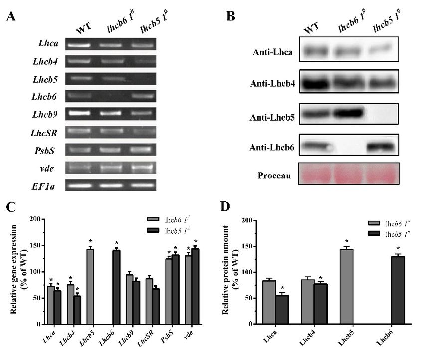
plants (Supplementary Figure S2B). To further characterize the photosynthetic apparatus, the light-
response curves of PSII quantum yield (Y(II)) and electron transport rate (ETR) were also analyzed.
After 3 h of high-light treatment, Y(II) and ETR in lhcb5 1# were significantly reduced compared with
wild-type plants (Figure 4A). These results indicated that the minor antenna protein Lhcb55 was
Int. J. Mol. Sci. 2019, 20, 3665 of 14
involved in excitation energy via non-photochemical pathways.
Figure
Figure 4.
4. Detailed
Detailed characterization
characterization ofof chlorophyll
chlorophyll fluorescence
fluorescence in in wild-type
wild-type and
and mutants.
mutants. (A) (A)Light-
Light-
response
response curves of PSII quantum yield and ETR in wild-type and mutants. (B) Time courses for
curves of PSII quantum yield and ETR in wild-type and mutants. (B) Time courses for
induction
induction and
and relaxation
relaxation of
of non-photochemical
non-photochemicalquenching
quenching(NPQ)(NPQ)before,
before,and
andafter,
after, 33 hh of
of high
high light
light
treatment.
treatment. (C)
(C) The maximal photochemical
The maximal photochemicalefficiency
efficiencyofofPSII.
PSII.HLHL represents
represents high
high light
light treatment
treatment for for
3 h.
3M1h. or
M1M3orrepresent
M3 represent
plantsplants
underunder
500 mM 500mannitol
mM mannitol foror1 3day
for 1 day or respectively.
days, 3 days, respectively.
The data The data
represent
represent
the meansthe± SD means ± SD
of three of threereplicates.
biological biologicalStatistical
replicates. Statistical compared
significance significance compared
with with p
the wild-type the
is
wild-type p is indicated by asterisks (* p ≤ 0.05,
indicated by asterisks (* p ≤ 0.05, Student’s t-test). Student’s t-test).
TheComplexes
2.3. PSII NPQ amplitude has been
Accumulation reported
Were to after
Affected be dependent
High-LightonTreatment
the lumen pH or on the concentration
of PsbS [13,23]. Proton pumping into the chloroplast lumen was influenced in lhcb5 1# rather than
After a short-term high-light treatment, the proteins of the PSII reaction center were damaged.
lhcb6 1# and wild-type (Supplementary Figure S2). This result is consistent with the hypothesis that
Then, a rapid repair and reassembly process occurred to enable photosynthetic electron
the limitation of NPQ in lhcb5 1# is partly associated with reduced acidification of the lumen under
transportation [24]. The defected photosynthesis in mutants was possibly caused by a reduced level
illumination. The impact of the loss of Lhcb6 or Lhcb5 on photosynthesis was investigated by measuring
of protein complexes in the electron transport chain. To further investigate the effects of Lhcb6 and
the electron transport activity (ETA) in CO2 -saturating conditions. The electron transport activity
Lhcb5 on the formation of thylakoid membrane protein complexes under abiotic stress, thylakoid
(ETA) in CO2 saturating conditions was lower in mutants compared with that in wild-type plants
membranes (with equal amounts of chlorophyll) in different plants were solubilized in 1% n-dodecyl-
(Supplementary Figure S2B). To further characterize the photosynthetic apparatus, the light-response
β-D-maltoside (DM) and the chlorophyll–protein complexes were separated by BN-PAGE. Six
curves of PSII quantum yield (Y(II)) and electron transport rate (ETR) were also analyzed. After 3 h of
protein complexes, PSII-LHCII super-complex, PSII dimer/PSI monomer, PSII core monomer, CP43-
high-light treatment, Y(II) and ETR in lhcb5 1# were significantly reduced compared with wild-type
less PSII core monomer, LHCII trimer, and unassembled protein were resolved (Figure 5). Under
plants (Figure 4A). These results indicated that the minor antenna protein Lhcb5 was involved in
high-light stress, the PSII core monomer of the lhcb5 1# mutant was reduced, suggesting Lhcb5 might
excitation energy via non-photochemical pathways.
associate with the formation and stability of the PSII complex. Under osmotic stress, the amount of
monomer and free pigment
2.3. PSII Complexes of mutants
Accumulation did not after
Were Affected showHigh-Light
significantTreatment
differences compared with wild-type
plants. These results suggest that light was a critical abiotic factor that limits the photosynthesis
Afterof
efficiency a short-term high-light treatment, the proteins of the PSII reaction center were damaged. Then,
PSII in Physcomitrella.
a rapid repair and reassembly process occurred to enable photosynthetic electron transportation [24].
The defected
2.4. Growth photosynthesis
Malformation in mutants
Phenotype was possibly caused by a reduced level of protein complexes
of Mutants
in the electron transport chain. To further investigate the effects of Lhcb6 and Lhcb5 on the formation
Even though
of thylakoid there was
membrane no significant
protein complexesdifference in thestress,
under abiotic protonemal filaments
thylakoid between
membranes mutants
(with equal
and wild-type plants, the leafy gametophyte showed large differences. The leafy gametophyte
amounts of chlorophyll) in different plants were solubilized in 1% n-dodecyl-β-D-maltoside (DM) and of
the chlorophyll–protein complexes were separated by BN-PAGE. Six protein complexes, PSII-LHCII
super-complex, PSII dimer/PSI monomer, PSII core monomer, CP43-less PSII core monomer, LHCII
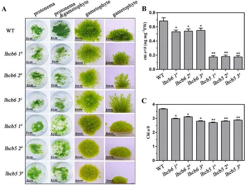
trimer, and unassembled protein were resolved (Figure 5). Under high-light stress, the PSII corelhcb6 plants grew slender and higher, with relatively narrower and longer leaves, while lhcb5 showed
a more serious malformation phenotype when compared with lhcb6 or wild-type plants (Figure 6A,
Supplementary Figure S4A). In addition, the absence of Lhcb5 or Lhcb6 resulted in reduced lateral
buds and chloroplast cells in protonema (Supplementary Figure S7). In addition, both mutants,
Int. J. Mol. Sci.
especially the2019, 20, 3665
lhcb5 6 of 14
plants, displayed a dramatically reduced chlorophyll a+b content and a noticeably
lower chlorophyll a/b ratio in comparison with wild-type plants (Figure 6B,C, Supplementary Figure
S4B,C).
monomer of the lhcb5 1# mutant was reduced, suggesting Lhcb5 might associate with the formation
In summary, the Lhcb5 or Lhcb6 deletion mutant showed severe growth inhibition and
and stability of the PSII complex. Under osmotic stress, the amount of monomer and free pigment of
developmental deficiency. The remarkable growth inhibition in different mutants suggested that
mutants did not show significant differences compared with wild-type plants. These results suggest
Lhcb6 and Lhcb5 played essential roles in leaf development of Physcomitrella.
that light was a critical abiotic factor that limits the photosynthesis efficiency of PSII in Physcomitrella.
Figure 5.5. Analysis
Figure Analysisofofpigment-protein
pigment-proteincomplexes
complexesin wild-type and mutants
in wild-type by BN-PAGE
and mutants gel. A freshly
by BN-PAGE gel. A
isolated thylakoid
freshly isolated membrane
thylakoid of wild-type
membrane and mutants
of wild-type andwere solubilized
mutants with 1% DM
were solubilized at a1%
with chlorophyll
DM at a
concentration of 25 µg. HL
chlorophyll concentration ofmeans
25 μg. high light stress
HL means for 3stress
high light h. M1foror3M3 means
h. M1 plants
or M3 meansunder
plants500under
mM
mannitol for 1 day or 3 days, respectively. Assignments of the thylakoid membrane
500 mM mannitol for 1 day or 3 days, respectively. Assignments of the thylakoid membrane macromolecular
protein complexes,
macromolecular indicated
protein on the right,
complexes, were on
indicated identified according
the right, to a previous
were identified studyto
according [25].
a previous
study [25].
2.4. Growth Malformation Phenotype of Mutants
Even though there was no significant difference in the protonemal filaments between mutants
and wild-type plants, the leafy gametophyte showed large differences. The leafy gametophyte of lhcb6
plants grew slender and higher, with relatively narrower and longer leaves, while lhcb5 showed a
more serious malformation phenotype when compared with lhcb6 or wild-type plants (Figure 6A,
Supplementary Figure S4A). In addition, the absence of Lhcb5 or Lhcb6 resulted in reduced lateral buds
and chloroplast cells in protonema (Supplementary Figure S7). In addition, both mutants, especially
the lhcb5 plants, displayed a dramatically reduced chlorophyll a+b content and a noticeably lower
chlorophyll a/b ratio in comparison with wild-type plants (Figure 6B,C, Supplementary Figure S4B,C).
In summary, the Lhcb5 or Lhcb6 deletion mutant showed severe growth inhibition and
developmental deficiency. The remarkable growth inhibition in different mutants suggested that Lhcb6
and Lhcb5 played essential roles in leaf development of Physcomitrella.Int. J. Mol. Sci. 2019, 20, 3665 7 of 14
Int. J. Mol. Sci. 2019, 20, x FOR PEER REVIEW 7 of 14
Figure
Figure 6. 6. The
Thephenotype
phenotypeand andchlorophyll
chlorophyll content
content of
of wild-type
wild-type and
and mutant
mutant plants.
plants. (A)
(A) The
The protonema
protonema
and
and gametophyte
gametophyte phenotypes
phenotypes of of wild-type
wild-type and mutants. The The protonema
protonema tissues
tissues of
of wild-type
wild-type andand
mutants
mutants were were grown
grown for one week on ammonium tartrate-free BCD medium. The The protonema
protonema and and
gametophyte tissueswere
gametophyte tissues weregrown
grownin in
thisthis medium
medium for four
for four weeks.
weeks. The gametophyte
The gametophyte tissuestissues were
were grown
grown in themedium
in the same same medium for six-weeks.
for six-weeks. The chlorophyll a+b content
The chlorophyll a+b content (B)chlorophyll
(B) and and chlorophyll a/b(C)
a/b ratio ratio
of
leafy
(C) ofgametophyte tissues.tissues.
leafy gametophyte FW, FreshFW,weight. The dataThe
Fresh weight. represent the means
data represent ± SD
the of three
means ± SDbiological
of three
replicates.replicates.
biological StatisticalStatistical
significance comparedcompared
significance with the wild-type is indicated
with the wild-type by asterisks
is indicated (** p ≤ 0.01,
by asterisks (**
p* p≤ ≤ 0.05,
0.01, * pStudent’s t-test). t-test).
≤ 0.05, Student’s
3. Discussion
3. Discussion
Light-harvesting complexes
Light-harvesting complexes (Lhc)
(Lhc) are
are members
members of of aa large
large multigene
multigene family.
family. They
They play
play important
important
roles in
roles inregulating
regulatingphotosynthesis
photosynthesis in in plants’
plants’ response
response to environmental
to environmental stress.
stress. The function
The function of minorof
minor chlorophyll-binding proteins is in bridging major LHCII antenna
chlorophyll-binding proteins is in bridging major LHCII antenna to a dimeric PSII core complex to a dimeric PSII core
complex [22,23,26].
[22,23,26]. Both Lhcb6Both andLhcb6
Lhcb5 and
haveLhcb5 have
a single a single
energy energy
transfer transfer
process fromprocess
Chl b from
to Chl Chl b to
a [27].
Chl a [27].analysis
Structural Structural
of theanalysis of the Lhcb supercomplex
Lhcb supercomplex and PSII
and PSII organization has organization
revealed thathas revealed
Lhcb5 that
is located
Lhcb5 is located between the strongly bound LHCII trimer and dimeric
between the strongly bound LHCII trimer and dimeric PSII core complex, whereas Lhcb6 is always PSII core complex, whereas
Lhcb6 is always
associated with the associated
connection with the connection
of LHCII of LHCII
trimers and the core trimers
of PSIIand the core
in higher of PSII
plants in higher
[2–4,28]. The
plants [2–4,28].has
Physcomitrella Physcomitrella has
Thedemonstrated demonstrated
a capacity a capacity for Lhc-dependent
for Lhc-dependent mechanisms inmechanisms
response to in
response to environmental conditions in previous
environmental conditions in previous research [17,29,30]. research [17,29,30].
To investigate
To investigate the the functions
functions ofof Lhcb6
Lhcb6 andand Lhcb5
Lhcb5 in Physcomitrella, the
in Physcomitrella, PpLhcb6 and
the PpLhcb6 PpLhcb5 were
and PpLhcb5 were
inactivated by a targeted gene replacement assay. The mRNA or protein
inactivated by a targeted gene replacement assay. The mRNA or protein levels of Lhcb6 and Lhcb5 levels of Lhcb6 and Lhcb5
were undetectable
were undetectable in in three
three independent
independent mutation
mutation lines
lines (Figure
(Figure 1).1). Moreover,
Moreover, the the expression
expression of Lhc
of Lhc
#
components in
components in both
both mutants
mutants werewere significantly
significantly changed
changed (Figure
(Figure 2).
2). Interestingly, the lhcb5
Interestingly, the lhcb5 11# showed
showed
an obvious increase in Lhcb6, PsbS and vde accumulation, in addition to
an obvious increase in Lhcb6, PsbS and vde accumulation, in addition to decreased Lhca and Lhcb4 decreased Lhca and Lhcb4
levels. The
The lhcb6 #
lhcb6 11# mutant
mutantshowed
showedsimilar
similar expression
expression levels
levels ofof pigment-protein
pigment-protein components.
components. Under Under
levels.
high-light stress, the efficiency of excitation energy trapping and non-photochemical
high-light stress, the efficiency of excitation energy trapping and non-photochemical quenching quenching in the
in
absence of the lhcb5 mutant were much lower than WT plant, while the
the absence of the lhcb5 mutant were much lower than WT plant, while the Lhcb6 deletion mutant Lhcb6 deletion mutant only
only displayed a dramatic decrease in non-photochemical quenching (Figure 4). PsbS and LhcSR
regulate excess energy dissipation by the xanthophyll cycle, which is an important anticipatoryInt. J. Mol. Sci. 2019, 20, 3665 8 of 14
displayed a dramatic decrease in non-photochemical quenching (Figure 4). PsbS and LhcSR regulate
excess energy dissipation by the xanthophyll cycle, which is an important anticipatory strategy against
photo-inhibition [31]. Lhc proteins are associated with scavenging of reactive oxygen speciecs (ROS)
species by switching back and forth between PSI and PSII. The direct quenching of chlorophyll triplet
states by xanthophyll is associated with VDE and LhcSR [12,30,32]. The mechanisms which balance
light absorption and the amount of electron transport acceptor substrates clarify the evolution of
oxygenic photosynthesis [31]. Here, the different expression levels of Lhc genes observed in different
mutants might be interpreted as compensatory mechanisms aimed at the dissipation of excess energy.
In higher plants, the minor antenna proteins Lhcb6 and Lhcb5 affect the interactions of PSII
subunits and the electron transport rate in grana membranes, especially for limiting plastoquinone
diffusion [22,23,26]. Here, the absence of either Lhcb5 or Lhcb6 influenced the ultrastructure of
chloroplast and the accumulation of thylakoid proteins, and particularly, reduced the number of
stacked grana and increased the amount of starch granules (Figure 3). The disordered arrangement of
thylakoid lamella and increased starch granules suggested that Lhcb6 and Lhcb5 might maintain the
chloroplast ultrastructure in vivo.
The structural analysis of the Lhc supercomplex and PSII organization revealed that Lhcb5
is located between the strongly bound LHCII trimer and dimeric PSII core complex, while Lhcb6
is associated with the connection of LHCII trimers and the core of PSII in higher plants [2–4,28].
The regulation of light-harvesting and energy transfer from LHCII to the PSII core does not require
Lhcb5 [3,23]. In the absence of Lhcb5, the NPQ and Y(II) of chlorophyll fluorescence after 3 h of
high-light treatment were lower than the plants lacking Lhcb6 (Figure 4, Supplementary Figure S6).
After 3 h of high-light treatment or 3 d of osmotic treatment, lhcb5 1# showed significantly reduced PSII
core monomer protein levels in comparison to WT (Figure 5). Under high-light stress conditions, the
Lhcb5 deletion mutant showed reduced PSII core monomer protein levels, and decreased levels of PSII
quantum yield, electron transport rate and NPQ, suggesting that the interaction between LHCII and
the PSII super-complex was altered. These results implied the function of the minor antenna protein,
Lhcb5, might be associated with the formation and stability of PSII super-complex assembly in the
thylakoid membrane.
The results described above demonstrated that Lhcb6 and Lhcb5 participated in the accumulation
of thylakoid proteins. These two minor antenna proteins probably interacted transiently with PSII
complex or some PSII subunits, in a manner similar to several PSII auxiliary factors that interacted with
PSII subunits. The cooperative interaction between the PSII core and minor Lhcbs is disrupted, which
leads to a decreased affinity when Lhcb6 or Lhcb5 is in low amounts [22]. Furthermore, the inactivation
of Lhcb6 or Lhcb5 displayed pleiotropic effects on photosynthetic electron transport. Chlorophyll
fluorescence measurements showed that Fv/Fm was slightly reduced in mutants, suggesting no
impairment of the PSII complexes (Figure 4), which is similar to previous evidence [33]. A dramatic
decrease in NPQ indicated that the photoprotection of PSII was severely inhibited in mutants. These
results demonstrated that minor antenna proteins Lhcb6 and Lhcb5 affected the chloroplast ultrastructure,
the accumulation of PSII complexes and PSII activity under abiotic stress conditions.
Under different abiotic stress conditions, chloroplasts, peroxisomes, and mitochondria
accumulated a large amount of reactive oxygen species, which led to cell death and cytotoxicity.
Multiple Lhc isoforms assemble with supramolecular photosynthetic complexes to dissipate excess
absorbed energy and scavenging ROS. Under high-light stress, Lhc components of the PSII antenna
system use non-photochemical quenching to limit the over-reduction of the electron transport chain
and the formation of toxic ROS [34–37]. PsbS and LhcSR regulate the excess energy dissipation by the
xanthophyll cycle, which is an important anticipatory strategy in the fight against photo-inhibition [31].
Lhc proteins associate with ROS scavenging by switching back and forth between PSI and PSII,
and the direct quenching of chlorophyll triplet states by xanthophyll is associated with VDE and
LhcSR [12,30,32]. Compared with the vascular plants, a stronger NPQ and higher capacity of dissipating
excitation energy as heat were observed in mosses [17,20,30]. In Physcomitrella, it has been shownInt. J. Mol. Sci. 2019, 20, 3665 9 of 14
that NPQ requires LhcsR and PsbS to regulate excess light energy absorption and dissipate light
damage [17,20,30,38]. NPQ is also viewed as a compensatory mechanism in response to overexcited
chlorophyll to prevent irreversible damage to PSII [39]. In our study, the mutants showed clear
differences in the Lhcb polypeptide composition, especially in PsbS and VDE. The expression of PsbS
and vde were increased, while Lhca, Lhcb4, and LhcSR were decreased in the Lhcb5-defective mutant.
The alteration of chloroplast organization and photosynthesis efficiency suggested that the absence
of Lhcb5 might alter the structure or stability of PSII super-complexes (Figure 3). NPQ activation,
in response to illumination with strong actinic light- of dark-adapted plants, was lower in lhcb5 1# ,
especially under high-light stress (Figure 4). The distinctly decreased NPQ suggested that the absence
of Lhcb5 resulted in the obstruction of excess light energy transfer between LhcII trimer and PSII
core complexes. PsbS and xanthophyll control the affinity of different LhcII antenna complexes for
protons to participate in the feed-back control of excess light energy that underlies non-photochemical
chlorophyll fluorescence quenching [40]. Although the individual antenna components were not
quantified in the analyses on the accumulation of PSII complexes, the chlorophyll a+b contents and
chlorophyll a/b ratios of mutants suggested there may be some minor changes in the antenna in response
to high-light treatment. Similarly, the distinguishable electron transport reflected the lower rate of
photosynthetic electron transport in mutants (Supplementary Figure S2). An alternative explanation
might reflect the role of Lhcb6 and Lhcb5 in channeling prtons away from PSII due to different pH
levels in these plants [3,22]. Partial electron transport reactions localized at the restricted step between
the plastoquinone site of PSII and the cytochrome b6 f complex for electron donors to cytochrome b6 f
are effective in sustaining NADP+ reduction [22]. A lack of Lhc proteins might result in the restriction
of PQH diffusion from the PSII QB site to the cytochrome b6 f complex.
In our findings, the absence of Lhcb5 or Lhcb6 resulted in similar growth-defect phenotypes, such
as reduced lateral buds and chloroplast cells in protonema (chloronema and caulonema) and thinner
leaves in mature plants (Figure 6, Supplementary Figures S4 and S7). Interestingly, the mutation of
Lhcb5 contributed to a more significant growth-defective phenotype when compared with Lhcb6. The
phenotype differences in protonema and gametophyte between WT and mutants implied that Lhcb5
may function slightly differently from Lhcb6. Lacking both genes contributed to a decreased growth
ratio in gametophyte, but not in protonema (Figure 6C, Supplementary Figure S5). Moreover, both
mutants also showed disordered lamella arrangements of thylakoid, and abnormal extension of starch
granules and plastoglobules in chloroplast, especially in lhcb5 (Figure 3). Excess sunlight damages
photosynthetic machinery and limits plant photosynthetic activity, growth, and productivity [39].
Spectroscopic characterization showed that the light-harvesting efficiency decreased more in mutants
(Supplementary Figure S1). Under high-light stress, the NPQ of lhcb6 and lhcb5 decreased sharply,
implying that the antenna proteins might participate in photo-protection by dissipating excess energy.
Under abiotic stress, only lhcb5 mutant showed any inhibition of NPQ, Y(II) and ETR, suggesting that the
minor antenna protein, Lhcb5, played an important role in the excitation energy via non-photochemical
pathways. The lower photo-inhibition PSII activity in Lhcb-defective mutants might connect with the
limited growth rate and malformed phenotype in gametophyte. These results indicated that minor
antenna proteins, Lhcb6 and Lhcb5, were involved in the gametophyte development in Physcomitrella.
Light may be the critical factor in influencing Physcomitrella to diverge from seed and hydrophyte
plants after land colonization.
In high-light conditions, Lhcb5 of Arabidopsis is activated in Zea-mediated photoprotection and
catalyzed quenching of the qI type, while it is not phosphorylated in the state transition [14,22,23]. It has
been reported that the deletion of Lhcb5 does not cause alterations in several photosynthetic parameters,
except in reduced growth [3,22,23]. In the present study, we found the minor antenna proteins Lhcb5
and Lhcb6 of Physcomitrella played an important role in the organization of photosynthetic complexes
in grana partitions, participated in electron transport, and excess excitation energy dissipation under
high-light stress in higher plants. Moreover, the loss of minor antenna protein Lhcb6 or Lhcb5 resulted
in a pale-green leaf and the developmental defect phenotype, which has not been reported in higherInt. J. Mol. Sci. 2019, 20, 3665 10 of 14
plants or other green alga. Function analysis of minor antenna proteins, Lhcb6 and Lhcb5 in P. patens,
provides evolutionary insights for the land plants.
4. Materials and Methods
4.1. Plant Materials and Treatments
The moss P. patens were cultured on modified BCD medium [41] in a growth chamber at 23 ± 1 ◦ C
with 16 h of light (55 µmol m−2 s−1 ) and 8 h of dark control light conditions. Uniform leafy shoots
and protonema were obtained as described [42]. Two-week-old gametophore colonies were gently
ground with a homogenizer, and transferred the homogenate to a cellophane overlay on solid BCD
medium, containing 0.75% (w/v) agar, with 5 mM ammonium tartrate, and 0.5% (w/v) glucose. After a
week, the cellophane overlay bearing regenerated protonema tissues was transferred onto ammonium
tartrate-free BCD medium. The extended leaves and rhizoids were obtained for experimental analysis
after three weeks. For light treatments, 5-day-old plants were transferred to 450 µmol m−2 s−1 for 3 h
(HL) [20]. For osmotic treatments, the plants were transferred onto fresh agar plates of BCD medium
containing 500 mM mannitol [43] for 1 day and 3 days, respectively (M1 and M3).
4.2. Protoplast Transformation and Mutant Identification
Genomic P. patens protonema DNA extraction was performed as previously described in [17],
and used as a template for PpLhcb5 (locus XM-001752760) and PpLhcb6 (locus XM-001757486) cloning.
Two homologous regions (upstream and downstream) of target coding sequences were amplified by
PCR from the cDNA library of P. patens tissue and sub-cloned into pTN182 vector (Figure 1A) [44].
The transformation followed, as described previously in [41], with minor modifications. One-week-old
protonemal tissues were collected for protoplast generation and PEG-mediated transformation. The
selection and generation of resistant colonies were described previously [17]. The defects of Lhcb5
and Lhcb6 mutants were studied in three independent lines and were parallel. Quantitative PCR and
immunoblotting showed that the genes or proteins were absent in both mutants. The primers used in
this study were listed in Table S1.
4.3. Chlorophyll Content Measurement and EM Scanning
Leaf total chlorophyll was extracted with 80% acetone from two-week-old tissues and measured
as previously described [22]. Samples from one-month-old tissues were fixed overnight in 3%
glutaraldehyde and 0.1 M sodium cacodylate buffer (pH 6.9) at 4 ◦ C. Ultrathin sections were cut with
an ultramicrotome (Reichert-Jung, Ultracut, Wetzlar, Germany) and observed with a transmission
electron microscope (FEI Tecnai G2 F20 S-TWIN, Hillsboro, OR, USA) operating at 75 kV [45].
4.4. Fluorescence Spectrum Analysis
The fluorescence emission spectrum was performed at room temperature using 436 nm excitation,
5 nm spectral bandwidth and 1 nm spectral resolution with a Hitachi F-4500 spectrofluorometer [46].
Samples were diluted at a chlorophyll concentration of 10 µg/mL in 20 mM Hepes/KOH, pH 7.8, 0.33 M
sorbitol, 5 mM MgCl2 and 70% glycerol (w/v).
4.5. The Electron Transport Activity Analysis
The electron transport measurements were performed with a Clark type oxygen electrode
(Hansatech) [47,48] at room temperature. The reaction mixture of whole chain electron transport
activity contained reaction buffer: 1 mM methyl viologen (Sigma-Aldrich, St. Louis, MO, USA), 10 mM
methylamine (Sigma-Aldrich), 1 mM NaN3 and thylakoid membranes. The reaction mixtures for
PSII-catalyzed electron transport activity contained reaction buffer with 5 mM NH4 C (Sigma-Aldrich),
5 µM DCPIP (Sigma-Aldrich) and thylakoid membranes. The PSI-catalyzed electron transport activity
assay mixture contained 2 mM ascorbate, 100 mM DCPIP, 1 mM MV and 1 mM NaN3 , 10 mMInt. J. Mol. Sci. 2019, 20, 3665 11 of 14
methylamine and 10 mM DCMU (Sigma-Aldrich). The thylakoid membranes concentration was
10 µg/mL.
4.6. Measurement of ∆pH
The kinetics of ∆pH formation across the thylakoid membrane were measured as previously
described in [22]. The reaction buffer was as follows: 30 mM Tricine/NaOH, pH 7.8, 0.1 M sorbitol,
5 mM MgCl2 , 10 mM NaCl, 20 mM KCl, 100 mM methyl viologen, and 2 mM 9-aminoacridine. The
chlorophyll concentration in the reaction buffer was 30 µg/mL.
4.7. The Fluorescence and NPQ Measurement
The chlorophyll fluorescence was measured with a pulse-amplitude modulated chlorophyll
fluorometer (mini-PAM) (Walz, Effeltrich, Germany) at room temperature, with saturating light of
6000 µmol·m−2 ·s−1 and actinic light of 830 µmol·m−2 ·s−1 . For measurements, plants were dark-adapted
for 30 min. Fm is the maximal fluorescence and F0 is the minimal fluorescence of dark-adapted
leaves, Fm’ is the maximal fluorescence and F0 0 is the minimal fluorescence of samples under light.
The parameters Fv, Fv/Fm, Y(II), NPQ, qP, and relative ETR were calculated as Fm-F0 , (Fm’-F0 )/Fm’,
(Fm’-F)/Fm’, (Fm-Fm’)/Fm’, (Fm’-F)/(Fm’-F0 0 ) and Y(II)·PAR, respectively [33].
4.8. BN-PAGE and Immunoblotting Analysis
Thylakoids and functional chloroplasts were isolated from three-week-old protonemal tissue
as previously described [49,50]. BN-PAGE was performed with slight modifications as described
previously in [25]. Thylakoid was solubilized with 1% (w/v) n-dodecyl-β-d-maltoside (DM) and
incubated on ice for 30 min. After centrifugation at 13,000× g for 10 min at 4 ◦ C, the supernatant was
supplemented with 0.1 vol sample buffer (100 mM BisTris/HCl, pH 7.0, 500 mM 6-amino-caproic acid,
30% (w/v) glycerol, 5% (w/v) Serva blue G) and subjected to BN-PAGE with a gradient of 5–13.5%
acrylamide in the separation gel. The electrophoresis was performed at 4 ◦ C, 125 V for 3–6 h.
Following Chen [9], isolated samples with 25 µg chlorophyll were loaded and electro-blotted on
nitrocellulose membranes. The antibody of Lhca, Lhcb4, Lhcb5, and Lhcb6 (Agrisera, Sweden) were
applied, and the signal was revealed by anti-rabbit IgG.
4.9. Measurement of Growth Rate
Analysis and identification of gametophyte and protonema tissue of Physcomitrella were
photographed directly in their growth media as previously described in [51]. The live-image acquisition
of protonema and gametophyte were performed with a light microscope (Nikon DS-Fi1c, Minato,
Japan) and a digital camera (Leica DFC495, Wetzlar, Germany), respectively.
The growth-rate analysis of gametophyte in mutants and wild-type plants was carried out as
previously described in [52]. The relative growth rate was estimated by measuring the fresh and dry
weight of petri dish tissues for the same growth time.
4.10. Statistical Analysis
A statistical analysis of the results was carried out from experiments with three or more mean
values using one-way analysis of variance (ANOVA). A difference was considered to be statistically
significant when p ≤ 0.05 or very significant when p ≤ 0.01.
Supplementary Materials: Supplementary materials can be found at http://www.mdpi.com/1422-0067/20/15/3665/
s1. Figure S1: Spectroscopic characterization of wild-type and mutants; Figure S2: Measurement of trans-thylakoid
∆pH and the activity of the electron transport chain in wild-type and mutants; Figure S3: Time courses for
induction and relaxation of NPQ of wild-type and mutants under 500 mM mannitol for 1 and 3 days; Figure S4:
The protonema and gametophyte tissues of wild-type and mutants; Figure S5: The relative growth rate (RGR)
of wild-type and mutants; Figure S6: Chlorophyll fluorescence parameters of WT and mutants; Figure S7:
Micrographs of protonema (chloronema and caulonema) cells and gametophyte of WT and mutants; Table S1:
Primers used in this study.Int. J. Mol. Sci. 2019, 20, 3665 12 of 14
Author Contributions: X.P., D.Z., and H.L. designed the experiments; X.P., X.D. and X.T. performed major
experiments of the study; T.T. and B.L. provided assistance in analyzing the data; X.P., X.D., D.Z. and H.L. wrote
the manuscript and contributed to discussions.
Funding: This work was supported by the National Basic Research Program of China (973 Program)
(2015CB150100), the National Research and Development Project of Transgenic Crops of China
(2016ZX08009-003-002), Sichuan Natural Science Foundation (2019YFS0457) and Sichuan Forage Grass Innovation
Team Program.
Acknowledgments: We thank Yikun He for the kind gift of pTN182 vector, and the wild-type of P. patens.
Conflicts of Interest: The authors declare that the research was conducted in the absence of any commercial or
financial relationship that could be construed as a potential or actual conflict of interest.
References
1. Chen, Y.E.; Zhao, Z.Y.; Zhang, H.Y.; Zeng, X.Y.; Yuan, S. The significance of cp29 reversible phosphorylation
in thylakoids of higher plants under environmental stresses. J. Exp. Bot. 2013, 64, 1167–1178. [CrossRef]
[PubMed]
2. Van Amerongen, H.; van Grondelle, R. Understanding the energy transfer function of lhcii, the major
light-harvesting complex of green plants. J. Phys. Chem. B 2001, 105, 604–617. [CrossRef]
3. Yakushevska, A.E.; Keegstra, W.; Boekema, E.J.; Dekker, J.P.; Andersson, J.; Jansson, S.; Ruban, A.V.; Horton, P.
The structure of photosystem ii in arabidopsis: Localization of the cp26 and cp29 antenna complexes.
Biochemistry 2003, 42, 608–613. [CrossRef] [PubMed]
4. Dekker, J.P.; Boekema, E.J. Supramolecular organization of thylakoid membrane proteins in green plants.
Biochim. Biophys. Acta, Bioenerg. 2005, 1706, 12–39. [CrossRef] [PubMed]
5. Ruban, A.V.; Murchie, E.H. Assessing the photoprotective effectiveness of non-photochemical chlorophyll
fluorescence quenching: A new approach. Biochim. Biophys. Acta, Bioenerg. 2012, 1817, 977–982. [CrossRef]
[PubMed]
6. Niyogi, K.K.; Truong, T.B. Evolution of flexible non-photochemical quenching mechanisms that regulate
light harvesting in oxygenic photosynthesis. Curr. Opin. Plant Biol. 2013, 16, 307–314. [CrossRef] [PubMed]
7. Depège, N.; Bellafiore, S.; Rochaix, J.-D. Role of chloroplast protein kinase stt7 in lhcii phosphorylation and
state transition in chlamydomonas. Science 2003, 299, 1572–1575. [CrossRef] [PubMed]
8. Li, X.P.; Gilmore, A.M.; Niyogi, K.K. Molecular and global time-resolved analysis of a psbs gene dosage
effect on ph- and xanthophyll cycle-dependent nonphotochemical quenching in photosystem ii. J. Biol. Chem.
2002, 277, 33590–33597. [CrossRef] [PubMed]
9. Chen, Y.E.; Yuan, S.; Du, J.B.; Xu, M.Y.; Zhang, Z.W.; Lin, H.H. Phosphorylation of photosynthetic antenna
protein cp29 and photosystem II structure changes in monocotyledonous plants under environmental stresses.
Biochemistry 2009, 48, 9757–9763. [CrossRef] [PubMed]
10. Havaux, M.; Dall’Osto, L.; Cuine, S.; Giuliano, G.; Bassi, R. The effect of zeaxanthin as the only xanthophyll
on the structure and function of the photosynthetic apparatus in arabidopsis thaliana. J. Biol. Chem. 2004,
279, 13878–13888. [CrossRef]
11. Betterle, N.; Ballottari, M.; Baginsky, S.; Bassi, R. High light-dependent phosphorylation of photosystem ii
inner antenna cp29 in monocots is stn7 independent and enhances nonphotochemical quenching. Plant Physiol.
2015, 167, 457–471. [CrossRef] [PubMed]
12. Morosinotto, T.; Baronio, R.; Bassi, R. Dynamics of chromophore binding to lhc proteins in vivo and in vitro
during operation of the xanthophyll cycle. J. Biol. Chem. 2002, 277, 36913–36920. [CrossRef] [PubMed]
13. Crimi, M.; Dorra, D.; Bösinger, C.S.; Giuffra, E.; Holzwarth, A.R.; Bassi, R. Time-resolved fluorescence
analysis of the recombinant photosystem ii antenna complex cp29. Eur. J. Biochem. 2001, 268, 260–267.
[CrossRef] [PubMed]
14. Dall’Osto, L.; Caffarri, S.; Bassi, R. A mechanism of nonphotochemical energy dissipation, independent
from psbs, revealed by a conformational change in the antenna protein cp26. Plant Cell 2005, 17, 1217–1232.
[CrossRef] [PubMed]
15. van Oort, B.; Alberts, M.; de Bianchi, S.; Dall’Osto, L.; Bassi, R.; Trinkunas, G.; Croce, R.; van Amerongen, H.
Effect of antenna-depletion in photosystem ii on excitation energy transfer in arabidopsis thaliana. Biophys. J.
2010, 98, 922–931. [CrossRef]
16. Schaefer, D.G. Gene targeting in physcomitrella patens. Curr. Opin. Plant Biol. 2001, 4, 143–150. [CrossRef]Int. J. Mol. Sci. 2019, 20, 3665 13 of 14
17. Alboresi, A.; Gerotto, C.; Giacometti, G.M.; Roberto, B.; Morosinotto, T. Physcomitrella patens mutants
affected on heat dissipation clarify the evolution of photoprotection mechanisms upon land colonization.
Proc. Natl. Acad. Sci. USA 2010, 107, 11128–11133. [CrossRef]
18. Alboresi, A.; Gerotto, C.; Cazzaniga, S.; Bassi, R.; Morosinotto, T. A red-shifted antenna protein associated
with photosystem ii in physcomitrella patens. J. Biol. Chem. 2011, 286, 28978–28987. [CrossRef]
19. Iwai, M.; Grob, P.; Iavarone, A.T.; Nogales, E.; Niyogi, K.K. A unique supramolecular organization of
photosystem i in the moss physcomitrella patens. Nat. Plants 2018, 4, 904–909. [CrossRef]
20. Gerotto, C.; Alboresi, A.; Giacometti, G.M.; Bassi, R.; Morosinotto, T. Role of psbs and lhcsr in physcomitrella
patens acclimation to high light and low temperature. Plant Cell Environ. 2011, 34, 922–932. [CrossRef]
21. Li, X.-P.; BjoÈrkman, O.; Shih, C.; Grossman, A.R.; Rosenquist, M.; Jansson, S.; Niyogi, K.K. A pigment-binding
protein essential for regulation of photosynthetic light harvesting. Nature 2000, 403, 391–395. [CrossRef]
[PubMed]
22. De Bianchi, S.; Dall’Osto, L.; Tognon, G.; Morosinotto, T.; Bassi, R. Minor antenna proteins cp24 and cp26
affect the interactions between photosystem ii subunits and the electron transport rate in grana membranes
of arabidopsis. Plant Cell 2008, 20, 1012–1028. [CrossRef] [PubMed]
23. Andersson, J.; Walters, R.G.; Horton, P.; Jansson, S. Antisense inhibition of the photosynthetic antenna
proteins cp29 and cp26: Implications for the mechanism of protective energy dissipation. Plant Cell 2001, 13,
1193–1204. [CrossRef] [PubMed]
24. Armbruster, U.; Ruhle, T.; Kreller, R.; Strotbek, C.; Zuhlke, J.; Tadini, L.; Blunder, T.; Hertle, A.P.; Qi, Y.;
Rengstl, B.; et al. The photosynthesis affected mutant68-like protein evolved from a psii assembly factor to
mediate assembly of the chloroplast nad(p)h dehydrogenase complex in arabidopsis. Plant Cell 2013, 25,
3926–3943. [CrossRef] [PubMed]
25. Zhang, L.; Duan, Z.; Zhang, J.; Peng, L. Biogenesis factor required for atp synthase 3 facilitates assembly of
the chloroplast atp synthase complex. Plant Physiol. 2016, 171, 1291–1306. [CrossRef] [PubMed]
26. Kovacs, L.; Damkjaer, J.; Kereiche, S.; Ilioaia, C.; Ruban, A.V.; Boekema, E.J.; Jansson, S.; Horton, P. Lack of
the light-harvesting complex cp24 affects the structure and function of the grana membranes of higher plant
chloroplasts. Plant Cell 2006, 18, 3106–3120. [CrossRef] [PubMed]
27. Marin, A.; Passarini, F.; Croce, R.; van Grondelle, R. Energy transfer pathways in the cp24 and cp26 antenna
complexes of higher plant photosystem ii: A comparative study. Biophys. J. 2010, 99, 4056–4065. [CrossRef]
[PubMed]
28. Marin, A.; Passarini, F.; van Stokkum, I.H.; van Grondelle, R.; Croce, R. Minor complexes at work:
Light-harvesting by carotenoids in the photosystem ii antenna complexes cp24 and cp26. Biophys. J. 2011,
100, 2829–2838. [CrossRef]
29. Alboresi, A.; Caffarri, S.; Nogue, F.; Bassi, R.; Morosinotto, T. In silico and biochemical analysis of
physcomitrella patens photosynthetic antenna: Identification of subunits which evolved upon land adaptation.
PLoS ONE 2008, 3, e2033. [CrossRef]
30. Pinnola, A.; Dall’Osto, L.; Gerotto, C.; Morosinotto, T.; Bassi, R.; Alboresi, A. Zeaxanthin binds to
light-harvesting complex stress-related protein to enhance nonphotochemical quenching in physcomitrella
patens. Plant Cell 2013, 25, 3519–3534. [CrossRef]
31. Ballottari, M.; Girardon, J.; Dall’osto, L.; Bassi, R. Evolution and functional properties of photosystem ii light
harvesting complexes in eukaryotes. Biochim. Biophys. Acta 2012, 1817, 143–157. [CrossRef] [PubMed]
32. Simionato, D.; Basso, S.; Zaffagnini, M.; Lana, T.; Marzotto, F.; Trost, P.; Morosinotto, T. Protein redox
regulation in the thylakoid lumen: The importance of disulfide bonds for violaxanthin de-epoxidase.
FEBS Lett. 2015, 589, 919–923. [CrossRef] [PubMed]
33. Baker, N.R. Chlorophyll fluorescence: A probe of photosynthesis in vivo. Annu. Rev. Plant Biol. 2008, 59,
89–113. [CrossRef] [PubMed]
34. Allen, D.J.; Ort, D.R. Impacts of chilling temperatures on photosynthesis in warm-climate plants. Trends
Plant Sci. 2001, 6, 1360–1385. [CrossRef]
35. Iwai, M.; Takizawa, K.; Tokutsu, R.; Okamuro, A.; Takahashi, Y.; Minagawa, J. Isolation of the elusive
supercomplex that drives cyclic electron flow in photosynthesis. Nature 2010, 464, 1210–1213. [CrossRef]
[PubMed]Int. J. Mol. Sci. 2019, 20, 3665 14 of 14
36. Takahashi, H.; Iwai, M.; Takahashi, Y.; Minagawa, J. Identification of the mobile light-harvesting complex
ii polypeptides for state transitions in chlamydomonas reinhardtii. Proc. Natl. Acad. Sci. USA 2006, 103,
477–482. [CrossRef]
37. Kargul, J.; Barber, J. Photosynthetic acclimation: Structural reorganisation of light harvesting antenna–role of
redox-dependent phosphorylation of major and minor chlorophyll a/b binding proteins. FEBS J. 2008, 275,
1056–1068. [CrossRef]
38. Peers, G.; Truong, T.B.; Ostendorf, E.; Busch, A.; Elrad, D.; Grossman, A.R.; Hippler, M.; Niyogi, K.K. An
ancient light-harvesting protein is critical for the regulation of algal photosynthesis. Nature 2009, 462, 518–521.
[CrossRef]
39. Takahashi, S.; Badger, M.R. Photoprotection in plants: A new light on photosystem ii damage. Trends Plant
Sci. 2011, 16, 53–60. [CrossRef]
40. Ruban, A.V.; Johnson, M.P.; Duffy, C.D. The photoprotective molecular switch in the photosystem ii antenna.
Biochim. Biophys. Acta 2012, 1817, 167–181. [CrossRef]
41. Nishiyama, T.; Hiwatashi, Y.; Sakakibara, K.; Kato, M.; Hasebe, M. Tagged mutagenesis and gene-trap in the
moss, physcomitrella patens by shuttle mutagenesis. DNA Res. 2000, 7, 9–17. [CrossRef] [PubMed]
42. Cui, S.; Hu, J.; Guo, S.; Wang, J.; Cheng, Y.; Dang, X.; Wu, L.; He, Y. Proteome analysis of physcomitrella
patens exposed to progressive dehydration and rehydration. J. Exp. Bot. 2012, 63, 711–726. [CrossRef]
[PubMed]
43. Minami, A.; Nagao, M.; Arakawa, K.; Fujikawa, S.; Takezawa, D. Abscisic acid-induced freezing tolerance
in the moss physcomitrella patens is accompanied by increased expression of stress-related genes. J. Plant
Physiol. 2003, 160, 475–483. [CrossRef] [PubMed]
44. Wang, H.; Jin, S.; Chen, X.; Gen, X.; He, Y. Target deletion of the aaa atpase ppcdc48ii in physcomitrella
patens results in freezing sensitivity after cold acclimation. Sci. China: Life Sci. 2012, 55, 150–157. [CrossRef]
[PubMed]
45. Xu, F.; Zhang, D.W.; Zhu, F.; Tang, H.; Lv, X.; Cheng, J.; Xie, H.F.; Lin, H.H. A novel role for cyanide in the
control of cucumber (cucumis sativus l.) seedlings response to environmental stress. Plant Cell Environ. 2012,
35, 1983–1997. [CrossRef] [PubMed]
46. Passarini, F.; Wientjes, E.; Hienerwadel, R.; Croce, R. Molecular basis of light harvesting and photoprotection
in cp24: Unique features of the most recent antenna complex. J. Biol. Chem. 2009, 284, 29536–29546.
[CrossRef] [PubMed]
47. Jacoby, R.P.; Che-Othman, M.H.; Millar, A.H.; Taylor, N.L. Analysis of the sodium chloride-dependent
respiratory kinetics of wheat mitochondria reveals differential effects on phosphorylating and
non-phosphorylating electron transport pathways. Plant Cell Environ. 2016, 39, 823–833. [CrossRef]
48. Kyle, D.; Ohad, I.; Arntzen, C. Membrane protein damage and repair: Selective loss of a quinone-protein
function in chloroplast membranes. Proc. Natl. Acad. Sci. USA 1984, 81, 4070–4074. [CrossRef]
49. Suorsa, M.; Regel, R.E.; Paakkarinen, V.; Battchikova, N.; Herrmann, R.G.; Aro, E.-M. Protein assembly of
photosystem ii and accumulation of subcomplexes in the absence of low molecular mass subunits psbl and
psbj. Eur. J. Biochem. 2003, 271, 96–107. [CrossRef]
50. Casazza, A.P.; Tarantino, D.; Soave, C. Preparation and functional characterization of thylakoids from
arabidopsis thaliana. Photosynth. Res. 2001, 68, 175–180. [CrossRef]
51. Perroud, P.F.; Demko, V.; Johansen, W.; Wilson, R.C.; Olsen, O.A.; Quatrano, R.S. Defective kernel 1 (dek1) is
required for three-dimensional growth in physcomitrella patens. New Phytol. 2014, 203, 794–804. [CrossRef]
[PubMed]
52. Cove, D.J.; Perroud, P.F.; Charron, A.J.; McDaniel, S.F.; Khandelwal, A.; Quatrano, R.S. Culturing the moss
physcomitrella patens. Cold Spring Harbor Protocols 2009, 2009, pdb-prot5136. [CrossRef] [PubMed]
© 2019 by the authors. Licensee MDPI, Basel, Switzerland. This article is an open access
article distributed under the terms and conditions of the Creative Commons Attribution
(CC BY) license (http://creativecommons.org/licenses/by/4.0/).You can also read