Evaluation of common carotid artery in type 1 diabetes mellitus patients through speckle tracking carotid strain ultrasonography
←
→
Page content transcription
If your browser does not render page correctly, please read the page content below
Diagn Interv Radiol 2021; 27: 195–205 C A R D I O VA S C U L A R I M A G I N G
© Turkish Society of Radiology 2021 ORIGINAL ARTICLE
Evaluation of common carotid artery in type 1 diabetes mellitus
patients through speckle tracking carotid strain ultrasonography
Mahi Nur Cerit
PURPOSE
Halit Nahit Şendur We aimed to evaluate the effectiveness of speckle tracking carotid strain (STCS) technique,
Başak Bolayır which enables measurement of arterial stiffness and strain parameters, in the detection of
early atherosclerotic findings in type 1 diabetes mellitus (T1DM).
Ethem Turgay Cerit
Emetullah Cindil METHODS
We prospectively enrolled 30 T1DM patients and 30 age- and sex-matched control partici-
Müjde Yaşım Aktürk pants with no history of cardiovascular disease. All study population underwent carotid ul-
Füsun Baloş Törüner trasonography. Radial and circumferential movement of the common carotid artery (CCA) in
the transverse plane as the well as the radial movement of the CCA in the longitudinal plane
Suna Özhan Oktar were calculated automatically by using the STCS method. In addition, the strain (%), strain
rate (per second), and peak circumferential and radial displacements (mm) were calculated.
Arterial stiffness parameters, such as elastic modulus, distensibility, arterial compliance, and
β-stiffness index, were calculated using the radial measurements. The mean value of the ca-
rotid intima media thickness (CIMT) was calculated semi-automatically for each CCA, in the
longitudinal plane. We also analyzed the patients’ overall body composition.
RESULTS
T1DM and control groups were compared in terms of strain and stiffness parameters and no
statistically significant difference was found (p > 0.05). CIMT was higher in diabetic patients
than in the control group (p = 0.039). In both groups, age was correlated with all arterial
stiffness and strain parameters (p < 0.05). The duration of diabetes was also correlated with
β-stiffness index, distensibility, and elastic modulus in the longitudinal plane (p < 0.05). In the
diabetic group, abdominal fat ratio, whole body fat ratio, and fat mass were correlated with
radial and circumferential displacement and strain parameters in transverse plane, and radial
displacement in longitudinal plane (p < 0.05, for each). Diabetic patients were divided into
subgroups according to the presence of nephropathy and dyslipidemia. Although no signif-
icant difference was found between the groups in terms of CIMT, patients with nephropathy
had higher values for transverse and longitudinal elastic modulus, pulse-wave velocity, and
longitudinal β-stiffness index, as well as lower values for longitudinal arterial compliance and
distensibility, compared with patients without nephropathy (p < 0.05). Also, patients with
dyslipidemia had higher longitudinal β-stiffness and elastic modulus values compared with
patients without dyslipidemia (p < 0.05).
CONCLUSION
STCS ultrasonography is an effective, easy, and noninvasive method for evaluating the arte-
rial elasticity. It may provide an early assessment of atherosclerosis in patients with T1DM,
especially in the presence of nephropathy and dyslipidemia; thus, together with CIMT mea-
surement, it may be used more frequently to detect subclinical damage and stratify athero-
From the Departments of Radiology (M.N.C. sclerosis.
mahinurp@yahoo.com, H.N.Ş., E.C., S.Ö.O.) and
R
Endocrinology (B.B., M.Y.A., F.B.T.), Gazi University
Faculty of Medicine, Ankara, Turkey; Department of esearchers have shown that the mortality rate in type 1 diabetes mellitus (T1DM) pa-
Endocrinology (E.T.C.), Ankara Memorial Hospital, tients is three times higher than in the general population (1). The major cause of
Ankara, Turkey. mortality in T1DM patients of both sexes is atherosclerosis; on average, cardiovas-
Received 23 January 2020; revision requested 24 cular events occur 10 years earlier in this population than in the general population (1, 2).
March 2020; last revision received 3 June 2020; Long-term nonenzymatic glycosylation of arterial wall proteins and excessive superoxide
accepted 22 June 2020.
production are thought to play a role in the etiology (3). Advanced glycation end-products
Published online 9 February 2021. (AGEs) accumulate at a high rate in T1DM and form abnormal protein-protein crosslinks
DOI 10.5152/dir.2021.20025 on the collagen molecule. AGEs-mediated crosslinks may prevent enzymatic digestion and
You may cite this article as: Cerit MN, Şendur HN, Bolayır B, et al. Evaluation of common carotid artery in type 1 diabetes mellitus patients through
speckle tracking carotid strain ultrasonography. Diagn Interv Radiol 2021; 27: 195–205
195
slow degradation, resulting in increased Another approach to evaluate arterial the measurement of changes in arterial
overall collagen content in the arterial wall wall stiffness is the speckle tracking carotid diameter created by each pulse pressure.
and collagen:elastin ratio (4). These factors strain (STCS) method, which enables both From the diameter and pressure measure-
are thought to play a role in the etiology, morphological and functional analysis ment, variables concerning arterial wall
cause endothelial dysfunction, stiffening of of the CCA by providing arterial stiffness stiffness such as distensibility, compliance,
the arterial wall, and consequently athero- measurements and CIMT measurements, elastic modulus index, β-stiffness index,
sclerotic changes (3). and which leads to earlier detection of car- and pulse wave velocity (PWV) and vari-
Studies have shown that the atheroscle- diovascular disease, especially for at-risk ables concerning arterial strain such as
rotic process typically manifest early in the populations (14). Studies have shown that displacement, strain and strain rate can
carotid arteries (5). Therefore, evaluation of the STCS method can be an alternative to be calculated. Studies have shown that
carotid arteries is very important in cardio- applanation tonometry in the evaluation subclinical signs of arterial wall stiffening
vascular risk assessment. Carotid B-mode of arterial wall stiffness (15, 16). The feasi- due to increased cardiovascular risk can be
ultrasonography allows for noninvasive bility of STCS method has been validated detected by the STCS method (10, 15, 17,
evaluations of atherosclerotic changes, in- by in vitro, in silico, and in vivo studies (15, 18, 23–25). Moreover, it has been proposed
cluding the measurement of carotid intima 17–22). Arterial analysis software using an that the STCS method allows determina-
media thickness (CIMT), as well as the pres- ultrasound-based STCS method provides tion of the vulnerability of atherosclerotic
ence and extent of carotid plaque and arte-
rial stiffness (6–9). The existence of carotid
plaque or thickened CIMT is indicative of a
the morphological changes that result from
the progression of atherosclerosis in the
carotid artery. The local common carotid ar-
tery (CCA) stiffness parameters such as the
elastic modulus, distensibility coefficient,
strain, strain rate reflect the functional char-
acteristics of atherosclerotic alterations in
the carotid artery; these functional alter-
ations manifest before structural alterations
such as the intimal thickening of the artery
wall and carotid plaque (10).
Applanation tonometry is the gold stan-
dard technique used to evaluate arterial wall
stiffness. However, this technique is time con-
suming, requires dedicated equipment and
is not widely used in clinical routine (11, 12).
Moreover, it assumes vascular homogeneity
and does not provide sufficient information
about the localized deformation characteris- b
tics of the arterial wall (13).
Main points
• In type 1 diabetes mellitus (T1DM) group,
abdominal fat ratio, whole-body fat ratio,
and fat mass were negatively correlated
with radial and circumferential displace-
ment and the strain parameters in the trans-
verse plane, as well as radial displacement in
the longitudinal plane.
• STCS ultrasonography is an effective, easy,
and noninvasive method for evaluating the
elastic properties of the arterial wall. STCS
technique may provide an early assessment
of atherosclerosis in patients with T1DM, par-
ticularly those with nephropathy and dyslip-
idemia.
• Duration of diabetes was correlated with
β-stiffness index, distensibility, and elastic
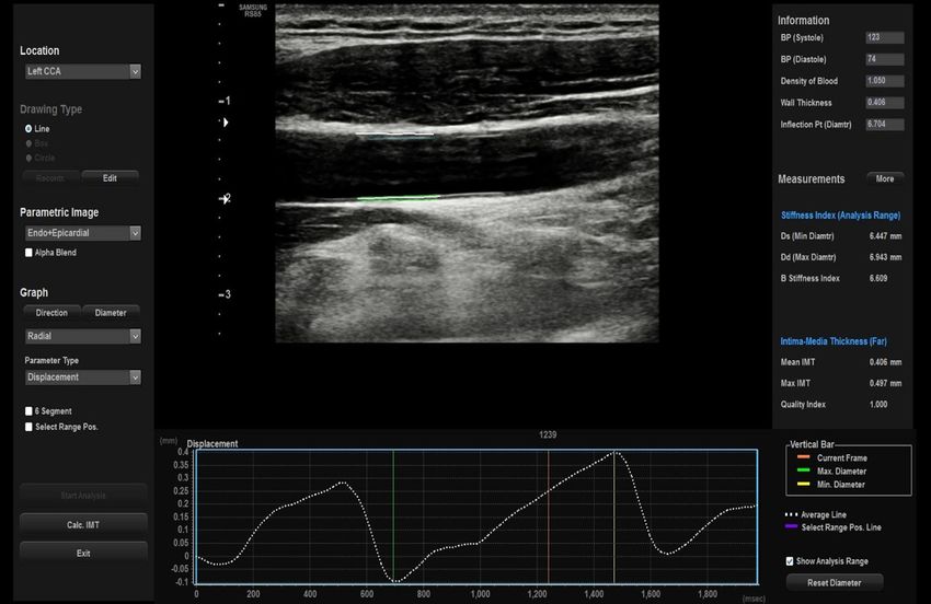
modulus in the longitudinal plane. Figure 1. a, b. Evaluation of the strain parameters of CCA in (a) transverse and (b) longitudinal planes
using the STCS method in carotid B-mode US.
196• March–April 2021 • Diagnostic and Interventional Radiology Cerit et al.plaques (26). Also, in another study, STCS Table 1. Terminology of arterial analysis software
imaging-based measurements have been
Parameter Definition Formula
shown to allow early assessment of poten-
tial effects of statins on vascular function Arterial compliance Absolute change in vessel diame- Difference between diastolic and
ter according to change in applied systolic diameter (ΔD)/ difference
(27). This suggests that early determination
pressure between systolic and diastolic
of arterial stiffness has the potential to be blood pressure (ΔP)
a valuable tool in the prevention of cardio-
Arterial distensibility The relative change in vessel diam- ΔD/(ΔP × vessel diameter in the
vascular events. In several studies, the STCS eter according to change in applied diastolic phase (D)) in mm Hg
method has been used to identify cardio- pressure
vascular risk in many different patient pop-
Elastic modulus index Alteration of pressure necessary for (ΔPxD)/ΔD in mm Hg
ulations such as atherothrombotic stroke, theoretical stretch from resting vessel
end-stage renal disease, rheumatoid arthri- diameter
tis, obstructive sleep apnea, and hyperten- β-stiffness index The ratio of the natural logarithm of log (SBP/DBP)/(ΔD/D)
sion (28–32). This method promises to be systolic/diastolic pressure to relative
beneficial, noninvasive, quick, and easily change in diameter
applicable, especially in the evaluation of Pulse wave velocity The speed of the pulse wave extends Stiffness index × diastolic blood
the CCA (11, 32). (PWV) through the length of the vessel pressure/(2 × blood density)
Arterial stiffness has been defined as an When the blood density is
early marker of atherosclerosis in T1DM assumed to be 1.050 g/cm3.
patients (3, 33). However, to the best of our
knowledge, no prior study evaluated the
chemical evaluation, early in the morning, Speckle tracking strain analysis of the
effectiveness of the STCS method and ar-
just before the carotid ultrasonography carotid artery
terial analysis software in the detection of
examination. Age, sex, heart rate (/min), Arterial Analysis software (Samsung Me-
early atherosclerotic signs in adult T1DM
central systolic and diastolic blood pressure dison Co., Ltd.), which was inherent to US
patients.
(mmHg), BMI (kg/m2), total cholesterol (mg/ device, was used to quantify CCA strain
We aimed to evaluate the effectiveness of
dL), LDL, HDL, apo B, triglycerides (mg/dL), and stiffness parameters. Vascular wall dis-
STCS technique, which enables measuring
smoking status, fasting blood sugar, HgA1c, placement was automatically calculated by
arterial stiffness and strain parameters in
visceral fat (kg), abdominal fat ratio (%), software to evaluate the functional capabil-
the detection of early atherosclerotic find-
whole-body fat ratio (%), fat mass (kg), fat- ities of vascular structures. For the analysis,
ings in T1DM.
free mass (kg), total blood weight (kg), cre- ≥5 mm of the CCA below the origin of the
atinin (mg/dL) values were recorded for all carotid bulb was used. The user-defined
Methods patients on the day of carotid ultrasound. control points in the vessel wall were set to
Study population Non-HDL cholesterol level was calculated follow the optical flow algorithm in a de-
The Institutional Review Board (24074710- as [total cholesterol - HDL cholesterol]. The termined frame automatically. The control
604.01.01-08) approved this prospective recommended level of non-HDL cholesterol points were limited to maintain the round
study. Written informed consent was ob- isTable 2. Baseline descriptive features and laboratory results of T1DM and control groups the systolic and diastolic blood pressure
was measured by a trained, experienced
T1DM (n=30) Control (n=30) p
technician with pulse wave analysis of the
Age (years) 34.27±9.12 34.23±8.58 0.988 brachial artery using an ERKA sphygmoma-
Men, n (%) 11 (36.6) 13 (43.3) 0.598 nometer (ERKA, D- 83646 Perfect Aneroid).
Heart rate (/min) 86.13±10.89 79.83±10.44 0.026 Blood pressure measurements were per-
formed on the elbow after the patients had
Central systolic blood pressure (mmHg) 123.13±12.96 125.3±11.21 0.410
rested in the supine position for 5 minutes.
Central diastolic blood pressure (mmHg) 80.53±9.75 78.43±9.07 0.392 The measured systolic and diastolic blood
Body mass index (kg/m2) 23.7±3.06 25.3±3.45 0.916 pressure was entered into the software, and
Total cholesterol (mg/dL) 187 (138–293) 188 (138–266) 0.968 all arterial stiffness parameters were then
Apo B 111±26.8 97.6±24.6 0.077
automatically calculated by the software
with respect to the pre-defined formula for
LDL (mg/dL) 117.84±31.04 115.72±26.86 0.781
each parameter. The definition and formu-
HDL (mg/dL) 59.57±12.32 53.03±11.00 0.036 la of each arterial stiffness parameter are
Non-HDL cholesterol (mg/dL) 133.96±35.97 137.48±32.69 0.702 presented in Table 1. An increase in elas-
Triglycerides (mg/dL) 82 (33–211) 90 (35–352) 0.212 tic modulus, β-stiffness indices and PWV
indicates an increase in arterial stiffness,
Smoking, n (%) 13 (43) 7 (23) 0.100
whereas an increase in arterial distensibility
Fasting blood glucose (mg/dL) 179 (28–384) 83 (72–117)Table 3. Comparison of parameters in the transverse and longitudinal plane between groups
Transverse plane Longitudinal plane
T1DM (n=30) Control (n=30) p T1DM (n=30) Control (n=30) p
CIMT (mm) 0.47±0.11 0.42±0.05 0.039
Minimum diameter (mm) 6.8 (5.8–8.5) 6.8 (5.8–7.8) 0.657 5.91±0.59 5.98±0.52 0.650
Maximum diameter (mm) 7.34±0.70 7.26±0.55 0.641 6.4 (5.4–7.9) 6.4 (5.6–7.7) 0.749
Stiffness parameters
β-stiffness index 5.7 (3.1–16.2) 5.9 (2.4–14.9) 0.824 4.3 (2.5–10.4) 5.3 (1.7–10.3) 0.160
Arterial compliance (mm/kPa) 0.98±0.34 0.99±0.413 0.947 1.02±0.42 0.96±0.49 0.642
Arterial distensibility (/kPa) 0.01 (0.01–0.03) 0.01 (0.01–0.03) 0.826 0.02 (0.01–0.04) 0.02 (0.01–1.01) 0.489
Elastic modulus (kPa) 78.4 (37.7–220) 84.8 (31.6–193) 0.965 58.6 (30.8–141) 68.2 (22.6–133.5) 0.308
Pulse wave velocity (m/s) 5.80±1.27 5.72±1.19 0.789 5.10± 1.05 5.17± 1.02 0.771
Strain parameters (radial)
Displacement (mm) 0.48±0.15 0.47±0.15 0.889 0.55±0.18 0.52± 0.19 0.590
Strain (%) 6.81±2.14 6.70±2.26 0.852 8.86±2.80 8.18±2.84 0.355
Strain rate (1/s) 0.72 (0.39–1.34) 0.77 (0.3–1.35) 0.929 0.84 (0.55–1.46) 0.77 (0.43–1.13) 0.165
Strain parameters (circumferential)
Displacement (mm) 0.06 (0.04–0.11) 0.06 (0.03–0.11) 0.953
Strain (%) 6.82±2.13 6.82±2.19 0.990
Strain rate (1/s) 0.72 (0.38–1.33) 0.75 (0.29–1.34) 0.965
T1DM, type 1 diabetes mellitus; CIMT, carotid intima media thickness.
Data are expressed as mean±standard deviation for normal distributions or median (min–max) for non-normalized variables.
and whole-body fat ratio on the parameters
Table 4. Comparison of stiffness parameters in T1DM patients with non-HDL cholesterol value >130
mg/dL and ≤ 130 mg/dL of carotid artery strain and arterial stiffness
were assessed using Pearson and Spearman
Non-HDL>130 mg/dL (n=15) Non-HDL≤130 mg/dL (n=15) p
correlation coefficient (two-tailed) for data
Longitudinal plane with normal and non-normal distribution,
CIMT (mm) 0.47±0.12 0.48±0.12 0.748 respectively. Significance level was set at α
β-stiffness index 4.83 (3.4–10.4) 3.66 (2.5–8.96) 0.049 = 0.05. We performed all the statistical analy-
ses using SPSS (Version 22.0, IBM). When the
Elastic modulus (kPa) 62.18 (39–141) 48.78 (30.8–119) 0.049
prevalence of T1DM was accepted as 0.08%,
T1DM, type 1 diabetes mellitus; HDL, high-density lipoprotein; CIMT, carotid intima media thickness.
the power of our study was 98.3% according
Data are expressed as mean±standart deviation if normally distributed or median (min-max) if non-normally
distributed. to the power analysis performed in the epi
info program by taking the alpha value 0.05,
within the 95% confidence interval (35).
wall interfaces that define the blood-intima Statistical analysis
boundaries in the carotid artery (at least 4 Categorical variables were presented as Results
spots in all) were marked on a still image, n (%), non-normally distributed continuous As shown in Table 2, T1DM and control
then the movement of the marked points variables were shown as “median (min-max)”, groups were similar in terms of age, sex, cen-
was automatically monitored by the soft- and normally distributed variables were tral systolic and diastolic blood pressure, BMI,
ware. The average of the right and left ca- shown as “mean ± standard deviation”. The total cholesterol, Apo B, LDL, non-HDL cho-
rotid artery CIMT values were used in the normality of data distribution in groups and lesterol, triglycerides, smoking, visceral fat,
study analysis (Fig. 1). subgroups was evaluated with the Kolmog- abdominal fat ratio, whole-body fat ratio, fat-
orov-Smirnov test and Shapiro Wilks test, re- free mass, total blood weight and creatinine.
Other physiological measurements spectively. Pearson’s chi-square test was used In the T1DM group, the heart rate, HDL, fast-
TANITA Body Composition Analyzer (BC- to compare the categorical variables; the ing blood glucose, and HbA1c were higher
418 MA, Tanita Corp.) was used for anthro- Mann-Whitney U test (for non-normal distri- than in the control group (p = 0.026, p = 0.036,
pometric and body composition measure- bution) or Student’s t test (for normal distri- p < 0.001, p < 0.001, respectively). In the con-
ments and Tanita AB-140 ViScan (Tanita bution) was used to compare the continuous trol group, the fat mass was higher than in the
Corp.) was used for abdominal fat ratio variables. In the diabetic group, the impact diabetic group (p = 0.012). In the T1DM group,
measurement. of age, duration of diabetes, CIMT, fat mass, nephropathy or neuropathy was present in
Speckle tracking carotid strain US in type 1 DM • 199Table 5. Comparison of strain and stiffness parameters in T1DM patients with and without nephropathy, neuropathy, and retinopathy
Nephropathy Nephropathy Neuropathy Neuropathy Retinopathy Retinopathy
(+) (-) (+) (-) (+) (-)
Transverse plane (n=12) (n=18) p (n=12) (n=18) p (n=8) (n=22) p
Elastic modulus (kPa) 98.12 72.35 0.039 80.79 75.95 0.553 77.05 80.57 0.574
(37.7–220) (37.7–219) (48.7–174.4) (37.7–219.8) (71.5–174.4) (37.7–219.8)
Pulse wave velocity (m/s) 5.98 5.26 0.031 5.79 5.55 0.374 5.44 5.57 0.439
(5.07–8.73) (3.92–8.9) (4.51–7.79) (3.92–8.9) (5.1–7.8) (3.92–8.9)
Longitudinal plane
Elastic modulus 84.1 55.5 0.016 60.0 56.9 0.735 70.8 56.9 0.302
(48.8–140.5) (30.8–116.7) (97–359) (30.8–140.5) (48.8–124.6) (30.8–140.5)
β-stiffness index 5.61 4.03 0.047 4.37 4.27 0.866 4.65 4.2 0.453
(3.4–10.4) (2.5–8.4) (2.54–10.3) (2.50–10.4) (3.35–10.2) (2.5–10.4)
Arterial compliance (mm/kPa) 0.83 1.15 0.042 1.03 0.99 0.687 0.87 1.09 0.398
(0.32–1.63) (0.46–1.94) (0.32–1.94) (0.41–1.85) (0.42–1.63) (0.32–1.94)
Distensibility (/kPa) 0.0100 0.015 0.026 0.02 0.02 0.965 0.02 0.02 0.243
(0.01–0.03) (0.01–0.04) (0.01–0.03) (0.01–0.04) (0.01–0.03) (0.01–0.04)
Pulse wave velocity (m/s) 5.58 4.5 0.038 5.04 4.71 0.268 5.29 4.74 0.464
(4.31–7.05) (3.54–6.53) (3.81–6.8) (3.54–7.05) (4.31–6.58) (3.54–7.05)
CIMT (mm) 0.50 0.46 0.362 0.51 0.44 0.138 0.50 0.46 0.241
(0.35–0.65) (0.28–0.76) (0.34–0.65) (0.28–0.76) (0.40–0.65) (0.28–0.76)
CIMT, carotid intima media thickness.
Data are expressed as mean±standart deviation if normally distributed or median (min-max) if non-normally distributed.
Table 6. Correlation of age and arterial analysis parameters in T1DM group
ferential strain, circumferential strain rate
evaluated in the transverse plane and lon-
Longitudinal plane Transverse plane
gitudinal plane (Table 3).
r p r p Diabetic patients were subdivided ac-
CIMT 0.624 130 mg/dL (n=15) or ≤130 mg/dL (n=15).
β-stiffness index 0.687a b c
d e
Figure 3. a–e. Correlation between whole
body fat ratio (%) and transverse radial
displacement (mm) (a), transverse radial strain
(%) (b), transverse circumferential displacement
(mm) (c), circumferential strain (%) (d), and
longitudinal radial displacement (mm) (e) in the
T1DM group.
a b c
d e
Figure 4. a–e. Correlation between fat mass
(kg) and transverse radial displacement (mm)
(a), transverse radial strain (%) (b), transverse
circumferential displacement (mm) (c),
circumferential strain (%) (d), and longitudinal
radial displacement (mm) (e) in the T1DM
group.
higher β-stiffness index values in the longi- ed in the longitudinal plane compared with with retinopathy or neuropathy and patients
tudinal plane compared with patients with- patients without nephropathy (p = 0.042, without those conditions (p > 0.05) (Table 5).
out nephropathy (p = 0.047). Patients with p = 0.026, respectively). In terms of the arte- In both groups, age was correlated with
nephropathy had lower arterial compliance rial stiffness indices and strain parameters, all arterial stiffness and strain parameters of
and distensibility values which were evaluat- there were no differences between patients the CCA in all planes (p < 0.05) (Table 6).
Speckle tracking carotid strain US in type 1 DM • 201a b HDL cholesterol had higher β-stiffness and
elastic modulus values which were evalu-
ated in the longitudinal plane. In terms of
CIMT, there was no difference between pa-
tients with higher versus normal non-HDL
cholesterol levels.
Arterial stiffness has been defined as an
early marker of atherosclerosis in T1DM pa-
tients (3, 33). Many studies have shown that
subclinical signs of arterial wall stiffening
due to increased cardiovascular risk can be
detected by speckle tracking techniques
(15, 17–22). Arterial analysis software using
c d ultrasound-based speckle tracking method
evaluates arterial wall stiffness by providing
measurement of regional mechanical prop-
erties of the arterial wall by ultrasound. To
the best of our knowledge, no prior study
has evaluated the effectiveness of the STCS
method for detecting early atherosclerotic
signs in adult T1DM patients. Several stud-
ies have revealed that the strain values ob-
tained using the STCS method can be used
to effectively evaluate alterations in the
elasticity of the carotid artery due to aging
or cardiovascular risk factors (17, 32, 36). In
Figure 5. a–d. Correlation between duration of diabetes (years) and longitudinal β-stiffness
index (a), longitudinal arterial compliance (mm/kPa) (b), longitudinal elastic modulus (kPa) (c), a study evaluating arterial stiffness using
and longitudinal pulse wave velocity (m/s) (d) in the T1DM group. speckle tracking method in 50 children with
T1DM, it was found that stiffness parame-
In the diabetic group, abdominal fat ratio, group and the diabetic patients. T1DM and ters (strain, strain rate) derived from STCS
whole-body fat ratio, and fat mass were neg- control groups were compared in terms of imaging were lower in diabetic children
atively correlated with radial displacement in carotid artery wall elasticity characteristics than in controls (11). In a study including
the longitudinal plane (r= -0.416 p = 0.028, r= by evaluating strain and stiffness param- 26 elderly diabetic patients (Type 1 and 2)
-0.434 p = 0.014, r= -0.410 p = 0.030, respec- and 26 healthy young volunteers, the mean
eters and no statistically significant dif-
amplitude value for diameter change and
tively), as well as with radial strain (r= -0.437 ference was found in arterial stiffness and
the longitudinal displacement of arterial
p = 0.020, r= -0.470 p = 0.012, r= -0.437 strain parameters measured in transverse
wall were lower in diabetes patients than in
p = 0.020, respectively), circumferential dis- and longitudinal planes. However, high-
healthy young volunteers (37). Kim et al. (27)
placement (r= -0.554 p = 0.002, r= -0.540 er CIMT values were found in the diabetic
previously found that carotid artery elas-
p = 0.003, r= -0.468 p = 0.012, respectively) group than in the control group. In both
ticity, as measured with a STCS technique,
and circumferential strain (r= -0.442 p = 0.019, groups, age was positively correlated with
significantly increased after short-term
r= -0.466 p = 0.013, r= -0.432 p = 0.022, re- the arterial stiffness indices but negatively
high-dose statin treatment, but they found
spectively) in the transverse plane (Figs. 2–4). correlated with the CCA strain parameters.
no change in CIMT or maximum plaque
In the diabetic group, the duration of In the T1DM group, duration of diabetes thickness as a result of this treatment. This
diabetes was correlated with β-stiffness was positively correlated with β-stiffness suggests that STCS imaging-based mea-
index (r= 0.503 p = 0.005), arterial disten- index, elastic modulus and PWV, but neg- surements allow for an early assessment of
sibility (r= -0.490 p = 0.006), arterial com- atively correlated with the arterial distensi- statins’ potential effects on vascular func-
pliance (r= -0.447 p = 0.013), elastic mod- bility. When T1DM patients with and with- tion. Furthermore, Seals et al. (38) reported
ulus (r= 0.493 p = 0.006) and PWV (r= 0.502 out diabetic nephropathy were compared, that regular exercise can attenuate age-re-
p = 0.006) in the longitudinal plane. Also, those with diabetic nephropathy had lated increase in arterial stiffness, and Pugh
the duration of diabetes was correlated with higher transverse and longitudinal elastic et al. (13) found that in comparison to con-
β-stiffness index (r= 0.390 p = 0.033), arterial modulus and PWV, as well as longitudinal ventional arterial stiffness measures, STCS is
compliance (r= -0.384 p = 0.036), elastic mod- β-stiffness index, but lower longitudinal superior to demonstrate cardiorespiratory
ulus (r= 0.387 p = 0.034) and PWV (r= 0.426 arterial compliance and distensibility. In fitness related alterations on arterial stiff-
p = 0.019) in the transverse plane (Fig. 5). terms of CIMT, there was no difference be- ness in young individuals. Taken together,
tween patients with and without nephrop- these findings suggest that STCS imaging
Discussion athy. When diabetic patients with non-HDL has the potential to be a valuable tool in the
In the present study, the STCS method cholesterol >130 mg/dL and ≤130 mg/dL determination of arterial stiffness and may
was effectively applied in both the control were compared, patients with high non- aid in prevention of cardiovascular events.
202 • March–April 2021 • Diagnostic and Interventional Radiology Cerit et al.Applanation tonometry is the gold stan- CIMT is an important marker of athero- Non-HDL cholesterol level has been iden-
dard technique used to evaluate arterial sclerosis and an independent predictor of tified as a significant predictor of persistent
wall stiffness. However, this technique is cardiovascular events. Studies have shown dyslipidemia and atherosclerosis for diabetic
time consuming, requires dedicated equip- that CIMT is increased in patients with T1DM patients (46). In our study, when diabetic pa-
ment and is not widely used in clinical (39). In this study, in accordance with the lit- tients were divided into subgroups according
routine (11, 12). Studies have shown that erature, we found that CIMT was higher in to the presence of dyslipidemia, no differ-
speckle tracking method can be an alterna- the T1DM group than in the control group, ence was found between the two groups in
tive to applanation tonometry in the eval- and CIMT was correlated with age (2, 40, 41). terms of CIMT, while arterial stiffness param-
uation of arterial wall stiffness (15–17). In a Studies have shown that advanced age eters were found to be higher in the group
study evaluating arterial stiffness using ap- can change the elastic composition of with high non-HDL cholesterol. This finding
planation tonometry and speckle tracking the arterial wall matrix (increase in colla- suggests that, in T1DM patients with dyslip-
method in 50 children with T1DM, stiffness gen-elastin ratio due to age-related degen- idemia, functional changes may exist, even in
parameters derived from tonometry and eration of elastin fibers and compensatory the absence of morphological changes.
sonography were significantly correlated increases in collagen), which causes the Weight problems and visceral body-fat
with each other (11). Unlike applanation stiffening of the large central arteries (4, deposition play leading roles in the de-
tonometry, measuring local stiffness with 13). In our study, age was positively cor- velopment of metabolic syndrome in the
speckle tracking method in CCA provides related with the arterial stiffness indices but overall population (47–49). Generally, pa-
additional information about arterial wall negatively correlated with the CCA strain tients with T1DM have normal weight, but
compliance and local changes in the het- parameters in both groups. These results researchers have associated central fat
erogeneous movement pattern, and in accumulation with insulin resistance and
are concordant with previous studies which
this way it assures to be a superior index metabolic syndrome in T1DM patients (50–
also demonstrated that CCA strain mea-
53). In this study’s T1DM group, abdominal
of whole artery wall stress (13, 17). Publica- surements, elastic modulus, and β-stiffness
fat ratio, whole-body fat ratio, and fat mass
tions which reported that 2D strain imaging index correlate significantly with increasing
all were negatively correlated with the ra-
is more sensitive than conventional stiff- age, which is a well-defined risk factor in
dial and circumferential displacement and
ness parameters, such as PWV, elastic mod- atherosclerosis (17–19, 42–44).
strain parameters in transverse plane, as
ulus, β-stiffness index, in detecting age-re- In the diabetic group, statistically signif-
well as with radial displacement in the lon-
lated changes in CCA elastic properties also icant differences were found between ar-
gitudinal plane.
support this (17). terial stiffness parameters between groups
Several researchers have proven a rela-
In this study, we compared individuals with and without high non-HDL choles-
tionship between atherosclerosis and dura-
with and without T1DM in terms of carot- terol. However, there was no statistically tion of diabetes (45, 53). In our study, in the
id artery wall elasticity characteristics by significant difference between the groups diabetic patient group; as the duration of
evaluating strain and stiffness parameters in terms of CIMT. This finding suggested diabetes increased, parameters indicating
using the STCS method and no significant that in T1DM patients with dyslipidemia, vascular stiffness such as β-stiffness index,
difference was found in arterial stiffness functional changes may exist, even in the elastic modulus and PWV values increased,
and strain parameters in both transverse absence of morphological changes, and the whereas parameters indicating strain abili-
and longitudinal planes between the dia- STCS technique may provide for the early ty values such as distensibility decreased.
betic and control groups. Our study popu- assessment of atherosclerosis in patients These results strongly support the deteri-
lation consisted of 30 T1DM patients with- with T1DM, especially in those with dyslip- oration of vascular function as duration of
out known cardiovascular disease and 30 idemia. diabetes increases.
non-diabetic sex, age, and BMI matched Mortality risk due to cardiovascular disease The main limitation of this study is that
controls without any known disease. There increases remarkably after the development the control group did not consist of nor-
were no individuals over 52 years of age of nephropathy (10). In addition, mortality mal healthy patients. The unsuitability of
in the study. There was no significant dif- rates increase in T1DM patients with prom- the control group to act as a true control
ference in the prevalence of hypertension, inent kidney disease (2, 45). Thus, we eval- masked the true CCA stiffness profile of the
smoking status, BMI, which could influence uated our T1DM patients for the presence T1DM patients. In addition, the small num-
the arterial stiffness and strain parameters, of nephropathy. Patients with diabetic ne- ber of patients in the diabetic subgroups,
between diabetic and control groups. phropathy had higher values in transverse varied disease duration, and the hetero-
However, in control individuals who were and longitudinal elastic modulus and PWV, geneous disease severity associated with
accepted as healthy at the beginning of as well as for longitudinal β-stiffness index, nephropathy, neuropathy, and retinopathy
the study, fat mass values, which were but lower values for longitudinal arterial are other limitations of the study. A large
found to have a negative correlation with compliance and distensibility, than patients group of diabetic patients with prespeci-
strain parameters, were higher than in the without nephropathy. Whereas, in terms of fied disease duration and homogeneous
diabetic patients. Moreover, HDL values CIMT there were no statistically significant disease severity and a control group with
were lower in the control group. These differences between patients with or without healthy individuals may be more valuable
unexpected findings of the control group diabetic nephropathy. Our findings suggest for demonstrating whether there are signif-
may have contributed to the inability to that, in T1DM patients with diabetic nephrop- icant differences between these groups.
clearly demontrate the effect of T1DM on athy, functional changes may exist, even in In conclusion, functional changes may
arterial stiffness. the absence of morphological changes. exist in T1DM patients with diabetic ne-
Speckle tracking carotid strain US in type 1 DM • 203phropathy and dyslipidemia, even in the 11. Podgórski M, Szatko K, Stańczyk M, et al. 23. Kawasaki T, Sasayama S, Yagi S, Asakawa T, Hirai T.
absence of morphological changes, and the Two-dimensional speckle tracking versus Non-invasive assessment of the age related chang-
applanation tonometry in evaluation of sub- es in stiffness of major branches of the human ar-
STCS technique may provide for the early as- clinical atherosclerosis in children with type teries. Cardiovasc Res 1987; 21:678–687. [Crossref]
sessment of atherosclerosis in patients with 1 diabetes mellitus. Med Sci Monit 2019; 24. Mattace-Raso FU, van der Cammen TJ, Hofman
T1DM, particularly those with nephropathy 25:7289–7294. [Crossref] A, et al. Arterial stiffness and risk of coronary
and dyslipidemia. Also, functional changes 12. Wang KL, Cheng HM, Chuang SY, et al. Cen- heart disease and stroke: the Rotterdam Study.
in the carotid artery may be demonstrated tral or peripheral systolic or pulse pressure: Circulation 2006; 113:657–663. [Crossref]
which best relates to target organs and future 25. Selzer RH, Mack WJ, Lee PL, et al. Improved
with the STCS technique in T1DM patients mortality? J Hypertens 2009; 27:461–467. common carotid elasticity and intima-media
with high abdominal fat ratio, whole-body [Crossref] thickness measurements from computer anal-
fat ratio, and fat mass. Thus, together with 13. Pugh CJA, Stone KJ, Stöhr EJ, et al. Carotid ar- ysis of sequential ultrasound frames. Athero-
CIMT measurement, STCS analyses may be tery wall mechanics in young males with high sclerosis 2001; 154:185–193. [Crossref]
cardiorespiratory fitness. Exp Physiol 2018; 26. Zhang L, Liu Y, Zhang PF, et al. Peak radial and cir-
used to detect subclinical damage and strat-
103:1277–1286. [Crossref] cumferential strain measured by velocity vector
ify atherosclerosis in patients with T1DM. 14. Laurent S, Cockcroft J, Van Bortel L, et al.; Eu- imaging is a novel index for detecting vulnerable
However, before implementing this technol- ropean Network for Non-invasive Investigation plaques in a rabbit model of atherosclerosis. Ath-
ogy in routine clinical applications, further of Large Arteries. Expert consensus document erosclerosis 2010; 211:146–152. [Crossref]
studies are necessary to validate its utility. on arterial stiffness: methodological issues and 27. Hae Kim C, Wang S, Park JB, et al. Assessing
clinical applications. Eur Heart J 2006; 27:2588– impact of high-dose pitavastatin on carotid
Conflict of interest disclosure 2605. [Crossref] artery elasticity with speckle-tracking strain
The authors declared no conflicts of interest. 15. Podgórski M, Grzelak P, Szymczyk K, et al. Pe- imaging. J Atheroscler Thromb 2018; 25:1137–
ripheral vascular stiffness, assessed with two-di- 1148. [Crossref]
mensional speckle tracking versus the degree of 28. Lee SE, Lee J, Yoo TH, Cho IJ, Chang HJ. End-
References stage renal disease impairs the multidirection-
coronary artery calcification, evaluated by tomo-
1. Khunti K, Davies M, Majeed A, Thorsted BL,
graphic coronary artery calcification index. Arch al movements of the common carotid artery:
Wolden ML, Paul SK. Hypoglycemia and risk of
Med Sci 2015; 11:122–129. [Crossref] assessment using dimensional speckle-track-
cardiovascular disease and all-cause mortality
16. Podgórski M, Grzelak P, Kaczmarska M, et al. ing carotid strain ultrasonography. J Cardio-
in insulin treated people with type 1 and type vasc Imaging 2018; 26:155–164. [Crossref]
Feasibility of two-dimensional speckle tracking
2 diabetes: a cohort study. Diabetes Care 2015; 29. Yoon JH, Han D, Kim S, et al. Assessment of mul-
in evaluation of arterial stiffness: Comparison
38:316–322. [Crossref] tidirectional movements of the common carotid
with pulse wave velocity and conventional so-
2. Schofield J. Ho J. Soran H. Cardiovascular risk artery in atherothrombotic stroke using dimen-
nographic markers of atherosclerosis. Vascular
in type 1 diabetes mellitus. Diabetes Ther 2019; 2018; 26:63–69. [Crossref] sional speckle tracking carotid ultrasonography:
10:773–789. [Crossref] 17. Bjallmark A, Lind B, Peolsson M, Shahgaldi K, A prospective, controlled cohort study. Echocar-
3. Duarte SV, de Souza Rajão J, Pinho JF, et al. Brodin LA, Nowak J. Ultrasonographic strain diography 2018; 35:957–964. [Crossref]
Changes in aortic pulse wave components, imaging is superior to conventional non-in- 30. Lee JH, Cho KI, Kim SM. Carotid arterial stiffness
pulse pressure amplification, and hemody- vasive measures of vascular stiffness in the in patients with rheumatoid arthritis assessed
namic parameters of children and adolescents detection of age-dependent differences in the by speckle tracking strain imaging: its asso-
with type 1 diabetes. Pediatr Diabetes 2019; mechanical properties of the common carotid ciation with carotid atherosclerosis. Clin Exp
20:202–209. [Crossref] artery. Eur J Echocardiogr 2010; 11:630–636. Rheumatol 2012; 30:720–728.
4. Wagenseil JE, Mecham RP. Elastin in large ar- [Crossref] 31. You M, Zhang L, Fang L, Li J, Xie M. Evaluation of
tery stiffness and hypertension. J Cardiovasc 18. Park HE, Cho GY, Kim HK, Kim YJ, Sohn DW. Val- carotid arterial elasticity in patients with obstruc-
Transl Res 2012; 5:264–273. [Crossref] idation of circumferential carotid artery strain tive sleep apnea hypopnea syndrome by two-di-
5. Nair SB, Malik R, Khattar RS. Carotid intima-me- as a screening tool for subclinical atheroscle- mensional speckle tracking imaging. Medicine
dia thickness: ultrasound measurement, prog- rosis. J Atheroscler Thromb 2012; 19:349–356. (Baltimore) 2017; 96:e8817. [Crossref]
nostic value and role in clinical practice. Post- [Crossref] 32. Yang EY, Brunner G, Dokainish H, et al. Appli-
grad Med J 2012; 88:694–699. [Crossref] 19. Catalano M, Lamberti-Castronuovo A, Cat- cation of speckle-tracking in the evaluation of
6. Gamble G, Zorn J, Sanders G, MacMahon S, alano A, Filocamo D, Zimbalatti C. Two-di- carotid artery function in subjects with hyper-
Sharpe N. Estimation of arterial stiffness, com- mensional speckle-tracking strain imaging tension and diabetes. J Am Soc Echocardiogr
pliance, and distensibility from M-mode ultra- in the assessment of mechanical properties 2013; 26:901–909.e1. [Crossref]
sound measurements of the common carotid of carotid arteries: feasibility and comparison 33. Sharrett AR, Ding J, Criqui MH, et al. Smoking,
artery. Stroke 1994; 25:11–16. [Crossref] with conventional markers of subclinical ath- diabetes, and blood cholesterol differ in their
7. O’Leary DH, Polak JF, Kronmal RA, Manolio TA, erosclerosis. Eur J Echocardiogr 2011; 12:528– associations with subclinical atherosclerosis: the
Burke GL, Wolfson SK Jr. Carotid-artery intima 535. [Crossref] multiethnic study of atherosclerosis (MESA). Ath-
and media thickness as a risk factor for myocar- 20. Larsson M, Kremer F, Claus P, et al. Ultra- erosclerosis 2006; 186:441–447. [Crossref]
dial infarction and stroke in older adults. Cardio- sound-based radial and longitudinal strain 34. Standards of medical care in diabetes 2020. Diabe-
vascular Health Study Collaborative Research estimation of the carotid artery: a feasibility tes Care 2020; 43 (Suppl 1):S163–S182. [Crossref]
Group. N Engl J Med 1999; 340:14–22. [Crossref] study. IEEE Trans Ultrason Ferroelectr Freq Con- 35. Yeşilkaya E, Cinaz P, Andıran N, et al. First report
8. Bots ML, Hoes AW, Koudstaal PJ, Hofman A, trol 2011; 58:2244–2251. [Crossref] on the nationwide incidence and prevalence
Grobbee DE. Common carotid intima-media 21. Larsson M, Heyde B, Kremer F, et al. Ultrasound of Type 1 diabetes among children in Turkey.
thickness and risk of stroke and myocardial speckle tracking for radial, longitudinal and Diabet Med 2017; 34:405–410. [Crossref]
infarction: the Rotterdam Study. Circulation circumferential strain estimation of the carotid 36. Myung Y, Seo HS, Jung IH, et al. The correlation
1997; 96:1432–1437. [Crossref] artery – An in vitro validation via sonomicrome- of carotid artery stiffness with heart function in
9. Wada T, Kodaira K, Fujishiro K, et al. Correlation try using clinical and highfrequency ultrasound. hypertensive patients. J Cardiovasc Ultrasound
of ultrasound-measured common carotid ar- Ultrasonics 2015; 56:399–408. [Crossref] 2012; 20:134–139. [Crossref]
tery stiffness with pathological findings. Arte- 22. Ribbers H, Lopata RGP, Holewijn S, et al. 37. Zahnd G, Boussel L, Marion A, et al. Measure-
rioscler Thromb 1994; 14:479–482. [Crossref] Noninvasive two-dimensional strain imag- ment of two-dimensional movement parame-
10. Kim SA, Park SM, Kim MN, et al. The relationship ing of arteries: validation in phantoms and ters of the carotid artery wall for early detec-
between mechanical properties of carotid artery preliminary experience in carotid arteries in tion of arteriosclerosis: a preliminary clinical
and coronary artery disease. Eur Heart J Cardio- vivo. Ultrasound Med Biol 2007; 33:530–540. study. Ultrasound Med Biol 2011; 37:1421–
vasc Imaging 2012; 13:568–573. [Crossref] [Crossref] 1429. [Crossref]
204 • March–April 2021 • Diagnostic and Interventional Radiology Cerit et al.38. Seals DR,Walker AE, Pierce GL, Lesniewski LA. 44. Yoon JH, Cho IJ, Chang HJ, et al. The value of 50. Thorn LM, Forsblom C, Fegerudd J, et al. Met-
Habitual exercise and vascular aging. J Physiol elastic modulus index as a novel surrogate abolic syndrome in type 1 diabetes. Diabetes
2009; 587:5541–5549. [Crossref] marker for cardiovascular risk stratification by Care 2005; 28:2019–2024. [Crossref]
39. Yamasaki Y, Kawamori R, Matsushima H, et dimensional speckle-tracking carotid ultra- 51. Dib SA. Insulin resistance and metabolic syn-
al. Atherosclerosis in carotid artery of young sonography. J Cardiovasc Ultrasound 2016; drome in type 1 diabetes. Arq Bras Endocrinol
IDDM patients monitored by ultrasound 24:215–222. [Crossref] Metabol 2006; 50:250–263. [Crossref]
high-resolution B-mode imaging. Diabetes 45. Lind M, Svensson AM, Kosiborod M, et al. Gly- 52. Kilpatrick ES, Rigby AS, Atkin SL. Insulin resis-
1994; 43:634–639. [Crossref] cemic control and excess mortality in type 1 tance, the metabolic syndrome, and complica-
40. Lyons TJ, Jenkins AJ, Zheng D, et al. Nuclear diabetes. N Engl J Med 2014; 371:1972–1982. tion risk in type 1 diabetes. Diabetes Care 2007;
magnetic resonance-determined lipoprotein [Crossref] 30:707–712. [Crossref]
subclass profile in the DCCT/EDIC cohort: asso- 46. Kissebah AH, Vydelingum N, Murray R, et al. 53. Rawshani A, Sattar N, Franzen S, et al. Excess
ciations with carotid intima–media thickness. Relation of body fat distribution to metabolic mortality and cardiovascular disease in young
Diabet Med 2006; 23:955–66. [Crossref] complication of obesity. J Clin Endocrinol Me- adults with type 1 diabetes in relation to age
41. Nathan DM, Lachin J, Cleary P, et al. Intensive tab 1982; 54:254–260. [Crossref] at onset: a nationwide, register-based cohort
diabetes therapy and carotid intima–media 47. Fujioka S, Matsuzawa Y, Tokunaga K, Tarui S. study. Lancet 2018; 392:477–486. [Crossref]
thickness in type 1 diabetes mellitus. N Engl J Contribution of intraabdominal fat accumula-
Med 2003; 348:2294–2303. [Crossref] tion to the impairment of glucose and lipid me-
42. Yuda S, Kaneko R, Muranaka A, et al. Quanti- tabolism in human obesity. Metabolism 1987;
tative measurement of circumferential carot- 36:54–59. [Crossref]
id arterial strain by two-dimensional speckle 48. Ribeiro Filho FF, Mariosa LS, Ferreira SRG,
tracking imaging in healthy subjects. Echocar- Zanella MT. Gordura visceral e síndrome me-
diography 2011; 28:899–906. [Crossref] tabólica: mais do que uma simples associação.
43. Oishi Y, Miyoshi H, Iuchi A, Nagase N, Ara N, Oki Arq Bras Endocrinol Metabol 2006; 50:230–
T. Vascular aging of common carotid artery and 238. [Crossref]
abdominal aorta in clinically normal individu- 49. Momesso DP, Bussade I, Balarini Lima GA, Fonse-
als and preclinical patients with cardiovascular ca LPC, Russo LAT, Kupfer R. Body composition,
risk factors: diagnostic value of two dimension- metabolic syndrome and insulin resistance in
al speckle-tracking echocardiography. Heart type 1 diabetes mellitus. Arq Bras Endocrinol
Vessels 2013; 28:222–228. [Crossref] Metab 2011; 55:189–193. [Crossref]
Speckle tracking carotid strain US in type 1 DM • 205You can also read