Prevalence and Characteristics of Listeria monocytogenes Isolates in Raw Milk, Heated Milk and Nunu, a Spontaneously Fermented Milk Beverage, in ...
←
→
Page content transcription
If your browser does not render page correctly, please read the page content below
beverages
Article
Prevalence and Characteristics of Listeria
monocytogenes Isolates in Raw Milk, Heated Milk
and Nunu, a Spontaneously Fermented Milk
Beverage, in Ghana
James Owusu-Kwarteng 1, *, Alhassan Wuni 2 , Fortune Akabanda 1 and Lene Jespersen 3                 ID

 1   Department of Applied Biology, Faculty of Applied Sciences, University for Development Studies,
     P.O. Box 24, Navrongo Campus, Ghana; fakabanda@uds.edu.gh
 2   Department of Biotechnology, Faculty of Agriculture, University for Development Studies, P. O. Box TL 1882,
     Nyankpala, Ghana; alhassanwuni@yahoo.com
 3   Department of Food Science, University of Copenhagen, Rolighedsvej 26, DK 1958 Frederiksberg C,
     1958 Copenhagen, Denmark; lj@food.ku.dk
 *   Correspondence: jowusukwarteng@uds.edu.gh; Tel.: +233-209-265-738

 Received: 20 April 2018; Accepted: 21 May 2018; Published: 23 May 2018

 Abstract: Listeria monocytogenes is a gram-positive food-borne pathogen that causes listeriosis in
 humans. Currently, there is little information on the prevalence of Listeria monocytogenes in raw
 milk and traditional yoghurt-like milk beverage, nunu, in Ghana. The purpose of this study was
 to investigate the prevalence of L. monocytogenes isolates in raw cow milk, boiled milk and nunu in
 Ghana, and to characterize these L. monocytogenes isolates according to their serogroups, virulence
 potentials and antibiotic susceptibility profiles. A total of 254 samples comprising 114 raw cow milk,
 56 boiled milk and 84 nunu were collected from dairy farms and market vendors for detection of
 L. monocytogenes. The overall prevalence of L. monocytogenes in raw milk, boiled milk and nunu
 was 5.5% (14/254). Listeria monocytogenes was prevalent in raw cow milk (8.8%; 10/114) and nunu
 (13.1%; 11/84), while no Listeria spp. was not detected in boiled milk. A total of 62 L. monocytogenes
 isolates were analysed to belong to molecular serogroups 1/2a-3a (32/62, 51.6%), 1/2b-3b-7 (14/62,
 22.6%), 4b-4d-4e (9/62, 14.5%) and 1/2c-3c (7/62, 11.3%). All 62 L. monocytogenes isolates harbored the
 virulence-associated genes inlA, inlB, inlC, inlJ, plcA, actA, hlyA, iap and prfA. All Listeria monocytogenes
 in the present study were generally susceptible to the tested antibiotics, except neomycin and
 tetracycline, for which phenotypic resistance was observed among isolates.

 Keywords: dairy products; food safety; virulence genes; antimicrobial resistance; listeria

1. Introduction
     Listeria monocytogenes is a gram positive, facultative intracellular food-borne pathogen that can
cause listeriosis in humans, especially in people of compromised immunity, including the elderly,
pregnant women and newborns [1,2]. The bacterium is widespread in nature and can survive and
grow under low temperatures and pH, high concentrations of salt or bile, oxidative stress, carbon
starvation, and other adverse conditions making it a potential hazard in foods [3]. L. monocytogenes has
been isolated from different raw and ready-to-eat (RTE) foods and in raw milk and dairy products in
different countries [4–6]. Several cases of listeriosis in humans are reported, sometimes with a high
case-fatality rates [7].
     Currently, thirteen (13) different serotypes of L. monocytogenes strains have been identified but
serotypes 1/2a, 1/2b, 1/2c and 4b are responsible for about 95% of human listeriosis [8,9]. To evaluate

Beverages 2018, 4, 40; doi:10.3390/beverages4020040                            www.mdpi.com/journal/beverages
Beverages 2018, 4, 40 2 of 10
the potential implications of isolated L. monocytogenes for food safety and public health, it is necessary to
differentiate between virulent and non-virulent strains [10]. Many supposed virulence markers such as
internalins (inlA, inlB, inlC, inlJ), listeriolysin O (hlyA), actin (actA), phosphatidylinositol-phospholipase
C (PI-PLC, plcA), Iap (invasion associated protein, iap) and virulence regulator (prfA) have been
implicated in the pathogenicity of L. monocytogenes [11–13].
Generally, L. monocytogenes isolates are susceptible to many antibiotics, especially
ampicillin/penicillin which are the primary antibiotics for treating listeriosis [14,15]. However,
resistance to single or multiple antibiotics have recently been reported for L. monocytogenes strains
isolated from various food and environmental sources [6,16–18]. It is, therefore, necessary to intensify
research aimed at increasing the available data on the prevalence and antibiotic susceptibility of
L. monocytogenes isolates from various food and environmental sources around the world.
In Ghana, cow milk may be consumed in its raw form, heated or processed by spontaneous
fermentation into a yoghurt-like beverage known as nunu [19–21]. Poor control measures in the
Ghanaian dairy chain potentially exposes raw cow milk and its products to possible contamination
by pathogenic microorganisms such as L. monocytogenes, which has implications for the safety of
consumers of raw milk and traditional dairy products in Ghana. Appiah et al. [20] reported on the
quantitative probabilistic assessment of L. monocytogenes exposure among consumers of milk in Ghana.
In general, however, there is limited data that can be used for the qualitative and/or quantitative
assessment of the risk of L. monocytogenes infection associated with the consumption of raw milk and
traditional dairy products in Ghana. The problem of limited data is compounded by the lack of a
proper surveillance systems for food-borne diseases to provide reliable information on the burden
of food-borne illnesses, particularly those involving L. monocytogenes associated with milk and milk
products in Ghana. Consequently, a number of illnesses or sporadic outbreaks associated with the
consumption of Listeria contaminated raw milk and milk products may go unreported. Therefore,
the objectives of this study were to determine the prevalence of L. monocytogenes in raw milk, heated
milk and nunu in Ghana and to characterize the isolated L. monocytogenes according to their molecular
serogroups, antibiotic susceptibility profiles and virulence potential.
2. Materials and Methods
2.1. Samples
A total of 326 samples comprising 114 raw cow milk, 56 boiled milk and 84 nunu (spontaneously
fermented yoghurt-like milk beverage) were purchased from dairy farms and open markets in the
Northern Region of Ghana between September 2015 and July 2016. Raw milk samples were collected
within 10 min. of milking at the dairy farm while boiled milk and nunu samples were collected at point
of sale in the market. Prior to sampling, all samples were stored at ambient temperatures (28 ± 2 ◦ C).
All samples were collected aseptically and immediately transported in a cool box stored at 4.0 ± 0.5 ◦ C
to the laboratory (Savana Agricultural Research Institute, Tamale-Ghana) for analysis. Samples were
kept at 4 ◦ C and analyzed within 24 h of collection. The pH of samples was determined upon delivery
to the laboratory using a pH meter (Crison basic 20, Barcelona, Spain) calibrated with standard buffers.
2.2. Isolation and Characterization of L. monocytogenes
Isolation and identification of L. monocytogenes in this study was carried out as described by
Becker et al. [22]. Briefly, a 25 g or 25 mL of each sample was aseptically homogenized in 225 mL
of Listeria enrichment broth base (CM 0862, Oxoid Ltd., Basingstoke, UK) with selective enrichment
supplement (SR 0141, Oxoid Ltd., Basingstoke, UK) in Stomacher bags (Seward Ltd., Worthing, UK) for
30 s using a Stomacher (BagMixer, Buch & Holm A/S, Interscience, 78860 St Nom, France), followed
by incubation at 30 ◦ C for 24 h. Second enrichment was done by adding 0.1 mL of the overnight
culture to 10 mL of Listeria enrichment broth base (Oxoid, Basingstoke, UK) with selective enrichment
supplement (Oxoid, Basingstoke, UK) and incubated at 37 ◦ C for 48 h. Subsequently, 0.1 mL of theBeverages 2018, 4, 40 3 of 10
enriched broth was surface plated on Listeria selective oxford agar (CM0856, Oxoid, Basingstoke, UK)
plates supplemented with Listeria selective supplement (SR0140, Oxoid, Basingstoke, UK). Inoculated
plates were incubated at 37 ◦ C for 24–48 h. Presumptive Listeria spp. were identified on oxford agar
plates after 24 h incubation as colonies with approximately 1 mm diameter, grey to black colonies
surrounded by a black halo. Following 48-h incubation period, typical Listeria species colonies were
approximately 2–3 mm diameter, black with a black halo and sunken center.
For biochemical identification of L. monocytogenes, up to five (5) presumptive colonies from each
positive plate were streaked on tryptic soy agar (Oxoid, Basingstoke, UK) supplemented with 0.6%
yeast extract (Oxoid, Basingstoke, UK) (TSA-YE) and incubated at 35 ◦ C for 24 h. The colonies from
TSA-YE plates were confirmed by biochemical tests including Gram staining, catalase, oxidase, triple
sugar iron (TSI), sulphide-idole-motility (SIM), methyl-red Voges-Proskauer (MR-VP) reaction, nitrate
reduction, and production of acid from rhamnose, xylose, mannitol and α-methyl-D-mannopyranoside.
Listeria monocytogenes ATCC 19115 was used as a reference strain for biochemical tests and PCR analysis.
2.3. PCR-Based Identification of L. monocytogenes
Following biochemical characterization of Listeria isolates, identities of L. monocytogenes were
further confirmed polymerase chain reaction (PCR). Crude DNA was extracted from Listeria isolated in
this study and L. monocytogenes ATCC 19115, used as positive control, by direct boiling of a suspension
of the cell lysates [23]. PCR was performed to detect the presence of Listeria spp. (LI1/U1) using the
specific primer sequences (50 -CTCCATAAAGGTGACCCT-30 ) and (50 -CAGCMGCCGCGGTAATC-30 )
and L. monocytogenes (LM1/LM2) primer sequences (50 -CCTAAGACGCCAATCGAA-30 ) and
(50 -AAGCGCTTGCAACTGCTC-30 ) [24]. The primers LI1/U1 and LM1/LM2 were used to amplify
the highly conserved 16S rRNA gene (938 bp) of all Listeria spp. and listeriolysin O gene (702 bp) to
detect L. monocytogenes, respectively. The PCR reaction was performed in an automatic thermal cycler
(Biotron, Göttingen, Germany) under the following optimized cycling conditions: initial denaturation
step of 4 min at 95 ◦ C; 30 cycles of denaturation at 95 ◦ C for 1 min, annealing at 52 ◦ C for 45 s, extension
at 72 ◦ C for 2 min; and a final elongation at 72 ◦ C for 8 min. The PCR products were separated by a
submerged 1.5% agarose gel electrophoresis, stained with ethidium bromide and visualized under
UV illumination. A PCR reaction mixture without DNA template was used as negative control for
extraneous nucleic acid contamination.
2.4. Molecular Serotyping of L. monocytogenes
Listeria monocytogenes were separated into four major serotypes (1/2a, 1/2c, 1/2b and 4b) by
multiplex-PCR described by Doumith et al. [25] using the five primers sets lmo0737, lmo1118, ORF2819,
ORF2110 and prs. Four groups of serotypes are observed based on PCR results. Serogroups 1/2a-3a
and 1/2b-3b-7 displays single fragments, 691 bp and 471 bp, with the primers lmo0737 and ORF2819
respectively. Serogroup 1/2c-3c displays two fragments (691 bp and 906 bp) when amplified with the
primer sets lmo0737 and lmo1118, respectively. Similarly, serogroup 4b-4d-4e displays two fragments
of size 471 bp and 597 bp when amplified with the primers ORF2819 and ORF2120 respectively. All
members of the genus Listeria show single fragment of 370 bp when amplified with prs primers.
2.5. Detection of Virulence Associated Genes in L. monocytogenes
Detection of the virulence-associated genes in L. monocytogenes was carried out in two separate
multiplex PCR assays. Internalin genes (inlA, inlB, inlC, and inlJ), were detected in a multiplex-PCR
using primers and cycling conditions described by Liu et al. [26]. For the detection of plcA, actA, hlyA,
iap and prfA, PCR assay was carried out as described by Kalorey et al. [27].
2.6. Determination Antimicrobial Resistance among L. monocytogenes Isolates
Sixty-two identified L. monocytogenes isolates were tested for their susceptibility to antibiotics
using the broth micro-dilution method recommended by the standard criteria of the Clinical andBeverages 2018, 4, x 4 of 10
Beverages 2018, 4, 40 4 of 10
2.6. Determination Antimicrobial Resistance among L. monocytogenes Isolates
Sixty-two
Laboratory identified
Standards L. monocytogenes
Institute isolates M45-A2
(CLSI) guidelines were tested
for L.formonocytogenes
their susceptibility to antibiotics
[28]. The antibiotics
using the broth micro-dilution method recommended by the standard criteria
used include Amoxicillin, Ampicillin, Cefepime, Chloramphenicol, Ciprofloxacin, Clindamycin, of the Clinical and
Laboratory
Doxycycline, Standards Institute
Erythromycin, (CLSI) guidelines
Gentamycin, M45-A2
Kanamycin, for L. monocytogenes
Neomycin, [28]. TheTetracycline,
Penicillin, Rifampin, antibiotics
used include Amoxicillin, Ampicillin, Cefepime, Chloramphenicol, Ciprofloxacin,
and Vancomycin. MIC was determined by two-fold dilution of antibiotics (in the range of 64 to Clindamycin,
Doxycycline,
0.06 µg/mL) Erythromycin, Gentamycin,
in cation-adjusted Kanamycin,
Mueller–Hinton brothNeomycin,
(CAMHB, Difco Penicillin, Rifampin,supplemented
Laboratories) Tetracycline,
and Vancomycin. MIC was determined by two-fold dilution of antibiotics (in the
with 5% lysed horse blood. The MIC breakpoints used for the interpretation of susceptibility range of 64 towere
0.06
µbased
g/mL)oninthose
cation-adjusted Mueller–Hinton broth (CAMHB, Difco
proposed by Bertsch et al. [16] and Acar et al. [29]. Laboratories) supplemented with
5% lysed horse blood. The MIC breakpoints used for the interpretation of susceptibility were based
on those proposed
3. Results by Bertsch et al. [16] and Acar et al. [29].
and Discussion
3.1.
3. Prevalence
Results and of L. monocytogenes in Raw Milk, Boiled Milk and Nunu
Discussion
In this study, a total of 254 samples of raw milk, boiled milk and nunu were examined for Listeria
3.1. Prevalence of L. monocytogenes in Raw Milk, Boiled Milk and Nunu
spp. and Listeria monocytogenes (Table 1). Overall, 44/254 (12.2%) samples were contaminated with
Listeria spp.study,
In this whereas 14/254
a total (5.5%)
of 254 samples
samples were
of raw positive
milk, boiledfor Listeria
milk monocytogenes.
and nunu The prevalence
were examined of
for Listeria
L. monocytogenes
spp. and Listeria among the various
monocytogenes samples
(Table were 8.8%
1). Overall, (10/114)
44/254 forsamples
(12.2%) raw cowwere
milkcontaminated
and 4.7% (4/84) for
with
nunu. No
Listeria spp.Listeria
whereas or L. monocytogenes
spp.14/254 (5.5%) samples was
weredetected in for
positive boiled milk.monocytogenes. The prevalence
Listeria
of L. monocytogenes among the various samples were 8.8% (10/114) for raw cow milk and 4.7% (4/84)
for nunu.Table
No1.Listeria spp.oforListeria
Prevalence spp. and Listeria
L. monocytogenes wasmonocytogenes in raw milk.
detected in boiled milk, boiled milk and nunu.
Table 1. PrevalencepH
Product of of
Listeria
Samples
Number
spp. and Listeria of
monocytogenes in rawspp.
Listeria milk, boiled milkListeria
(%) and nunu.
Samples monocytogenes (%)
Product
Raw cow milkpH of Samples 6.7 ± 0.2Number of Samples114 Listeria spp. (%) Listeria monocytogenes
20 (17.5) 10 (8.8) (%)
Raw cow milk
Boiled milk 6.7 ± 0.2
6.3 ± 0.4 114 56 20 (17.5)
ND 10 (8.8)
ND
milk a
BoiledNunu 6.3 ± 0.4
4.1 ± 0.9 56 84 ND11 (13.1) ND
4 (4.7)
NunuTotal
a 4.1 ± 0.9 NA 84 254 11 (13.1)
44 (12.2) 4 14
(4.7)
(5.5)
Total ND: Not detected;
NA NA: Not applicable; 254 Spontaneously fermented
a 44 (12.2)yoghurt-like milk beverage.
14 (5.5)
ND: Not detected; NA: Not applicable; a Spontaneously fermented yoghurt-like milk beverage.
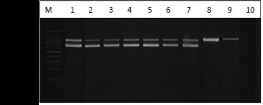
All Listeria spp. and L. monocytogenes isolates from raw milk and nunu samples were confirmed
All Listeria
by both spp. and
biochemical testsL.and
monocytogenes
duplex PCRisolates
(Figurefrom
1). raw milk and nunu samples were confirmed
by both biochemical tests and duplex PCR (Figure 1).
Figure 1. Representative PCR product detecting the highly conserved 16Sr RNA gene sequence for
Figure 1. Representative PCR product detecting the highly conserved 16Sr RNA gene sequence for
Listeria spp. and listeriolysin O gene sequence for L. monocytogenes. Lane M, 100 bp molecular size
Listeria spp. and listeriolysin O gene sequence for L. monocytogenes. Lane M, 100 bp molecular size DNA
DNA marker; lane 1, L. monocytogenes ATCC 19115 (positive control); lanes 2–7, L. monocytogenes
marker; lane 1, L. monocytogenes ATCC 19115 (positive control); lanes 2–7, L. monocytogenes isolated
isolated from raw cow milk and nunu; lanes 8 and 9, non-L. monocytogenes isolated from raw cow milk
from raw cow milk and nunu; lanes 8 and 9, non-L. monocytogenes isolated from raw cow milk and
and nunu; lane 10, negative control (PCR mixture without DNA template).
nunu; lane 10, negative control (PCR mixture without DNA template).
Among other human pathogenic microorganisms, L. monocytogenes is considered a major
Among other
microbiological human
hazard pathogenic
associated with the consumption ofL.
microorganisms, monocytogenes
raw is considered
cow milk [30,31]. a major
The prevalence of
microbiological hazard associated with the consumption of raw cow milk [30,31]. The prevalence
Listeria spp. and Listeria monocytogenes in milk and dairy products, has been widely reported [17,31– of
Listeria spp. and Listeria monocytogenes in milk and dairy products, has been widely reported [17,31–33].
33]. The prevalence of L. monocytogenes in milk varies considerably among different survey reports
The prevalence
and of L. monocytogenes
has been attributed to factors in milkasvaries
such considerably
geographic among different
(environmental) surveyfarm
conditions, reports and
size, has
farmBeverages 2018, 4, 40 5 of 10
been attributed to factors such as geographic (environmental) conditions, farm size, farm management
practices, sampling and detection methods used among others [30]. In this report, the prevalence
of L. monocytogenes in raw cow milk collected from small dairy farms in northern Ghana was 8.8%
while Appiah et al. [20] reported prevalence of over 42% for raw cow milk sampled from farms in
Accra (southern Ghana). According to other previous survey reports, the frequency of detection of
L. monocytogenes in raw cow milk can vary from 0% [34] to more than 45% [35–40]. Despite the widely
reported variation in the prevalence of L. monocytogenes in milk, all the surveys demonstrate that raw
cow milk and dairy products can be a source of Listeria infections in humans [30,32].
Listeria monocytogenes occurs widely in agricultural and food processing environments [41] and
can contaminate raw milk during milking processes [31]. Additionally, L. monocytogenes is widely
distributed in food processing environments with the capacity to tolerate and grow in the temperature
range of 0–45 ◦ C in media with pH range of 4.4–9.4 [42–44].
Due to the observed high prevalence of L. monocytogenes in raw cow milk from dairy farms in
this study, it is possible that the pathogen could enter the processing environments and persist during
processing, resulting in the contamination of the dairy products. Nunu is produced by spontaneously
fermenting raw cow milk at ambient temperature (28–35 ◦ C) for about 18–24 h under uncontrolled
conditions, bringing the pH to 4.1 ± 0.9 (Table 1). The fermented yoghurt-like milk beverage, nunu,
can then be consumed within 4 days after fermentation without refrigeration or refrigerated at 4 ◦ C for
several weeks [21]. The observed prevalence of L. monocytogenes in nunu may be attributed to factors
such as post-processing contamination, improper handling of raw milk, or inadequate fermentation to
bring the pH below tolerance levels for L. monocytogenes. Thus, the production of organic acids and
bacteriocins which are important natural antimicrobials in nunu, resulting from lactic acid fermentation
of milk, might not be sufficient to eliminate L. monocytogenes in the product and therefore should not
replace good hygiene practices (practices) during processing. On the other hand, Listeria was not
detected in boiled milk, which undergoes thermal treatment without further processing. Boiling is
one method that is known to reduce or even eliminate the microbiological risk associated with the
consumption of raw milk [30].
3.2. Molecular Serogroups of L. monocytogenes Isolates
Based on PCR serotyping, L. monocytogenes isolates belonged to molecular serogroups 1/2a-3a
(32/62, 51.6%), 1/2b-3b-7 (14/62, 22.6%), 1/2c-3c (7/62, 11.3%) and 4b-4d-4e (9/62, 14.5%). Previous
reports indicate that over 95% of the isolates originating from human listeriosis and food samples
belonged to serotypes 1/2a, 1/2b, 1/2c, and 4b [6,11,45–47]. Consistent with the previous reports,
51.6% of L. monocytogenes isolates in this study belonged to serogroup 1/2a-3a, followed by 1/2b-3b-7
(22.6%), 4b-4d-4e (14.5%) and the 1/2c-3c (11.3%). The high prevalence and enhanced ability of
serotype 1/2a to persist in food environments has been ascribed to the fact that members of this
group might be carrying more plasmids which often confer resistance to toxic compounds [48]. Major
outbreaks of invasive forms of listeriosis have been associated with serotype 4b [49], which has
also been isolated from animal-derived foods [6]. Thus, the frequent detection of serotype 4b in
human listeriosis outbreaks and sporadic cases indicate that these strains may be more virulent than
other serotypes.
3.3. Prevalence of Virulence-Associated Markers
Irrespective of serotype, all of the sixty-two (62) L. monocytogenes tested harbored the
virulence-associated genes inlA, inlB, inlC, and inlJ inlA, inlB, inlC, inlJ, plcA, actA, hlyA, iap and
prfA. Listeriolysin O is reported to be the main bacterial determinant for the escape of L. monocytogenes
from both primary and secondary vacuoles and is one of its main virulent factors [50,51]. The detection
of internalin genes (inlA, inlB, inlC, and inlJ) in almost all examined L. monocytogenes isolates from
food samples have been reported [52–55]. The virulence markers InlA, InlC and InlJ, all of which play
various roles in L. monocytogenes infections and pathogenesis, are important virulence factors of L.Beverages 2018, 4, 40 6 of 10
monocytogenes [11]. In L. monocytogenes, InlA and InlB have evidently been shown to be important for
host cell invasion and virulence [56,57]. Furthermore, null mutations in the internalin genes (inlA, inlB,
inlC, and inlJ) result in reduced invasion or virulence in tissue culture or animal model studies [58–60],
underscoring the important role of these genes in virulence and pathogenesis. In general, the presence
of the internalin genes and the other virulence-associated genes in L. monocytogenes isolated from milk
and dairy products in Ghana indicates that these L. monocytogenes strain could be potentially virulent.
3.4. Antibiotic Resistance among L. monocytogenes Isolates
Phenotypic resistance to 15 antimicrobial agents by 62 L. monocytogenes isolates from raw
milk and nunu in Ghana are shown in Table 2. Based on the MIC breakpoints used in this
study, L. monocytogenes isolates were generally susceptible to amoxicillin (100%), ampicillin (100%),
erythromycin (100%), gentamycin (100%), penicillin (100%), rifampicin (100%) and vancomycin (100%).
However, phenotypic resistances were observed against neomycin (61.3%) and tetracycline (24.2%),
while intermediate susceptibilities were obtained for chloramphenicol, ciprofloxacin, clindamycin,
doxycycline, kanamycin, neomycin, streptomycin and tetracycline.
Table 2. Antimicrobial susceptibility profile of L. monocytogenes isolates.
a MIC Breakpoints b Interpretations n (%)
Antimicrobial
S (≤) R (≥) S I R
Amoxicillin 2 8 62 (100) 0 0
Ampicillin 4 8 62 (100) 0 0
Chloramphenicol 8 32 60 (96.8) 2 (3.2) 0
Ciprofloxacin 2 8 55 (88.7) 7 (11.3) 0
Clindamycin 2 8 51 (82.3) 11 (17.7) 0
Doxycycline 4 16 58 (93.5) 4 (6.5) 0
Erythromycin 2 4 62 (100) N/A 0
Gentamycin 2 8 62 (100) 0 0
Kanamycin 16 64 57 (91.9) 5 (8.1) 0
Neomycin 8 32 21 (33.9) 3 (4.8) 38 (61.3)
Penicillin 4 8 62 (100) 0 0
Rifampicin 1 2 62 (100) N/A 0
Streptomycin 8 32 61 (98.4) 1 (1.6) 0
Tetracycline 2 8 44 (70.9) 3 (4.8) 15 (24.2)
Vancomycin 8 32 62 (100) 0 0
a MIC breakpoint values for all antibiotics against L. monocytogenes were according to the description of Bertsch et
al., 2014, except doxycycline and neomycin for which the breakpoints proposed by Acar et al., 1998 were used. b S:
susceptible; I: intermediate susceptibility; R: resistant.
Listeria monocytogenes is naturally reported to be sensitive to a wide range of antibiotics.
However, recent reports show a continuous pattern of emergence of resistant strains [6,14,48].
Listeria monocytogenes from food sources have generally been reported to be highly susceptible
to ampicillin, gentamycin, penicillin, rifampicin and vancomycin. On the other hand, varying
prevalence of resistance to tetracycline by L. monocytogenes isolates from different food sources have
also been reported [16,53,61,62]. The high number of L. monocytogenes isolates showing intermediate
susceptibility to ciprofloxacin (11.3%) and clindamycin (17.7%) has similarly been reported by
Lyon et al. [63] and Bertsch et al. [16]. Natural populations of several Listeria species have been
described as intermediately susceptible to ciprofloxacin and clindamycin [64].
4. Conclusions
The results presented in this report demonstrates Listeria monocytogenes is prevalent in raw milk
and nunu, a spontaneously fermented yoghurt-like milk beverage, in Ghana. L. monocytogenes isolates
from raw milk and nunu in Ghana harbored virulence-associated genes. However, the L. monocytogenesBeverages 2018, 4, 40 7 of 10
isolates were generally susceptible to the different antibiotics tested except tetracycline and neomycin
for which phenotypic resistance was observed. However, genotypic assessment of the prevalence of
genetic markers associated with antimicrobial resistance for the genetic basis of resistance phenotypes
observed in this study would provide a better baseline for further antimicrobial resistance molecular
surveillance of L. monocytogenes milk and dairy products in Ghana. Implementation of adequate
sanitation program and good hygiene/manufacturing practices (GHP/GMP) for milk production
and processing of fermented dairy beverages in Ghana could reduce microbial contamination of
these products.
Author Contributions: J.O.-K. conceived the research concept, participated in laboratory experiments, analyzed
and interpreted the data and wrote the manuscript. A.W. carried out most of the laboratory experiments and
participated in analysis and interpretation of data. F.A. participated in interpretation of data and reviewed the
manuscript. L.J. supervised the project and corrected the manuscript. All authors read and approved the content
of the manuscript.
Acknowledgments: The work was supported with funding from Danida (Danish International Development
Agency, Ministry of Foreign Affairs) and the Government of Ghana (GoG) through the collaborative research
project ‘Preserving African Food Microorganisms for Green Growth’ DFC No. 13-04KU. The authors are also
grateful to management of the Savana Agricultural Research Institute (SARI), Tamale-Ghana and Navrongo
Health Research Centre (NHRC), Ghana, for granting access to their laboratory facilities.
Conflicts of Interest: The authors declare no conflict of interest in this work.
References
1. Kang, Q.; Guo, Y.; Hong, S.; Jiang, L. Listeria monocytogenes meningitis in infants: Report of two cases. Chin. J.
Contemp. Pediatr. 2013, 15, 1150–1152.
2. Thomas, M.K.; Vriezen, R.; Farber, J.M.; Currie, A.; Schlech, W.; Fazil, A. Economic cost of a Listeria
monocytogenes outbreak in Canada, 2008. Foodborne Pathog. Dis. 2015, 12, 966–971. [CrossRef] [PubMed]
3. Razavilar, V.; Genigeorgis, C. Prediction of Listeria spp. growth as affected by various levels of chemicals,
pH, temperature and storage time in a model broth. Int. J. Food Microbiol. 1998, 40, 149–157. [CrossRef]
4. Jamali, H.; Chai, L.C.; Thong, K.L. Detection and isolation of Listeria spp. and Listeria monocytogenes in
ready-to-eat foods with various selective culture media. Food Control 2013, 32, 19–24. [CrossRef]
5. Rahimi, E.; Ameri, M.; Momtaz, H. Prevalence and antimicrobial resistance of Listeria species isolated from
milk and dairy products in Iran. Food Control 2010, 21, 1448–1452. [CrossRef]
6. Wu, S.; Wu, Q.; Zhang, J.; Chen, M.; Hu, H. Listeria monocytogenes prevalence and characteristics in retail raw
foods in China. PLoS ONE 2015, 10, e0136682. [CrossRef] [PubMed]
7. Scallan, E.; Crim, S.M.; Runkle, A.; Henao, O.L.; Mahon, B.E.; Hoekstra, R.M.; Griffin, P.M. Bacterial
enteric infections among older adults in the united states: Foodborne diseases active surveillance network,
1996–2012. Foodborne Pathog. Dis. 2015, 12, 492–499. [CrossRef] [PubMed]
8. Kathariou, S. Listeria monocytogenes virulence and pathogenicity, a food safety perspective. J. Food Prot. 2002,
65, 1811–1829. [CrossRef] [PubMed]
9. Salcedo, C.; Arreaza, L.; Alcala, B.; De La Fuente, L.; Vazquez, J. Development of a multilocus sequence
typing method for analysis of Listeria monocytogenes clones. J. Clin. Microbiol. 2003, 41, 757–762. [CrossRef]
[PubMed]
10. Jensen, A.; Thomsen, L.E.; Jørgensen, R.L.; Larsen, M.H.; Roldgaard, B.B.; Christensen, B.B.; Vogel, B.F.;
Gram, L.; Ingmer, H. Processing plant persistent strains of Listeria monocytogenes appear to have a lower
virulence potential than clinical strains in selected virulence models. Int. J. Food Microbiol. 2008, 123, 254–261.
[CrossRef] [PubMed]
11. Shen, J.; Rump, L.; Zhang, Y.; Chen, Y.; Wang, X.; Meng, J. Molecular subtyping and virulence gene analysis
of Listeria monocytogenes isolates from food. Food Microbiol. 2013, 35, 58–64. [CrossRef] [PubMed]
12. Soni, D.K.; Singh, M.; Singh, D.V.; Dubey, S.K. Virulence and genotypic characterization of Listeria
monocytogenes isolated from vegetable and soil samples. BMC Microbiol. 2014, 14, 241. [CrossRef] [PubMed]
13. Wu, S.; Wu, Q.; Zhang, J.; Chen, M.; Guo, W. Analysis of multilocus sequence typing and virulence
characterization of Listeria monocytogenes isolates from Chineseretail ready-to-eat food. Front. Microbiol. 2016,
7, 168. [CrossRef] [PubMed]Beverages 2018, 4, 40 8 of 10
14. Pesavento, G.; Ducci, B.; Nieri, D.; Comodo, N.; Nostro, A.L. Prevalence and antibiotic susceptibility of
Listeria spp. isolated from raw meat and retail foods. Food Control 2010, 21, 708–713. [CrossRef]
15. Wieczorek, K.; Dmowska, K.; Osek, J. Prevalence, characterization, and antimicrobial resistance of Listeria
monocytogenes isolates from bovine hides and carcasses. Appl. Environ. Microbiol. 2012, 78, 2043–2045.
[CrossRef] [PubMed]
16. Bertsch, D.; Muelli, M.; Weller, M.; Uruty, A.; Lacroix, C.; Meile, L. Antimicrobial susceptibility and antibiotic
resistance gene transfer analysis of foodborne, clinical, and environmental Listeria spp. isolates including
Listeria monocytogenes. MicrobiologyOpen 2014, 3, 118–127. [CrossRef] [PubMed]
17. Chen, M.; Wu, Q.; Zhang, J.; Wu, S.; Guo, W. Prevalence, enumeration, and pheno-and genotypic
characteristics of Listeria monocytogenes isolated from raw foods in South China. Front. Microbiol. 2015, 6,
1026. [CrossRef] [PubMed]
18. Soni, D.K.; Singh, R.K.; Singh, D.V.; Dubey, S.K. Characterization of Listeria monocytogenes isolated from
Ganges water, human clinical and milk samples at Varanasi, India. Infect. Genet. Evol. 2013, 14, 83–91.
[CrossRef] [PubMed]
19. Akabanda, F.; Owusu-Kwarteng, J.; Tano-Debrah, K.; Glover, R.L.; Nielsen, D.S.; Jespersen, L. Taxonomic and
molecular characterization of lactic acid bacteria and yeasts in nunu, a Ghanaian fermented milk product.
Food Mcrobiol. 2013, 34, 277–283. [CrossRef] [PubMed]
20. Appiah, J.; Tano-Debrah, K.; Annor, G.A.; Alpha, M.M.; Makita, K.; Grace, D. Quantitative probabilistic
assessment of the risk of Listeriosis from the consumption of milk from informal markets in Ghana. In
Proceedings of the International Congress of Pathogens at the Human-Animal Interface (ICOPHAI), Addis
Ababa, Ethiopia, 15–17 September 2011; International Livestock Research Institute (ILRI): Nairobi, Kenya, 2011.
21. Owusu-Kwarteng, J.; Akabanda, F.; Johansen, P.; Jespersen, L.; Nielsen, D.S. Nunu, a West-African fermented
yogurt-like milk product. In Yogurt in Health and Disease Prevention; Shah, N.P., Ed.; Elsevier: New York, NY,
USA, 2017; pp. 275–283.
22. Becker, B.; Schuler, S.; Lohneis, M.; Sabrowski, A.; Curtis, G.D.; Holzapfel, W.H. Comparison of two
chromogenic media for the detection of Listeria monocytogenes with the plating media recommended by
EN/DIN 11290-1. Int. J. Food Microbiol. 2006, 109, 127–131. [CrossRef] [PubMed]
23. Thong, K.; Lai, M.; Teh, C.; Chua, K. Simultaneous detection of methicillin-resistant Staphylococcus aureus,
Acinetobacter baumannii, Escherichia coli, Klebsiella pneumoniae and Pseudomonas aeruginosa by multiplex PCR.
Trop. Biomed. 2011, 28, 21–31. [PubMed]
24. Rossmanith, P.; Krassnig, M.; Wagner, M.; Hein, I. Detection of Listeria monocytogenes in food using a
combined enrichment/real-time PCR method targeting the prfA gene. Res. Microbiol. 2006, 157, 763–771.
[CrossRef] [PubMed]
25. Doumith, M.; Buchrieser, C.; Glaser, P.; Jacquet, C.; Martin, P. Differentiation of the major Listeria
monocytogenes serovars by multiplex PCR. J. Clin. Microbiol. 2004, 42, 3819–3822. [CrossRef] [PubMed]
26. Liu, D.; Lawrence, M.L.; Austin, F.W.; Ainsworth, A.J. A multiplex PCR for species-and virulence-specific
determination of Listeria monocytogenes. J. Microbiol. Meth. 2007, 71, 133–140. [CrossRef] [PubMed]
27. Kalorey, D.; Kurkure, N.; Warke, S.; Rawool, D.; Malik, S.; Barbuddhe, S. Isolation of pathogenic Listeria
monocytogenes in faeces of wild animals in captivity. Comp. Immunol. Microbiol. 2006, 29, 295–300. [CrossRef]
[PubMed]
28. CLSI. Methods for Antimicrobial Dilution and Disk Susceptibility Testing of Infrequently Isolated or Fastidious
Bacteria; Approved Guideline (M45-A2); Wayne, P.A., Ed.; Clinical and Laboratory Standards Institute:
Wayne, PA, USA, 2010.
29. Acar, J.F.; Bergogne-Berezin, E.; Chabbert, Y.; Cluzel, R.; Courtieu, A.; Courvalin, P.; Veron, M. Communiqué
1992 du Comité de L’Antibiogramme de la Societé Francaise de Microbiologie. Pathol. Biol. 1998, 40, 741–748.
30. Claeys, W.L.; Cardoen, S.; Daube, G.; De Block, J.; Dewettinck, K.; Dierick, K.; De Zutter, L.; Huyghebaert, A.;
Imberechts, H.; Thiange, P. Raw or heated cow milk consumption: Review of risks and benefits. Food Control
2013, 31, 251–262. [CrossRef]
31. Verraes, C.; Vlaemynck, G.; Van Weyenberg, S.; De Zutter, L.; Daube, G.; Sindic, M.; Uyttendaele, M.;
Herman, L. A review of the microbiological hazards of dairy products made from raw milk. Int. Dairy J.
2015, 50, 32–44. [CrossRef]Beverages 2018, 4, 40 9 of 10
32. Buchanan, R.L.; Gorris, L.G.; Hayman, M.M.; Jackson, T.C.; Whiting, R.C. A review of Listeria monocytogenes:
An update on outbreaks, virulence, dose-response, ecology, and risk assessments. Food Control 2017, 75, 1–13.
[CrossRef]
33. Nayak, D.N.; Savalia, C.; Kalyani, I.; Kumar, R.; Kshirsagar, D. Isolation, identification, and characterization
of Listeria spp. from various animal origin foods. Vet. World 2015, 8, 695. [CrossRef] [PubMed]
34. Stephan, R.; Buhler, K. Prevalence of Campylobacter spp., Salmonella spp. and Listeria monocytogenes in
bulk-tank milk samples from north-east Switzerland. Arch. Lebensmittelhyg. 2002, 53, 62–65.
35. De Reu, K.; Grijspeerdt, K.; Herman, L. A Belgian survey of hygiene indicator bacteria and pathogenic
bacteria in raw milk and direct marketing of raw milk farm products. J. Food Safety 2004, 24, 17–36. [CrossRef]
36. Domínguez, R.L.; Fernández, G.J.; Vazquez, B.J.; Rodriguez, F.E.; Suarez, F.G. Isolation of micro-organisms
of the species Listeria from raw milk intended for human consumption. Can. J. Microbiol. 1985, 31, 938–941.
37. El Marnissi, B.; Bennani, L.; Cohen, N.; Lalami, A.E.O.; Belkhou, R. Presence of Listeria monocytogenes in raw
milk and traditional dairy products marketed in the north-central region of Morocco. Afr. J. Food Sci. 2013, 7,
87–91. [CrossRef]
38. Fenlon, D.; Stewart, T.; Donachie, W. The incidence, numbers and types of Listeria monocytogenes isolated
from farm bulk tank milks. Lett. Appl. Microbiol. 1995, 20, 57–60. [CrossRef] [PubMed]
39. Meyer-Broseta, S.; Diot, A.; Bastian, S.; Rivière, J.; Cerf, O. Estimation of low bacterial concentration: Listeria
monocytogenes in raw milk. Int. J. Food Microbiol. 2003, 80, 1–15. [CrossRef]
40. Waak, E.; Tham, W.; Danielsson-Tham, M.-L. Prevalence and fingerprinting of Listeria monocytogenes strains
isolated from raw whole milk in farm bulk tanks and in dairy plant receiving tanks. Appl. Environ. Microbiol.
2002, 68, 3366–3370. [CrossRef] [PubMed]
41. Strawn, L.K.; Fortes, E.D.; Bihn, E.A.; Nightingale, K.K.; Gröhn, Y.T.; Worobo, R.W.; Wiedmann, M.;
Bergholz, P.W. Landscape and meteorological factors affecting prevalence of three food-borne pathogens in
fruit and vegetable farms. Appl. Environ. Microbiol. 2013, 79, 588–600. [CrossRef] [PubMed]
42. Carpentier, B.; Cerf, O. Persistence of Listeria monocytogenes in food industry equipment and premises. Int. J.
Food Microbiol. 2011, 145, 1–8. [CrossRef] [PubMed]
43. Ferreira, V.; Wiedmann, M.; Teixeira, P.; Stasiewicz, M. Listeria monocytogenes persistence in food-associated
environments: Epidemiology, strain characteristics, and implications for public health. J. Food Prot. 2014, 77,
150–170. [CrossRef] [PubMed]
44. WHO. Risk Assessment of Listeria monocytogenes in Ready-to-Eat Foods: Interpretative Summary, Food and
Agriculture Organization: Rome, Italy, 2004.
45. Althaus, D.; Lehner, A.; Brisse, S.; Maury, M.; Tasara, T.; Stephan, R. Characterization of Listeria monocytogenes
strains isolated during 2011–2013 from human infections in Switzerland. Foodborne Pathog. Dis. 2014, 11,
753–758. [CrossRef] [PubMed]
46. Pontello, M.; Guaita, A.; Sala, G.; Cipolla, M.; Gattuso, A.; Sonnessa, M.; Gianfranceschi, M.V. Listeria
monocytogenes serotypes in human infections (Italy, 2000–2010). Ann. Dell’ist. Super. Sanitã 2012, 48, 146–150.
[CrossRef]
47. Yu, T.; Jiang, X. Prevalence and characterization of Listeria monocytogenes isolated from retail food in Henan,
China. Food Control 2014, 37, 228–231. [CrossRef]
48. Korsak, D.; Borek, A.; Daniluk, S.; Grabowska, A.; Pappelbaum, K. Antimicrobial susceptibilities of Listeria
monocytogenes strains isolated from food and food processing environment in Poland. Int. J. Food Microbiol.
2012, 158, 203–208. [CrossRef] [PubMed]
49. Buchrieser, C.; Brosch, R.; Catimel, B.; Rocourt, J. Pulsed-field gel electrophoresis applied for comparing
Listeria monocytogenes strains involved in outbreaks. Can. J. Microbiol. 1993, 39, 395–401. [CrossRef] [PubMed]
50. Camejo, A.; Carvalho, F.; Reis, O.; Leitão, E.; Sousa, S.; Cabanes, D. The arsenal of virulence factors deployed
by Listeria monocytogenes to promote its cell infection cycle. Virulence 2011, 2, 379–394. [CrossRef] [PubMed]
51. Schnupf, P.; Portnoy, D.A. Listeriolysin O: A phagosome-specific lysin. Microbes Infect. 2007, 9, 1176–1187.
[CrossRef] [PubMed]
52. Indrawattana, N.; Nibaddhasobon, T.; Sookrung, N.; Chongsa-nguan, M.; Tungtrongchitr, A.; Makino, S.-I.;
Tungyong, W.; Chaicumpa, W. Prevalence of Listeria monocytogenes in raw meats marketed in Bangkok
and characterization of the isolates by phenotypic and molecular methods. J. Health Popul. Nutr. 2011, 29, 26.
[CrossRef] [PubMed]Beverages 2018, 4, 40 10 of 10
53. Jamali, H.; Paydar, M.; Ismail, S.; Looi, C.Y.; Wong, W.F.; Radmehr, B.; Abedini, A. Prevalence, antimicrobial
susceptibility and virulotyping of Listeria species and Listeria monocytogenes isolated from open-air fish
markets. BMC Microbiol. 2015, 15, 144. [CrossRef] [PubMed]
54. Lomonaco, S.; Patti, R.; Knabel, S.J.; Civera, T. Detection of virulence-associated genes and epidemic clone
markers in Listeria monocytogenes isolates from PDO Gorgonzola cheese. Int. J. Food Microbiol. 2012, 160,
76–79. [CrossRef] [PubMed]
55. Sant’Ana, A.S.; Igarashi, M.C.; Landgraf, M.; Destro, M.T.; Franco, B.D. Prevalence, populations and
pheno-and genotypic characteristics of Listeria monocytogenes isolated from ready-to-eat vegetables marketed
in São Paulo, Brazil. Int. J. Food Microbiol. 2012, 155, 1–9. [CrossRef] [PubMed]
56. Dramsi, S.; Biswas, I.; Maguin, E.; Braun, L.; Mastroeni, P.; Cossart, P. Entry of Listeria monocytogenes into
hepatocytes requires expression of inIB, a surface protein of the internalin multigene family. Mol. Microbiol.
1995, 16, 251–261. [CrossRef] [PubMed]
57. Gaillard, J.-L.; Berche, P.; Frehel, C.; Gouln, E.; Cossart, P. Entry of L. monocytogenes into cells is mediated by
internalin, a repeat protein reminiscent of surface antigens from gram-positive cocci. Cell 1991, 65, 1127–1141.
[CrossRef]
58. Dramsi, S.; Dehoux, P.; Lebrun, M.; Goossens, P.L.; Cossart, P. Identification of four new members of the
internalin multigene family of Listeria monocytogenes EGD. Infect. Immun. 1997, 65, 1615–1625. [PubMed]
59. Raffelsbauer, D.; Bubert, A.; Engelbrecht, F.; Scheinpflug, J.; Simm, A.; Hess, J.; Kaufmann, S.; Goebel, W. The
gene cluster inlC2DE of Listeria monocytogenes contains additional new internalin genes and is important for
virulence in mice. Mol. Gen. Genet. 1998, 260, 144–158. [CrossRef] [PubMed]
60. Sabet, C.; Lecuit, M.; Cabanes, D.; Cossart, P.; Bierne, H. LPXTG protein InlJ, a newly identified internalin
involved in Listeria monocytogenes virulence. Infect. Immun. 2005, 73, 6912–6922. [CrossRef] [PubMed]
61. Al-Nabulsi, A.A.; Osaili, T.M.; Shaker, R.R.; Olaimat, A.N.; Jaradat, Z.W.; Elabedeen, N.A.Z.; Holley, R.A.
Effects of osmotic pressure, acid, or cold stresses on antibiotic susceptibility of Listeria monocytogenes.
Food Microbiol. 2015, 46, 154–160. [CrossRef] [PubMed]
62. Conter, M.; Paludi, D.; Zanardi, E.; Ghidini, S.; Vergara, A.; Ianieri, A. Characterization of antimicrobial
resistance of foodborne Listeria monocytogenes. Int. J. Food Microbiol. 2009, 128, 497–500. [CrossRef] [PubMed]
63. Lyon, S.A.; Berrang, M.E.; Fedorka-Cray, P.J.; Fletcher, D.L.; Meinersmann, R.J. Antimicrobial resistance
of Listeria monocytogenes isolated from a poultry further processing plant. Foodborne Pathog. Dis. 2008, 5,
253–259. [CrossRef] [PubMed]
64. Troxler, R.; Von Graevenitz, A.; Funke, G.; Wiedemann, B.; Stock, I. Natural antibiotic susceptibility of
Listeria species: L. grayi, L. innocua, L. ivanovii, L. monocytogenes, L. seeligeri and L. welshimeri strains.
Clin. Microbiol. Infect. 2000, 6, 525–535. [CrossRef] [PubMed]
© 2018 by the authors. Licensee MDPI, Basel, Switzerland. This article is an open access
article distributed under the terms and conditions of the Creative Commons Attribution
(CC BY) license (http://creativecommons.org/licenses/by/4.0/).You can also read