The Winged Helix Domain of CSB Regulates RNAPII Occupancy at Promoter Proximal Pause Sites
←
→
Page content transcription
If your browser does not render page correctly, please read the page content below
International Journal of
               Molecular Sciences

Article
The Winged Helix Domain of CSB Regulates RNAPII
Occupancy at Promoter Proximal Pause Sites
Nicole L. Batenburg, Shixin Cui, John R. Walker, Herb E. Schellhorn and Xu-Dong Zhu *

                                          Department of Biology, McMaster University, Hamilton, ON L8S 4K1, Canada; Batenbn@mcmaster.ca (N.L.B.);
                                          cuis19@mcmaster.ca (S.C.); jwalker@mcmaster.ca (J.R.W.); schell@mcmaster.ca (H.E.S.)
                                          * Correspondence: zhuxu@mcmaster.ca; Tel.: +1-905-525-9140 (ext. 27737)

                                          Abstract: Cockayne syndrome group B protein (CSB), a member of the SWI/SNF superfamily, resides
                                          in an elongating RNA polymerase II (RNAPII) complex and regulates transcription elongation. CSB
                                          contains a C-terminal winged helix domain (WHD) that binds to ubiquitin and plays an important
                                          role in DNA repair. However, little is known about the role of the CSB-WHD in transcription
                                          regulation. Here, we report that CSB is dependent upon its WHD to regulate RNAPII abundance
                                          at promoter proximal pause (PPP) sites of several actively transcribed genes, a key step in the
                                          regulation of transcription elongation. We show that two ubiquitin binding-defective mutations in
                                          the CSB-WHD, which impair CSB’s ability to promote cell survival in response to treatment with
                                          cisplatin, have little impact on its ability to stimulate RNAPII occupancy at PPP sites. In addition,
                                          we demonstrate that two cancer-associated CSB mutations, which are located on the opposite side
                                          of the CSB-WHD away from its ubiquitin-binding pocket, impair CSB’s ability to promote RNAPII
                                occupancy at PPP sites. Taken together, these results suggest that CSB promotes RNAPII association
                                   with PPP sites in a manner requiring the CSB-WHD but independent of its ubiquitin-binding activity.
Citation: Batenburg, N.L.; Cui, S.;       These results further imply that CSB-mediated RNAPII occupancy at PPP sites is mechanistically
Walker, J.R.; Schellhorn, H.E.; Zhu,      separable from CSB-mediated repair of cisplatin-induced DNA damage.
X.-D. The Winged Helix Domain of
CSB Regulates RNAPII Occupancy at         Keywords: Cockayne syndrome group B (CSB); RNAPII; promoter proximal pause sites; winged
Promoter Proximal Pause Sites. Int. J.    helix domain; CSB cancer mutations
Mol. Sci. 2021, 22, 3379.
https://doi.org/10.3390/ijms
22073379

                                          1. Introduction
Academic Editor: Alexande Baykov
                                                The Cockayne syndrome group B protein (CSB), a member of the SWI/SNF2 (switch/
                                          sucrose non-fermenting) family, is a multifunctional protein that participates in a wide
Received: 18 February 2021
Accepted: 24 March 2021
                                          range of cellular processes. CSB was first described for its role in transcription-coupled
Published: 25 March 2021
                                          nucleotide excision repair (TC-NER) [1,2]. Impaired TC-NER is considered to be an un-
                                          derlying cause of photosensitivity seen in clinical cases of Cockayne syndrome (CS), the
Publisher’s Note: MDPI stays neutral
                                          majority of which are caused by germline mutations of the ERCC6 gene encoding CSB [3–5].
with regard to jurisdictional claims in
                                          However, CS patients do not exhibit an increased risk to skin cancer. Aside from pho-
published maps and institutional affil-   tosensitivity, CS is also characterized by severe impairment of physical development,
iations.                                  progressive neurological degeneration, cataracts, hearing loss and segmental premature
                                          ageing [6]. These phenotypes cannot be fully explained by a deficiency in TC-NER. It
                                          has been reported that CSB-deficient cells derived from CS patients exhibit defects in
                                          transcription [7,8], oxidative damage repair [9], mitochondria function [10,11], cell divi-
Copyright: © 2021 by the authors.
                                          sion [12], telomere maintenance [13,14] and DNA double-stranded break repair pathway
Licensee MDPI, Basel, Switzerland.
                                          choice [15–17], indicative of a role of these additional cellular processes in the develop-
This article is an open access article
                                          ment of CS. CSB has also been implicated in transcription regulation [18,19] as well as
distributed under the terms and           transcription recovery following genotoxic stress [20,21]. It has been suggested that defects
conditions of the Creative Commons        in transcription regulation may underlie some of the neurological features of CS [22–24].
Attribution (CC BY) license (https://           CSB resides in an elongating RNA polymerase II (RNAPII) complex [25–27]. RNAPII
creativecommons.org/licenses/by/          arrested at bulky DNA lesions is thought to serve as the initiating signal for TC-NER [28–32].
4.0/).                                    It has been suggested that in TC-NER, CSB binds stalled RNAPII and that this binding

Int. J. Mol. Sci. 2021, 22, 3379. https://doi.org/10.3390/ijms22073379                                       https://www.mdpi.com/journal/ijms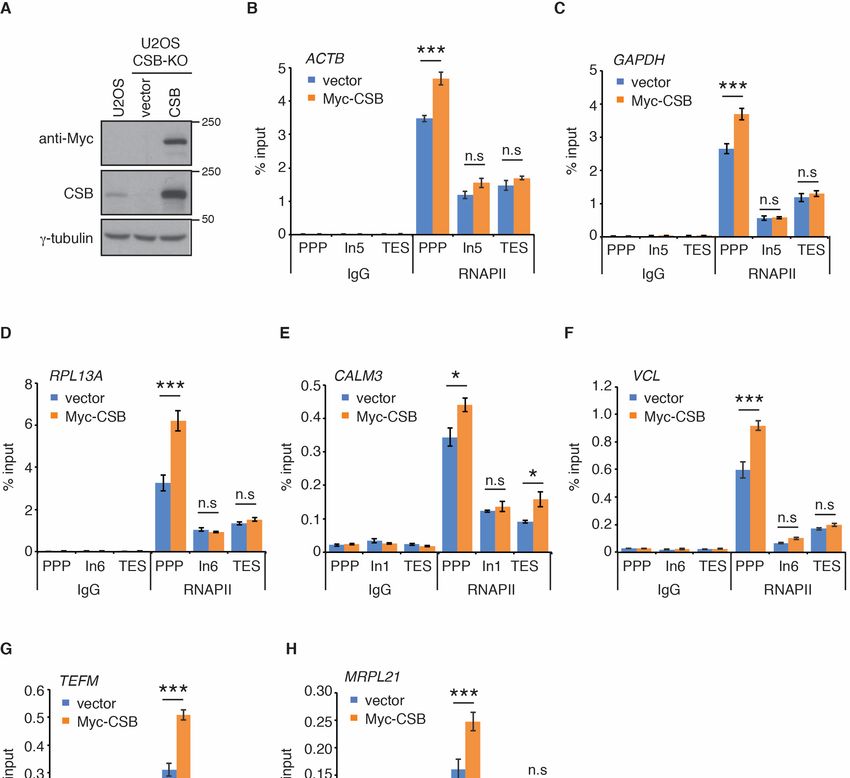
Int. J. Mol. Sci. 2021, 22, 3379                                                                                         2 of 14

                                   presents new protein interaction interfaces that facilitate recruitment of downstream repair
                                   factors [33]. Recent cryo-EM studies of the structure of the yeast homolog of CSB, Rad26,
                                   in complex with stalled/paused RNAPII suggest that although Rad26 does not promote
                                   efficient transcriptional bypass of bulky DNA lesions, it promotes paused RNAPII for-
                                   ward translocation on non-damaged templates to stimulate transcription elongation by
                                   RNAPII [33]. Promoter proximal pausing of RNAPII represents a key step in regulating
                                   transcription elongation at protein-coding genes in metazoans [34]. These findings suggest
                                   that CSB may regulate the paused RNAPII at promoter proximal pause (PPP) sites.
                                         CSB contains a N-terminal region, a central ATPase domain and a C-terminal region.
                                   The last 76 amino acids of CSB constitute a winged helix domain (WHD) [15,35]. This
                                   domain, a protein-protein interaction module, mediates the CSB interaction with several
                                   proteins including MRE11/RAD50/NBS1 [16], RIF1 [15] as well as RNAPII [36]. In addition,
                                   it has been reported that the WHD of CSB binds ubiquitin [35]. The crystal structure of the
                                   CSB-WHD in complex with ubiquitin has revealed that the ubiquitin-binding activity of the
                                   CSB-WHD is dependent upon residues in α2 and the C-terminal extremity [35]. Aspartic
                                   acid at position 1425 (D1425) and phenylalanine at position 1437 (F1437), both part of α2,
                                   make direct interactions with ubiquitin and mutating either D1425 to alanine or F1437 to
                                   aspartic acid abrogates binding of the CSB-WHD to ubiquitin [35]. The ubiquitin-binding
                                   activity of the WHD has been implicated in efficient TC-NER [36,37], however little is
                                   known about the WHD and its ubiquitin-binding activity in transcription regulation. The
                                   CSB-WHD is not known to be associated with any CS-causing mutations although the
                                   vast majority of CS-causing CSB mutations are nonsense mutations, frameshift mutations
                                   and deletions [3]. While much attention has been given to CS-causing CSB mutations
                                   over the past decades, recent work suggests that CSB plays a role in cancer prognosis
                                   and treatment [38–41]. Cancer genomics databases of both BioPortal and COSMIC have
                                   revealed the presence of cancer-associated mutations in the CSB-WHD albeit their roles
                                   have not been characterized.
                                         In this report, we show that CSB is enriched at PPP sites and promotes RNAPII
                                   association with PPP sites of several actively transcribed genes through its WHD. We
                                   show that the previously reported ubiquitin binding-defective mutations in the CSB-WHD,
                                   which impair CSB’s ability to promote cell survival in response to treatment with cisplatin,
                                   do not affect its ability to mediate RNAPII association with PPP sites of ACTB, GAPDH and
                                   RPL13A genes. In addition, we demonstrate that two cancer-associated mutations, arginine
                                   to glutamine at position 1467 (R1467Q) and glycine to arginine at position (G1484R), both
                                   of which are located on the opposite side of the CSB-WHD away from its ubiquitin-binding
                                   pocket, impair its ability to promote RNAPII occupancy at PPP sites of ACTB, GAPDH
                                   and RPL13A genes, providing the first evidence that CSB somatic mutations affect RNAPII
                                   association with PPP sites. Taken together, these results suggest that the CSB-WHD
                                   mediates RNAPII association with PPP sites in a manner independent of its ubiquitin-
                                   binding activity. These results further imply that CSB-mediated RNAPII association with
                                   PPP sites is mechanistically separable from CSB-mediated repair of cisplatin-induced
                                   DNA damage.

                                   2. Results
                                   2.1. CSB Promotes RNAPII Accumulation at PPP Sites of Several Actively Transcribed Genes
                                        Recent cryo-EM studies of the structure of the yeast homolog of CSB, Rad26, in
                                   complex with stalled/paused RNAPII suggest that CSB promotes paused RNAPII on non-
                                   damaged templates to stimulate transcription elongation [33]. Therefore, we investigated
                                   if CSB regulates RNAPII occupancy at PPP sites of seven actively transcribed genes. ACTB,
                                   GAPDH, RPL13A, CALM3, VCL, TEFM, and MRPL21 are actively transcribed genes that
                                   have previously been used for studies of RNAPII occupancy [42]. Using the same primer
                                   pair sets previously reported [42] (also shown in Supplementary Table S1), we measured
                                   RNAPII occupancy at PPP sites, introns and transcription end sites (TES) of these seven
                                   genes in human osteosarcoma U2OS cells that are either wild type (WT) or knockout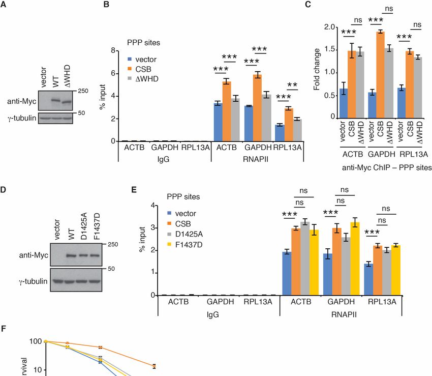
Int. J. Mol. Sci. 2021, 22, x FOR PEER REVIEW 3 of 13
ACTB, GAPDH, RPL13A, CALM3, VCL, TEFM, and MRPL21 are actively transcribed genes
Int. J. Mol. Sci. 2021, 22, 3379 that have previously been used for studies of RNAPII occupancy [42]. Using the3 of same
14
primer pair sets previously reported [42] (also shown in Supplementary Table S1), we
measured RNAPII occupancy at PPP sites, introns and transcription end sites (TES) of
these seven genes in human osteosarcoma U2OS cells that are either wild type (WT) or
(KO) for CSB
knockout (KO)[15,16] (Figure
for CSB [15,16]1A). ChIP-qPCR
(Figure analysis revealed
1A). ChIP-qPCR that loss that
analysis revealed of CSB
lossled to
of CSB
aled
pronounced reduction in RNAPII occupancy at PPP sites of all seven
to a pronounced reduction in RNAPII occupancy at PPP sites of all seven genes exam- genes examined
(Figure 1B–H).1B–H).
ined (Figure On the Onother
thehand,
otherloss of CSB
hand, losshad littlehad
of CSB impact
littleonimpact
RNAPII onassociation with
RNAPII associa-
introns and transcription end sites (TES) of all seven genes (Figure 1B–H).
tion with introns and transcription end sites (TES) of all seven genes (Figure 1B–H). To To substantiate
this finding, we
substantiate generated
this finding,U2OS CSB-KO cells
we generated U2OS stably
CSB-KOcomplemented
cells stably with either the vector
complemented with
alone
eitherorthe
Myc-tagged
vector alone wildortype CSB (Figure
Myc-tagged wild2A) and
type CSBperformed
(Figure 2A) rescue
and experiments. Myc-
performed rescue
CSB is highly overexpressed
experiments. Myc-CSB is highlyin U2OS CSB-KO cellsin
overexpressed compared
U2OS CSB-KO to endogenous CSB in U2OS
cells compared to en-
parental cells (Figure 2A). ChIP-qPCR analysis revealed that while
dogenous CSB in U2OS parental cells (Figure 2A). ChIP-qPCR analysis revealed that reintroduction of wild
while
type CSB had little
reintroduction of impact on RNAPII
wild type CSB hadassociation
little impactwith onintrons
RNAPII of association
all seven genes,
withitintrons
rescuedof
RNAPII
all sevenoccupancy at PPP RNAPII
genes, it rescued sites of all seven genes
occupancy at PPPin U2OS
sites ofCSB-KO
all sevencells
genes(Figure
in U2OS2B–H).
CSB-
We also investigated RNAPII occupancy at PPP sites in GM16095,
KO cells (Figure 2B–H). We also investigated RNAPII occupancy at PPP sites in GM16095, a SV40-transformed
CSB-deficient CS cell line [1,13]. We focused on RNAPII occupancy in ACTB, GAPDH and
a SV40-transformed CSB-deficient CS cell line [1,13]. We focused on RNAPII occupancy
RPL13A genes since these three genes contained a higher level of RNAPII at their PPP sites
in ACTB, GAPDH and RPL13A genes since these three genes contained a higher level of
than the other four genes (TEFM, MRPL21, CALM3, and VCL). ChIP-qPCR analysis showed
RNAPII at their PPP sites than the other four genes (TEFM, MRPL21, CALM3, and VCL).
that overexpression of Myc-CSB in GM16095 cells enhanced RNAPII occupancy at PPP
ChIP-qPCR analysis showed that overexpression of Myc-CSB in GM16095 cells enhanced
sites of (Supplementary Figure S1). These results altogether suggest that CSB promotes
RNAPII occupancy at PPP sites of (Supplementary Figure S1). These results altogether
RNAPII occupancy at PPP sites.
suggest that CSB promotes RNAPII occupancy at PPP sites.
Figure1.1.Loss
Figure LossofofCSB
CSB impairs
impairs RNAPII
RNAPII occupancy
occupancy at PPP
at PPP sites.
sites. (A)(A) Western
Western analysis
analysis of U2OS
of U2OS CSB-
CSB-WT
WT and CSB-KO cells. Immunoblotting was performed with anti-CSB and anti-γ-tubulin antibod-
and CSB-KO cells. Immunoblotting was performed with anti-CSB and anti-γ-tubulin antibodies. The
ies. The γ-tubulin blot was used as a loading control in this and subsequent figures. (B–H) RNAPII
γ-tubulin blot was used as a loading control in this and subsequent figures. (B–H) RNAPII ChIP
ChIP analyses of ACTB, GAPDH, RPL13A, CALM3, VCL, TEFM and MRPL21 genes in U2OS CSB-
analyses of ACTB, GAPDH, RPL13A, CALM3, VCL, TEFM and MRPL21 genes in U2OS CSB-WT
and CSB-KO cells. Standard errors from three independent experiments are shown. p values were
derived using the 2-way ANOVA test. *** p < 0.001, n.s.: not significant. Abbreviations used in this
and subsequent figures—PPP: promoter proximal pause site; ln: intron; TES: transcription end site.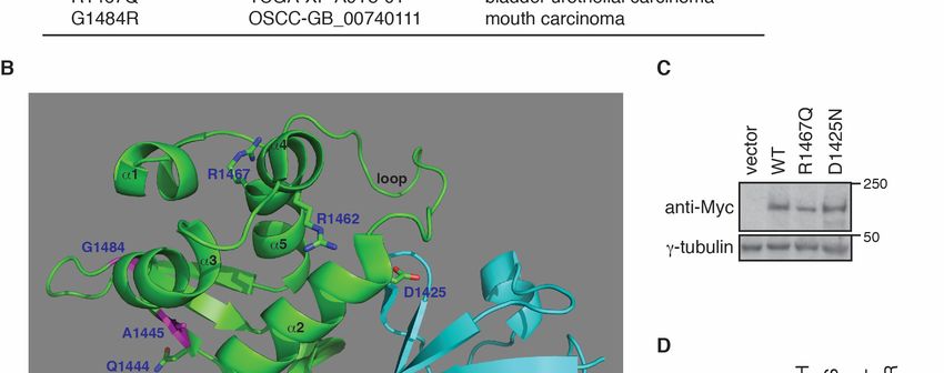
Int. J. Mol. Sci. 2021, 22, x FOR PEER REVIEW 4 of 13
Int. J. Mol. Sci. 2021, 22, 3379 WT and CSB-KO cells. Standard errors from three independent experiments are shown. p values4 of 14
were derived using the 2-way ANOVA test. *** p < 0.001. Abbreviations used in this and subse-
quent figures—PPP: promoter proximal pause site; ln: intron; TES: transcription end site.
Figure 2. Re-introduction of wild type CSB rescues RNAPII occupancy at PPP sites. (A) Western analysis of U2OS cells as
Figure 2. Re-introduction of wild type CSB rescues RNAPII occupancy at PPP sites. (A) Western analysis of U2OS cells
well as U2OS CSB-KO cells complemented with the vector alone or Myc-CSB. Immunoblotting was performed with anti-
as well as U2OS CSB-KO cells complemented with the vector alone or Myc-CSB. Immunoblotting was performed with
Myc, anti-CSB, and anti-γ-tubulin antibodies. (B–H) RNAPII ChIP analyses of ACTB, GAPDH, RPL13A, CALM3, VCL,
anti-Myc,
TEFM andanti-CSB,
MRPL21and anti-γ-tubulin
genes antibodies.
in U2OS CSB-KO cells(B–H) RNAPII
expressing theChIP analyses
vector of ACTB,
alone or GAPDH,
Myc-CSB. RPL13A,
Standard CALM3,
errors VCL,
from three
TEFM and MRPL21 genes in U2OS CSB-KO cells expressing the vector alone or Myc-CSB. Standard errors from
independent experiments are shown. P values were derived using the 2-way ANOVA test. * p < 0.05; *** p < 0.001. three
independent experiments are shown. p values were derived using the 2-way ANOVA test. * p < 0.05; *** p < 0.001, n.s.:
not significant. 2.2. The WHD of CSB Is Necessary for Its Ability to Promote RNAPII Association with PPP
Sites
2.2. The WHD of CSB Is Necessary for Its Ability to Promote RNAPII Association with PPP Sites
CSB contains a C-terminal WHD [15,35], which has been implicated in its interaction
CSB contains a C-terminal WHD [15,35], which has been implicated in its interaction
with RNAPII in TC-NER [36]. Therefore, we asked if CSB might be dependent upon its
with RNAPII in TC-NER [36]. Therefore, we asked if CSB might be dependent upon
WHD
its WHD to regulate RNAPII
to regulate RNAPIIassociation with with
association PPP sites. To address
PPP sites. this question,
To address we gener-
this question, we
ated U2OS CSB-KO cells stably complemented with the vector alone,
generated U2OS CSB-KO cells stably complemented with the vector alone, Myc-CSB Myc-CSB or Myc-
or
CSB lacking
Myc-CSB the WHD
lacking (Myc-CSB-∆WHD)
the WHD (Myc-CSB-∆WHD) (Figure 3A). 3A).
(Figure We chose to focus
We chose on RNAPII
to focus on RNAPIIoc-
cupancy
occupancy in in
ACTB,
ACTB,GAPDH
GAPDH and
andRPL13A
RPL13Agenes
genesininthese
thesecomplemented
complementedcell cell lines
lines because
because
these
these three
three genes
genes contained
contained aa higher
higher level
level of
of RNAPII
RNAPII at at their
their PPP
PPP sites
sites than
than the
the other
other four
four
genes
genes (TEFM, MRPL21, CALM3, and VCL) (Figure 1). ChIP-qPCR analysis revealed that
(TEFM, MRPL21, CALM3, and VCL) (Figure 1). ChIP-qPCR analysis revealed that
while
while overexpression
overexpression of of Myc-CSB
Myc-CSB inin U2OS
U2OS CSB-KO
CSB-KO cells promoted RNAPII
cells promoted RNAPII association
association
with PPP sites of all three genes examined, overexpression of Myc-CSB-∆WHD in U2OS
CSB-KO cells failed to do so (Figure 3B). The defect of Myc-CSB-∆WHD to promote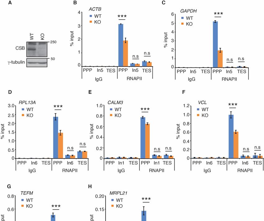
Int. J. Mol. Sci. 2021, 22, 3379                                                                                           5 of 14

                                   RNAPII association with PPP sites was unlikely due to a change in its expression since
                                   Myc-CSB-∆WHD was expressed at a comparable level to Myc-CSB (Figure 3A). Formally
                                   it was possible that a defect in the ability of Myc-CSB-∆WHD to be recruited to PPP sites
                                   might have contributed to its deficiency to promote RNAPII association with PPP sites. To
                                   address this possibility, we measured CSB occupancy at PPP sites in U2OS CSB-KO cells
                                   complemented with the vector alone, Myc-CSB or Myc-CSB-∆WHD. ChIP-qPCR analysis
                                   revealed an enrichment of Myc-CSB at PPP sites of all three genes ACTB, GAPDH and
                                   RPL13A (Figure 3C), indicating that CSB is associated with PPP sites. Deleting the WHD
                                   did not affect Myc-CSB association with any of the three PPP sites (Figure 3C), suggesting
                                   that the CSB-WHD is dispensable for CSB association with PPP sites. Taken together, these
                                   results suggest that CSB is dependent upon the CSB-WHD to promote RNAPII occupancy
                                   at PPP sites of ACTB, GAPDH and RPL13A genes.

                                   2.3. Ubiquitin Binding-Defective Mutations of CSB Sensitize Cells to Cisplatin But Have No
                                   Effect on RNAPII Occupancy at PPP Sites
                                        It has been reported that the CSB-WHD binds to ubiquitin and that this binding
                                   activity is abrogated by either a D1425A or a F1437D mutation [35]. To investigate if
                                   the ubiquitin-binding activity of the CSB-WHD might be required to promote RNAPII
                                   occupancy at PPP sites, we generated U2OS CSB-KO cells stably expressing the vector
                                   alone, Myc-CSB, Myc-CSB carrying a ubiquitin binding-defective D1425A mutation (Myc-
                                   CSB-D1425A) or Myc-CSB carrying a ubiquitin binding-defective F1437D mutation (Myc-
                                   CSB-F1437D) (Figure 3D). ChIP-qPCR analysis revealed that both Myc-CSB-D1425A and
                                   Myc-CSB-F1437D behaved like Myc-CSB in rescuing RNAPII association with PPP sites
                                   of ACTB, GAPDH and RPL13A genes in U2OS CSB-KO cells (Figure 3E). It has been
                                   reported that both D1425A and F1437D mutations impair the function of CSB in UV
                                   repair [35]. To verify if both D1425A and F1437D mutations affected CSB’s function in DNA
                                   repair, we performed clonogenic survival assays. We observed that when overexpressed
                                   in U2OS CSB-KO cells, both Myc-CSB-D1425A and Myc-CSB-F1437D were defective in
                                   promoting cell survival in response to treatment with cisplatin (Figure 3F), a platinum-
                                   based chemotherapeutic drug that is known to induce bulky DNA adducts, in agreement
                                   with the previous report [35]. This defect was unlikely due to a change in expression since
                                   both Myc-CSB-D1425A and Myc-CSB-F1437D were expressed at a comparable level to
                                   Myc-CSB (Figure 3D). Collectively, these results suggest that the ubiquitin-binding activity
                                   of CSB is dispensable for RNAPII association with PPP sites of ACTB, GAPDH and RPL13A
                                   genes. These results further imply that CSB-mediated RNAPII association with PPP sites is
                                   mechanistically separable from CSB-mediated repair of cisplatin-induced DNA damage.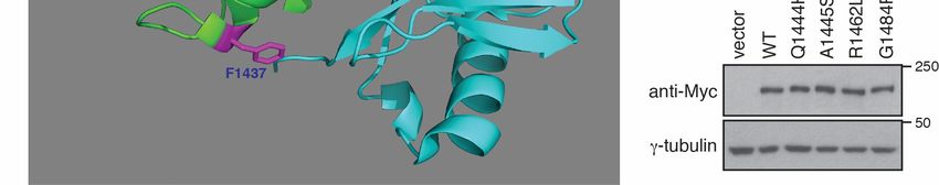
Int. J. Mol. Sci. 2021, 22, 3379 6 of 14
Int. J. Mol. Sci. 2021, 22, x FOR PEER REVIEW 6 of 13
Figure 3. The
Figure CSB-WHD
3. The CSB-WHD mediates
mediatesRNAPII occupancyatat
RNAPII occupancy PPP
PPP sites
sites in ain a manner
manner independent
independent of itstoability
of its ability bind totoubiquitin.
bind to ubiq-
uitin.(A)
(A)Western
Western analysis
analysis of U2OS
of U2OS CSB-KO CSB-KO cells expressing
cells expressing the vector
the vector alone, Myc-CSB alone,
WT Myc-CSB WT or Myc-CSB-∆WHD.
or Myc-CSB-∆WHD. Immunoblotting Im-
munoblotting
was performed was performed
with anti-Mycwith
and anti-Myc and anti-γ-tubulin
anti-γ-tubulin antibodies. (B)antibodies.
RNAPII ChIP (B)analyses
RNAPII ofChIP
ACTB,analyses
GAPDH of andACTB,
RPL13A GAPDH
and RPL13A genesCSB-KO
genes in U2OS in U2OScells
CSB-KO cells complemented
complemented with the vectorwith theMyc-CSB
alone, vector alone,
WT or Myc-CSB
Myc-CSB-∆WHD.WT or Myc-CSB-∆WHD.
Standard errors fromStand-
ard errors from three independent
three independent experiments areexperiments are shown.
shown. p values P values
were derived usingwere derived
the 2-way using test.
ANOVA the 2-way ANOVA
*** p < 0.001; ** p 0.05. ChIP
(C) Anti-Myc
analyses ofChIP analyses
of ACTB, GAPDHof ofand
ACTB,
RPL13AGAPDH genesand RPL13A
in U2OS CSB-KOgenes in complemented
cells U2OS CSB-KO cells
complemented
with the vectorwithalone,
the vector
Myc-CSBalone,
WT orMyc-CSB WT or Myc-CSB-∆WHD.
Myc-CSB-∆WHD. Standard errors from Standard errors fromexperiments
three independent three independent
are shown. experi-
ments are shown.
p values P valuesusing
were derived werethe
derived
2-way using
ANOVA thetest.
2-way
*** pANOVA test.
< 0.001; ns: p> ***0.05.
p < (D)
0.001; ns: p >analysis
Western 0.05. (D) Western
of U2OS analysis of
CSB-KO
U2OScellsCSB-KO cellsthe
expressing expressing the Myc-CSB
vector alone, vector alone, Myc-CSB WT, Myc-CSB-D1425A
WT, Myc-CSB-D1425A or Myc-CSB-F1437D. or Immunoblotting
Myc-CSB-F1437D. wasImmunoblotting
performed
was performed
with anti-Myc and anti-γ-tubulin antibodies. (E) RNAPII ChIP analyses of ACTB, GAPDH and RPL13A genes inand
with anti-Myc and anti-γ-tubulin antibodies. (E) RNAPII ChIP analyses of ACTB, GAPDH U2OSRPL13A
genesCSB-KO
in U2OS CSB-KO
cells cells complemented
complemented with the vectorwith the
alone, vector alone,
Myc-CSB Myc-CSB WT, Myc-CSB-D1425A
WT, Myc-CSB-D1425A or Myc-CSB-F1437D.or Myc-CSB-F1437D.
Standard errors
Standard
from errors from three independent
three independent experiments areexperiments are shown.
shown. p values were pderived
values using
were thederived
2-wayusing
ANOVA the 2-way ANOVA
test. *** p < 0.001;test. ***
p < 0.001;
ns: p ns: p > (F)
> 0.05. 0.05. (F) Cisplatin
Cisplatin clonogenic
clonogenic survival survival
assays of assays of U2OS
U2OS CSB-KO CSB-KO
cells cells complemented
complemented with the vectorwith the
alone, vector alone,
Myc-CSB
Myc-CSB WT, Myc-CSB-D1425A
WT, Myc-CSB-D1425A or Myc-CSB-F1437D.
or Myc-CSB-F1437D. Standard from
Standard deviations deviations from three experiments
three independent independent areexperiments
indicated. are indi-
cated.
Int. J. Mol. Sci. 2021, 22, 3379 7 of 14
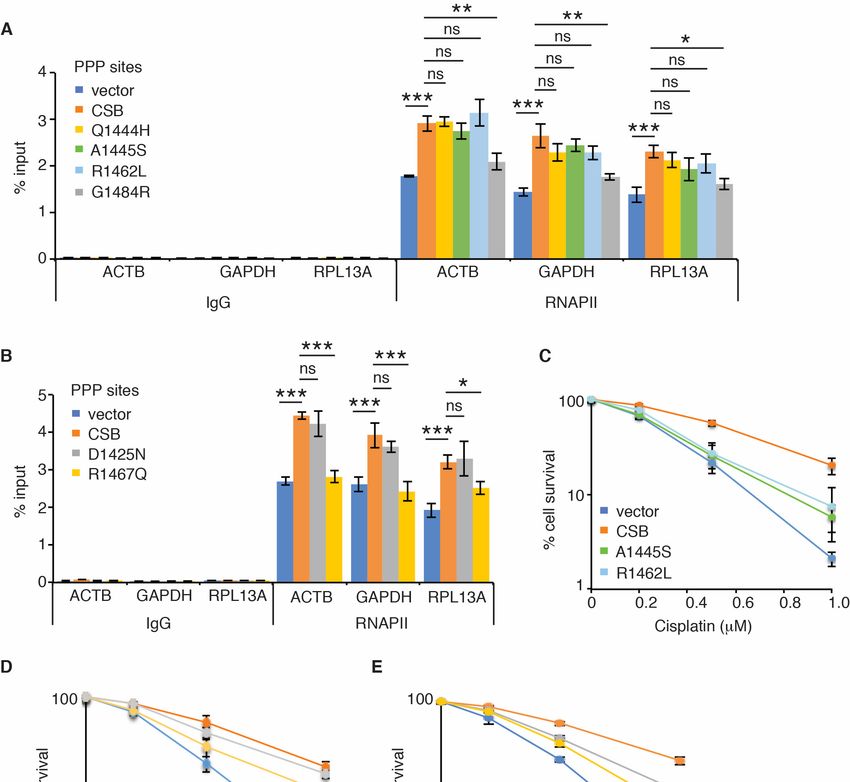
2.4. Cancer-Associated R1467Q and G1484R Mutations Impair CSB’s Ability to Promote RNAPII
Occupancy at PPP Sites of ACTB, GAPDH and RPL13A Genes
No CS mutations have been reported to reside in the CSB-WHD, however analysis
of cancer genomics databases of both BioPortal and COSMIC revealed the presence of
somatic mutations in the CSB-WHD. We have shown that the ubiquitin-binding activity
of the CSB-WHD is dispensable for RNAPII occupancy at PPP sites of ACTB, GAPDH
and RPL13A genes. To gain further insight into the ubiquitin-binding independent role of
CSB-WHD in regulating RNAPII occupancy at PPP sites, we selected five cancer-associated
CSB mutations, glutamine to histidine at position 1444 (Q1444H), alanine to serine at
position 1445 (A1445S), arginine to leucine at position 1462 (R1462L), arginine to glutamine
at position 1467 (R1467Q) and glycine to arginine at position 1484 (G1484R) (Figure 4A),
which are located distal to the ubiquitin-binding pocket of the CSB-WHD according to
the previously reported crystal structure of the CSB-WHD in complex with ubiquitin [35]
(Figure 4B). In addition, we also included in our study the CSB somatic mutation, aspartic
acid to asparagine at position 1425 (D1425N), which is from uterine endometrioid carci-
noma according to both BioPortal and COSMIC (Figure 4A). We reasoned that the D1425N
mutation could serve as a control for a ubiquitin binding-defective somatic mutation since it
has been reported that D1425 is directly engaged in binding to ubiquitin [35]. To investigate
if any of these six CSB somatic mutations might affect RNAPII occupancy at PPP sites, we
generated U2OS CSB-KO cells stably expressing the vector alone, Myc-CSB WT, Myc-CSB-
D1425N, Myc-CSB-Q1444H, Myc-CSB-A1445S, Myc-CSB-R1462L, Myc-CSB-R1467Q or
Myc-CSB-G1484R. All six CSB mutants were expressed at a comparable level to Myc-CSB
(Figure 4C,D). ChIP-qPCR analysis revealed that R1467Q and G1484R mutations but not
D1425N, Q1444H, A1445S and R1462L mutations impaired the ability of Myc-CSB to rescue
RNAPII occupancy at PPP sites of ACTB, GAPDH and RPL13A genes in U2OS CSB-KO
cells (Figure 5A,B). Clonogenic survival assays revealed that all six cancer-associated CSB
mutations except for G1484R impaired the ability of Myc-CSB to rescue survival of U2OS
CSB-KO cells in response to treatment with cisplatin (Figure 5C–E), suggesting that the
observed lack of an effect of D1425N, Q1444H, A1445S and R1462L mutations on RNAPII
occupancy at PPP sites is unlikely due to their being silent mutations. Taken together,
these results suggest that CSB relies on amino acids R1467 and G1484 but not amino acids
D1425, Q1444, A1445 and R1462 to promote RNAPII association with PPP sites. These
results also support the notion that CSB-mediated RNAPII association with PPP sites can
be mechanistically separated from CSB-mediated repair of cisplatin-induced DNA damage.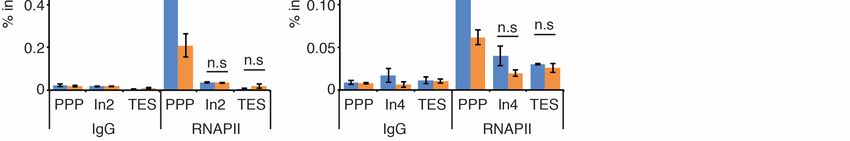
Int. J. Mol. Sci. 2021, 22, 3379                                                                                                                 8 of 14
 Int. J. Mol. Sci. 2021, 22, x FOR PEER REVIEW                                                                                                  8 of 13

      Figure 4.
     Figure    4. Cancer-associated
                  Cancer-associated CSB
                                      CSB mutations
                                           mutationsinin the
                                                          the CSB-WHD
                                                              CSB-WHD havehave little
                                                                                little effect
                                                                                       effect on
                                                                                              on CSB
                                                                                                  CSB expression.
                                                                                                       expression. (A)
                                                                                                                    (A) Six
                                                                                                                         Six cancer-associated
                                                                                                                             cancer-associated
      CSB mutations taken from both BioPortal and COSMIC databases. (B) Cartoon representation of the structure of the CSB-
     CSB mutations taken from both BioPortal and COSMIC databases. (B) Cartoon representation of the structure of the
      WHD in complex with ubiquitin (PDB code 6A6I). The CSB-WHD is coloured green, and ubiquitin is coloured cyan.
     CSB-WHD in complex with ubiquitin (PDB code 6A6I). The CSB-WHD is coloured green, and ubiquitin is coloured cyan.
      Sidechains for D1425, F1437, Q1444, A1445, R1462, R1467 and G1484 are shown in stick representation, with nitrogen and
     Sidechains
      oxygen atoms for D1425,
                       colouredF1437,  Q1444,
                                 in blue       A1445,
                                          and red,     R1462, R1467
                                                   respectively.       and G1484
                                                                   Sidechains       are shown
                                                                              of F1437           in stickalong
                                                                                          and A1445,       representation,  with nitrogen
                                                                                                                with the position   of G1484  and
                                                                                                                                                on
     oxygen    atoms
      the cartoon      colouredare
                    backbone,     incoloured
                                     blue andmagenta.
                                               red, respectively.   Sidechainsαof
                                                         For the CSB-WHD,           F1437are
                                                                                 helices    and   A1445, along
                                                                                               numbered,         with
                                                                                                             and the    the position
                                                                                                                     N-terminal    loopof isG1484
                                                                                                                                             iden-
     on   the The
      tified. cartoon   backbone,
                   figure           are coloured
                           was generated          magenta.
                                           using PyMOL        For the CSB-WHD,
                                                           (www.pymol.org).            helices are
                                                                                 (C)αWestern        numbered,
                                                                                                 analysis         andCSB-KO
                                                                                                            of U2OS    the N-terminal     loop is
                                                                                                                               cells expressing
      the vector The
     identified.   alone,  Myc-CSB
                        figure        WT, Myc-CSB-R1467Q
                               was generated                    or Myc-CSB-D1425N.
                                               using PyMOL (www.pymol.org,                Immunoblotting
                                                                                     accessed   on 10 February was2021).
                                                                                                                   performed    with anti-Myc
                                                                                                                         (C) Western    analysis
      and
     of     anti-γ-tubulin
         U2OS   CSB-KO cells antibodies.
                               expressing (D)
                                            theWestern  analysis
                                                vector alone,      of U2OS
                                                               Myc-CSB   WT,CSB-KO       cells expressing
                                                                              Myc-CSB-R1467Q                 the vector alone,
                                                                                                     or Myc-CSB-D1425N.         Myc-CSB WT,
                                                                                                                              Immunoblotting
      Myc-CSB-Q1444H,        Myc-CSB-A1445S,     Myc-CSB-R1462L       or Myc-CSB-G1484R.        Immunoblotting     was
     was performed with anti-Myc and anti-γ-tubulin antibodies. (D) Western analysis of U2OS CSB-KO cells expressing     performed    with anti-
                                                                                                                                               the
      Myc   and   anti-γ-tubulin  antibodies.
     vector alone, Myc-CSB WT, Myc-CSB-Q1444H, Myc-CSB-A1445S, Myc-CSB-R1462L or Myc-CSB-G1484R. Immunoblotting
     was performed with anti-Myc and anti-γ-tubulin antibodies.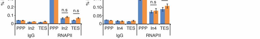
Int. J. Mol. Sci. 2021, 22, 3379 9 of 14
Int. J. Mol. Sci. 2021, 22, x FOR PEER REVIEW 9 of 13
Figure 5.
Figure 5. Cancer-associated
Cancer-associatedR1467Q
R1467QandandG1484R
G1484Rmutations
mutations ofof
CSBCSBimpair RNAPII
impair RNAPII pausing
pausingat PPP sites.sites.
at PPP (A) RNAPII ChIP
(A) RNAPII
analyses
ChIP of ACTB,
analyses GAPDH
of ACTB, and RPL13A
GAPDH genesgenes
and RPL13A in U2OS CSB-KO
in U2OS cells expressing
CSB-KO the vector
cells expressing alone,alone,
the vector Myc-CSB WT, Myc-
Myc-CSB WT,
CSB-Q1444H, Myc-CSB-R1462L,
Myc-CSB-Q1444H, Myc-CSB-R1467Q
Myc-CSB-R1462L, Myc-CSB-R1467Q or Myc-CSB-G1484R.
or Myc-CSB-G1484R. Standard errors errors
Standard from three
fromindependent experi-
three independent
ments are shown. P values were derived using the 2-way ANOVA test. * p < 0.05; ** p < 0.01; *** p < 0.001; ns: p > 0.05. (B)
experiments are shown. p values were derived using the 2-way ANOVA test. * p < 0.05; ** p < 0.01; *** p < 0.001; ns: p > 0.05.
RNAPII ChIP analyses of ACTB, GAPDH and RPL13A genes in U2OS CSB-KO cells expressing the vector alone, Myc-CSB
(B) RNAPII ChIP analyses of ACTB, GAPDH and RPL13A genes in U2OS CSB-KO cells expressing the vector alone, Myc-CSB
WT, Myc-CSB-D1425N or Myc-CSB-R1467Q. Standard errors from three independent experiments are shown. p values
WT,
wereMyc-CSB-D1425N
derived using theor2-way
Myc-CSB-R1467Q.
ANOVA test. Standard
* p < 0.05;errors from***
** p < 0.01; three
p < independent
0.001; ns: P > experiments are shown.
0.05. (C) Cisplatin p values
clonogenic were
survival
derived using the 2-way ANOVA test. * p < 0.05; ** p < 0.01; *** p < 0.001; ns: p > 0.05. (C) Cisplatin
assays of U2OS CSB-KO cells expressing the vector alone, Myc-CSB WT, Myc-CSB-A1445S or Myc-CSB-R1462L. Standard clonogenic survival
assays of U2OS
deviations fromCSB-KO cells expressing
three independent the vector
experiments arealone, Myc-CSB
indicated. WT, Myc-CSB-A1445S
(D) Cisplatin or Myc-CSB-R1462L.
clonogenic survival assays of U2OSStandard
CSB-KO
cells expressing the vector alone, Myc-CSB WT, Myc-CSB-Q1444H or Myc-CSB-G1484R. Standard deviations
deviations from three independent experiments are indicated. (D) Cisplatin clonogenic survival assays of U2OS from three
CSB-KO
independent
cells expressing experiments are indicated.
the vector alone, Myc-CSB (E)WT,
Cisplatin clonogenic survival
Myc-CSB-Q1444H assays of U2OS
or Myc-CSB-G1484R. CSB-KO
Standard cells expressing
deviations the
from three
vector alone, Myc-CSB WT, Myc-CSB-D1425N or Myc-CSB-R1467Q. Standard deviations from three independent
independent experiments are indicated. (E) Cisplatin clonogenic survival assays of U2OS CSB-KO cells expressing the vector experi-
ments Myc-CSB
alone, are indicated.
WT, Myc-CSB-D1425N or Myc-CSB-R1467Q. Standard deviations from three independent experiments
are indicated.
3. Discussion
3. Discussion
It has been suggested that CSB tracks paused RNAPII on non-damage templates to
It hastranscription
stimulate been suggested that however
[33,43], CSB tracks paused
it has RNAPII
not been on non-damage
previously templates
demonstrated. to
In this
stimulate transcription [33,43], however it has not been previously demonstrated.
report, we have provided evidence demonstrating that CSB promotes RNAPII association In this
with PPP sites of several actively transcribed genes in cultured cells, supporting the notion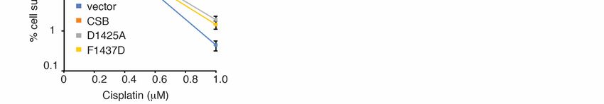
Int. J. Mol. Sci. 2021, 22, 3379                                                                                         10 of 14

                                   report, we have provided evidence demonstrating that CSB promotes RNAPII association
                                   with PPP sites of several actively transcribed genes in cultured cells, supporting the notion
                                   that CSB regulates paused RNAPII for transcription elongation. CSB contains a C-terminal
                                   winged helix domain (WHD), which binds to ubiquitin [35]. It has been reported that
                                   the ubiquitin-binding activity of the CSB-WHD is important for efficient TC-NER [35].
                                   However, several lines of evidence presented here suggest that the ubiquitin-binding
                                   activity of the CSB-WHD is dispensable for RNAPII association with PPP sites of ACTB,
                                   GAPDH and RPL13A genes. Firstly, both D1425A and F1437D mutations, which have
                                   previously been reported to not only abrogate binding of the CSB-WHD to ubiquitin but
                                   also sensitize cells to treatment with UV [35], have little effect on the ability of CSB to
                                   promote RNAPII occupancy at PPP sites of ACTB, GAPDH and RPL13A genes. Secondly,
                                   a cancer-associated D1425N mutation of CSB, which sensitizes cells to cisplatin, does
                                   not affect RNAPII occupancy at PPP sites of ACTB, GAPDH and RPL13A genes. D1425
                                   has been reported to be directly engaged in binding to ubiquitin [35]. Thirdly, the two
                                   cancer-associated CSB mutations R1467Q and G1484R, located on the opposite side of the
                                   CSB-WHD away from its ubiquitin-binding pocket, impair RNAPII association with PPP
                                   sites of ACTB, GAPDH and RPL13A genes. Our finding suggests that the surface of the
                                   CSB-WHD where R1467 and G1484 reside may play a role in regulating RNAPII association
                                   with PPP sites of ACTB, GAPDH and RPL13A genes, which requires further investigation.
                                         While much attention has been given to CS-causing CSB mutations over the past
                                   decades, little is known about CSB somatic mutations that have been identified in cancer
                                   cell lines and tumor samples. The work presented here has provided the first evidence
                                   that CSB somatic mutations affect CSB function in cultured cells. Increasing evidence
                                   suggests that CSB plays a role in cancer prognosis and treatment [38–41]. Our finding that
                                   five of six cancer-associated CSB mutations located in the CSB-WHD, D1425N, Q1444H,
                                   A1445S, R1462L and R1467Q, sensitize cells to treatment with cisplatin, a platinum-based
                                   chemotherapeutic drug commonly used to treat a number of cancers [44–46], suggests that
                                   these mutations can serve as biomarkers for cancer diagnosis and treatment.
                                         CSB is a multifunctional protein that participates in a number of nuclear processes,
                                   including transcription regulation [7,8], UV repair [1,2], oxidative DNA damage [9] and
                                   DNA DSB repair [15–17]. We have previously reported that regulation of CSB function in
                                   UV repair is separable from that in DNA DSB repair [37]. Deletion of the first 30 amino
                                   acids in the N-terminal region of CSB does not affect its ability to repair UV-induced DNA
                                   damage [37] but abrogates its ability to promote cell survival in response to treatment
                                   with olaparib, a PARP inhibitor known to be toxic to cells deficient in homologous recom-
                                   bination [15]. In addition, it has been reported that the function of CSB in UV repair is
                                   differentially regulated from its role in oxidative DNA damage [47,48]. The work presented
                                   in this report suggests that CSB-mediated RNAPII association with PPP sites of ACTB,
                                   GAPDH and RPL13A genes is mechanistically separable from CSB-mediated repair of
                                   cisplatin-induced DNA damage. Our finding adds to the growing list of evidence that
                                   CSB function in various nuclear processes is distinctively regulated from one another. Our
                                   finding that five of six cancer-associated CSB mutations, D1425N, Q1444H, A1445S, R1462L
                                   and G1484R, differentially affect CSB’s ability to regulate RNAPII association with ACTB,
                                   GAPDH and RPL13A genes as well as to promote cell survival in response to cisplatin-
                                   induced DNA damage suggests the complexity of the impact of CSB somatic mutations.
                                   Future studies are needed to catalog the effect of CSB somatic mutations on its function,
                                   which would be expected to aid cancer diagnosis and treatment.

                                   4. Materials and Methods
                                   4.1. Plasmids and Antibodies
                                       Mammalian expression constructs of Myc-tagged CSB and CSB-∆WHD have been
                                   described [15,17]. Wild type CSB was used as a template to generate, via site-directed
                                   mutagenesis, CSB mutants D1425A, F1437D, D1425N, Q1444H, A1445S, R1462L, R1467Q
Int. J. Mol. Sci. 2021, 22, 3379 11 of 14
and G1484R, which were cloned into the retroviral expression vector pLPC-NMyc [13]. The
primers used to generate these constructs are included in Supplementary Table S2.
Antibodies used include CSB/ERCC6 (A301-345A, Bethyl Laboratories, Montgomery,
TX, USA); CSB (A301-347A, Bethyl Laboratories, Montgomery, TX, USA); anti-Myc (9E10,
MilliporeSigma, Darmstadt, Germany); RNAPII (05-623, MilliporeSigma, Darmstadt, Ger-
many); γ-tubulin (GTU88, MilliporeSigma, Darmstadt, Germany).
4.2. Cell Culture, Transfection, Retroviral Infection and Treatment
All cells were grown in DMEM medium with 10% fetal bovine serum supplemented
with non-essential amino acids, L-glutamine, 100 U/mL penicillin and 0.1 mg/mL strepto-
mycin. Cell lines used: Phoenix [13], U2OS [49] (ATCC), U2OS-CSB knockout (KO) [15],
GM16095 (Coriell Institute for Medical Research, Camden, NJ, USA). GM16095 is a SV40-
transformed cell line derived from GM739 [1]. Cell cultures were routinely fixed, stained
with DAPI, and examined for mycoplasma contamination. Retroviral gene delivery was
carried out as described [50,51] to generate stable cell lines. DNA transfection was car-
ried out with JetPrime transfection reagent (Polyplus, Illkirch, France) according to the
manufacturer’s instructions.
4.3. Chromatin Immunoprecipitation (ChIP)
ChIP assays were carried out as described [15]. For each ChIP, 200 µL of the cell
lysate was diluted 1:5 in IP dilution buffer [1% Triton X-100, 2 mM EDTA, 20 mM Tris-
HCl pH 8.1, 150 mM NaCl]. Out of 1 mL diluted lysate, 20 µL was set aside as input
control and the remaining was precleared with protein G sepharose beads (GE Healthcare,
Chicago, IL, USA), which were preblocked with BSA and tRNA, and then incubated with
primary antibody (1 µg) overnight at 4 ◦ C. The final IP DNA precipitated with ethanol
in the presence of 20 µg glycogen (Roche) was resuspended in sterile ddH2 O. DNA was
then analyzed by qPCR using previously described primer pair sets [42] (also shown
in Supplementary Table S1) and SensiFAST SYBR NO-ROX kit (Bioline, Alvinston, ON,
Canada). The threshold cycle (Ct) value of the qPCR reactions for the indicated genes of
each ChIP DNA was normalized to that of the input DNA, giving rise to the percentage of
input for each ChIP reaction.
4.4. Immunoblotting
Immunoblotting was performed using whole cell extracts as described [52]. Briefly, cell
extracts were fractionated by either 6% (for CSB) or 8% (for γ-tubulin) SDS-polyacrylamide
gel electrophoresis and then transferred to nitrocellulose membranes. Following im-
munoblotting, membranes were exposed to Amersham hyperfilms (Cytiva 28906838, Marl-
borough, MA, USA). Development of the hyperfilm was done on Konica Medical Film
Processor SRX-101A, Tokyo, Japan.
4.5. Clonogenic Survival Assays
Clonogenic survival assays were done as described [17]. Twenty-four hours post
seeding, cells were washed with PBS, treated with varying doses of cisplatin and left to
grow in the presence of cisplatin for the entirety of the experiments. Ten days post treatment,
colonies were fixed and stained with a solution containing 50% methanol, 7% acetic acid
and 0.1% Coomasie blue for 10 min at room temperature. Colonies consisting of more than
32 cells were manually scored on a microscope (Leica EZ4, Concord, ON, Canada).
Supplementary Materials: The following are available online at https://www.mdpi.com/1422-0
067/22/7/3379/s1, Figure S1: Uncropped Images, Table S1: Q-PCR Primers, Table S2: Oligos for
Site-Directed Mutagenesis.
Author Contributions: Conceptualization, X.-D.Z.; methodology, N.L.B.; investigation, N.L.B., S.C.,
J.R.W., X.-D.Z.; resources, H.E.S.; writing—original draft preparation, X.-D.Z.; writing—review andInt. J. Mol. Sci. 2021, 22, 3379 12 of 14
editing, N.L.B., S.C., J.R.W.; supervision, X.-D.Z.; funding acquisition, X.-D.Z. All authors have read
and agreed to the published version of the manuscript.
Funding: This research was funded by Natural Sciences and Engineering Research Council of
Canada, grant number RGPIN-05110-2016 and Canadian Institutes of Health Research, grant number
PJT159793. S.C. was a holder of Mitacs Globalink Graduate Fellowship.
Institutional Review Board Statement: Not applicable.
Informed Consent Statement: Not applicable.
Data Availability Statement: All data are contained within this manuscript.
Conflicts of Interest: The authors declare no conflict of interest.
References
1. Troelstra, C.; van Gool, A.; de Wit, J.; Vermeulen, W.; Bootsma, D.; Hoeijmakers, J.H. ERCC6, a member of a subfamily of putative
helicases, is involved in Cockayne’s syndrome and preferential repair of active genes. Cell 1992, 71, 939–953. [CrossRef]
2. Van der Horst, G.T.; van Steeg, H.; Berg, R.J.; van Gool, A.J.; de Wit, J.; Weeda, G.; Morreau, H.; Beems, R.B.; van Kreijl,
C.F.; de Gruijl, F.R.; et al. Defective transcription-coupled repair in Cockayne syndrome B mice is associated with skin cancer
predisposition. Cell 1997, 89, 425–435. [CrossRef]
3. Laugel, V. Cockayne syndrome: The expanding clinical and mutational spectrum. Mech. Ageing Dev. 2013, 134, 161–170.
[CrossRef]
4. Laugel, V.; Dalloz, C.; Durand, M.; Sauvanaud, F.; Kristensen, U.; Vincent, M.C.; Pasquier, L.; Odent, S.; Cormier-Daire, V.; Gener,
B.; et al. Mutation update for the CSB/ERCC6 and CSA/ERCC8 genes involved in Cockayne syndrome. Hum. Mutat. 2009, 31,
113–126. [CrossRef]
5. Calmels, N.; Botta, E.; Jia, N.; Fawcett, H.; Nardo, T.; Nakazawa, Y.; Lanzafame, M.; Moriwaki, S.; Sugita, K.; Kubota, M.; et al.
Functional and clinical relevance of novel mutations in a large cohort of patients with Cockayne syndrome. J. Med. Genet. 2018,
55, 329–343. [CrossRef] [PubMed]
6. Karikkineth, A.C.; Scheibye-Knudsen, M.; Fivenson, E.; Croteau, D.L.; Bohr, V.A. Cockayne syndrome: Clinical features, model
systems and pathways. Ageing Res. Rev. 2017, 33, 3–17. [CrossRef]
7. Balajee, A.S.; May, A.; Dianov, G.L.; Friedberg, E.C.; Bohr, V.A. Reduced RNA polymerase II transcription in intact and
permeabilized Cockayne syndrome group B cells. Proc. Natl. Acad. Sci. USA 1997, 94, 4306–4311. [CrossRef] [PubMed]
8. Dianov, G.L.; Houle, J.-F.; Iyer, N.; Bohr, V.A.; Friedberg, E.C. Reduced RNA polymerase II transcription in extracts of cockayne
syndrome and xeroderma pigmentosum/Cockayne syndrome cells. Nucleic Acids Res. 1997, 25, 3636–3642. [CrossRef] [PubMed]
9. Dianov, G.; Bischoff, C.; Sunesen, M.; Bohr, V.A. Repair of 8-oxoguanine in DNA is deficient in Cockayne syndrome group B cells.
Nucleic Acids Res. 1999, 27, 1365–1368. [CrossRef]
10. Aamann, M.D.; Sorensen, M.M.; Hvitby, C.; Berquist, B.R.; Muftuoglu, M.; Tian, J.; de Souza-Pinto, N.C.; Scheibye-Knudsen, M.;
Wilson, D.M., III; Stevnsner, T.; et al. Cockayne syndrome group B protein promotes mitochondrial DNA stability by supporting
the DNA repair association with the mitochondrial membrane. FASEB J. 2010, 24, 2334–2346. [CrossRef]
11. Berquist, B.R.; Canugovi, C.; Sykora, P.; Wilson, D.M., III; Bohr, V.A. Human Cockayne syndrome B protein reciprocally
communicates with mitochondrial proteins and promotes transcriptional elongation. Nucleic Acids Res. 2012, 40, 8392–8405.
[CrossRef]
12. Paccosi, E.; Costanzo, F.; Costantino, M.; Balzerano, A.; Monteonofrio, L.; Soddu, S.; Prantera, G.; Brancorsini, S.; Egly, J.-M.;
Proietti-De-Santis, L. The Cockayne syndrome group A and B proteins are part of a ubiquitin–proteasome degradation complex
regulating cell division. Proc. Natl. Acad. Sci. USA 2020, 117, 30498–30508. [CrossRef]
13. Batenburg, N.L.; Mitchell, T.R.H.; Leach, D.M.; Rainbow, A.J.; Zhu, X.-D. Cockayne Syndrome group B protein interacts with
TRF2 and regulates telomere length and stability. Nucleic Acids Res. 2012, 40, 9661–9674. [CrossRef]
14. Feng, E.; Batenburg, N.L.; Walker, J.R.; Ho, A.; Mitchell, T.R.H.; Qin, J.; Zhu, X.-D. CSB cooperates with SMARCAL1 to maintain
telomere stability in ALT cells. J. Cell Sci. 2020, 133, jcs234914. [CrossRef] [PubMed]
15. Batenburg, N.L.; Walker, J.R.; Noordermeer, S.M.; Moatti, N.; Durocher, D.; Zhu, X.D. ATM and CDK2 control chromatin
remodeler CSB to inhibit RIF1 in DSB repair pathway choice. Nat. Commun. 2017, 8, 1921. [CrossRef] [PubMed]
16. Batenburg, N.L.; Walker, J.R.; Coulombe, Y.; Sherker, A.; Masson, J.Y.; Zhu, X.D. CSB interacts with BRCA1 in late S/G2 to
promote MRN- and CtIP-mediated DNA end resection. Nucleic Acids Res. 2019, 47, 10678–10692. [CrossRef] [PubMed]
17. Batenburg, N.L.; Thompson, E.L.; Hendrickson, E.A.; Zhu, X.D. Cockayne syndrome group B protein regulates DNA double-
strand break repair and checkpoint activation. EMBO J. 2015, 34, 1399–1416. [CrossRef] [PubMed]
18. Newman, J.C.; Bailey, A.D.; Weiner, A.M. Cockayne syndrome group B protein (CSB) plays a general role in chromatin
maintenance and remodeling. Proc. Natl. Acad. Sci. USA 2006, 103, 9613–9618. [CrossRef]
19. Lake, R.J.; Boetefuer, E.L.; Tsai, P.-F.; Jeong, J.; Choi, I.; Won, K.-J.; Fan, H.-Y. The Sequence-Specific Transcription Factor c-Jun
Targets Cockayne Syndrome Protein B to Regulate Transcription and Chromatin Structure. PLoS Genet. 2014, 10, e1004284.
[CrossRef] [PubMed]Int. J. Mol. Sci. 2021, 22, 3379 13 of 14
20. Kristensen, U.; Epanchintsev, A.; Rauschendorf, M.A.; Laugel, V.; Stevnsner, T.; Bohr, V.A.; Coin, F.; Egly, J.M. Regulatory interplay
of Cockayne syndrome B ATPase and stress-response gene ATF3 following genotoxic stress. Proc. Natl. Acad. Sci. USA 2013, 110,
E2261–E2270. [CrossRef]
21. Epanchintsev, A.; Costanzo, F.; Rauschendorf, M.A.; Caputo, M.; Ye, T.; Donnio, L.M.; de Santis, L.P.; Coin, F.; Laugel, V.; Egly, J.M.
Cockayne’s Syndrome A and B Proteins Regulate Transcription Arrest after Genotoxic Stress by Promoting ATF3 Degradation.
Mol. Cell 2017, 68, 1054–1066. [CrossRef]
22. Ciaffardini, F.; Nicolai, S.; Caputo, M.; Canu, G.; Paccosi, E.; Costantino, M.; Frontini, M.; Balajee, A.S.; Proietti-De-Santis, L.
The cockayne syndrome B protein is essential for neuronal differentiation and neuritogenesis. Cell Death Dis. 2014, 5, e1268.
[CrossRef]
23. Wang, Y.; Chakravarty, P.; Ranes, M.; Kelly, G.; Brooks, P.J.; Neilan, E.; Stewart, A.; Schiavo, G.; Svejstrup, J.Q. Dysregulation
of gene expression as a cause of Cockayne syndrome neurological disease. Proc. Natl. Acad. Sci. USA 2014, 111, 14454–14459.
[CrossRef]
24. Wang, Y.; Jones-Tabah, J.; Chakravarty, P.; Stewart, A.; Muotri, A.; Laposa, R.R.; Svejstrup, J.Q. Pharmacological Bypass of
Cockayne Syndrome B Function in Neuronal Differentiation. Cell Rep. 2016, 14, 2554–2561. [CrossRef]
25. Van Gool, A.J.; Citterio, E.; Rademakers, S.; Van Os, R.; Vermeulen, W.; Constantinou, A.; Egly, J.-M.; Bootsma, D.; Hoeijmakers, J.H.
The Cockayne syndrome B protein, involved in transcription-coupled DNA repair, resides in an RNA polymerase II-containing
complex. EMBO J. 1997, 16, 5955–5965. [CrossRef]
26. Selby, C.P.; Sancar, A. Cockayne syndrome group B protein enhances elongation by RNA polymerase II. Proc. Natl. Acad. Sci.
USA 1997, 94, 11205–11209. [CrossRef] [PubMed]
27. Tantin, D.; Kansal, A.; Carey, M. Recruitment of the putative transcription-repair coupling factor CSB/ERCC6 to RNA polymerase
II elongation complexes. Mol. Cell. Biol. 1997, 17, 6803–6814. [CrossRef] [PubMed]
28. Hanawalt, P.C.; Spivak, G. Transcription-coupled DNA repair: Two decades of progress and surprises. Nat. Rev. Mol. Cell Biol.
2008, 9, 958–970. [CrossRef]
29. Svejstrup, J.Q. Contending with transcriptional arrest during RNAPII transcript elongation. Trends Biochem. Sci. 2007, 32, 165–171.
[CrossRef] [PubMed]
30. Lainé, J.-P.; Egly, J.-M. Initiation of DNA repair mediated by a stalled RNA polymerase IIO. EMBO J. 2006, 25, 387–397. [CrossRef]
[PubMed]
31. Lindsey-Boltz, L.A.; Sancar, A. RNA polymerase: The most specific damage recognition protein in cellular responses to DNA
damage? Proc. Natl. Acad. Sci. USA 2007, 104, 13213–13214. [CrossRef] [PubMed]
32. Saxowsky, T.T.; Doetsch, P.W. RNA Polymerase Encounters with DNA Damage: Transcription-Coupled Repair or Transcriptional
Mutagenesis? Chem. Rev. 2006, 106, 474–488. [CrossRef] [PubMed]
33. Xu, J.; Lahiri, I.; Wang, W.; Wier, A.; Cianfrocco, M.A.; Chong, J.; Hare, A.A.; Dervan, P.B.; DiMaio, F.; Leschziner, A.E.; et al.
Structural basis for the initiation of eukaryotic transcription-coupled DNA repair. Nat. Cell Biol. 2017, 551, 653–657. [CrossRef]
34. Adelman, K.; Lis, J.T. Promoter-proximal pausing of RNA polymerase II: Emerging roles in metazoans. Nat. Rev. Genet. 2012, 13,
720–731. [CrossRef] [PubMed]
35. Takahashi, T.S.; Sato, Y.; Yamagata, A.; Goto-Ito, S.; Saijo, M.; Fukai, S. Structural basis of ubiquitin recognition by the winged-helix
domain of Cockayne syndrome group B protein. Nucleic Acids Res. 2019, 47, 3784–3794. [CrossRef]
36. Sin, Y.; Tanaka, K.; Saijo, M. The C-terminal Region and SUMOylation of Cockayne Syndrome Group B Protein Play Critical Roles
in Transcription-coupled Nucleotide Excision Repair. J. Biol. Chem. 2016, 291, 1387–1397. [CrossRef]
37. Batenburg, N.L.; Qin, J.; Walker, J.R.; Zhu, X.-D. Efficient UV repair requires disengagement of the CSB winged helix domain
from the CSB ATPase domain. DNA Repair. 2018, 68, 58–67. [CrossRef]
38. Zhao, Z.; Zhang, G.; Li, W. Elevated Expression of ERCC6 Confers Resistance to 5-Fluorouracil and Is Associated with Poor
Patient Survival in Colorectal Cancer. DNA Cell Biol. 2017, 36, 781–786. [CrossRef]
39. Lu, Y.; Mani, S.; Kandimalla, E.; Yu, N.; Agrawal, S.; States, J.; Bregman, D. The Cockayne syndrome group B DNA repair protein
as an anti-cancer target. Int. J. Oncol. 2001, 19, 1089–1097. [CrossRef] [PubMed]
40. Caputo, M.; Frontini, M.; Velez-Cruz, R.; Nicolai, S.; Prantera, G.; Proietti-De-Santis, L. The CSB repair factor is overexpressed in
cancer cells, increases apoptotic resistance, and promotes tumor growth. DNA Repair. 2013, 12, 293–299. [CrossRef]
41. Proietti-De-Santis, L.; Balzerano, A.; Prantera, G. CSB: An Emerging Actionable Target for Cancer Therapy. Trends Cancer 2018, 4,
172–175. [CrossRef]
42. Shivji, M.K.K.; Renaudin, X.; Williams, C.H.; Venkitaraman, A.R. BRCA2 Regulates Transcription Elongation by RNA Polymerase
II to Prevent R-Loop Accumulation. Cell Rep. 2018, 22, 1031–1039. [CrossRef]
43. Geijer, M.E.; Marteijn, J.A. What happens at the lesion does not stay at the lesion: Transcription-coupled nucleotide excision
repair and the effects of DNA damage on transcription in cis and trans. DNA Repair. 2018, 71, 56–68. [CrossRef]
44. Wang, D.; Lippard, S.J. Cellular processing of platinum anticancer drugs. Nat. Rev. Drug Discov. 2005, 4, 307–320. [CrossRef]
45. Jung, Y.; Lippard, S.J. Direct Cellular Responses to Platinum-Induced DNA Damage. Chem. Rev. 2007, 107, 1387–1407. [CrossRef]
[PubMed]
46. Basu, A.; Krishnamurthy, S. Cellular Responses to Cisplatin-Induced DNA Damage. J. Nucleic Acids 2010, 2010, 201367. [CrossRef]Int. J. Mol. Sci. 2021, 22, 3379 14 of 14
47. Selzer, R.R.; Nyaga, S.; Tuo, J.; May, A.; Muftuoglu, M.; Christiansen, M.; Citterio, E.; Brosh, R.M., Jr.; Bohr, V.A. Differential
requirement for the ATPase domain of the Cockayne syndrome group B gene in the processing of UV-induced DNA damage and
8-oxoguanine lesions in human cells. Nucleic Acids Res. 2002, 30, 782–793. [CrossRef]
48. Ranes, M.; Boeing, S.; Wang, Y.; Wienholz, F.; Menoni, H.; Walker, J.; Encheva, V.; Chakravarty, P.; Mari, P.-O.; Stewart, A.; et al. A
ubiquitylation site in Cockayne syndrome B required for repair of oxidative DNA damage, but not for transcription-coupled
nucleotide excision repair. Nucleic Acids Res. 2016, 44, 5246–5255. [CrossRef]
49. Wilson, F.R.; Ho, A.; Walker, J.R.; Zhu, X.-D. Cdk-dependent phosphorylation regulates TRF1 recruitment to PML bodies and
promotes C-circle production in ALT cells. J. Cell Sci. 2016, 129, 2559–2572. [CrossRef] [PubMed]
50. Wu, Y.; Xiao, S.; Zhu, X.-D. MRE11–RAD50–NBS1 and ATM function as co-mediators of TRF1 in telomere length control. Nat.
Struct. Mol. Biol. 2007, 14, 832–840. [CrossRef] [PubMed]
51. Wu, Y.; Mitchell, T.R.; Zhu, X.-D. Human XPF controls TRF2 and telomere length maintenance through distinctive mechanisms.
Mech. Ageing Dev. 2008, 129, 602–610. [CrossRef] [PubMed]
52. Zhu, X.D.; Kuster, B.; Mann, M.; Petrini, J.H.; Lange, T. Cell-cycle-regulated association of RAD50/MRE11/NBS1 with TRF2 and
human telomeres. Nat. Genet. 2000, 25, 347–352. [CrossRef] [PubMed]You can also read