Moringa Oleifera aqueous leaf extract down-regulates nuclear factor-kappaB and increases cytotoxic effect of chemotherapy in pancreatic cancer cells
←
→
Page content transcription
If your browser does not render page correctly, please read the page content below
Berkovich et al. BMC Complementary and Alternative Medicine 2013, 13:212
http://www.biomedcentral.com/1472-6882/13/212

 RESEARCH ARTICLE                                                                                                                                Open Access

Moringa Oleifera aqueous leaf extract
down-regulates nuclear factor-kappaB and
increases cytotoxic effect of chemotherapy
in pancreatic cancer cells
Liron Berkovich, Gideon Earon, Ilan Ron, Adam Rimmon, Akiva Vexler and Shahar Lev-Ari*

  Abstract
  Background: Fewer than 6% patients with adenocarcinoma of the pancreas live up to five years after diagnosis.
  Chemotherapy is currently the standard treatment, however, these tumors often develop drug resistance over time.
  Agents for increasing the cytotoxic effects of chemotherapy or reducing the cancer cells’ chemo-resistance to the drugs
  are required to improve treatment outcome. Nuclear factor kappa B (NF-kB), a pro-inflammatory transcription factor,
  reportedly plays a significant role in the resistance of pancreatic cancer cells to apoptosis-based chemotherapy. This
  study investigated the effect of aqueous Moringa Oleifera leaf extract on cultured human pancreatic cancer cells - Panc-1,
  p34, and COLO 357, and whether it can potentiates the effect of cisplatin chemotherapy on these cells.
  Methods: The effect of Moringa Oleifera leaf extract alone and in combination with cisplatin on the survival of cultured
  human pancreatic cancer cells was evaluated by XTT-based colorimetric assay. The distribution of Panc-1 cells in the cell
  cycle following treatment with Moringa leaf extract was evaluated by flow cytometry, and evaluations of protein levels
  were via immunoblotting. Data of cell survival following combined treatments were analyzed with Calcusyn software.
  Results: Moringa Oleifera leaf extract inhibited the growth of all pancreatic cell lines tested. This effect was significant in
  all cells following exposure to ≥0.75 mg/ml of the extract. Exposure of Panc-1 cells to Moringa leaf extract induced an
  elevation in the sub-G1 cell population of the cell-cycle, and reduced the expression of p65, p-IkBα and IkBα proteins in
  crude cell extracts. Lastly, Moringa Oleifera leaf extract synergistically enhanced the cytotoxic effect of cisplatin on Panc-1
  cells.
  Conclusion: Moringa Oleifera leaf extract inhibits the growth of pancreatic cancer cells, the cells NF-κB signaling
  pathway, and increases the efficacy of chemotherapy in human pancreatic cancer cells.
  Keywords: Moringa Oleifera, Pancreatic cancer, NF-kB, cisplatin

Background                                                                           origin, among them taxanes (docetaxel, paclitaxel), Vinca
Natural products from plants provide an important source                             alkaloids (vindesine, vinblastine, vincristine), anthracyclines
of new drugs and potential pharmaceutical "lead" com-                                (idarubicin, daunorubicin, epirubicin), and others. Thus,
pounds. Natural products or natural product-derived drugs                            there is a promising future for the use of natural products
include 28% of all new chemical entities launched between                            derived from plants as anti-tumor agents.
1981 and 2002, and 24% of them are semi-synthetic natural                               Adenocarcinoma of the pancreas, the most common
product analogues or synthetic compounds based on nat-                               form of pancreatic cancer, is the fourth commonest
ural product pharmacophores [1]. Furthermore, many                                   cause of cancer-related mortality worldwide [2]. This
anti-tumor agents in current clinical use are of natural                             cancer is often diagnosed at advanced stages and has a
                                                                                     poor prognosis, with fewer than 6% of those patients liv-
* Correspondence: Shaharl@tasmc.health.gov.il                                        ing as long as five years after diagnosis [2]. The basis of
Laboratory of Herbal Medicine and Cancer Research, Tel-Aviv Sourasky                 current pancreatic cancer therapy is targeting DNA
Medical Center, Tel-Aviv, Israel

                                       © 2013 Berkovich et al.; licensee BioMed Central Ltd. This is an Open Access article distributed under the terms of the Creative
                                       Commons Attribution License (http://creativecommons.org/licenses/by/2.0), which permits unrestricted use, distribution, and
                                       reproduction in any medium, provided the original work is properly cited.
Berkovich et al. BMC Complementary and Alternative Medicine 2013, 13:212                                                  Page 2 of 7
http://www.biomedcentral.com/1472-6882/13/212

synthesis using gemcitabine, with or without a second                  Methods
agent like 5-FU or a platinum based agent [3]. Unfortu-                Preparation of Moringa Oleifera aqueous extract
nately, this treatment is limited by a resistance of the               Leaves of Moringa Oleifera were received from Moringa
cancer cells to these therapies as well as a somatic tox-              Arava Ltd, Israel. Moringa Arava grows the Moringa
icity [4]. The drug resistance is attributed to several                Oleifera plant in the Dead Sea area, Israel, where it is
mechanisms: drugs exclusion from the cells, changes in                 grown in rich mineral soil. The plant derived aqueous ex-
the enzymes metabolizing the drugs, or the cells becom-                tract tested in this study was prepared in our laboratory
ing more resistant to stress and apoptosis [4].                        by mixing 1g dried and powdered leaves of Moringa
   Nuclear factor kappa B (NF-κB) is an essential regula-              Oleifera with 10 mL boiling water for 5 minutes. The mix-
tor of the innate and adaptive immunity that, under                    ture was then filtered twice through a 2 μm pore sterile fil-
most conditions, supports proliferation and survival of                ter paper into a sterile tube. The aqueous extract stock
cells via inhibition of apoptosis [5]. Constitutively active           solution (100 mg/mL) was freshly prepared for each set of
NF-κB signaling that strengthens the malignant cells’                  experiments and stored at 4°C for up to 5 days.
ability to survive has been demonstrated in most tumor
cell lines as well as in a variety of patient-derived tumor            Cell lines and culture conditions
tissues, including those from pancreatic cancer [6,7].                 Human pancreatic cancer cell lines (Panc-1 and COLO-
NF-κB activation has also been directly linked to pancre-              357) were kindly provided by Prof. Ziv Gil (Laboratory
atic cancer metastatic potential [8]. Although the reason              for Applied Cancer Research, Tel Aviv Sourasky Medical
for its constitutive activation in malignant cells has not             Center, Israel). Human pancreatic cancer cell line p34
been fully elucidated, suppression of NF-κB in the ma-                 [14], developed from pleural effusion of a pancreatic
jority of these tumors leads to the induction of apoptosis             cancer patient, was kindly provided by Dr. Alex Starr
and the subsequent generation of cell death. In addition,              (Laboratory of Lung Biology, Lung and Allergy Institute,
several lines of evidence indicate that NF-κB plays a sig-             Tel Aviv Sourasky Medical Center, Israel). All cells were
nificant role in pancreatic cancer resistance to apoptosis-            grown in Dulbecco modified Eagle medium (DMEM)
based chemotherapies, leading to NF-kB being suggested                 supplemented by 10% heat-inactivated bovine serum,
as a potential molecular target for pancreatic cancer                  4.5 g/L glucose, 200 μM L-glutamine, 10 units/ml penicil-
therapy [8].                                                           lin and 10 μg/mL streptomycin (Biological Industries, Beit
   Moringa Oleifera, Lam. (Moringaceae) is a tree that                 HaEmek, Israel). The cells were incubated at 37°C in a 5%
grows widely in the tropics and subtropics of Asia and Af-             CO2 humidified atmosphere. The cells were harvested by
rica. Its leaves have been traditionally consumed by Asian             trypsin/EDTA solution with 1–2 passages per week in a
village people, but it is a relatively novel food material in          split ratio of 1:3–5. The 24-hour cell cultures were used in
the western world [9]. Moringa Oleifera contains several               all the experiments.
phytochemicals, some of which are of special interest be-
cause of their medicinal properties. Leaves of Moringa                 Colorimetric tetrazolium salt (XTT) assay for cell survival
Oleifera contain flavonoid pigments, such as kaempferol,               The effect of the treatments tested on the survival of
rhamnetin, isoquercitrin and kaempferitrin. In addition,               cultured cells was evaluated by XTT-based colorimetric
these leaves are rich in a group of the glycoside com-                 assay (Biological Industries). Typically, 200 μl with 1.5–
pounds, glucosinolates and isothiocyanates [10] as well as             2x10³ cells per well from exponentially growing cultures
beta-sitosterol, glycerol-1-(9-octadecanoate), 3-O-(6'-                were plated in 96 micro-well flat-bottom plates. After 24
O-oleoyl-beta-D-glucopyranosyl), beta-sitosterol and beta-             hours had elapsed, the agents tested were added in cal-
sitosterol-3-O-beta-D-glucopyranoside, all of which have               culated concentrations each in three replicate wells and
demonstrated anti-cancer properties in-vitro [11]. An in-              incubated for 72 hours. At the end of the experiment, a
vitro study using human KB cells as a cancer model has                 freshly prepared mixture of XTT and an activation re-
shown that Moringa Oleifera leaf extract exerts strong                 agent (PMS) was added into each well (50 μL). Following
anti-tumor activity [12]. In addition, different leaf extracts         2 hours of incubation at 37°C, the optical density (OD)
of Moringa Oleifera generate significant cytotoxic effects             readings were measured at 450 nm using a microplate
on human multiple myeloma cultured cell lines [13].                    reader (TECAN Sunrise™ , Switzerland). The measure-
   The present study examined the effect of Moringa                    ments were repeated following 3 and 4 hours of incuba-
Oleifera aqueous leaf extract on the viability and cell cycle          tion, and the time point at which the assay showed
of cultured human pancreatic cancer cells, and evaluated               optimal OD readings was chosen to calculate the effect
its ability to modify the expression of key proteins of the            of the treatment. When more than one time point fitted
NF-kB signaling pathway. Lastly, this study examined the               these criteria, the results for the different time points
effect of combined treatments with Moringa Oleifera aque-              were normalized and averaged. Cell survival following
ous leaf extract and cisplatin chemotherapy on these cells.            treatment was expressed as a percentage of viable cells
Berkovich et al. BMC Complementary and Alternative Medicine 2013, 13:212                                                  Page 3 of 7
http://www.biomedcentral.com/1472-6882/13/212

relative to control value. All the experiments were                    U.S.A), NF-κB, p65, IκBα and GAPDH (Santa Cruz
conducted at least twice, in triplicate wells each, and the            Biotechnology, Inc., Santa Cruz, CA). Membranes were
results of each experiment were averaged. The preliminary              washed in TBS 0.05% Tween 20, and incubated with
experiments on the selected pancreatic cancer cell lines               either goat, anti-rabbit or rat secondary antibodies
had demonstrated that the OD readings correlated well                  (1:1000) conjugated to horseradish peroxidase (Santa
(r>0.97–0.99) with the number of seeded cells/well.                    Cruz Biotechnology, Inc. U.S.A). Detection was by
                                                                       SuperSignal® West Pico from Pierce BioLynx Inc.
Analysis of synergistic effect of combined treatment                   (Brockville, Ontario, Canada) reagent.
The IC50 value of each treatment was calculated on the
basis of dose–response curves produced by the XTT as-                  Flow cytometry (FACS) analysis of cell cycle
says. In order to determine whether the combined treat-                The distribution of Panc-1 cells (intact and treated with
ment is synergistic or additive, the data on cell survival             Moringa Oleifera leaf extract for 24 hours) in the cell
for each treatment alone and for combined treatment were               cycle was evaluated by flow cytometry. FACS analysis
analyzed with Calcusyn software (Biosoft, Cambridge, UK)               was used to detect sub-diploid apoptotic cells, and try-
that is based on the Chou and Talalay's equations [15]. This           pan blue assay was used to discriminate between nec-
software evaluates the combined effect of Moringa Oleifera             rotic and apoptotic cells. The cells (0.5-1.0x10⁶ cells/
leaf extract and chemotherapeutic agents by calculating the            plate) were collected following treatment, washed with
combination index (CI) using the following equation:                   cold PBS, fixed in ice cold 70% ethanol and kept at −20°C
                                                                       for 24–48 hours. The pellets were washed in cold PBS
            ðDÞ1   ðDÞ2                                                and suspended in PBS containing 0.1% Triton-X and
    CI ¼         þ
           ðDx Þ1 ðDx Þ2                                               30 mg/mL DNAse-free RNAse A (Sigma-Aldrich, Israel)
                                                                       for 6 hours at room temperature. Approximately one mi-
  Where (D)1 and (D)2 are the doses of drug 1 and drug                 nute before the cells were analyzed, propidium iodide
2 in a mixture that inhibits the system x percent and                  (Sigma) in PBS was added at a final concentration of
where (Dx)1 and (Dx)2 are the doses of the drugs that                  10 μg/mL. The samples were analyzed using a FACSCallibur
were given individually that inhibited the system x per-               instrument (BD Bioscience). Data were processed with BD
cent. According to the software, CI ≤1 indicates syner-                Bioscience software.
gism, CI = 1 indicates an additive effect, and CI ≤1
indicates drug antagonism.                                             Statistical analysis
                                                                       The results for each variant of treatment in these experi-
Immunoblotting analysis                                                ments were represented as an average of 2–4 experi-
The cells were plated in 10-cm tissue culture dishes for               ments, and each arm was performed in triplicate. The
24 hours at 37°C in a 5% CO2 humidified atmosphere.                    mean values and standard errors were calculated for
The culture medium was then replaced with a fresh                      each time point from the pooled normalized data. The
medium supplemented with Moringa Oleifera leaf extract                 statistical significance of the difference between groups
for 24 hours. Whole-cell protein samples for immunoblot-               was determined by the two-tailed Student’s t-test. Values
ting were prepared using a proteoJET™ mammalian cell lysis             of p < 0.05 were considered significant.
reagent according to standard protocol (Fermentas Life Sci-
ences, Thermo Fisher Scientific). The cytoplasmic and nu-              Results
clear extracts for immunoblotting were prepared using                  The antiproliferative and apoptotic effects of the aque-
NucBuster™ protein extraction kit (Novagen® ,U.S.A).                   ous leaf extract of Moringa oleifera have been previously
   Determination of the protein concentrations was                     evaluated in KB cultured human tumor cells [12], where
performed using the Bradford assay. Protein from                       the extract was used in up to 200 μg/mL final concentra-
each sample (100 μg) was subjected to electrophor-                     tion. Thus, for our survival experiments of cultured pan-
esis in 10% SDS-PAGE and transferred to pure nitro-                    creatic cancer cells we started looking for an effect
cellulose blotting membranes (Millipore, Bedford,                      around 0.1 mg/mL and upraised it according to our
M.A, U.S.A). Membranes were blocked for 1 hour at                      results.
room temperature with Tris (hydroxymethyl) amino
methane saline (TBS) containing 0.05% Tween 20                         Effect of Moringa Oleifera leaf extract on survival and cell
and 5% non-fat skim milk (BD Bioscience, San Jose,                     cycle of pancreatic cancer cells
CA). The membranes were then incubated at room                         The effect of Moringa Oleifera leaf extract (0.1-2.0 mg/mL)
temperature in phosphate-buffered solution (PBS)                       on the survival of cultured pancreatic cancer cells was eval-
containing 5% milk and the following antibodies (1:200 di-             uated following exposure for 72 hours. As shown in
lutions): phospho-IκBα (Cell Signaling Technology®, M.A,               Figure 1, Moringa extract inhibited the growth of all
Berkovich et al. BMC Complementary and Alternative Medicine 2013, 13:212 Page 4 of 7
http://www.biomedcentral.com/1472-6882/13/212
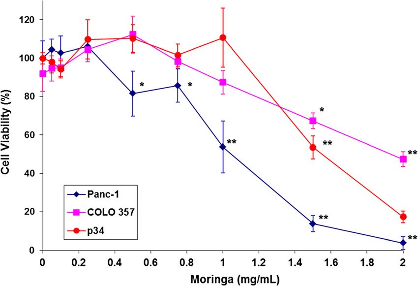
Figure 1 Effect of Moringa Oleifera leaf extract on the viability of Panc-1, COLO 357 and p34 pancreatic cancer cells. The cells were
plated in 96-well plates and treated in triplicates with Moringa Oleifera leaf extract for 3 days. Cell survival was assessed using XTT-based cell
proliferation assay, and is indicated after normalization to the control group. The significance of the effect is indicated as *p ≤ 0.05 and
**p ≤ 0.001.
three tested cell lines. Panc-1 cells were more susceptible reduced or eliminated more significantly the presence of
to the treatment (IC50 = 1.1 mg/ml) compared to COLO all three proteins of the NF-κB signaling pathway. More-
357 (IC50 =1.8 mg/ml) and p34 cells (IC50 = 1.5 mg/ml). over, p65 protein subunit levels have decreased in Panc-1
There was a significant inhibition of Panc-1 cell survival cells nuclei as a result of treatments with 0.1 - 1.5 mg/mL
at an extract concentration of 0.75 mg/mL. There was also Moringa extract. These data suggest that Moringa extract
a significant inhibitory effect at a higher concentration attenuated pancreatic cancer cell’s survival ability, at least
(1.5 mg/mL) in the two other cell lines. Moreover, treat- in part, by targeting the NF-κB signaling pathway.
ment with 2 mg/mL Moringa extract resulted in a 98%
reduction of Panc-1 cell survival. Combined effect of Moringa Oleifera leaf extract and
We also evaluated the distribution of Panc-1 cells in the cisplatin
cell cycle following 24 hours exposure to Moringa Oleifera Based on the ability of Moringa extract to inhibit the
leaf extract using flow cytometry analysis of propidium NF-κB signaling pathway, we hypothesized that its ex-
iodide-stained cells. It revealed a dose-dependent signifi- tract treatment would sensitize pancreatic cancer cells to
cant increase of the percentage of cells in the sub-G1 chemotherapy. We therefore tested the cytotoxic effect
phase, characterized by a very low DNA content, following of several combinations of Moringa Oleifera leaf extract
Moringa extract treatment (Figure 2A&B). This indicated a with cisplatin on Panc-1 cells. Since a synergistic effect
fragmentation of the DNA as a result of progressive cell was expected, both the agents were used in the concen-
apoptosis. Importantly, Moringa extract treatment also in- trations of a low inhibitory effect on proliferation of
duced significant (p < 0.05) apoptosis in Panc-1 cancer cells Panc-1 cells. As shown in Figure 4, all the tested combi-
at the minimal evaluated concentration of 0.25 mg/mL. nations demonstrated an inhibitory effect higher than
The induction of apoptosis was especially high (up to 30%) the effects of each agent alone. The analysis of these data
at a concentration of 0.75 mg/mL. by Calcusyn software, presented in Figure 4 and summa-
rized in the Table 1, clearly demonstrated that the com-
Effect of Moringa Oleifera leaf extract on NF-κB signaling bined Moringa Oleifera leaf extract and cisplatin
pathway in pancreatic cancer cells regimen resulted in strong synergistic (CI = 0.1–0.3) or
Moringa Oleifera leaf extract treatment of Panc-1 cells synergistic (CI = 0.3–0.7) effects.
down-regulated the expression of key NF-κB signaling
pathway proteins (Figure 3). The treatment of the cells Discussion
with 0.25 mg/mL extract for 24 hours resulted in a The results of this study show that Moringa Oleifera leaf
down-regulation of p65, phospho-IκBα and IκBα pro- extract can significantly inhibit the growth of cultured
teins levels compared to untreated cells (Figure 3A). human pancreatic carcinoma cells as well as its cell-
Treatments with 0.75 and 1.5 mg/mL Moringa extract cycle progression, in a concentration-dependent manner.Berkovich et al. BMC Complementary and Alternative Medicine 2013, 13:212 Page 5 of 7 http://www.biomedcentral.com/1472-6882/13/212 Figure 2 Effect of Moringa Oleifera leaf extract on the distribution of Panc-1 cells in the cell cycle. (A) Representative histograms of cell distribution following Moringa Oleifera leaf extract treatments. (B) Data summarized from three independent experiments (%). The cells were treated with the extract for 24 hours, collected, washed and fixed in 70% ice-cold ethanol. Prior to the analysis, the cells were suspended in 0.1% Triton-X with 30 mg/ml DNAse-free RNAse A for 6 hrs. Propidium iodide was added several minutes before cell analysis by flow cytometry. The significance of the effect, relatively to non-treated cells (control), is indicated as *p ≤ 0.05 and **p ≤ 0.001. Notably, the reduction of Panc-1 cells viability reached of apoptosis and thus generation of cell death [8]. In- 100% following exposure to 2 mg/mL of Moringa extract hibition of the NF-κB signaling cascade by Moringa ex- (Figure 1). To the best of our knowledge, this is the first tract explains, at least in part, its attenuating effect on time that Moringa Oleifera was shown to have an effect the survival of pancreatic cancer cells, as observed on on pancreatic carcinoma cells. The exposure of Panc-1 the viability assay (Figure 1). cells to Moringa Oleifera leaf extract also reduced the Several lines of evidence indicate that NF-κB plays a sig- overall expression of key NF-κB family proteins in the nificant role in the resistance of pancreatic cancer to cells, as well as the levels of p65 protein subunit in the cell apoptosis-based chemotherapies. Therefore, the NF-κB nuclei (Figure 3). Consequently, this extract could inhibit signaling pathway was suggested as a potential molecular the NF-κB signaling cascade execution of target gene target for combined therapy of pancreatic cancer [8,16]. transcription. Active NF-κB signaling was shown to Cisplatin is a platinum-based chemotherapeutic agent that strengthen the pancreatic cancer cell’s ability to sur- is known to have a minimal clinical efficacy in pancreatic vive, and that suppression of NF-κB leads to induction cancer due to tumor chemoresistance mechanisms [4]. It Figure 3 Effect of Moringa Oleifera leaf extract on the expression of p65 NF-kB, IkB and p-IkB proteins in Panc-1. (A) Protein expressions in crude lysates of the cells treated with Moringa Oleifera leaf extract (0–1.5 mg/mL) for 24 hours. (B) Expression of p65 in nuclear extracts of the cells treated with Moringa Oleifera leaf extract. Protein expression was analyzed by Western blot. GAPDH and ß-actin were used to demonstrate the quantity of standard proteins in the samples tested.
Berkovich et al. BMC Complementary and Alternative Medicine 2013, 13:212                                                                      Page 6 of 7
http://www.biomedcentral.com/1472-6882/13/212

                                                                          Table 1 Calculated Combination Index (CI) of the effect of
                                                                          combined treatments with Moringa Oleifera leaf extract
                                                                          and cisplatin on Panc-1 cells viability
                                                                          Combination index (CI)             Cisplatin (μM)           Moringa (mg/mL)
                                                                                    0.348                           0.3                       0.25
                                                                                    0.517                           0.3                       0.50
                                                                                    0.652                           0.3                       0.75
                                                                                    0.218                           1.0                       0.25
                                                                                    0.379                           1.0                       0.50
                                                                                    0.539                           1.0                       0.75
                                                                                    0.156                           3.0                       0.25
                                                                                    0.375                           3.0                       0.50
                                                                                    0.504                           3.0                       0.75
                                                                          The cells were treated as described in Figure 4. Cell viability was evaluated
                                                                          using XTT-based cell proliferation assay. Data was analyzed using
                                                                          Calcusyn software.

                                                                          that the effect of Moringa Oleifera leaf extract on the in-
                                                                          hibition of the NF-κB pathway may increase the efficacy of
                                                                          cisplatin. As shown in Figure 4 and summarized in Table 1,
                                                                          combined therapy of Moringa Oleifera leaf extract with cis-
                                                                          platin demonstrated strong (CI = 0.1–0.3) or moderate
                                                                          (CI = 0.3–0.7) synergistic effects in Panc-1 cells, re-
                                                                          spectively. This indicates that the treatment of pancre-
                                                                          atic cancer with cisplatin in combination with Moringa
                                                                          Oleifera leaf extract may be a clinical therapeutic op-
                                                                          tion for this chemoresistant cancer.
                                                                            The past decade has witnessed the establishment of
                                                                          herbal medicine as a rich source of new "western medicine"
                                                                          drugs for multiple conditions, including cancer. These
                                                                          herbs are often traditionally consumed by particular popu-
                                                                          lations, presenting an advantage in terms of laying rest to
                                                                          toxicity concerns. Herbs and their bioactive metabolites
                                                                          have been reported to be anti-neoplastic both in ex-
                                                                          perimental and clinical studies [19,20]. Moringa Oleifera
                                                                          leaves contain flavonoid pigments, such as kaempferol,
                                                                          rhamnetin, isoquercitrin and kaempferitrin. Flavonoid com-
                                                                          pounds have various biological activities, including anti-
 Figure 4 Combined treatments effect of Moringa Oleifera leaf
                                                                          inflammatory and anti-cancer ones [21], and may be
 extract with cisplatin on the viability of Panc-1 cells. (A) Cell        mediating, at least in part, the effects shown here. In addition,
 viability following treatments of Moringa Oleifera leaf extract,         the Moringa Oleifera leaves are rich in a group of the glyco-
 cisplatin and combined treatments of increasing concentrations of        side compounds, glucosinolates and isothiocyanates [10], as
 the extract and cisplatin for 3 days. (B) Normalized isobologram of      well as in beta-sitosterol, glycerol-1-(9-octadecanoate),
 Moringa Oleifera leaf extract with cisplatin treatments shown in
 compound A. (C) Median-effect plot curve of Moringa Oleifera leaf
                                                                          3-O-(6'-O-oleoyl-beta-D-glucopyranosyl), beta-sitosterol and
 extract, cisplatin and Moringa Oleifera leaf extract with cisplatin      beta-sitosterol-3-O-beta-D-glucopyranoside, all of which
 shown in compound A. The cells were treated with each agent              have demonstrated anti-cancer properties in-vitro [11] and
 alone or in combination for 3 days. Cell viability was evaluated using   may have also contributed to the effects we exhibit. Since
 XTT-based cell proliferation assay. Data was analyzed by                 this work does not include the isolation of bioactive
 Calcusyn software.
                                                                          compounds from the Moringa Oleifera leaf extract, we
                                                                          are not able to propose one or more specific active
has been shown in other human cancer models that in-                      compounds to be mediating the cellular anti-cancer ef-
creased efficacy of cisplatin may be achieved by NF-κB in-                fects demonstrated in our results. Moreover, it is pos-
hibitors in cancer cells [17,18]. Therefore, we hypothesized              sible that our findings can be attributed to additive or
Berkovich et al. BMC Complementary and Alternative Medicine 2013, 13:212 Page 7 of 7
http://www.biomedcentral.com/1472-6882/13/212
synergistic effects of several bioactive compounds from 7. Van Waes C: Nuclear factor-kappaB in development, prevention, and
the extract and not to a single one. Further research on therapy of cancer. Clin Cancer Res 2007, 13:1076–1082.
8. Holcomb B, Yip-Schneider M, Schmidt CM: The role of nuclear factor
anti-cancer effects of specific bioactive metabolites derived kappaB in pancreatic cancer and the clinical applications of targeted
from Moringa Oleifere is warranted. therapy. Pancreas 2008, 36:225–235.
Toxicity has been a limiting factor in clinical pancre- 9. Shih MC, Chang CM, Kang SM, Tsai ML: Effect of different parts (leaf, stem
and stalk) and seasons (summer and winter) on the chemical
atic cancer treatment: combined therapy with cisplatin compositions and antioxidant activity of Moringa Oleifera. Int J Mol Sci
and gemcitabine has shown promising results in phase II 2011, 12:6077–6088.
and III trials, but it was followed by severe toxicity and 10. Bose CK: Possible role of Moringa Oleifera Lam. root in epithelial ovarian
cancer. Med Gen Med 2007, 9:26.
therefore has not been approved as a standard of care 11. Guevara AP, Vargas C, Sakurai H, Fujiwara Y, Hashimoto K, et al: An anti-tumor
[4]. This should not be the case regarding Moringa promoter from Moringa Oleifera Lam. Mutat Res 1999, 440:181–188.
Oleifera Lam, since an acute toxicity test carried out 12. Sreelatha S, Jeyachitra A, Padma PR: Antiproliferation and induction of
apoptosis by Moringa Oleifera leaf extract on human cancer cells.
using male Wistar albino mice, estimated the LD(50) of Food Chem Toxicol 2011, 49:1270–1275.
orally administered aqueous leaf extract of Moringa 13. Parvathy MVS, Umamaheshwari A: Cytotoxic effect of Moringa Oleifera leaf
oleifera Lam. to be around 1585 mg/kg for a single ad- extracts on human multiple myeloma cultured cell lines. Trends in
Medical Research 2 2007, 2(1):44–50.
ministration [22]. The aqueous leaf extract of Moringa 14. Lev-Ari S, Vexler A, Starr A, Ashkenazy-Voghera M, Greif J, Aderka D, Ben-
oleifera is, therefore, considered relatively safe when ad- Yosef R: Curcumin augments gemcitabine cytotoxic effect on pancreatic
ministered orally and its acquisition of permission for adenocarcinoma cell lines. Cancer Invest 2007, 25:411–418.
15. Chou T-C, Talalay P: Analysis of combined drug effect: a new look at a
clinical use highly probable. very old problem. Trends Pharmacol Sci 1983, 4:150–154.
Still, future studies need to be conducted to evaluate 16. Sclabas GM, Fujioka S, Schmidt C, Fan Z, Evans DB, et al: Restoring
and quantify the therapeutic index of Moringa Oleifera apoptosis in pancreatic cancer cells by targeting the nuclear factor-kB
signaling pathway with the anti-epidermal growth factor antibody IMC-
leaf extract applicable to pancreatic cancer patients in C225. J Gastrointest Surg 2003, 7:37–43.
the clinical setting. 17. Mabuchi S, Ohmichi M, Nishio Y, Hayasaka T, Kimura A, et al: Inhibition of
NF-kappaB increases the efficacy of cis-platin in in vitro and in vivo
ovarian cancer models. J Biol Chem 2004, 279:23477–23485.
Conclusions 18. Venkatraman M, Anto RJ, Nair A, Varghese M, Karunagaran D: Biological and
Moringa Oleifera leaf extract inhibits the NF-κB signal- chemical inhibitors of NF-kappaB sensitize SiHa cells to cisplatin-induced
ing pathway and increases the efficacy of chemotherapy apoptosis. Mol Carcinog 2005, 44:51–59.
19. Wu M, Yao B: Advances in TCM treatment of gastric cancer and studies
in human pancreatic cancer cells. on the apoptosis. J Tradit Chin Med 2002, 22:303–307.
20. Engdal S, Klepp O, Nilsen OG: Identification and exploration of herb-drug
Abbreviations combinations used by cancer patients. Integr Cancer Ther 2009, 8:29–36.
NF-κB: Nuclear factor kappa-B; XTT: 2,3-bis-(2-methoxy-4-nitro-5-sulfophenyl)- 21. Lee ER, Kang GH, Cho SG: Effect of flavonoids on human health: old
2H-tetrazolium-5-carboxanilide; CI: Combination Index; PBS: Phosphate- subjects but new challenges. Recent Pat Biotechnol 2007, 1(2):139–150.
buffered solution; GAPDH: Glyceraldehyde 3-phosphate dehydrogenase; 22. Awodele O, Oreagba IA, Odoma S, da Silva JA, Osunkalu VO: Toxicological
OD: Optical density. evaluation of the aqueous leaf extract of Moringa oleifera Lam.
(Moringaceae). J Ethnopharmacol 2012, 139(2):330–336.
Competing interest
The authors declare that they have no competing interests.
doi:10.1186/1472-6882-13-212
Cite this article as: Berkovich et al.: Moringa Oleifera aqueous leaf
Authors’ contributions extract down-regulates nuclear factor-kappaB and increases
LB planned the study’s experiments, conducted the cell culture experiments, cytotoxic effect of chemotherapy in pancreatic cancer cells. BMC
analyzed the signaling assays and prepared the manuscript. SLA, AR and AV Complementary and Alternative Medicine 2013 13:212.
performed the statistical analysis and data calculations. GE and IR assisted
with the manuscript. SLA conceived the study and gave final approval of the
manuscript. All authors read and approved the final manuscript.
Financial support
This study was supported by the Chaya and Kadish Shermeister Endowment.
Received: 21 February 2013 Accepted: 8 August 2013
Published: 19 August 2013
Submit your next manuscript to BioMed Central
References and take full advantage of:
1. Newman DJ, Cragg GM, Snader KM: Natural products as sources of new
drugs over the period 1981–2002. J Nat Prod 2003, 66:1022–1037.
• Convenient online submission
2. Hariharan D, Saied A, Kocher HM: Analysis of mortality rates for pancreatic
cancer across the world. HPB (Oxford) 2008, 10:58–62. • Thorough peer review
3. Oberstein PE, Wasif Saif M: First-line treatment for advanced pancreatic • No space constraints or color figure charges
cancer. J Pancreas (Online) 2011, 12:96–100.
• Immediate publication on acceptance
4. Greenhalf W, Thomas A: Combination therapy for the treatment of
pancreatic cancer. Anticancer Agents Med Chem 2011, 11:418–426. • Inclusion in PubMed, CAS, Scopus and Google Scholar
5. Karin M, Lin A: NF-kappaB at the crossroads of life and death. Nat • Research which is freely available for redistribution
Immunol 2002, 3:221–227.
6. Aggarwal BB: Nuclear factor-kappaB: the enemy within. Cancer Cell 2004,
6:203–208. Submit your manuscript at
www.biomedcentral.com/submitYou can also read