Diclofenac impairs the proliferation and glucose metabolism of triple negative breast cancer cells by targeting the c Myc pathway
←
→
Page content transcription
If your browser does not render page correctly, please read the page content below
EXPERIMENTAL AND THERAPEUTIC MEDICINE 21: 584, 2021

  Diclofenac impairs the proliferation and glucose metabolism of
triple‑negative breast cancer cells by targeting the c‑Myc pathway
                    LIHUI YANG1*, JIACHEN LI2*, YONGZHUO LI3*, YONGLI ZHOU2, ZIQIAN WANG2,
                               DAHAO ZHANG2, JINLU LIU4 and XIAODONG ZHANG4

             1
            Department of Nursing, Guangxi Medical University Nursing College; 2Department of Clinical Medicine,
       Guangxi Medical University The First Clinical Medical College, Nanning, Guangxi 530021; 3Department of Medicine
       Guangxi University Medical College, Nanning, Guangxi 530004; 4Department of Gastrointestinal and Gland Surgery,
               The First Affiliated Hospital of Guangxi Medical University, Nanning, Guangxi 530021, P.R. China

                                      Received June 17, 2020; Accepted February 26, 2021

                                                  DOI: 10.3892/etm.2021.10016

Abstract. Triple‑negative breast cancer (TNBC) cells obtain         GLUT1 protein expression and HK activity through the c‑Myc
energy mainly through aerobic glycolysis, and their glycolytic      pathway.
rate is significantly higher compared with that of non‑TNBC
cells. Glucose transporter 1 (GLUT1) is a transmembrane             Introduction
transporter necessary for the entry of glucose into tumor
cells, hexokinase (HK) is a key enzyme in the glycolytic            Breast cancer is one of the most common malignant tumours
pathway, and both are targets of the transcription factor           in female patients, with an annually increasing trend. In addi‑
c‑Myc. c‑Myc can promote aerobic glycolysis by upregulating         tion, breast cancer is a heterogeneous disease with multiple
GLUT1 expression and enhancing HK activity. c‑Myc and               subtypes; triple‑negative breast cancer (TNBC) accounts for
GLUT1 are highly expressed in TNBC. The non‑steroidal               15‑20% of cases, worldwide (1). The typical characteristics
anti‑inflammatory drug diclofenac can inhibit glycolysis in         of TNBC are the absence of oestrogen receptor (ER), proges‑
melanoma cells and thereby promote apoptosis by downregu‑           terone receptor and human epidermal growth factor receptor
lating c‑Myc and GLUT1. To explore the effect of diclofenac         2 expression (1). TNBC is highly invasive, has a poor clinical
on the energy metabolism of TNBC cells and determine the            prognosis and rapidly recurs (2,3). Consequently, it is neces‑
underlying mechanism, a comparative study in two TNBC cell          sary to further explore the biological characteristics of TNBC
lines (MDA‑MB‑231 and HCC1937) and one non‑TNBC cell                and then identify novel, effective and safe antitumor drugs to
line (MCF‑7) was conducted. Cell proliferation was detected         improve the survival rate of patients.
by Cell Counting Kit‑8 (CCK‑8) and flow cytometric assays;              At present, the treatment for TNBC is still radiotherapy
GLUT1 and c‑Myc expression was measured by western                  and chemotherapy, which is due to the lack of reliable specific
blotting. Diclofenac significantly inhibited cell proliferation,    targets to develop targeted drugs (2,4). The reprogramming of
downregulated GLUT1 and c‑Myc expression, and decreased             energy metabolism can be used as a sign of the physiology
HK activity in TNBC cells compared with non‑TNBC cells. In          of several cancer types, including TNBC (5). In normal cells,
conclusion, the studies suggested that diclofenac inhibited cell    most pyruvate enters mitochondria and is oxidized through the
glycolysis and suppressed TNBC cell growth by decreasing            tricarboxylic acid cycle to produce adenosine triphosphate and
                                                                    meet the energy needs of the cell. However, in cancer cells,
                                                                    most pyruvate is reduced to lactic acid by lactate dehydroge‑
                                                                    nase rather than entering the mitochondria. This process is
                                                                    called ‘aerobic glycolysis’ and is referred to as the ‘Warburg
                                                                    effect’ (6). Like most cancer cells, breast cancer cells also have
Correspondence to: Mr. Xiaodong Zhang or Mr. Jinlu Liu,             abnormal glucose metabolism with high glucose absorption
Department of Gastrointestinal and Gland Surgery, The First
                                                                    and glycolysis rates (7).
Affiliated Hospital of Guangxi Medical University, 6 ShuangYong
                                                                        Glucose transporters (GLUTs) are transmembrane
Road, Nanning, Guangxi 530021, P.R. China
E‑mail: 15177788067@163.com                                         transporters that are necessary for the entry of glucose into
E‑mail: hanjiangsnow@163.com                                        cells (8). Fourteen types of GLUTs are expressed in humans.
                                                                    GLUT1, GLUT2, GLUT3, GLUT4, GLUT5 and GLUT12
*
    Contributed equally                                             have been successively identified in breast cancer (9‑12). As
                                                                    the most invasive breast cancer subtype, TNBC exhibits higher
Key words: diclofenac, triple‑negative breast cancer, glycolysis,   levels of GLUT1 compared with other subtypes of breast
c‑Myc, GLUT1                                                        cancer; however, diclofenac can significantly decrease GLUT1
                                                                    expression and glucose uptake (13). In addition, c‑Myc is a
                                                                    driving factor of glucose uptake and aerobic glycolysis (14,15).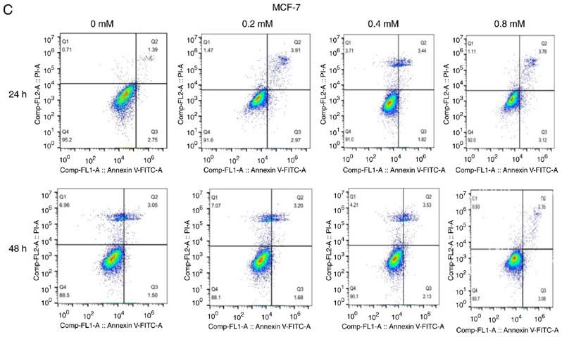
2                          YANG et al: DICLOFENAC IMPAIRS TNBC CELLS TARGETING C-MYC PATHWAY

The most recent research shows that diclofenac can control         addition of CCK‑8 (Beyotime Institute of Biotechnology) at
glycolysis in melanoma cells by inhibiting c‑Myc, down‑            5 time points. After brief shaking, the absorbance values at
regulating the expression of GLUT1 and suppressing glucose         450 nm were immediately measured using a microplate reader.
metabolism (16). In addition, hexokinase (HK) participates
in the first step of glycolysis as the key rate‑limiting enzyme.   Assessment of apoptosis. The three cell lines were treated with
Therefore, the decrease in HK expression and activity can          0, 0.2, 0.4 and 0.8 mM diclofenac for 24 and 48 h and were then
inhibit glycolysis. Studies have shown that HK is also a target    stained with 5 µl of Annexin‑V‑Fluorescein Isothiocyanate
of c‑Myc (17,18).                                                  (Annexin‑V‑FITC) and 10 µl of Propidium Iodide (PI) (both
    Recently, a team found that compared with other types          obtained from BD Biosciences) according to the manufac‑
of breast cancer cells, TNBC cells have a unique molecular         turer's instructions. The number of sample cells was 1x10⁵.
mechanism; the high levels of c‑Myc and low levels of              Before the assay, three groups of negative control samples,
TXNIP in TNBC can promote cancer cell proliferation. High          including blank, Annexin V‑FITC‑stained and PI‑stained,
c‑Myc/low TXNIP gene expression is associated with a lower         were analysed; this was repeated 3 times. Flow cytometric
overall survival and metastasis‑free survival rates of patients    analyses were performed with a Fluorescence activated Cell
with TNBC. Furthermore, c‑Myc and TXNIP can compete                Sorting (FACS) Calibur (BD Biosciences) using BD Cell
with each other; c‑Myc promotes glucose uptake and its use         Quest Pro 5.1 software for data acquisition and analysis.
in tumor cells to maintain proliferation, while TXNIP does
the opposite (2). Based on these results, diclofenac is expected   Measurement of HK activity. Cells in logarithmic growth
to inhibit c‑Myc transcription and downregulate GLUT1              phase were inoculated in 6‑well plates at 2.5x10⁶ cells/well and
expression, subsequently suppressing glycolysis and inducing       were then incubated overnight with diclofenac (0, 0.2, 0.4 and
apoptosis in TNBC cells.                                           0.8 mM) at 37˚C in 5% CO2. For the detection of HK activity,
    To verify this hypothesis, the present study investigated      each group was cultured for both 24 and 48 h. Detection was
the effects of diclofenac on aerobic glycolysis in TNBC            performed with a Hexokinase Activity Detection kit (Beijing
by establishing cell models with human TNBC cell lines             Solarbio Science & Technology Co., Ltd.) according to the
(MDA‑MB‑231 and HCC1937) and a non‑TNBC cell line                  manufacturer's instructions. The reagents were mixed in
(MCF‑7). After the cells were treated with diclofenac              proportional amounts into the solution and preheated at 37˚C
for 24 and 48 h, the effects of diclofenac were evaluated,         for 10 min. The absorbance values at 340 nm at 20 sec (A1)
including alterations in cell proliferation and apoptosis, using   after sample addition and 5 min after water bath immersion
Cell Counting Kit‑8 (CCK‑8) and flow cytometric assays.            (A2) were measured in a spectrophotometer (Thermo Fisher
In addition, the expression levels of GLUT1 and c‑Myc              Scientific, Inc.). These data were input into the formula
were analysed by western blotting to further elucidate the         HK(U/10 4 cell)=[Δ AxV total/(εxd)x109]÷(500xV sample/V
underlying mechanism by which diclofenac inhibits TNBC             sample total)/T=2.572xΔA (ΔA=A2‑A1; V Total, total volume
cell proliferation and induces TNBC cell apoptosis.                of reaction system, 2x10 ‑4 l; ε, NADPH molar extinction
                                                                   coefficient, 6.22x10³ l/mol/cm; d, 96‑well plate optical path,
Materials and methods                                              0.5 cm; V sample, sample volume 0.01 ml; V sample total,
                                                                   extract volume 1 ml; T, reaction time 5 min) to calculate HK
Cells and cell culture. TNBC cell lines (MDA‑MB‑231 and            activity. U is defined as 1 nmol of NADPH produced per
HCC1937) and a non‑TNBC cell line (MCF‑7) were purchased           minute per 10,000 bacteria or cells, and is considered to be an
from the The Cell Bank of Type Culture Collection of The           enzyme activity unit.
Chinese Academy of Sciences (Shanghai, China). The cryovials
containing the frozen cells were removed from liquid nitrogen      Western blot analysis. The three cell lines were treated with
storage and immediately placed into a 37˚C water bath. Then,       0, 0.4 and 0.8 mM diclofenac for 24 and 48 h, washed twice
complete growth medium, consisting of DMEM (Thermo                 with cold phosphate buffer saline (PBS) after collection, and
Fisher Scientific, Inc.) supplemented with 10% (v/v) fetal         lysed with radio immunoprecipitation assay (RIPA) buffer
bovine serum (Biological Industries Israel Beit Haemek Ltd.)       (Beyotime Institute of Biotechnology). Then, the lysates were
and 1% (v/v) penicillin‑streptomycin (Beijing Solarbio Science     centrifuged at 12,000 x g and 4˚C for 30 min. The supernatant
& Technology Co., Ltd.), was added to resuspend the cells, and     was collected, and sodium dodecyl sulphate‑polyacrylamide
the suspension was centrifuged at ~300 x g for 5‑10 min at         gel electrophoresis (SDS‑PAGE) sample loading buffer
37˚C. The supernatant was decanted, and the cells were gently      (Beyotime Institute of Biotechnology) was added at a
resuspended in 5 ml of complete growth medium. Then, the           buffer:lysate ratio of 1:4, and proteins were denatured in a
cells were transferred into a culture flask and incubated at       thermal cycler at 100˚C for 10 min. The protein concentration
37˚C in 5% CO2.                                                    was determined using a BCA Protein Assay kit (cat. no. 23227;
                                                                   Thermo Fisher Scientific, Inc.). The proteins (30 µg per lane)
Assessment of cell proliferation. The concentration of the three   were separated on a denaturing 12% SDS‑PAGE gel and
cell suspensions was adjusted to 5x10⁴ cells/ml, and 100 µl of     transferred to a polyvinylidene fluoride (PVDF) membrane
each suspension was added to 96‑well plates and cultured in an     for western blotting. The membrane was sequentially probed
incubator at 37˚C and 5% CO2. Cells were treated with diclof‑      with antibodies against GLUT1 (1:1,000; cat. no. 12939; Cell
enac (Sigma‑Aldrich; Merck KGaA) at the concentrations of          Signaling Technologies, Inc.), MYC (1:1,000; cat. no. 9402;
0, 0.2, 0.4, 0.8 mM for 24, 48, 72, 96, 120 h. Then, cells were    Cell Signaling Technologies, Inc.), and β ‑actin (1:1,000;
incubated at 37˚C and 5% CO2 for another 2 h, following the        cat. no. 3700; Cell Signaling Technologies, Inc.) diluted by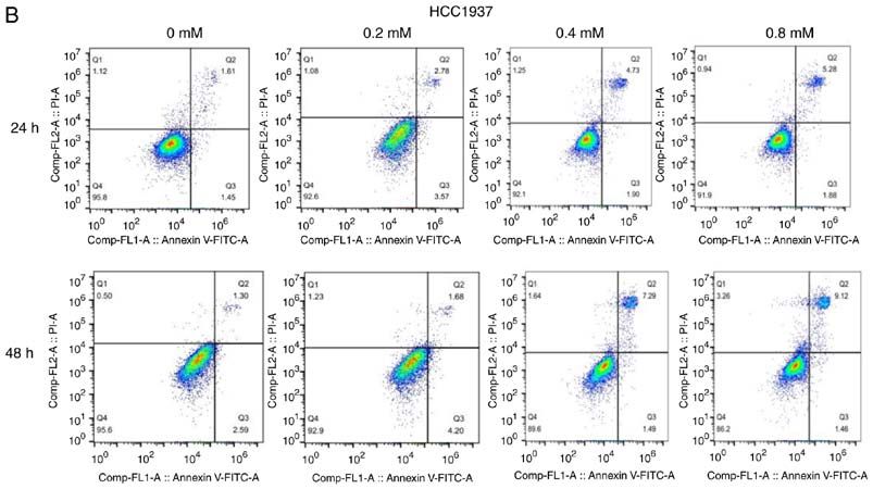
EXPERIMENTAL AND THERAPEUTIC MEDICINE 21: 584, 2021                                            3

Primary Antibody Dilution buffer (Beyotime Institute of             downregulated in a dose‑dependent manner in the TNBC
Biotechnology). After incubation with the primary antibodies,       cell lines (MDA‑MB‑231 and HCC1937). HK activity was
the membrane was washed 3 times for 5 min by tris‑buffered          most significantly downregulated under treatments with 0.4
saline with 0.1% tween20 (TBST) and was then incubated              and 0.8 mM diclofenac. However, no difference was observed
with the secondary antibody (1:15,000; IRDye® 800CW Goat            in HK activity in the non‑TNBC cell line (MCF‑7) groups
anti‑Rabbit IgG Secondary Antibody cat. no. 926‑32211;              compared with the corresponding control groups.
IRDye® 800CW Goat anti‑Mouse IgG Secondary Antibody
cat. no. 926‑32210; LI‑COR Biosciences) for 2 h. Protein            Effect of diclofenac on the protein expression levels of
bands were visualized using Odyssey Infrared Imaging                c‑Myc and GLUT1. To study the effect of diclofenac on the
System (LI‑COR Biosciences). Finally, the greyscale values of       relative expression levels of GLUT1 and c‑Myc, TNBC cells
the protein bands were determined by Image Studio Lite 5.2.5        (MDA‑MB‑231 and HCC1937) and non‑TNBC cells (MCF‑7)
(LI‑COR Biosciences).                                               were exposed to diclofenac (0, 0.4 and 0.8 mM) for 24 and
                                                                    48 h, and the protein expression levels of GLUT1 and c‑Myc
Statistical analysis. All results are presented as the              were measured by western blotting. As shown in Fig. 4, after
means ± standard deviations and were analysed with                  incubation with diclofenac for 24 and 48 h, the protein expres‑
SPSS 17.0 statistical software (SPSS, Inc.). Statistical analysis   sion levels of both c‑Myc and GLUT1 were decreased in a
was performed with one‑way ANOVA with post hoc contrasts            dose‑ and time‑dependent manner in the TNBC cell lines
by Bonferroni's test. P≤0.05 was considered to indicate a           (MDA‑MB‑231 and HCC1937). The protein expression levels
statistically significant difference. The experiments were          of c‑Myc and GLUT1 were most significantly decreased under
performed in triplicate.                                            treatment with 0.4 and 0.8 mM diclofenac. In the non‑TNBC
                                                                    cell line (MCF‑7) groups, although the protein expression levels
Results                                                             of c‑Myc and GLUT1 were decreased in a time‑dependent
                                                                    manner, the differences were not significant.
Diclofenac inhibits breast cancer cell proliferation in vitro.
The addition of diclofenac, a member of the arylacetic acid         Discussion
group of non‑steroidal anti‑inflammatory drugs (NSAIDs),
at clinically relevant concentrations (see http://www.drugs.        TNBC is a unique subtype of breast cancer with a 5‑year
com/pro/diclofenac.html) led to significant effects on TNBC         survival rate of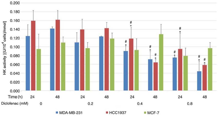
4                               YANG et al: DICLOFENAC IMPAIRS TNBC CELLS TARGETING C-MYC PATHWAY

Figure 1. In vitro effects of diclofenac on the proliferation of human breast cancer cell lines. The TNBC cell lines MDA‑MB‑231 (A) and HCC1937 (B) and
the non‑TNBC cell line MCF‑7 (C) were incubated with different concentrations (0.2, 0.4 and 0.8 mM) of diclofenac, and proliferation was assessed at 5 time
points. The results are presented as the mean ± SD of the results from 3 independent experiments. *P
EXPERIMENTAL AND THERAPEUTIC MEDICINE 21: 584, 2021   5

                  Figure 2. Continued.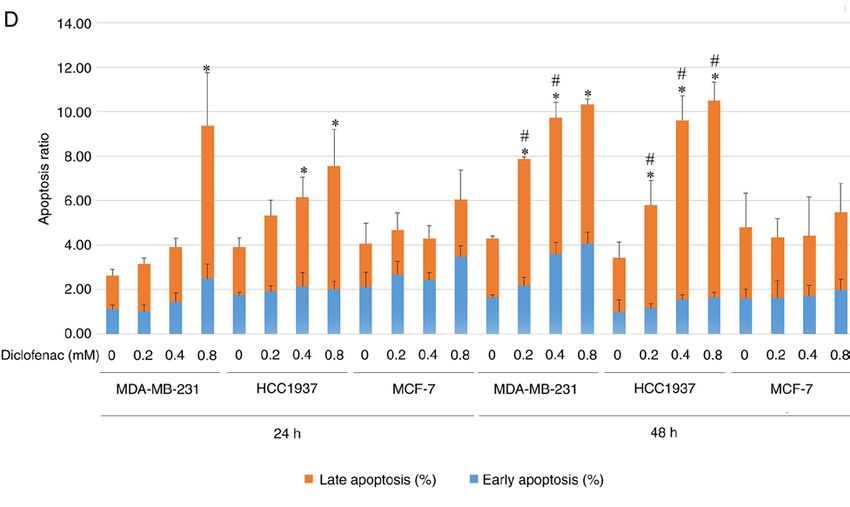
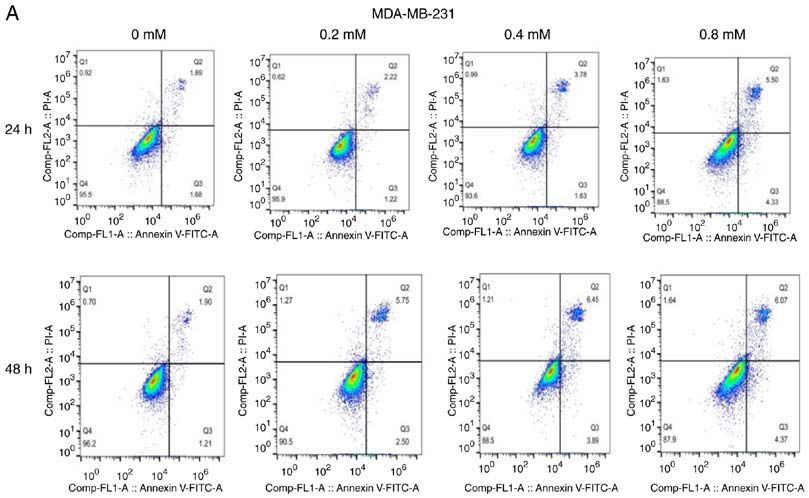
6                                YANG et al: DICLOFENAC IMPAIRS TNBC CELLS TARGETING C-MYC PATHWAY

Figure 2. Effect of different concentrations of diclofenac on the apoptosis of breast cancer cells. After incubation with diclofenac (0, 0.2, 0.4 and 0.8 mM) for
24 and 48 h, the percentage of apoptotic TNBC MDA‑MB‑231 (A) and HCC1937 (B) cells increased in a dose‑ and time‑dependent manner (D). However, the
non‑TNBC cell line MCF‑7 (C) did not exhibit this pattern. The data are presented as the means ± SD (n=3). *P
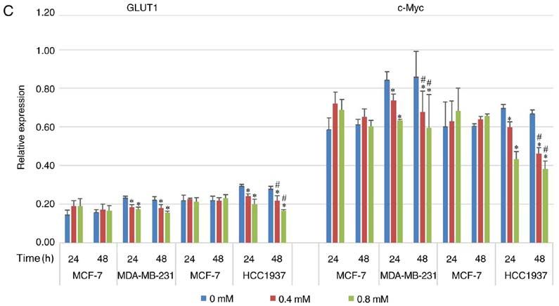
EXPERIMENTAL AND THERAPEUTIC MEDICINE 21: 584, 2021                                                           7

Figure 4. Effect of diclofenac on the relative protein expression levels of c‑Myc and GLUT1. (A and C) The human breast cancer cell lines MCF‑7 and
MDA‑MB‑231 and (B and C) MCF‑7 and HCC1937 were incubated with diclofenac (0, 0.4 and 0.8 mM) for 24 and 48 h, and the protein expression levels of
c‑Myc and GLUT1 were measured by western blotting. All data are presented as the mean ± SD of the results from 3 independent experiments. *P
8                          YANG et al: DICLOFENAC IMPAIRS TNBC CELLS TARGETING C-MYC PATHWAY

and that diclofenac downregulated GLUT1 protein expression.        metabolism. In recent years, it has been proposed as a target
Moreover, this downregulating effect was stronger in TNBC          for diabetes and cancer (39). Its inhibitors have been proved
cells compared with non‑TNBC cells. HK is a key rate‑limiting      to decrease the proliferation of breast cancer cells (40). Drugs
enzyme in the first step of glycolysis in tumor tissues, and its   have always been multi‑targeted. Diclofenac in the present
expression and activity increase significantly in tumor tissues    study has an impact on the glucose uptake and proliferation
to ensure a sufficient energy supply, even under anaerobic         of cancer cells. It also has the possibility of targeting other
conditions. The expression of HK is upregulated in most malig‑     glycolysis‑associated genes including SGLT2, but this requires
nant tumors because most malignant tumors prefer aerobic           more extensive research.
glycolysis. A recent study demonstrated that the expression of         In summary, the present study results provide evidence that
HK was higher in MDA‑MB‑231 cells compared with MCF‑7              diclofenac can decrease the protein expression of GLUT1 and
cells (27). The present study demonstrated that diclofenac can     HK activity in cancer cells by downregulating the expression
decrease the activity of HK and showed that the activity of        of c‑Myc, lowering glucose uptake, preventing the supply of
HK in tumor cells was decreased in a dose‑dependent manner         energy and inhibiting glycolysis, eventually inhibiting the
with increasing doses of diclofenac. However, this effect was      growth of TNBC cells. Changes in energy metabolism in
not seen in non‑TNBC cells. Therefore, it was concluded            tumours significantly affect tumour proliferation and metas‑
that diclofenac inhibits the proliferation of TNBC cells by        tasis. For the first time, the effect and potential mechanism
inhibiting glycolytic enzymes such as GLUT1 and HK.                of diclofenac in TNBC cells was explored, and the potential
    c‑Myc is an important member of the Myc family. The            value of glycolysis inhibitors in the treatment of TNBC
c‑Myc gene can promote cell division and acts as a ‘switch’        was proposed. The use of glycolysis inhibitors alone or in
that determines the entry of cells into S phase from G 0/G1        combination with chemotherapeutic drugs has been currently
phase. The protein encoded by c‑Myc is closely associated          proposed in the clinic. Further studies on the effects of drugs
with cell proliferation (28). The c‑Myc oncogene is deregulated    targeting c‑Myc can provide basic data on abnormal energy
in >50% of human cancer types, and this deregulation is            metabolism and new therapeutic targets for drugs inhibiting
frequently associated with poor prognosis and unfavourable         energy metabolism in TNBC.
patient survival outcomes (29). c‑Myc has a central role in
almost every aspect of the oncogenic process, orchestrating        Acknowledgements
proliferation, apoptosis, differentiation and metabolism (29).
As the core of the glycolytic metabolism of cancer cells,          Not applicable.
c‑Myc controls the metabolism of cancer cells through a
variety of ways. For example, in recent reports, AMPK factor       Funding
can regulate c‑Myc bidirectionally, AMPK pathway positively
regulates the expression of oncogene c‑Myc to promote cancer       This study was supported by the National Natural Science
cell apoptosis (30), and AMPK can also reversely regulate          Foundation of China (grant no. 8176100111) and Basic Ability
c‑Myc to promote cancer autophagy (31). Furthermore, AMPK          Enhancement Project of Young and Middle‑aged Teachers in
is considered to be a factor associated with glucose‑mediated      Guangxi Universities (grant no. 2019KY0145).
cancer progression (32‑34). Recent studies have shown that the
molecular mechanism of TNBC cells is unique compared with          Availability of data and materials
that of other types of breast cancer cells: TNBC cells have
higher expression of c‑Myc and lower expression of TXNIP,          The datasets used and/or analysed during the current study are
and c‑Myc can promote glucose uptake and its use in tumor          available from the corresponding author on reasonable request.
cells, which in turn accelerates cancer cell proliferation (2).
In addition, other studies have shown that several pathways        Authors' contributions
of cell metabolism are regulated by c‑Myc and that the key
enzymes in glucose metabolism, such as GLUT1 and HK,               LY and JiaL participated in the preliminary experimental
are targets of c‑Myc (35‑38). In the present study, TNBC           design, preliminary experiment, main experiment operation
cell lines (MDA‑MB‑231 and HCC1937) and a non‑TNBC                 including cell culture, western blotting and flow cytometry
cell line (MCF‑7) were treated with diclofenac at different        detection, data analysis, manuscript writing and revision. YL,
concentrations and measured c‑Myc protein expression in            YZ, ZW and DZ participated in data analysis, manuscript
these cells by western blotting. The protein expression level of   writing and revision. JinL and XZ participated in early experi‑
c‑Myc in TNBC cells was significantly higher compared with         mental design including selecting drugs and designing possible
that in non‑TNBC cells. Moreover, diclofenac decreased the         signal pathways. LY and JiaL confirm the authenticity of all the
protein expression level of c‑Myc, and this effect was stronger    raw data. All authors read and approved the final manuscript.
in TNBC cells compared with non‑TNBC cells.
    Glycolysis plays a central role in tumor metabolism and        Ethics approval and consent to participate
growth, and this is reflected in a high rate of glucose uptake.
But in fact, in addition to c‑Myc, GLUT1, and HK in the            Not applicable.
present study, there are various other factors that play a role
in tumour metabolism and growth, which is reflected by a           Patient consent for publication
high rate of glucose uptake effect. For example, SGLT2, which
is a sodium‑glucose cotransporter, is involved in glucose          Not applicable.
EXPERIMENTAL AND THERAPEUTIC MEDICINE 21: 584, 2021 9
Competing interests 21. Butt JH, Barthel JS, Hosokawa MC and Moore RA: NSAIDs: A
clinical approach to the problems of gastrointestinal side‑effects.
Aliment Pharmacol Ther 2 (Suppl 1): S121‑S129, 1988.
The authors declare that they have no competing interests. 22. Grosser T, Ricciotti E and FitzGerald GA: The cardiovascular
pharmacology of nonsteroidal anti‑inflammatory drugs. Trends
Pharmacol Sci 38: 733‑748, 2017.
References 23. Barron CC, Bilan PJ, Tsakiridis T and Tsiani E: Facilitative
glucose transporters: Implications for cancer detection, prognosis
1. Foulkes WD, Smith IE and Reis‑Filho JS: Triple‑negative breast and treatment. Metabolism 65: 124‑139, 2016.
cancer. N Engl J Med 363: 1938‑1948, 2010. 24. Zhou YX, Zhou KM, Liu Q, Wang H, Wang W, Shi Y and
2. Shen L, O'Shea JM, Kaadige MR, Cunha S, Wilde BR, Ma YQ: The effect of Glut1 and c‑myc on prognosis in esopha‑
Cohen AL, Welm AL and Ayer DE: Metabolic reprogramming geal squamous cell carcinoma of Kazakh and Han patients.
in triple‑negative breast cancer through Myc suppression of Future Oncol 14: 1801‑1815, 2018.
TXNIP. Proc Natl Acad Sci USA 112: 5425‑5430, 2015. 25. Carvalho KC, Cunha IW, Rocha RM, Ayala FR, Cajaíba MM,
3. Luo C, Wang Y, Wei C, Chen Y and Ji Z: The anti‑migration and Begnami MD, Vilela RS, Paiva GR, Andrade RG and Soares FA:
anti‑invasion effects of Bruceine D in human triple‑negative breast GLUT1 expression in malignant tumors and its use as an
cancer MDA‑MB‑231 cells. Exp Ther Med 19: 273‑279, 2020. immunodiagnostic marker. Clinics (Sao Paulo) 66: 965‑972, 2011.
4. Yin L, Qi XW, Liu XZ, Yang ZY, Cai RL, Cui HJ, Chen L and 26. Vander Heiden MG, Cantley LC and Thompson CB:
Yu SC: Icaritin enhances the efficacy of cetuximab against Understanding the Warburg effect: The metabolic requirements
triple‑negative breast cancer cells. Oncol Lett 19: 3950‑3958, 2020. of cell proliferation. Science 324: 1029‑1033, 2009.
5. Li C, Li X, Li G, Sun L, Zhang W, Jiang J and Ge Q: Identification 27. Geng C, Li J, Ding F, Wu G, Yang Q, Sun Y, Zhang Z, Dong T and
of a prognosis‑associated signature associated with energy Tian X: Curcumin suppresses 4‑hydroxytamoxifen resistance in
metabolism in triple‑negative breast cancer. Oncol Rep 44: breast cancer cells by targeting SLUG/Hexokinase 2 pathway.
819‑837, 2020. Biochem Biophys Res Commun 473: 147‑153, 2016.
6. Warburg O: On the origin of cancer cells. Science 123: 309‑314, 1956. 28. Chauhan A, Paul R, Debnath M, Bessi I, Mandal S, Schwalbe H
7. Long JP, Li XN and Zhang F: Targeting metabolism in breast and Dash J: Synthesis of fluorescent binaphthyl amines that bind
cancer: How far we can go? World J Clin Oncol 7: 122‑130, 2016. c‑MYC G‑quadruplex DNA and repress c‑MYC expression.
8. Deng D and Yan N: GLUT, SGLT, and SWEET: Structural and J Med Chem 59: 7275‑7281, 2016.
mechanistic investigations of the glucose transporters. Protein 29. Chen H, Liu H and Qing G: Targeting oncogenic Myc as a strategy
Sci 25: 546‑558, 2016. for cancer treatment. Signal Transduct Target Ther 3: 5, 2018.
9. Godoy A, Ulloa V, Rodríguez F, Reinicke K, Yañez AJ, García 30. Kfoury A, Armaro M, Collodet C, Sordet‑Dessimoz J, Giner MP,
Mde L, Medina RA, Carrasco M, Barberis S, Castro T, et al: Christen S, Moco S, Leleu M, de Leval L, Koch U, et al:
Differential subcellular distribution of glucose transporters AMPK promotes survival of c‑Myc‑positive melanoma cells by
GLUT1‑6 and GLUT9 in human cancer: Ultrastructural local‑ suppressing oxidative stress. EMBO J 37: e97673, 2018.
ization of GLUT1 and GLUT5 in breast tumor tissues. J Cell 31. Jayasooriya RGPT, Dilshara MG, Karunarathne WAHM,
Physiol 207: 614‑627, 2006. Molagoda IMN, Choi YH and Kim GY: Camptothecin enhances
10. Rogers S, Docherty SE, Slavin JL, Henderson MA and Best JD: c‑Myc‑mediated endoplasmic reticulum stress and leads to
Differential expression of GLUT12 in breast cancer and normal autophagy by activating Ca 2+ ‑mediated AMPK. Food Chem
breast tissue. Cancer Lett 193: 225‑233, 2003. Toxicol 121: 648‑656, 2018.
11. Garrido P, Morán J, Alonso A, González S and González C: 32. Duan Q, Li H, Gao C, Zhao H, Wu S, Wu H, Wang C, Shen Q and
17β ‑estradiol activates glucose uptake via GLUT4 trans‑ Yin T: High glucose promotes pancreatic cancer cells to escape
location and PI3K/Akt signaling pathway in MCF‑7 cells. from immune surveillance via AMPK‑Bmi1‑GATA2‑MICA/B
Endocrinology 154: 1979‑1989, 2013. pathway. J Exp Clin Cancer Res 38: 192, 2019.
12. Krzeslak A, Wojcik‑Krowiranda K, Forma E, Jozwiak P, 33. Gutiérrez‑Salmerón M, García‑Martínez JM, Martínez‑Useros J,
Romanowicz H, Bienkiewicz A and Brys M: Expression of Fernández‑Aceñero MJ, Viollet B, Olivier S, Chauhan J,
GLUT1 and GLUT3 glucose transporters in endometrial and Lucena SR, De la Vieja A, Goding CR, et al: Paradoxical acti‑
breast cancers. Pathol Oncol Res 18: 721‑728, 2012. vation of AMPK by glucose drives selective EP300 activity in
13. Choi J, Jung WH and Koo JS: Metabolism‑related proteins are colorectal cancer. PLoS Biol 18: e3000732, 2020.
differentially expressed according to the molecular subtype of 34. Li Y, Liang R, Sun M, Li Z, Sheng H, Wang J, Xu P, Liu S,
invasive breast cancer defined by surrogate immunohistochem‑ Yang W, Lu B, et al: AMPK‑dependent phosphorylation of
istry. Pathobiology 80: 41‑52, 2013. HDAC8 triggers PGM1 expression to promote lung cancer cell
14. Hsieh AL, Walton ZE, Altman BJ, Stine ZE and Dang CV: MYC and survival under glucose starvation. Cancer Lett 478: 82‑92, 2020.
metabolism on the path to cancer. Semin Cell Dev Biol 43: 11‑21, 2015. 35. Osthus RC, Shim H, Kim S, Li Q, Reddy R, Mukherjee M, Xu Y,
15. Palaskas N, Larson SM, Schultz N, Komisopoulou E, Wong J, Wonsey D, Lee LA and Dang CV: Deregulation of glucose
Rohle D, Campos C, Yannuzzi N, Osborne JR, Linkov I, et al: transporter 1 and glycolytic gene expression by c‑Myc. J Biol
18F‑fluorodeoxy‑glucose positron emission tomography marks Chem 275: 21797‑21800, 2000.
MYC‑overexpressing human basal‑like breast cancers. Cancer 36. O'Connell BC, Cheung AF, Simkevich CP, Tam W, Ren X,
Res 71: 5164‑5174, 2011. Mateyak MK and Sedivy JM: A large scale genetic analysis
16. Gottfried E, Lang SA, Renner K, Bosserhoff A, Gronwald W, of c‑Myc‑regulated gene expression patterns. J Biol Chem 278:
Rehli M, Einhell S, Gedig I, Singer K, Seilbeck A, et al: New 12563‑12573, 2003.
aspects of an old drug‑diclofenac targets MYC and glucose 37. Dang CV, O'Donnell KA, Zeller KI, Nguyen T, Osthus RC and
metabolism in tumor cells. PLoS One 8: e66987, 2013. Li F: The c‑Myc target gene network. Semin Cancer Biol 16:
17. Penny HL, Sieow JL, Adriani G, Yeap WH, See Chi Ee P, San 253‑264, 2006.
Luis B, Lee B, Lee T, Mak SY, Ho YS, et al: Warburg metabolism in 38. Liu Y, Xiang F, Huang Y, Shi L, Hu C, Yang Y, Wang D, He N,
tumor‑conditioned macrophages promotes metastasis in human pancre‑ Tao K, Wu K and Wang G: Interleukin‑22 promotes aerobic glycol‑
atic ductal adenocarcinoma. Oncoimmunology 5: e1191731, 2016. ysis associated with tumor progression via targeting hexokinase‑2
18. Guo X, Zhang X, Wang T, Xian S and Lu Y: 3‑Bromopyruvate in human colon cancer cells. Oncotarget 8: 25372‑25383, 2017.
and sodium citrate induce apoptosis in human gastric cancer 39. Koepsell H: The Na+ ‑D‑glucose cotransporters SGLT1 and
cell line MGC‑803 by inhibiting glycolysis and promoting SGLT2 are targets for the treatment of diabetes and cancer.
mitochondria‑regulated apoptosis pathway. Biochem Biophys Pharmacol Ther 170: 148‑165, 2017.
Res Commun 475: 37‑43, 2016. 40. Komatsu S, Nomiyama T, Numata T, Kawanami T, Hamaguchi Y,
19. Johnson J, Rychahou P, Sviripa VM, Weiss HL, Liu C, Watt DS and Iwaya C, Horikawa T, Fujimura‑Tanaka Y, Hamanoue N,
Evers BM: Induction of AMPK activation by N,N'‑diarylurea FND‑4b Motonaga R, et al: SGLT2 inhibitor ipragliflozin attenuates
decreases growth and increases apoptosis in triple negative and breast cancer cell proliferation. Endocr J 67: 99‑106, 2020.
estrogen‑receptor positive breast cancers. PLoS One 14: e0209392,
2019. This work is licensed under a Creative Commons
20. Pelicano H, Zhang W, Liu J, Hammoudi N, Dai J, Xu RH, Attribution-NonCommercial-NoDerivatives 4.0
Pusztai L and Huang P: Mitochondrial dysfunction in some International (CC BY-NC-ND 4.0) License.
triple‑negative breast cancer cell lines: Role of mTOR pathway
and therapeutic potential. Breast Cancer Res 16: 434, 2014.You can also read