LNCRNA NR_027471 FUNCTIONS AS A CERNA FOR MIRNA-8055 LEADING TO SUPPRESSION OF OSTEOSARCOMA BY REGULATING THE EXPRESSION OF - TP53INP1 - FRONTIERS
←
→
Page content transcription
If your browser does not render page correctly, please read the page content below
ORIGINAL RESEARCH
                                                                                                                                         published: 29 September 2020
                                                                                                                                        doi: 10.3389/fonc.2020.563255

                                              LncRNA NR_027471 Functions as a
                                              ceRNA for miRNA-8055 Leading to
                                              Suppression of Osteosarcoma by
                                              Regulating the Expression of
                                              TP53INP1
                                              Jiajia Chen 1,2† , Wujun Miao 3† , Saishuai Yang 4† , Mengchen Yin 5 , Jianning Zhao 3* and
                                              Dianwen Song 2,6*
                                              1
                                                Department of Spine Surgery, The Second Affiliated Hospital of Nantong University, Nantong, China, 2 Department of
                                              Orthopedics, Shanghai General Hospital of Nanjing Medical University, Shanghai, China, 3 Department of Orthopedics,
                                              School of Medicine, Jinling Hospital, Nanjing University, Nanjing, China, 4 Department of Anesthesiology, The Second
                                              Affiliated Hospital of Nantong University, Nantong, China, 5 Department of Orthopaedics, LongHua Hospital, Shanghai
                                              University of Traditional Chinese Medicine, Shanghai, China, 6 Department of Orthopedics, Shanghai General Hospital,
                                              School of Medicine, Shanghai Jiaotong University, Shanghai, China

                            Edited by:
                                              Osteosarcoma is a malignancy with high aggressiveness and poor prognosis, which
                            Zhi Sheng,
           Virginia Tech, United States       occurs mainly in children. The therapeutic strategy against osteosarcoma includes
                        Reviewed by:          surgery combined with chemotherapy and radiotherapy. Although the treatment of
                           Hanqing Liu,       osteosarcoma has been improved in recent years, there is a large proportion of
              Jiangsu University, China
                 Kevin James Pridham,
                                              patients with incurable osteosarcoma. Investigation of the mechanism of osteosarcoma
           Virginia Tech, United States       progression would be of great help in discovering therapeutic targets for this disease.
                   *Correspondence:           Long non-coding RNAs play critical roles in the pathogenesis of different types of cancer.
                         Jianning Zhao
                                              The current study showed that long non-coding RNA NR_027471 was downregulated in
                 zhaojn1234@163.com
                        Dianwen Song          osteosarcoma cells. In vitro and in vivo studies indicated that upregulation of NR_027471
                  sdwhai123@163.com           impeded the viability, proliferation, and invasion of osteosarcoma, as well as induced cell
     † These   authors have contributed       cycle arrest at G1. In addition, binding of miR-8055 to NR_027471 was demonstrated,
                    equally to this work      thereby influencing the expression of tumor protein p53 inducible nuclear protein 1
                                              (TP53INP1). Knockdown of NR_027471 promoted epithelial–mesenchymal transition
                   Specialty section:
         This article was submitted to        by inhibiting E-cadherin and increasing the expression of zinc finger E-box-binding
        Cancer Molecular Targets and          homeobox 1 (ZEB1), Snail, and fibronectin. These results suggested that overexpression
                         Therapeutics,
               a section of the journal
                                              of NR_027471 upregulated TP53INP1 by sponging to miR-8055, leading to suppression
                 Frontiers in Oncology        of osteosarcoma cell proliferation and progression.
            Received: 27 June 2020            Keywords: osteosarcoma, TP53INP1, lncRNA, miR-8055, proliferation
         Accepted: 26 August 2020
      Published: 29 September 2020

                            Citation:         INTRODUCTION
      Chen J, Miao W, Yang S, Yin M,
  Zhao J and Song D (2020) LncRNA             Osteosarcoma is the most common malignancy of bone (1). It is predominantly diagnosed in
NR_027471 Functions as a ceRNA for
                                              children and adolescents aged 10–25 years (2). The current treatment of osteosarcoma involves the
miRNA-8055 Leading to Suppression
  of Osteosarcoma by Regulating the
                                              combination of surgical resection with radiotherapy and chemotherapy. Although the treatment of
            Expression of TP53INP1.           osteosarcoma has improved in recent years, the survival rate and prognosis of such patients remain
            Front. Oncol. 10:563255.          poor (3). Identification of the mechanisms underlying the progression of osteosarcoma would help
     doi: 10.3389/fonc.2020.563255            to find promising therapeutic strategies and ameliorate clinical outcomes.

Frontiers in Oncology | www.frontiersin.org                                         1                                      September 2020 | Volume 10 | Article 563255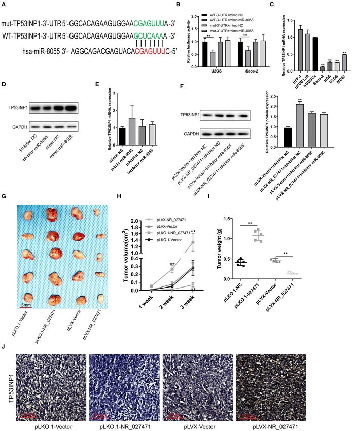
Chen et al.                                                                               LncRNA NR_027471 Suppressed Osteosarcoma Proliferation

   Previous studies have revealed that non-coding DNA accounts            60◦ C, 34 s; program 3: 95◦ C, 5 s, 1 cycle, 65◦ C, 60 s, 97◦ C,
for the majority of the human genome, and this is transcribed             1 s; and program 4: 42◦ C, 30 s, 1 cycle. The relative gene
into non-coding RNA (4). Long non-coding RNAs (lncRNAs)                   expression was calculated by the 2−11CT method, and the
are defined as transcripts longer than 200 nucleotides without            expression of glyceraldehyde-3-phosphate dehydrogenase was
evident protein-coding function (5). It has been shown that               used to normalize the expression of mRNA.
lncRNAs regulate biological functions, including cell growth,
differentiation, progression, and apoptosis (6). Dysregulation of         Construction of pLKO.1-Vectors
lncRNA expression is associated with different types of cancer,           The pLKO.1-Vector plasmid were purchased from Addgene.
including gastric (7), colon (8), lung (9), and breast (10) cancer.       The siRNA target to NR_027471 were designed by BLOCK-
   The aim of the present study was to elucidate the biological           iTTM RNAi Designer. The sequence of siRNA with with the
functions of the lncRNA NR_027471 in osteosarcoma, as well as             highest knockdown efficiency and control siRNA was as follows:
the underlying molecular mechanisms. This study may improve               NR_027471-siRNA,          5′ -TGTTGTTGTTGTTGTTATA-3′ ;
our understanding of the role of NR_027471 in osteosarcoma                Control-siRNA, 5 -TGTTTGTTGTTGTTTGATA-3′ ; The top
                                                                                          ′
and aid the development of treatment strategies, resulting                strand 5′ -CCGGTGTTGTTGTTGTTGTTATACGAATATAAC
in decreased recurrence rates and increased survival rates in             AACAACAACAACATTTTTTGGTACC-3′ ; and the bottom
patients with this disease.                                               strand 3′ -CACAACAACAACAACAATATGCTTATATTGTT
                                                                          GTTGTTGTTGTAAAAAAAAACCATGGTTAA-5′ was cloned
MATERIALS AND METHODS                                                     into pLKO.1-Vector plasmid.
Cell Culture
Osteosarcoma cell lines (U2OS, Saos-2, MG-63, and HOS)                    Cell Migration and Invasion Assays
were cultured in Dulbecco’s modified Eagle’s medium (DMEM)                Cells were inoculated into a six-well culture plate (50,000 cells
supplemented with 10% fetal bovine serum, 100 U/ml penicillin,            per well). When the cell grew to 100% confluency, 1 ml of blue
and 100 mg/ml streptomycin. The human fetal osteoblast cell               gun head was used to perform a scratch. Subsequently, the
line (hFOB1.19) was cultured in medium with DMEM/F-12 (1:1)               culture medium and suspension cells were removed, and replaced
containing 10% fetal bovine serum and 0.3 mg/l G418. Human                with serum-free culture medium (0 h). Following 24 h of culture,
foreskin fibroblast-1 (HFF-1) cells and human bone marrow stem            images were captured. The cell migration rate was calculated by
cells (hBMSCs) were maintained in DMEM supplemented with                  comparing the healing degree between 0 and 24 h.
10% of fetal bovine serum, 100 U/ml penicillin, and 100 µg/ml                After 1:1 mixing of DMEM medium and Matrigel, 50 µl of
streptomycin (all from Gibco, Carlsbad, CA, USA). All these cell          the mixture was evenly spread in the Transwell cell and placed
lines were cultured at 37◦ C in a humidified incubator containing         in a 37◦ C incubator for 45 min. The cells were divided into four
5% CO2 .                                                                  groups: pLVX-Vector; pLVX-NR_027471; pLKO.1-Vector; and
                                                                          pLKO.1-NR_027471 groups. The cells (20,000 cells per well) were
Cell Transfection                                                         inoculated into the upper chamber of the Transwell. The upper
Transfections were performed using Lipofectamine R 3000                   chamber of the Transwell contained serum-free medium, and 700
(Invitrogen; Thermo Fisher Scientific, Inc.) according to the             µl of medium containing 5% serum was added into the lower
instructions provided by the manufacturer. For overexpression,            chamber. The cells in the upper chamber were removed after
the NR_027471 overexpression vector was established using a               being cultured in the incubator for 12 h. The adherent cells in the
pLVX-IRES-puro vector backbone produced by Sangon Biotech                 lower chamber of the Transwell were stained with crystal violet
Co., Ltd. For knockdown, two shRNAs targeting NR_027471                   and counted after obtaining images.
were purchased from Shanghai GeneChem Co., Ltd. (Shanghai,
China). The miR-8055 mimics and inhibitor were purchased                  Cell Counting Kit-8 (CCK-8) Assay
from Genepharma (Shanghai, China).                                        Cells were inoculated into a 96-well culture plate (4,000 cells per
                                                                          well). Cells were divided into four groups: pLVX-Vector; pLVX-
RNA Isolation and Quantitative Reverse                                    NR_027471; pLKO.1-Vector; and pLKO.1-NR_027471groups.
Transcription-Polymerase Chain Reaction                                   The activity of cells was detected by CCK-8 at 24, 48, 72, or 96 h
(PCR)                                                                     after inoculation. CCK-8 solution (10 µl) was added into each
The total RNA of cells was extracted using the Trizol                     pore, and the absorbance at 450 nm was detected after incubation
reagent (ThermoFisher, Shanghai, China #15596018), and the                at 37◦ C for 2 h using an enzyme scale.
RNA concentration was detected by Nanodrop (ThermoFisher,
Shanghai, China). RNA (1 µg) was used along with the RNA                  Colony Formation Assay
reverse transcription kit. Real-time fluorescent quantitative PCR         Cells were inoculated into six-well plates (400 cells per well).
reaction was performed using the SYBR Green RT-qPCR Master                The cells were divided into four groups: pLVX-Vector; pLVX-
Mix kit, and the sample adding reaction was conducted according           NR_027471; pLKO.1-Vector; and pLKO.1-NR_027471groups.
to the instructions provided by the manufacturer (Takara, Dalian,         Following inoculation, the culture solution was changed once
China #RR420A). The reaction conditions were as follows:                  every 3 days. After 14 days of culture, 4% paraformaldehyde was
program 1: 95◦ C, 30 s, 1 cycle; program 2: 95◦ C, 5 s, 50 cycle,         used for fixation for 15 min. Subsequently, 0.1% crystal violet

Frontiers in Oncology | www.frontiersin.org                           2                                September 2020 | Volume 10 | Article 563255
Chen et al.                                                                                 LncRNA NR_027471 Suppressed Osteosarcoma Proliferation

staining solution was added for staining for 5 min. Phosphate-              supernatant was collected and the protein concentration was
buffered saline (PBS) was used to remove non-specific staining.             measured through the bicinchoninic acid method. A sodium
The number of clones was calculated after capturing images.                 dodecyl sulfate-polyacrylamide gel electrophoresis gel (10%) was
                                                                            prepared, and 20 µg of protein was loaded into each pore.
Flow Cytometry Analysis                                                     Following electrophoresis, 300 mA was transferred to 120 min
To detect the apoptotic rate, the transfected cells were stained            membrane, the protein was transferred to the polyvinylidene
with annexin V-fluorescein isothiocyanate and propidium iodide              difluoride membrane, 5% bovine serum albumin (BSA) was
using an annexin V-fluorescein isothiocyanate/propidium iodide              used for blocking for 1 h. The membrane was incubated with
apoptosis detection kit (Becton, Dickinson and Company,                     4% BSA overnight, washed thrice with tris-buffered saline
Franklin Lakes, NJ, USA). The cells were analyzed by a Gallios              with Tween 20, two was incubated at room temperature for
Flow Cytometer (Beckman Coulter, USA) to quantify the                       1 h, re-washed thrice with tris-buffered saline with Tween
percentage of apoptotic cells. For the cell cycle analysis, cells           20, and visualized using a development solution and an
were analyzed using a Cycletest Plus DNA Reagent kit (Becton,               enhanced chemiluminescence developer.
Dickinson and Company) according to the instructions provided
by the manufacturer. Following 15 min of incubation with the
Cycletest Plus DNA Reagent kit, cells were examined using                   Bioinformatics Prediction
Gallios Flow Cytometer to quantify the proportion of cells in each          A differential analysis was performed on the gene data of
stage of the cell cycle (S, G1, and G2/M).                                  patients with osteosarcoma (GSE85537), downloaded from
                                                                            the Gene Expression Omnibus (GEO) database (https://www.
Luciferase Reporter Assay                                                   ncbi.nlm.nih.gov/geo/). The TargetScan website (http://www.
Cells were seeded into a 96-well-plate for 24 h and subsequently            targetscan.org/vert_72/) predicted the target gene of miR-8055.
co-transfected with: pmirGLO-NR_027471-wildtype and miR-                    The MiRcode website (http://www.mircode.org/) predicted the
negative control; pmirGLO-NR_027471-wildtype and miR-8055;                  potential miRNAs bound to NR_027471.
pmirGLO-NR_027471-mutant and miR-negative control;
or pmirGLO-NR_027471-mutant and miR-8055 (Promega
Corporation, Madison, WI, USA), using Lipofectamine R 3000,
respectively. Following 48 h at 37◦ C, Firefly and Renilla luciferase
                                                                            Tumor Xenograft Experiment
                                                                            All experimental operations were based on the European
activities were measured using the Dual-Luciferase Reporter
                                                                            Union Directive 2010/63/EU for animal experimentation
assay system (Promega Corporation) according to the
                                                                            (http://ec.europa.eu/environment/chemicals/lab_animals/
instructions provided by the manufacturer. Firefly luciferase
                                                                            legislation_en.htm). Six-week-old female BALB/c nude mice
activity was normalized to Renilla luciferase activity.
                                                                            were purchased from Better Biotechnology Co., Ltd. (Nanjing,
                                                                            China). Transfected cells (1 × 10⁷ ) were resuspended in 100 µl of
RNA Immunoprecipitation (IP)                                                PBS and subcutaneously inoculated into the flank of nude mice
The cells were harvested, resuspended in nuclear isolation
                                                                            (n = 5 per group). Tumor size was recorded once every week,
buffer, and maintained on ice with frequent mixing for
                                                                            and tumor volume was calculated using the following formula:
20 min. The nuclei were pelleted by centrifugation at 2,500 g
                                                                            tumor volume = (length × width2 )/2. After 3 weeks, all mice
for 15 min. Radioimmunoprecipitation assay (RIPA) buffer
                                                                            were sacrificed, and the tumor bulks were resected and weighed.
was used to resuspend the nuclear pellet, which was split
equally into two fractions (for mock and IP). Chromatin was
mechanically sheared using a Dounce homogenizer with 15–20
strokes. The nuclear membrane and debris were separated after               Immunohistochemistry
centrifugation. Anti-MS2b (10 µg) was added to the supernatant              The tumor tissue was fixed in 4% paraformaldehyde for
(10 mg), which was incubated for 2 h at 4◦ C. Protein A/G beads             4 h, embedded in paraffin, cut into 10 µm paraffin sections,
(40 µl) were added to the mixture, which was incubated for 1 h              dewaxed, digested with pepsin for 45 min, incubated with
at 4◦ C. The beads were subsequently pelleted and washed in                 3% hydrogen peroxide for 15 min, and sealed with 5% BSA
RIPA, followed by washing with PBS. Coprecipitated RNAs were                for 1 h. Subsequently, the tissue was incubated overnight
isolated by resuspending the beads in Trizol reagent.                       with the primary antibody, followed by incubation with the
                                                                            secondary antibody for 1 h. The tissues were stained with
Western Blotting                                                            3,3′ -diaminobenzidine for 10 min, and hematoxylin staining
The cells were divided into four groups: pLVX-Vector; pLVX-                 was performed.
NR_027471; pLKO.1-Vector; and pLKO.1-NR_027471 groups.
After the culture supernatant was removed, the cells were
washed once with PBS, and 100 µl RIPA lysate containing                     Statistical Analysis
1 mmol/l phenylmethylsulfonyl fluoride was added into each                  The Student’s t-test was performed using the SPSS software
pore. The cells were detached using a cell scraper on ice, split            version 19.0 (IBM Corp.). All data are expressed as the mean ±
on ice for 30 min, centrifuged at 12,000 r/min 4◦ C for 15 min              standard deviation of three independent experiments. P < 0.05
(the centrifugation radius measured 11 cm). Subsequently, the               indicates a statistically significant difference.

Frontiers in Oncology | www.frontiersin.org                             3                                September 2020 | Volume 10 | Article 563255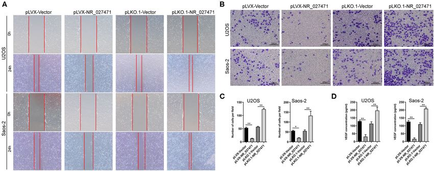
Chen et al. LncRNA NR_027471 Suppressed Osteosarcoma Proliferation
RESULTS group. Notably, the pLKO.1-NR_027471 group demonstrated
increased invasion compared with the pLKO.1-Vector group
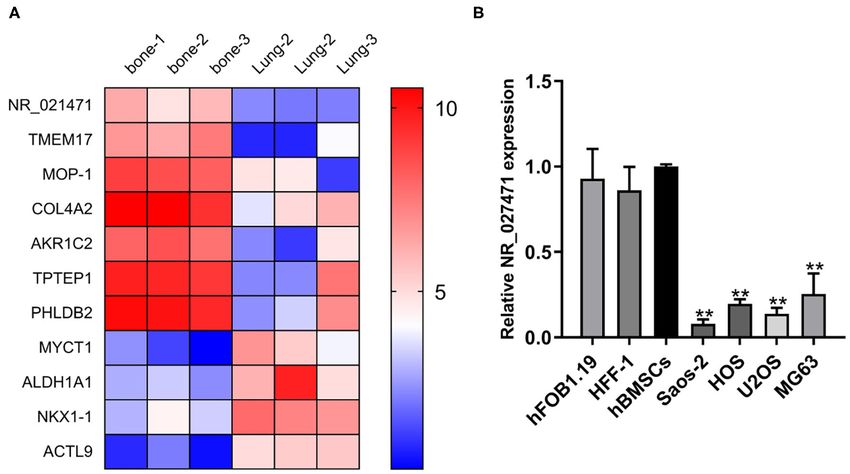
LncRNA NR_027471 Was Downregulated in (Figures 3B,C). The correlation of NR_027471 and EMT
Osteosarcoma Cell Lines was investigated using western blotting. The expression of E-
A bioinformatic analysis from the GEO database showed that cadherin was upregulated, while that of zinc finger E-box-binding
NR_027471 was downregulated in lung metastasis compared homeobox 1 (ZEB1), Snail, and Fibronectin was downregulated
with in situ lesions of osteosarcoma (Figure 1A). The expression in lncRNA NR_027471-overexpressing osteosarcoma cells
of lncRNA NR_027471 was confirmed by quantitative reverse compared to pLVX-Vector group (Figure 3D). Reversely,
transcription-PCR. The results indicated that NR_027471 was after knockdown of lncRNA NR_027471, the expression of
significantly downregulated in osteosarcoma cell lines (U2OS, E-cadherin was downregulated, whereas that of ZEB1, Snail, and
Saos-2, MG-63, and HOS) compared with hFOB1.19, HFF-1, and fibronectin was upregulated in osteosarcoma cells compared to
hBMSCs (Figure 1B). pLKO.1-Vector group (Figure 3D).
LncRNA NR_027471 Inhibited the Overexpression of NR_027471 Inhibited the
Proliferation of Osteosarcoma Cell Lines Effect of Osteosarcoma on the Migration
The expression of lncRNA NR_027471 in U2OS and Saos-2 and Invasion of Endothelial Cells
cells was significantly increased after transfection with pLVX- To investigate the impact of NR_027471 on endothelial
NR_027471 compared to pLVX-Vector group, whereas that cells, human umbilical vein endothelial cells (HUVECs) were
was significantly decreased after transfection with pLKO.1- cultured with culture media derived from osteosarcoma cell
NR_027471 compared to pLKO.1-Vector group (Figure 2A). with NR_027471 overexpression or knockdown. In the scratch
CCK-8 analysis showed that osteosarcoma cell viability was assay, the scratch wound was larger when HUVECs were
suppressed at 48, 72, and 96 h when lncRNA NR_027471 incubated culture media derived from osteosarcoma cell with
was overexpressed compared to pLVX-Vector group and NR_027471 overexpression compared that with culture media
osteosarcoma cell viability was upregulated at 48, 72, and 96 h derived from osteosarcoma cell with pLVX-Vector. In contrast,
with lncRNA NR_027471 knockdown compared to pLKO.1- the scratch wound was smaller when HUVECs were incubated
group (Figure 2B). Colony formation assay was performed with NR_027471-knockdown media compared with the pLKO.1-
to determine the proliferation ability of osteosarcoma cells. Vector group (Figure 4A). The Transwell invasion assay showed
The results indicated that osteosarcoma cells transfected with that fewer invasive HUVECs were observed at the bottom of
pLVX-NR_027471 had significantly decreased proliferation the insert following incubation with culture media derived from
compared with those transfected with the pLVX-Vector. osteosarcoma cell with NR_027471 overexpression compared
Osteosarcoma cells transfected with pLKO.1-NR_027471 that with culture media derived from osteosarcoma cell with
showed significantly increased proliferation compared with pLVX-Vector, whereas more invasive cells were observed after
those transfected with the pLKO.1-Vector (Figure 2C). Flow incubation with NR_027471-knockdown media compared with
cytometry showed that overexpression of lncRNA NR_027471 the pLKO.1-Vector group (Figures 4B,C). The concentration
induced G1 cell cycle arrest compared to pLVX-NR_027471 of vascular endothelial growth factor in the supernatant of
group, while knockdown of lncRNA NR_027471 reduced the the culture media of U2OS and Saos-2 cells was determined.
proportion of cells at G1 compared to pLKO.1-NR_027471 The results showed that vascular endothelial growth factor was
group (Figure 2D). Flow cytometry showed that there was no significantly decreased in pLVX-NR_027471 group compared
significant difference between the pLVX-NR_027471 and pLVX- to pLVX-Vector group and increased in pLKO.1-NR_027471
Vector groups regarding the apoptotic rate of osteosarcoma group compared to pLKO.1-Vector group (Figure 4D). Thus, the
cells. Moreover, there was no significant difference between the results indicate that overexpression of NR_027471 inhibits the
pLKO.1-NR_027471 and pLKO.1-Vector groups (Figure 2E). migration and invasion of endothelial cells.
LncRNA NR_027471 Inhibited the LncRNA NR_027471 Regulates the
Migration, Invasion, and Function of Osteosarcoma by Sponging
Epithelial–Mesenchymal Transition (EMT) miR-8055
of Osteosarcoma Cell Lines The mechanism through which lncRNA NR_027471 regulates
Moreover, the migration ability of osteosarcoma cells was osteosarcoma was investigated. The MS2bs-based RNA IP assay
determined by a scratch assay. The experiment revealed that indicated that lncRNA NR_027471 combined with miR-8055
osteosarcoma cells transfected with pLVX-NR_027471 showed (Figure 5A). These results were also confirmed by the luciferase
decreased migration in comparison with those transfected with reporter assay (Figure 5B). In addition, we conducted a biotin-
the pLVX-Vector. Also, osteosarcoma cells transfected with miRNA RNA IP assay, which showed that miR-8055 combined
pLKO.1-NR_027471 showed increased migration vs. those with lncRNA NR_027471 (Figure 5C). The CCK-8 assay showed
transfected with the pLKO.1-Vector (Figure 3A). The Transwell that overexpression of miR-8055 promoted cell viability, whereas
Matrigel assay showed that the pLVX-NR_027471 group had its inhibition suppressed cell viability (Figures 5D,E). The
decreased invasion ability compared with the pLVX-Vector Transwell assay revealed that overexpression of miR-8055
Frontiers in Oncology | www.frontiersin.org 4 September 2020 | Volume 10 | Article 563255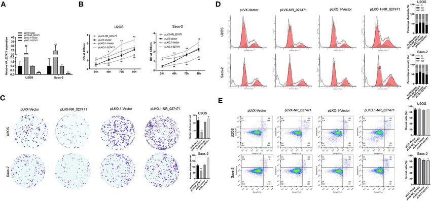
Chen et al.                                                                                           LncRNA NR_027471 Suppressed Osteosarcoma Proliferation

 FIGURE 1 | NR_027471 was downregulated in osteosarcoma cells. (A) A heatmap showing the expression of different genes at in situ lesions or lung metastasis
 osteosarcoma tissue. (B) The expression of lncRNA NR_027471 in hFOB1.19, HFF-1, hBMSCs, and osteosarcoma cell lines (Saos-2, HOS, U2OS, and MG63) was
 detected by qRT-PCR. Data are expressed as the mean ± SD of three independent experiments.** P < 0.01. lncRNA, long non-coding RNA; hFOB, human fetal
 osteoblast; HFF-1, human foreskin fibroblast-1; hBMSC, human bone marrow stem cell; qRT-PCR, quantitative reverse transcription-polymerase chain reaction; SD,
 standard deviation.

 FIGURE 2 | LncRNA NR_027471 inhibited the proliferation of osteosarcoma cells. (A) The expression of lncRNA NR_027471 in osteosarcoma cells was determined
 by qRT-PCR after transfection with pLVX-NR_027471 or pLKO.1-NR_027471. (B) The viability of osteosarcoma cells after overexpression or knockdown of lncRNA
 NR_027471 was determined by CCK-8. (C) The proliferation ability of osteosarcoma cells was determined by colony formation assay. (D) Cell cycle analysis of
 osteosarcoma cells after overexpression or knockdown of lncRNA NR_027471 using flow cytometry. (E) Apoptotic rate of osteosarcoma cells analyzed by flow
 cytometry. Data are expressed as the mean ± SD of three independent experiments.** P < 0.01 compared with the pLVX-Vector group. ## P < 0.01 compared with
 the pLKO.1-Vector group. lncRNA, long non-coding RNA; qRT-PCR, quantitative reverse transcription-polymerase chain reaction; CCK-8, Cell Counting Kit-8; SD,
 standard deviation.

Frontiers in Oncology | www.frontiersin.org                                     5                                    September 2020 | Volume 10 | Article 563255
Chen et al.                                                                                              LncRNA NR_027471 Suppressed Osteosarcoma Proliferation

 FIGURE 3 | NR_027471 inhibited the migration, invasion, and EMT of osteosarcoma cells. (A) The migration ability of osteosarcoma cells after overexpression or
 knockdown of lncRNA NR_027471 was determined using the scratch assay. (B) The invasion ability of osteosarcoma cells after overexpression or knockdown of
 lncRNA NR_027471 was determined using the Transwell Matrigel assay. (C) Quantitative analysis of the invasion ability of osteosarcoma cells after overexpression or
 knockdown of lncRNA NR_027471 was determined using the Transwell Matrigel assay. (D) The protein expression of E-cadherin, ZEB1, Snail, and fibronectin in
 osteosarcoma cell lines. Data are expressed as the mean ± SD of three independent experiments. ** P < 0.01 compared with the pLVX-Vector group. ## P < 0.01
 compared with the pLKO.1-Vector group. EMT, epithelial–mesenchymal transition; lncRNA, long non-coding RNA; ZEB1, zinc finger E-box-binding homeobox 1; SD,
 standard deviation.

 FIGURE 4 | Overexpression of lncRNA NR_027471 inhibited the effect of osteosarcoma on the migration and invasion of endothelial cells. (A) Scratch assay and (B)
 Transwell invasion assay performed to analyze the effect of culture supernatant obtained from osteosarcoma cells after overexpression or knockdown of lncRNA
 NR_027471 on the migration and invasion ability of HUVECs. (C) Histogram presenting the number of invasive cells. (D) Concentration of VEGF in the supernatant of
 osteosarcoma cells after overexpression or knockdown of lncRNA NR_027471, analyzed by ELISA. All data are expressed as mean ± SD of three independent
 experiments. * p < 0.05, ** P < 0.01. lncRNA, long non-coding RNA; HUVEC, human umbilical vein endothelial cell; VEGF, vascular endothelial growth factor; ELISA,
 enzyme-linked immunosorbent assay; SD, standard deviation.

Frontiers in Oncology | www.frontiersin.org                                       6                                      September 2020 | Volume 10 | Article 563255
Chen et al. LncRNA NR_027471 Suppressed Osteosarcoma Proliferation
promoted cell invasion compared to miRNA mimic NC group, that miR-8055 could combine with the wildtype-3′ UTR of
whereas miR-8055 inhibition suppressed cell invasion compared TP53INP1 mRNA in osteosarcoma cells, but not with the
to miRNA inhibitor NC group (Figures 5F,H). Following the mutant-3′ UTR of TP53INP1 mRNA (Figure 6B). The mRNA
inhibition of miR-8055 function by a miR-8055 inhibitor, expression of TP53INP1 in osteosarcoma cells was confirmed
the impact of NR_027471 on cell invasion was weakened by quantitative reverse transcription-PCR. The expression of
(Figures 5G,I). This finding indicated that the regulatory effect TP53INP1 was downregulated in osteosarcoma cell lines (U2OS,
of NR_027471 on invasion was miR-8055-dependent. Saos-2, MG-63, and HOS) compared with HFF-1, hFOB1.19, and
hBMSCs (Figure 6C). The protein expression of TP53INP1 was
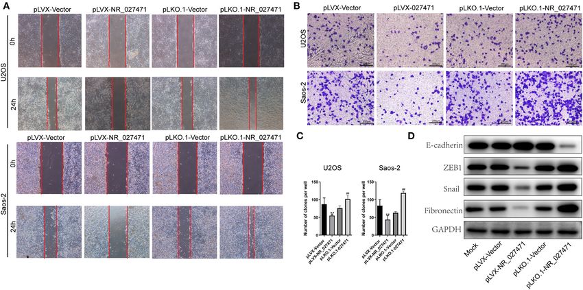
LncRNA NR_027471 Inhibited the Tumor inhibited following overexpression of miR-8055, whereas it was
Growth of Osteosarcoma in vivo by enhanced after knockdown of miR-8055 (Figure 6D). However,
the mRNA expression of TP53INP1was not significantly
Modulating the Expression of Tumor influenced after overexpression or knockdown of miR-8055
Protein p53 Inducible Nuclear Protein 1 (Figure 6E). Inhibition of the function of miR-8055 using a
(TP53INP1) miR-8055 inhibitor, weakened the impact of lncRNA NR_027471
A putative binding site between miR-8055 and the 3′ untranslated on the protein expression of TP53INP1 (Figure 6F). This finding
region (3′ UTR) of TP53INP1 mRNA was predicted by indicated that the regulatory effect of lncRNA NR_027471 on
TargetScan (Figure 6A). The luciferase reporter assay showed TP53INP1 protein was miR-8055-dependent.
FIGURE 5 | LncRNA NR_027471 regulates the function of osteosarcoma cells by sponging miR-8055. (A) MS2b-based RIP assay with anti-GFP antibody
(cross-reacting with YFP) in U2OS 48 h after transfection with MS2bp-YFP plasmid along with MS2bs-NR_027471, or MS2bs-Rluc (control vectors). (B) Binding of
lncRNA NR_027471 and miR-8055 detected using the luciferase assay. (C) Binding of lncRNA NR_027471 and miR-8055 using the biotin-miRNA RIP assay. (D)
Viability of U2OS cells transfected with mimic miR-8055 or inhibitor miR-8055 using CCK-8. (E) Viability of Saos-2 cells transfected with a mimic or inhibitor of
miR-8055 using CCK-8. (F) Transwell invasion assay determined the invasion ability of U2OS and Saos-2 cells transfected with a mimic or inhibitor of miR-8055. (G)
The Transwell invasion assay determined the invasion ability of U2OS and Saos-2 cells after co-transfection with the miR-8055 inhibitor and pLKO.1-NR_027471.
(H,I) Histogram presenting the number of invasive cells. Data are expressed as the mean ± SD of three independent experiments. ** P < 0.01. lncRNA, long
non-coding RNA; RIP, RNA immunoprecipitation; YFP, yellow fluorescent protein; GFP, green fluorescent protein; CCK-8, Cell Counting Kit-8; SD, standard deviation.
Frontiers in Oncology | www.frontiersin.org 7 September 2020 | Volume 10 | Article 563255Chen et al. LncRNA NR_027471 Suppressed Osteosarcoma Proliferation
FIGURE 6 | LncRNA NR_027471 inhibited the growth of osteosarcoma in vivo by modulating the expression of TP53INP1. (A) The WT-3′ UTR of TP53INP1 mRNA
was predicted as the target of miR-8055 and the mut-3′ UTR of TP53INP1 mRNA. (B) The mRNA of TP53INP1 was identified as the target of miR-8055 using the
(Continued)
Frontiers in Oncology | www.frontiersin.org 8 September 2020 | Volume 10 | Article 563255Chen et al. LncRNA NR_027471 Suppressed Osteosarcoma Proliferation
FIGURE 6 | luciferase assay. (C) qRT-PCR analysis revealed the mRNA expression of TP53INP1 in HFF-1, hFOB1.19, hBMSCs, and osteosarcoma cell lines (Saos-2,
HOS, U2OS, and MG63). (D) Western blotting analyzed the protein expression of TP53INP1 in U2OS transfected with a mimic or inhibitor of miR-8055. (E) qRT-PCR
analysis determined the expression of TP53INP1 in U2OS cells transfected with a mimic or inhibitor of miR-8055. (F) Western blotting analyzed the protein expression
of TP53INP1 in U2OS cell co-transfected with pLVX-NR_027471 and miR-8055 inhibitor. (G) The tumor xenografts in nude mice were excised at the end of week 3 (n
= 5 per group). (H) Tumor volume recorded at the end of weeks 1, 2, and 3. (I) Tumor weight of xenografts measured at the end of week 3. (J) Immunohistochemical
detection of the expression of TP53INP1 in tumor tissue. Data are expressed as the mean ± SD of three independent experiments. ** P < 0.01. lncRNA, long
non-coding RNA; TP53INP1, tumor protein p53 inducible nuclear protein 1; WT, wildtype; 3′ UTR, 3′ untranslated region; mut, mutant; qRT-PCR, quantitative reverse
transcription-polymerase chain reaction; hFOB, human fetal osteoblast; hff, human foreskin fibroblast; hBMSC, human bone marrow stem cell; SD, standard deviation.
Subcutaneous tumor xenografts were developed in nude mice. (SOX2-OT) was identified as an oncogene in osteosarcoma cells,
The tumor volumes were measured at the end of weeks 1, regulating the migration, invasion, and expression of cancer
2, and 3. The analysis showed that the tumor volume was stem cell biomarkers. In addition, it was recognized as a
significantly smaller in the pLVX-NR_027471 group vs. the prognostic biomarker in patients with osteosarcoma (16).
pLVX-Vector group at the end of week 3. In contrast, the tumor In a study conducted by Zhao et al. (17), BMSC-derived
volume was significantly larger in the pLKO.1-NR_027471 group exosomes encapsulated lncRNA PVTl and transported it into
vs. the pLKO.1-Vector group at the end of weeks 2 and 3 osteosarcoma cells, promoting tumor growth and metastasis
(Figure 6H). The xenografts were weighed and photographed by inhibiting ubiquitination and upregulating the expression
at the end of week 3 (Figure 6G). The results showed that of ERG in these cells. Cong and Jing (18) reported a tumor
tumor weight was significantly lower in the pLVX-NR_027471 suppressor, tumor suppressor candidate 7 (TUSC7), which
group vs. the pLVX-Vector group, while it was significantly inhibited the proliferation and migration of osteosarcoma cells,
higher in the pLKO.1-NR_027471 group vs. the pLKO.1-Vector promoted cellular apoptosis, and was largely mediated by
group (Figure 6I). The data indicated that overexpression of miR-211. In the present study, NR_027471 was downregulated
NR_027471 inhibits tumor growth of osteosarcoma in vivo. in osteosarcoma cells and suppressed their proliferation
Immunohistochemistry showed that the expression of TP53INP1 and invasion. NR_027471 plays a tumor suppressive role
in the pLVX-NR_027471 group was significantly higher than that in osteosarcoma.
observed in the pLVX-Vector group. Moreover, the expression LncRNAs exert their function in several manners,
of TP53INP1 in the pLKO.1-NR_027471 group was significantly including the transcriptional and translational levels (19).
lower than that measured in the pLKO.1-Vector group At the transcriptional level, lncRNAs may act as ceRNA by
(Figure 6J). binding several miRNAs and inhibiting their activities (20).
For instance, lncRNA prostate cancer associated transcript
6 (PCAT6) promote the progression of osteosarcoma
DISCUSSION through function as ceRNA of miR-185-5p (21). LncRNA
HIF1A antisense RNA 2 (HIF1A-AS2) was identified as
In our study, the expression levels of NR_027471 were ceRNA by sponging miR-33b-5p to facilitate cell survival
investigated in osteosarcoma cell lines. Significantly lower and migration and modulate the expression of sirtuin 6
expression of NR_027471 was observed in osteosarcoma cell (SIRT6) in osteosarcoma (22). The bioinformatics analysis
lines compared with osteoblast, fibroblast, and BMSC cell performed in the current study revealed that NR_027471
lines. Gain- and loss-of-function experiments demonstrated contains a putative binding site for miR-8055, and TP53INP1
that NR_027471 overexpression inhibited the cell proliferation was predicted as the target of miR-8055. RNA pull-down
and invasion of osteosarcoma cells, and also significantly and luciferase reporter assays were used to validate the
induced cell cycle arrest at G1 of the osteosarcoma cells. sequence-specific correlation between miR-8055 and
These results suggested a negative association between NR_027471.NR_027471 in the regulation of TP53INP1
NR_027471 and progression of osteosarcoma. Moreover, by sponging miR-8055. The present study revealed a
the mechanism through which NR_027471 regulated tumor novel lncRNA-miRNA target pair that is dysregulated in
proliferation and invasion was investigated. NR_027471 osteosarcoma cells.
positively regulated TP53INP1 by competitively binding TP53INP1, known as a tumor suppressor, is involved in a
with miR-8055. This was the first report investigating the series of biological activities. It has been evidenced that miR-182
role of NR_027471 in cancer research. The results of this promotes drug resistance in cisplatin-treated hepatocellular
study may improve our understanding of osteosarcoma carcinoma cells by downregulating TP53INP1 (23). By
progression and help to find therapeutic targets against antagonizing TP53INP1 and Yes1 associated transcriptional
this disease. regulator (YAP1), upregulated miR-200a enhances drug
LncRNAs play important roles in the formation and resistance in breast cancer (24). In colorectal cancer, miR-221
progression of osteosarcoma (11–14). LINC00612 functions promoted cell proliferation via the inhibition of autophagy and
as a competitive endogenous RNA (ceRNA) for miR-214-5p targeted TP53INP1 (25). In this study, TP53INP1 was predicted
to promote the proliferation and invasion of osteosarcoma in and confirmed to be a target of miR-8055. The upregulation
vitro and in vivo (15). LncRNA SOX2 overlapping transcript of NR_027471 increased the protein expression of TP53INP1.
Frontiers in Oncology | www.frontiersin.org 9 September 2020 | Volume 10 | Article 563255Chen et al. LncRNA NR_027471 Suppressed Osteosarcoma Proliferation
Therefore, both NR_027471 and TP53INP1 appear to play tumor ETHICS STATEMENT
suppressive roles in osteosarcoma.
The animal study was reviewed and approved by Animal Care
and Use Committee of Shanghai General Hospital of Nanjing
CONCLUSION
Medical University.
In this study, a newly identified regulatory mechanism of
the NR_027471/miR-8055/ TP53INP1 axis was systematically
AUTHOR CONTRIBUTIONS
studied in osteosarcoma. NR_027471 suppresses the proliferation
and invasion of osteosarcoma cells and induces cell cycle DS and JZ: conception and design: JC, WM, and SY: experiments
arrest at G1. NR_027471 inhibited EMT by increasing and data analysis: JZ: intellectual input and supervision: JC,
E-cadherin and decreasing ZEB1, Snail, and fibronectin. MY, and DS: manuscript writing. All authors approved the final
NR_027471 regulates the protein expression of TP53INP1 by version of the manuscript.
sponging miR-8055. This may improve our understanding
of epigenetic regulation via NR_027471 and miR-8055 in
osteosarcoma and may provide a novel insight into potential FUNDING
therapeutic strategies.
This study was supported by the National Natural Science
DATA AVAILABILITY STATEMENT Foundation of China (grant numbers: 81702216 and 81571828),
Nantong Health and Family Planning Commission Research
The datasets generated for this study are available on request to Project (grant number: MSZ18209), and the Project for Medical
the corresponding author. Key Youth Talent of Nantong (grant number: Youth 022).
REFERENCES 12. Xie W, Yuan S, Sun Z, Li Y. Long noncoding and circular RNAs
in lung cancer: advances and perspectives. Epigenomics. (2016) 8:1275–
1. Esiashvili N, Goodman M, Marcus RB Jr. Changes in incidence and 87. doi: 10.2217/epi-2016-0036
survival of Ewing sarcoma patients over the past 3 decades: Surveillance 13. Chu F, Xue L, Miao H. Long noncoding RNA TP73-AS1 in human
Epidemiology and End Results data. J Pediatr Hematol Oncol. (2008) 30:425– cancers. Clin Chim Acta. (2020) 500:104–8. doi: 10.1016/j.cca.2019.
30. doi: 10.1097/MPH.0b013e31816e22f3 09.024
2. Mirabello L, Troisi RJ, Savage SA. Osteosarcoma incidence and survival 14. Ghafouri-Fard S, Mohammad-Rahimi H, Taheri M. The role of long non-
rates from 1973 to 2004: data from the Surveillance, Epidemiology, and coding RNAs in the pathogenesis of thyroid cancer. Exp Mol Pathol. (2020)
End Results Program. Cancer. (2009) 115:1531–43. doi: 10.1002/cncr. 112:104332. doi: 10.1016/j.yexmp.2019.104332
24121 15. Zhou Y, Li X, Yang H. LINC00612 functions as a ceRNA for miR-214-
3. Zheng W, Huang Y, Chen H, Wang N, Xiao W, Liang Y, et al. 5p to promote the proliferation and invasion of osteosarcoma in vitro
Nomogram application to predict overall and cancer-specific survival in and in vivo. Exp Cell Res. (2020) 392:112012. doi: 10.1016/j.yexcr.2020.
osteosarcoma. Cancer Manag Res. (2018) 10:5439–50. doi: 10.2147/CMAR.S1 112012
77945 16. Wang Z, Tan M, Chen G, Li Z, Lu X. LncRNA SOX2-OT is a novel
4. Pereira Zambalde E, Mathias C, Rodrigues AC, de Souza Fonseca prognostic biomarker for osteosarcoma patients and regulates osteosarcoma
Ribeiro EM, Fiori Gradia D, Calin GA, et al. Highlighting transcribed cells proliferation and motility through modulating SOX2. IUBMB Life. (2017)
ultraconserved regions in human diseases. Wiley Interdiscip Rev RNA. (2020) 69:867–76. doi: 10.1002/iub.1681
11:e1567. doi: 10.1002/wrna.1567 17. Zhao W, Qin P, Zhang D, Cui X, Gao J, Yu Z, et al. Long non-
5. Batista PJ, Chang HY. Long noncoding RNAs: cellular address coding RNA PVT1 encapsulated in bone marrow mesenchymal stem
codes in development and disease. Cell. (2013) 152:1298– cell-derived exosomes promotes osteosarcoma growth and metastasis
307. doi: 10.1016/j.cell.2013.02.012 by stabilizing ERG and sponging miR-183-5p. Aging. (2019) 11:9581–
6. Liao D, Lv G, Wang T, Min J, Wang Y, Liu S. Prognostic value of long non- 96. doi: 10.18632/aging.102406
coding RNA BLACAT1 in patients with papillary thyroid carcinoma. Cancer 18. Cong M, Jing R. Long non-coding RNA TUSC7 suppresses
Cell Int. (2018) 18:47. doi: 10.1186/s12935-018-0544-9 osteosarcoma by targeting miR-211. Biosci Rep. (2019)
7. Li S, Zhang M, Zhang H, Hu K, Cai C, Wang J, et al. Exosomal long noncoding 39:BSR20190291. doi: 10.1042/BSR20190291
RNA lnc-GNAQ-6:1 may serve as a diagnostic marker for gastric cancer. Clin 19. Zhang X, Wang W, Zhu W, Dong J, Cheng Y, Yin Z, et al. Mechanisms and
Chim Acta. (2020) 501:252–257. doi: 10.1016/j.cca.2019.10.047 functions of long non-coding RNAs at multiple regulatory levels. Int J Mol Sci.
8. Liu KL, Wu J, Li WK, Li NS, Li Q, Lao YQ. LncRNA SNHG7 is an oncogenic (2019) 20:5573. doi: 10.3390/ijms20225573
biomarker interacting with microRNA-193b in colon carcinogenesis. Clin Lab. 20. Militello G, Weirick T, John D, Doring C, Dimmeler S, Uchida S. Screening
(2019) 65:2199–204. doi: 10.7754/Clin.Lab.2019.190501 and validation of lncRNAs and circRNAs as miRNA sponges. Brief Bioinform.
9. Li S, Teng Y, Yuan MJ, Ma TT, Ma J, Gao XJ. A seven long-noncoding RNA (2017) 18:780–8. doi: 10.1093/bib/bbw053
signature predicts prognosis of lung squamous cell carcinoma. Biomark Med. 21. Zhu C, Huang L, Xu F, Li P, Li P, Hu F. LncRNA PCAT6 promotes
(2020) 14:53–63. doi: 10.2217/bmm-2019-0282 tumor progression in osteosarcoma via activation of TGF-beta pathway
10. Arshi A, Raeisi F, Mahmoudi E, Mohajerani F, Kabiri H, Fazel R, et al. A by sponging miR-185-5p. Biochem Biophys Res Commun. (2020) 521:463–
Comparative study of HOTAIR expression in breast cancer patient tissues and 70. doi: 10.1016/j.bbrc.2019.10.136
cell lines. Cell J. (2020) 22:178–84. doi: 10.22074/cellj.2020.6543 22. Lin H, Zhao Z, Hao Y, He J, He J. Long noncoding RNA HIF1A-AS2
11. Bhan A, Soleimani M, Mandal SS. Long noncoding RNA facilitates cell survival and migration by sponging miR-33b-5p to modulate
and cancer: a new paradigm. Cancer Res. (2017) 77:3965– SIRT6 expression in osteosarcoma. Biochem Cell Biol. (2020) 98:284–
81. doi: 10.1158/0008-5472.CAN-16-2634 92. doi: 10.1139/bcb-2019-0171
Frontiers in Oncology | www.frontiersin.org 10 September 2020 | Volume 10 | Article 563255Chen et al. LncRNA NR_027471 Suppressed Osteosarcoma Proliferation
23. Li Q, Han Y, Wang C, Shan S, Wang Y, Zhang J, et al. MicroRNA-125b Conflict of Interest: The authors declare that the research was conducted in the
promotes tumor metastasis through targeting tumor protein 53-induced absence of any commercial or financial relationships that could be construed as a
nuclear protein 1 in patients with non-small-cell lung cancer. Cancer Cell Int. potential conflict of interest.
(2015) 15:84. doi: 10.1186/s12935-015-0233-x
24. Yu SJ, Yang L, Hong Q, Kuang XY, Di GH, Shao ZM. MicroRNA- Copyright © 2020 Chen, Miao, Yang, Yin, Zhao and Song. This is an open-access
200a confers chemoresistance by antagonizing TP53INP1 and YAP1 in article distributed under the terms of the Creative Commons Attribution License (CC
human breast cancer. BMC Cancer. (2018) 18:74. doi: 10.1186/s12885-017- BY). The use, distribution or reproduction in other forums is permitted, provided
3930-0 the original author(s) and the copyright owner(s) are credited and that the original
25. Liao D, Li T, Ye C, Zeng L, Li H, Pu X, et al. miR-221 inhibits autophagy publication in this journal is cited, in accordance with accepted academic practice.
and targets TP53INP1 in colorectal cancer cells. Exp Ther Med. (2018) No use, distribution or reproduction is permitted which does not comply with these
15:1712–7. doi: 10.3892/etm.2017.5522 terms.
Frontiers in Oncology | www.frontiersin.org 11 September 2020 | Volume 10 | Article 563255You can also read