Radiation exposure triggers the malignancy of non small cell lung cancer cells through the activation of visfatin/Snail signaling
←
→
Page content transcription
If your browser does not render page correctly, please read the page content below
ONCOLOGY REPORTS 45: 1153-1161, 2021

    Radiation exposure triggers the malignancy of non‑small cell lung
      cancer cells through the activation of visfatin/Snail signaling
              LIANG XIAO1*, YIWEN MAO2*, ZHUTING TONG1, YE ZHAO2, HAO HONG1 and FAN WANG1

                   1
                   Department of Radiation Oncology, The First Affiliated Hospital of Anhui Medical University;
         2
          Teaching and Research Section of Nuclear Medicine, Anhui Medical University, Hefei, Anhui 230022, P.R. China

                                      Received April 10, 2020; Accepted December 2, 2020

                                                     DOI: 10.3892/or.2021.7929

Abstract. It is estimated that one‑half of patients with             and radiotherapy are widely used for the clinical treatment of
non‑small cell lung cancer (NSCLC) undergo radiotherapy              patients with NSCLC (4).
worldwide. However, the outcome of radiotherapy alone is                 During the course of treatment, one‑half of all patients with
not always satisfactory. The aim of the present study was to         NSCLC patients undergo radiotherapy (5). It has been reported
evaluate the effects of radiotherapy on the malignancy of            that early‑stage patients can benefit from stereotactic body
NSCLC cells. It was demonstrated that radiation therapy could        radiation therapy (6). Stereotactic body radiation therapy has
increase the migration and invasion of NSCLC cells in vitro.         been used as a standard care modality in medically inoperable
Moreover, the upregulation of visfatin, a 52‑kDa adipokine,          patients with early‑stage NSCLC (7). However, previous studies
mediated radiation‑induced cell motility. A neutralizing             have indicated that the outcome of radiotherapy alone is not
antibody specific for visfatin blocked radiation‑induced cell        always satisfactory (8), which may be due to the development of
migration. Radiation and visfatin induced the expression of          radiotherapy resistance (9,10). Moreover, laboratory and clin-
Snail, a key molecule that regulates epithelial to mesenchymal       ical data indicated that radiotherapy may cause clinical adverse
transition in NSCLC cells. Furthermore, visfatin positively          effects for patients with NSCLC. For example, radiotherapy
regulated the mRNA stability of Snail in NSCLC cells, but had        can increase tumor invasion and metastasis by upregulating
no effect on its protein degradation. This may be explained by       TGFβ‑1 expression (11). Cancer cells treated with radiation
visfatin‑mediated downregulation of microRNA (miR)‑34a,              acquire mesenchymal‑like morphology (12). X‑ray irradiation
which was shown to bind the 3' untranslated region of Snail          can also increase the invasion of NSCLC cells (13). In addition,
mRNA to promote its decay. Collectively, these findings              radiation can stimulate the malignancy of residual, incom-
suggested that radiation could induce cell motility in NSCLC         pletely treated and viable tumors surrounding the treatment
cells through visfatin/Snail signaling.                              zone (14,15). Thus, understanding the mechanisms involved
                                                                     in radiation‑induced malignancy in NSCLC cells is needed in
Introduction                                                         order to improve the therapeutic efficacy of radiotherapy.
                                                                         Cytokines, such as visfatin, are involved in the progression
As the leading cause of cancer mortality, lung cancer accounts       of NSCLC (16). It has been reported that visfatin medi-
for 18.2% of total cancer deaths worldwide (1). Furthermore,         ates doxorubicin (Dox) resistance of human NSCLC cells
~80% of patients with lung cancer patients have non‑small‑cell       through Akt‑mediated upregulation of ATP‑binding cassette
lung cancer (NSCLC). The incidence of NSCLC has increased            subfamily C member 1 (17). Furthermore, visfatin triggers
in women during the last two decades worldwide (2). It               cell motility of NSCLC cells by increasing the expression of
has been reported that the 5‑year survival rate of NSCLC             matrix metalloproteinases (MMPs) (18). Targeted inhibition of
is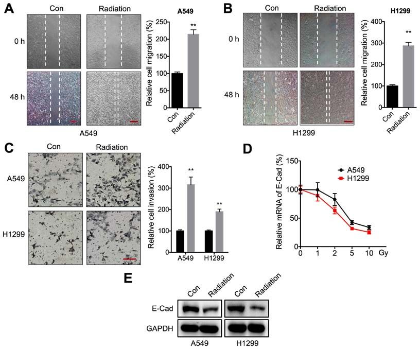
1154                       XIAO et al: RADIATION TRIGGERS MALIGNANCY OF NSCLC VIA VISFATIN/SNAIL

of Snail is essential for EMT induction in NSCLC cells (29).           selected fields of view was quantified using ImageJ software
In the present study, the expression levels of cytokines, such         version 1.47 (National Institutes of Health). The relative inva-
as IL‑6, IL‑8, IL‑10, VEGFA, TGF‑β, TNF‑ α and visfatin                sion rate was calculated by dividing the number of stained
were examined in NSCLC cell lines treated with radiation.              cells by the number of stained cells in the control group.
Further, the potential roles and mechanisms of visfatin in
radiation‑induced migration and invasion of NSCLC cells                Reverse transcription‑quantitative (RT‑q) PCR analysis. Total
were investigated.                                                     RNA was isolated using TRIzol® reagent (Invitrogen; Thermo
                                                                       Fisher Scientific, Inc.). cDNA was generated by using the
Materials and methods                                                  PrimeScript RT reagent kit with gDNA Eraser (Takara
                                                                       Biotechnology Co., Ltd.) for mRNA at 37˚C for 15 min, or the
Cell culture and radiation treatment. The human NSCLC                  qScript microRNA cDNA synthesis kit (Quantabio) for miRNA
A549 and H1299 cell lines and human bronchial epithelial               at 37˚C for 60 min followed by 5 min at 70˚C, respectively. For
cells (HBEpC) were purchased from American Type Culture                mRNA targets, qPCR was conducted using a SYBR‑Green
Collection and maintained in DMEM (Invitrogen; Thermo                  PCR Kit (Qiagen GmbH) on the Step‑One Plus Real‑Time PCR
Fisher Scientific, Inc.) supplemented with 10% FBS (Gibco;             System (Applied Biosystems, Inc.). The thermocycling condi-
Thermo Fisher Scientific, Inc.), penicillin and streptomycin           tions consisted of an initial denaturation at 95˚C for 5 min,
at 37˚C in 5% CO2. Mycoplasma contamination was moni-                  followed by 50 cycles at 95˚C for 15 sec and 60˚C for 30 sec. The
tored weekly during experiments. Radiation treatment                   primer sequences were as follows: i) IL‑6 forward, 5'‑CCT
was conducted according to a previous study (30). Briefly,             CCAGAACAGATTTGAGAGTAGT‑3' and reverse, 5'‑GGG
5x10⁵ cells/well were seeded in a 6‑well plate and were irradi-        TCAGGGGTGGTTATTGC‑3'; ii) IL‑8 forward, 5'‑GAGAGT
ated at room temperature using a 6 MV X‑ray linear accelerator         GATTGAGAGTGGACCAC‑3' and reverse, 5'‑CACAACCCT
(Varian 23Ex; Varian Inc.) at a dose rate of 300 cGy/min for           CTGCACCCAGTTT‑3'; iii) IL‑10 forward, 5'‑GTGGCATTC
the time required to apply the dose used in each assay. In order       AAGGAGTACCTC‑3' and reverse, 5'‑TGATGGCCTTCGAT
to determine the role of visfatin, anti‑visfatin neutralizing anti-    TCTGGATT‑3'; iv) VEGFA forward, 5'‑TACCTCCACCATGCC
body (100 ng/ml) or recombinant‑visfatin (rVisfatin;100 ng/ml)         AAGTGGT‑3' and reverse, 5'‑AGGACGGCTTGAAGATG
were added into culture medium for 24 h at 37˚C in 5% CO2,             TAC‑3'; v) TGF‑β forward, 5'‑GGCCAGATCCTGTCCAAGC‑3'
then irradiated at room temperature.                                   and reverse, 5'‑GTGGGTTTCCACCATTAGCAC‑3';
                                                                       vi) TNF‑α forward, 5'‑CCTCTCTCTAATCAGCCCTCTG‑3'
Reagents. Scramble negative control (NC) microRNA (miR,                and reverse, 5'‑GAGGACCTGGGAGTAGATGAG‑3';
5'‑UUCUCCGAACGUGUCACGUTT‑3'), miR‑34a inhibitor                        vii) visfatin forward, 5'‑AGGGTTACAAGTTGCTGCCACC‑3'
(5'‑AAGCUCCAUUUCGCAACCUUAC‑3'), small interfering                      and reverse, 5'‑CTCCACCAGAACCGAAGGCAAT‑3';
RNA (siRNA) NC (si‑NC; 5'‑GCACAACAAGCCGAAUACA‑3')                      viii) Snail forward, 5'‑GACCACTATGCCGCGCTCTT‑3' and
and siRNA targeting Snail (5'‑CAUCCGAAGCCACACG                         reverse, 5'‑TCGCTGTAGTTAGGCTTCCGATT‑3'; ix) Slug
CUG‑3') were purchased from Sigma‑Aldrich; Merck KGaA.                 forward, 5'‑AGCAGTTGCACTGTGATGCC‑3' and reverse,
The neutralizing antibody specific for visfatin (anti‑Visfatin; cat.   5'‑ACACAGCAGCCAGATTCCTC‑3'; x) Twist forward, 5'‑CGG
no. A300‑778A) and recombinant visfatin (cat. no. RP‑75758)            ACAAGCTGAGCAAGATT‑3' and reverse, 5'‑CCTTCTCT
were obtained from Invitrogen; Thermo Fisher Scientific, Inc.          GGAAACAATGAC‑3'; xi) Zeb1 forward, 5'‑GCACCTGAAGA
                                                                       GGACCAGAG‑3' and reverse, 5'‑TGCATCTGGTGTTCCAT
Wound healing and Transwell Matrigel™ assay. Cells                     TTT‑3'; xii) GAPDH forward, 5'‑GTCAACGGATTTGGTCTGT
(1.5x10 6 cells per well) were plated in 12‑well plates and            ATT‑3' and reverse, 5'‑AGTCTTCTGGGTGGCAGTGAT‑3'.
cultured to 80% confluence in complete medium. The cell                    For miR targets (miR‑137, miR‑34a, miR‑153 and miR‑22),
layer was scratched with a 200 µl pipette tip, washed twice            qPCRs were performed using the NCode miRNA qRT‑PCR
with PBS, then cultured with medium containing 0.5% FBS,               analysis kit (Invitrogen; Thermo Fisher Scientific, Inc.). The
with or without the indicated treatments, as described in              thermocycling conditions included an initial denaturation at
Figure legends. The migration distance was recorded in the             95˚C for 3 min followed by 40 cycles at 95˚C for 15 sec and
same visual fields under a phase‑contrast microscope. The              60˚C for 30 sec. The forward primer is the exact sequence
relative migration rate was calculated according to a previous         of the mature miRNA. The forward primer for U6 was
study (31), using the following formula: [(scratch area at             5'‑TGCGGGTGCTCGCTTCGCAGC‑3'. Gene expression levels
0 h‑scratch area at 48 h)/scratch area at 0 h] x 100%.                 were calculated using the 2‑ΔΔCq method (32) and standardized to
    Cell invasion was assessed using a Transwell Matrigel inva-        GAPDH and U6 for mRNA and miR targets, respectively. All
sion chamber (8‑µm pore filters; Corning, Inc.) according to the       RT‑qPCR reactions were performed three times.
manufacturer's instructions. A total of 2x10⁵ cells was seeded
into the upper chamber of a 24‑well chamber with FBS‑free              Western blot analysis. Cells were lysed using RIPA buffer
medium. The bottom chamber received 0.6 ml complete                    (Beyotime Institute of Biotechnology) and protein extracts
medium. After the indicated treatment and culture for 24 h, the        were collected. Protein concentration was measured using a
invading cells were fixed using methanol for 15 min at room            BCA Protein Assay Kit (Pierce™; Thermo Fisher Scientific,
temperature, dried under a laminar flow safety cabinet, stained        Inc.). After denaturation in boiling water for 10 min, proteins
with 0.5% crystal violet (Sigma‑Aldrich; Merck KGaA) for 2 h           (20 µg per lane) were separated by SDS‑PAGE on 10% gels,
at room temperature, then observed under an inverted optical           then transferred to PVDF membranes. The membranes were
microscope. The number of invading cells in five randomly              blocked with 5% skimmed milk at room temperature for 2 h,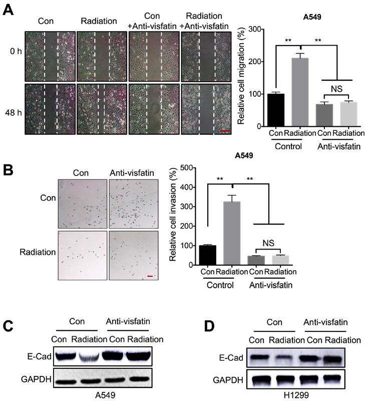
ONCOLOGY REPORTS 45: 1153-1161, 2021                                              1155

and then incubated with primary antibodies at 4˚C for at least     were assessed using unpaired Student's t‑test (two‑tailed).
15 h. The primary antibodies used were specific for GAPDH          One‑way ANOVA followed by Tukey's post hoc test was used
(cat. no. ab9485; Abcam; 1:1,000), E‑Cad (cat. no. 14472S;         when making pairwise comparisons among ≥3 groups. P
1156                            XIAO et al: RADIATION TRIGGERS MALIGNANCY OF NSCLC VIA VISFATIN/SNAIL

Figure 1. Radiation increases the motility of NSCLC cells. (A and B) Wound healing assay for (A) A549 or (B) H1299 cells treated with 5 Gy radiation
(Radiation) or incubated without radiation (Con) for 48 h. Scale bar, 100 µm. (C) Transwell Matrigel invasion assay for A549 and H1299 cells treated with 5 Gy
radiation (Radiation) or incubated without radiation (Con) for 48 h. Scale bar, 100 µm. (D) E‑Cad mRNA expression levels following treatment with increasing
doses of radiation for 24 h. (E) E‑Cad protein expression levels following treatment with 5 Gy radiation (Radiation) or incubation without radiation (Con) for
24 h. Data are presented as the mean ± SD of three independent experiments. **P
ONCOLOGY REPORTS 45: 1153-1161, 2021                                                               1157

Figure 3. Visfatin is essential for radiation‑induced cell motility. (A and B) A549 cells treated with 5 Gy radiation (Radiation) or incubated without radiation
(Con) were further incubated with or without 100 ng/ml anti‑visfatin antibody for 48 h. (A) Migration was measured using a wound healing assay. Scale bar, 100 µm.
(B) Invasion was evaluated using a Transwell Matrigel assay. (C and D) A549 and H1299 cells treated with 5 Gy radiation (Radiation) or incubated without radiation
(Con) were further incubated with or without 100 ng/ml anti‑visfatin antibody for 48 h. E‑Cad protein expression levels were measured in (C) A549 and (D) H1299
cells. Data are presented as the mean ± SD of three independent experiments. **P
1158                           XIAO et al: RADIATION TRIGGERS MALIGNANCY OF NSCLC VIA VISFATIN/SNAIL

Figure 4. Snail is involved in radiation‑ and visfatin‑induced cell migration. mRNA expression levels of Snail, Slug, Twist and Zeb1 in (A) A549 or (B) H1299
cells treated with 5 Gy radiation (Radiation) or incubated without radiation (Con) for 24 h. (C) Protein expression levels of Snail in A549 or H1299 cells treated
with 5 Gy radiation (Radiation) or incubated without radiation (Con) for 24 h. (D and E) Snail mRNA expression levels in (D) A549 and (E) H1299 cells treated
with 5 Gy radiation (Radiation) or incubated without radiation (Con) and further incubated with or without 100 ng/ml anti‑visfatin antibody for 48 h. (F) Snail
and E‑Cad protein expression levels in A549 cells transfected with si‑NC or si‑Snail for 12 h, then further treated with 5 Gy radiation (Radiation) or incubated
without radiation (Con) for 24 h. Data are presented as the mean ± SD of three independent experiments. **P
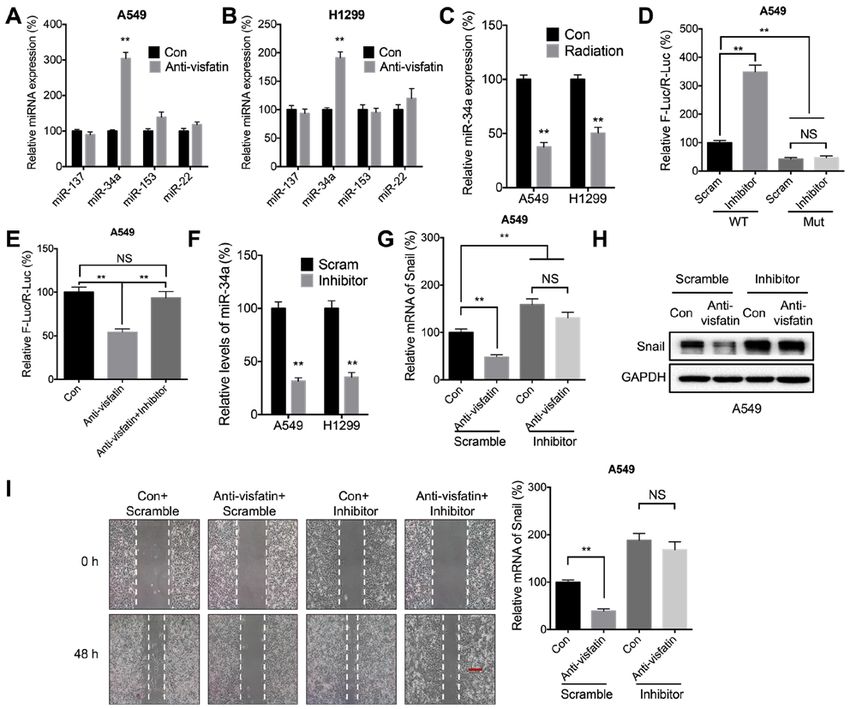
ONCOLOGY REPORTS 45: 1153-1161, 2021                                                              1159

Figure 6. miR‑34a may be involved in visfatin‑regulated expression of Snail. (A and B) miR expression levels in (A) A549 and (B) H1299 cells treated with
(Anti‑visfatin) or without (Con) 100 ng/ml anti‑visfatin antibody for 48 h. (C) miR‑34a expression levels in cells treated with (Radiation) or without (Con)
5 Gy radiation for 24 h. (D) Luciferase activity in A549 cells co‑transfected with WT or MUT Snail‑3'‑UTR Firefly luciferase reporter plasmid and Scram or
miR‑34a inhibitor for 24 h. (E) A549 cells were co‑transfected with WT Snail‑3'‑UTR Firefly luciferase reporter plasmid with or without miR‑34a inhibitor for
12 h. The Anti‑visfatin and Anti‑visfatin + inhibitor groups were treated with 100 ng/ml anti‑visfatin antibody for 24 h. (F) miR‑34a expression levels in cells
transfected with Scram miR or miR‑34a inhibitor for 24 h. (G‑I) A549 cells were pre‑transfected with Scram or miR‑34a inhibitor for 12 h, then treated with
(Anti‑visfatin) or without (Con) 100 ng/ml anti‑visfatin antibody for 48 h. (G) mRNA and (H) protein expression levels of Snail. (I) Relative cell migration.
Scale bar, 100 µm. Data are presented as the mean ± SD of three independent experiments. **P
1160                      XIAO et al: RADIATION TRIGGERS MALIGNANCY OF NSCLC VIA VISFATIN/SNAIL

hepatocellular carcinoma (54) and colorectal carcinoma (55).         Patient consent for publication
It is also involved in resistance to Dox treatment in colorectal
cancer (56). Visfatin is upregulated in lung cancer cells and        Not applicable.
tissues (42); it also regulates the EMT and invasion of NSCLC
cells (18), as well as Dox sensitivity (17). In the present study,   Competing interests
visfatin was upregulated after radiation therapy, and visfatin
blockade with a neutralizing attenuated radiation‑induced            The authors declare that they have no competing interests.
cell motility. This suggested that visfatin could promote the
malignancy of lung cancer.                                           References
     The present study revealed that miR‑34a/Snail is involved
in visfatin‑regulated radiotherapy‑induced malignancy of              1. Askoxylakis V, Thieke C, Pleger ST, Most P, Tanner J, Lindel K,
NSCLC cells. Moreover, it has been reported that visfatin can            Katus HA, Debus J and Bischof M: Long‑term survival of cancer
upregulate the expression of Snail to induce EMT‑like prop-              patients compared to heart failure and stroke: A systematic
                                                                         review. BMC Cancer 10: 105, 2010.
erties in osteosarcoma cells (57) and colorectal cancer (55).         2. Schwartz AG, Wenzlaff AS, Prysak GM, Murphy V, Cote ML,
In NSCLC cells, visfatin upregulates Snail expression by                 Brooks SC, Skafar DF and Lonardo F: Reproductive factors,
increasing its mRNA stability, likely by decreasing the expres-          hormone use, estrogen receptor expression and risk of non
                                                                         small‑cell lung cancer in women. J Clin Oncol 25: 5785‑5792, 2007.
sion of miR‑34a. Previous studies have indicated that Snail was       3. Gondos A, Bray F, Brewster DH, Coebergh JW, Hakulinen T,
the direct target of miR‑34a in gastric (58), pancreatic (59), and       Janssen‑Heijnen ML, Kurtinaitis J and Brenner H; EUNICE
ovarian (35) cancer cells. In addition, Snail directly induces           Survival Working Group: Recent trends in cancer survival across
                                                                         Europe between 2000 and 2004: A model‑based period analysis
ZNF281 transcription and represses miR‑34a/b/c, thereby                  from 12 cancer registries. Eur J Cancer 44: 1463‑1475, 2008.
alleviating ZNF281 mRNA from direct down‑regulation by                4. Molina JR, Yang P, Cassivi SD, Schild SE and Adjei AA:
miR‑34 and promoting the EMT of lung cancer cells (60).                  Non‑small cell lung cancer: Epidemiology, risk factors, treatment,
                                                                         and survivorship. Mayo Clin Proc 83: 584‑594, 2008.
However, the mechanisms underlying visfatin‑regulated                 5. Remon J, Ahn MJ, Girard N, Johnson M, Kim DW, Lopes G,
expression of miR‑34a remain unclear.                                    Pillai RN, Solomon B, Villacampa G and Zhou Q: Advanced‑stage
     In summary, the present study demonstrated that radiation           non‑small cell lung cancer: Advances in thoracic oncology 2018.
                                                                         J Thorac Oncol 14: 1134‑1155, 2019.
increased the malignancy of NSCLC cells through visfatin              6. Antonia SJ, Villegas A, Daniel D, Vicente D, Murakami S,
upregulation. miR‑34a‑regulated mRNA stability of Snail                  Hui R, Yokoi T, Chiappori A, Lee KH, de Wit M, et al; PACIFIC
and was involved in radiation/visfatin regulated motility of             investigators: Durvalumab after chemoradiotherapy in stage III
                                                                         non‑small‑cell lung cancer. N Engl J Med 377: 1919‑1929, 2017.
NSCLC cells. Although the detailed mechanisms underlying              7. Shinde A, Li R, Kim J, Salgia R, Hurria A and Amini A:
the role of visfatin and miR‑34a remain to be determined,                Stereotactic body radiation therapy (SBRT) for early‑stage lung
these findings highlighted the adverse effects of radiotherapy           cancer in the elderly. Semin Oncol 45: 210‑219, 2018.
                                                                      8. Draghiciu O, Walczak M, Hoogeboom BN, Franken KL,
on NSCLC cell motility.                                                  Melief KJ, Nijman HW and Daemen T: Therapeutic immu-
                                                                         nization and local low‑dose tumor irradiation, a reinforcing
Acknowledgements                                                         combination. Int J Cancer 134: 859‑872, 2014.
                                                                      9. Lee S, Lim MJ, Kim MH, Yu CH, Yun YS, Ahn J and Song JY:
                                                                         An effective strategy for increasing the radiosensitivity of human
No applicable.                                                           lung cancer cells by blocking Nrf2‑dependent antioxidant
                                                                         responses. Free Radic Biol Med 53: 807‑816, 2012.
                                                                     10. Provencio M, Sánchez A, Garrido P and Valcárcel F: New
Funding                                                                  molecular targeted therapies integrated with radiation therapy in
                                                                         lung cancer. Clin Lung Cancer 11: 91‑97, 2010.
This work was supported by The National Natural Science              11. Rube CE, Uthe D, Schmid KW, Richter KD, Wessel J, Schuck A,
                                                                         Willich N and Rube C: Dose‑dependent induction of trans-
Foundation of China (grant no. 81673099).                                forming growth factor beta (TGF‑beta) in the lung tissue of
                                                                         fibrosis‑prone mice after thoracic irradiation. Int J Radiat Oncol
Availability of data and materials                                       Biol Phys 47: 1033‑1042, 2000.
                                                                     12. Tsukamoto H, Shibata K, Kajiyama H, Terauchi M, Nawa A
                                                                         and Kikkawa F: Irradiation‑induced epithelial‑mesenchymal
The datasets used and/or analyzed during the current study are           transition (EMT) related to invasive potential in endometrial
available from the corresponding author on reasonable request.           carcinoma cells. Gynecol Oncol 107: 500‑504, 2007.
                                                                     13. Zhao Q, Mao A, Guo R, Zhang L, Yan J, Sun C, Tang J, Ye Y,
                                                                         Zhang Y and Zhang H: Suppression of radiation‑induced migration
Authors' contributions                                                   of non‑small cell lung cancer through inhibition of Nrf2‑Notch
                                                                         Axis. Oncotarget 8: 36603‑36613, 2017.
                                                                     14. Kroeze SG, van Melick HH, Nijkamp MW, Kruse FK, Kruijssen LW,
LX, YM, ZT, YZ, HH and FW conceived and designed the                     van Diest PJ, Bosch JL and Jans JJ: Incomplete thermal ablation
study. LX, YM and FW acquired the data. LX, YM and HH                    stimulates proliferation of residual renal carcinoma cells in a trans-
analyzed and interpreted the data. LX, HH and FW wrote,                  lational murine model. BJU Int 110: E281‑E286, 2012.
                                                                     15. Shiozawa K, Watanabe M, Takahashi M, Wakui N, Iida K and
reviewed and revised the manuscript. All authors read and                Sumino Y: Analysis of patients with rapid aggressive tumor
approved the final manuscript. LX and FW confirm the                     progression of hepatocellular carcinoma after percutaneous
authenticity of the raw data. All authors read and approved the          radiofrequency ablation. Hepatogastroenterology 56: 1689‑1695,
                                                                         2009.
final manuscript.                                                    16. Daly ME, Monjazeb AM and Kelly K: Clinical trials integrating
                                                                         immunotherapy and radiation for non‑small‑cell lung cancer. J
Ethics approval and consent to participate                               Thorac Oncol 10: 1685‑1693, 2015.
                                                                     17. Cao Z, Liang N, Yang H and Li S: Visfatin mediates doxorubicin
                                                                         resistance in human non‑small‑cell lung cancer via Akt‑mediated
Not applicable.                                                          up‑regulation of ABCC1. Cell Prolif 50: e12366, 2017.
ONCOLOGY REPORTS 45: 1153-1161, 2021 1161
18. Wang G, Tian W, Liu Y, Ju Y, Shen Y, Zhao S, Zhang B and 40. Strieter RM, Belperio JA, Burdick MD, Sharma S, Dubinett SM and
Li Y: Visfatin triggers the cell motility of non‑small cell lung Keane MP: CXC chemokines: Angiogenesis, immunoangiostasis, and
cancer via up‑regulation of matrix metalloproteinases. Basic metastases in lung cancer. Ann NY Acad Sci 1028: 351‑360, 2004.
Clin Pharmacol Toxicol 119: 548‑554, 2016. 41. Santaniello A, Napolitano F, Servetto A, De Placido P, Silvestris N,
19. Okumura S, Sasaki T, Minami Y and Ohsaki Y: Nicotinamide Bianco C, Formisano L and Bianco R: Tumour microenvironment
phosphoribosyltransferase: A potent therapeutic target in non‑small and immune evasion in EGFR addicted NSCLC: Hurdles and
cell lung cancer with epidermal growth factor receptor‑gene possibilities. Cancers (Basel) 11: 1419, 2019.
mutation. J Thorac Oncol 7: 49‑56, 2012. 42. Liu T, Miao Z, Jiang J, Yuan S, Fang W, Li B and Chen Y: Visfatin
20. Laiguillon MC, Houard X, Bougault C, Gosset M, Nourissat G, mediates SCLC cells migration across brain endothelial cells
Sautet A, Jacques C, Berenbaum F and Sellam J: Expression and through upregulation of CCL2. Int J Mol Sci 16: 11439‑11451, 2015.
function of visfatin (Nampt), an adipokine‑enzyme involved in 43. Uddin A and Chakraborty S: Role of miRNAs in lung
inflammatory pathways of osteoarthritis. Arthritis Res Ther 16: cancer. J Cell Physiol: Apr 20, 2018 (Epub ahead of print).
R38, 2014. doi: 10.1002/jcp.26607.
21. Gujar AD, Le S, Mao DD, Dadey DY, Turski A, Sasaki Y, Aum D, 44. Wang Z and Liu C: MiR‑153 regulates metastases of gastric
Luo J, Dahiya S, Yuan L, et al: An NAD+‑dependent transcrip- cancer through Snail. Tumour Biol 37: 15509‑15515, 2015.
tional program governs self‑renewal and radiation resistance in 45. Zhang K, Li XY, Wang ZM, Han ZF and Zhao YH: MiR‑22
glioblastoma. Proc Natl Acad Sci USA 113: E8247‑E8256, 2016. inhibits lung cancer cell EMT and invasion through targeting
22. Xiang B, Han L, Wang X, Tang L, Li K, Li X, Zhao X, Xia M, Snail. Eur Rev Med Pharmacol Sci 21: 3598‑3604, 2017.
Zhou X, Zhang F, et al: Nicotinamide phosphoribosyltransferase 46. Cui YH, Suh Y, Lee HJ, Yoo KC, Uddin N, Jeong YJ, Lee JS,
upregulation by phenylephrine reduces radiation injury in subman- Hwang SG, Nam SY, Kim MJ, et al: Radiation promotes
dibular gland. Int J Radiat Oncol Biol Phys 96: 538‑546, 2016. invasiveness of non‑small‑cell lung cancer cells through gran-
23. Thiery JP: Epithelial‑mesenchymal transitions in tumour ulocyte‑colony‑stimulating factor. Oncogene 34: 5372‑5382, 2015.
progression. Nat Rev Cancer 2: 442‑454, 2002. 47. Qian L‑W, Mizumoto K, Urashima T, Nagai E, Maehara N,
24. Lu W and Kang Y: Epithelial‑mesenchymal plasticity in cancer Sato N, Nakajima M and Tanaka M: Radiation‑induced increase
progression and metastasis. Dev Cell 49: 361‑374, 2019. in invasive potential of human pancreatic cancer cells and its
25. Batlle E, Sancho E, Francí C, Domínguez D, Monfar M, Baulida J blockade by a matrix metalloproteinase inhibitor, CGS27023.
and García De Herreros A: The transcription factor snail is a Clin Cancer Res 8: 1223‑1227, 2002.
repressor of E‑cadherin gene expression in epithelial tumour 48. Nambiar DK, Rajamani P and Singh RP: Silibinin attenuates
cells. Nat Cell Biol 2: 84‑89, 2000. ionizing radiation‑induced pro‑angiogenic response and EMT in
26. Xu J, Lamouille S and Derynck R: TGF‑beta‑induced epithelial prostate cancer cells. Biochem Biophys Res Commun 456: 262‑268,
to mesenchymal transition. Cell Res 19: 156‑172, 2009. 2015.
27. Wang G, Ma W, Li Y, Jiang Y, Ma G, Zhang X, Meng L and 49. Zhou YC, Liu JY, Li J, Zhang J, Xu YQ, Zhang HW, Qiu LB,
Du J: Prognostic value of Twist, Snail and E‑cadherin expression Ding GR, Su XM, Mei‑Shi, et al: Ionizing radiation promotes
in pathological N0 non‑small‑cell lung cancer: A retrospective migration and invasion of cancer cells through transforming
cohort study. Eur J Cardiothorac Surg 54: 237‑245, 2018. growth factor‑beta‑mediated epithelial‑mesenchymal transition.
28. Yanagawa J, Walser TC, Zhu LX, Hong L, Fishbein MC, Mah V, Int J Radiat Oncol Biol Phys 81: 1530‑1537, 2011.
Chia D, Goodglick L, Elashoff DA, Luo J, et al: Snail promotes 50. Wang L, Zhao Y, Xiong Y, Wang W, Fei Y, Tan C and Liang Z:
CXCR2 ligand‑dependent tumor progression in non‑small cell K‑ras mutation promotes ionizing radiation‑induced invasion
lung carcinoma. Clin Cancer Res 15: 6820‑6829, 2009. and migration of lung cancer in part via the Cathepsin L/CUX1
29. Zhang Y, Zhang X, Ye M, Jing P, Xiong J, Han Z, Kong J, Li M, pathway. Exp Cell Res 362: 424‑435, 2018.
Lai X, Chang N, et al: FBW7 loss promotes epithelial‑to‑mesen- 51. Cho JH, Hong WG, Jung YJ, Lee J, Lee E, Hwang SG, Um HD and
chymal transition in non‑small cell lung cancer through the Park JK: γ‑Ionizing radiation‑induced activation of the EGFR‑p38/
stabilization of Snail protein. Cancer Lett 419: 75‑83, 2018. ERK‑STAT3/CREB‑1‑EMT pathway promotes the migration/
30. Lin Y, Bai X, Zhou W, He Y, Wu Y and Wang X: Radiation invasion of non‑small cell lung cancer cells and is inhibited by
exposure triggers the progression of triple negative breast cancer podophyllotoxin acetate. Tumour Biol 37: 7315‑7325, 2016.
via stabilizing ZEB1. Biomed Pharmacother 107: 1624‑1630, 2018. 52. Romacho T, Sánchez‑Ferrer CF and Peiró C: Visfatin/Nampt: An
31. Wang Q, Yang G, Jiang Y, Luo M, Li C, Zhao Y, Xie Y, Song K and adipokine with cardiovascular impact. Mediators Inflamm 2013:
Zhou J: XB130, regulated by miR‑203, miR‑219, and miR‑4782‑3p, 946427, 2013.
mediates the proliferation and metastasis of non‑small‑cell lung 53. Carbone F, Liberale L, Bonaventura A, Vecchiè A, Casula M,
cancer cells. Mol Carcinog 59: 557‑568, 2020. Cea M, Monacelli F, Caffa I, Bruzzone S, Montecucco F, et al:
32. Livak KJ and Schmittgen TD: Analysis of relative gene expression Regulation and function of extracellular nicotinamide phospho-
data using real‑time quantitative PCR and the 2(‑Delta Delta ribosyltransferase/visfatin. Compr Physiol 7: 603‑621, 2017.
C(T)) method. Methods 25: 402‑408, 2001. 54. Liang N, Chen Y, Yang L, He S and Liu T: Visfatin increases miR‑21
33. Pecoraro A, Carotenuto P, Russo G and Russo A: Ribosomal to promote migration in HCC. Cell Mol Biol 64: 48‑52, 2018.
protein uL3 targets E2F1 and Cyclin D1 in cancer cell response 55. Yang J, Zhang K, Song H, Wu M, Li J, Yong Z, Jiang S, Kuang X
to nucleolar stress. Sci Rep 9: 15431, 2019. and Zhang T: Visfatin is involved in promotion of colorectal
34. Lu L, Chen Z, Lin X, Tian L, Su Q, An P, Li W, Wu Y, Du J, carcinoma malignancy through an inducing EMT mechanism.
Shan H, et al: Inhibition of BRD4 suppresses the malignancy of Oncotarget 7: 32306‑32317, 2016.
breast cancer cells via regulation of Snail. Cell Death Differ 27: 56. Yan X, Zhao J and Zhang R: Visfatin mediates doxorubicin resis-
255‑268, 2020. tance in human colorectal cancer cells via up regulation of multidrug
35. Dong P, Xiong Y, Watari H, Hanley SJ, Konno Y, Ihira K, resistance 1 (MDR1). Cancer Chemother Pharmacol 80: 395‑403,
Yamada T, Kudo M, Yue J and Sakuragi N: MiR‑137 and 2017.
miR‑34a directly target Snail and inhibit EMT, invasion and 57. Wang D, Qian G, Wang J, Wang T, Zhang L, Yang P and Lin F:
sphere‑forming ability of ovarian cancer cells. J Exp Clin Cancer Visfatin is involved in the cisplatin resistance of osteosarcoma
Res 35: 132, 2016. cells via upregulation of Snail and Zeb1. Cancer Biol Ther 20:
36. Kim R‑K, Kaushik N, Suh Y, Yoo KC, Cui YH, Kim MJ, Lee HJ, 999‑1006, 2019.
Kim IG and Lee SJ: Radiation driven epithelial‑mesenchymal 58. Zhang Y, Yuan Y, Zhang Y, Cheng L, Zhou X and Chen K:
transition is mediated by Notch signaling in breast cancer. SNHG7 accelerates cell migration and invasion through regulating
Oncotarget 7: 53430‑53442, 2016. miR‑34a‑Snail‑EMT axis in gastric cancer. Cell Cycle 19: 142‑152,
37. Feys L, Descamps B, Vanhove C, Vral A, Veldeman L, Vermeulen S, 2020.
De Wagter C, Bracke M and De Wever O: Radiation‑induced lung 59. Tang Y, Tang Y and Cheng YS: miR‑34a inhibits pancreatic cancer
damage promotes breast cancer lung‑metastasis through CXCR4 progression through Snail1‑mediated epithelial‑mesenchymal
signaling. Oncotarget 6: 26615‑26632, 2015. transition and the Notch signaling pathway. Sci Rep 7: 38232, 2017.
38. Lagadec C, Vlashi E, Della Donna L, Dekmezian C and Pajonk F: 60. Hahn S, Jackstadt R, Siemens H, Hünten S and Hermeking H: SNAIL
Radiation‑induced reprogramming of breast cancer cells. Stem and miR‑34a feed‑forward regulation of ZNF281/ZBP99 promotes
Cells 30: 833‑844, 2012. epithelial‑mesenchymal transition. EMBO J 32: 3079‑3095, 2013.
39. Rodriguez‑Ruiz ME, Vitale I, Harrington KJ, Melero I and
Galluzzi L: Immunological impact of cell death signaling driven This work is licensed under a Creative Commons
by radiation on the tumor microenvironment. Nat Immunol 21: Attribution-NonCommercial-NoDerivatives 4.0
120‑134, 2020. International (CC BY-NC-ND 4.0) License.You can also read