Caffeic Acid Phenethyl Ester Protects against Experimental Autoimmune Encephalomyelitis by Regulating T Cell Activities
←
→
Page content transcription
If your browser does not render page correctly, please read the page content below
Hindawi
Oxidative Medicine and Cellular Longevity
Volume 2020, Article ID 7274342, 13 pages
https://doi.org/10.1155/2020/7274342

Research Article
Caffeic Acid Phenethyl Ester Protects against Experimental
Autoimmune Encephalomyelitis by Regulating T Cell Activities

 YiFan Zhou,1 Jingqi Wang,1 Yanyu Chang,1 Rui Li,1 Xiaobo Sun,1 Lisheng Peng,1
 WenHua Zheng ,2 and Wei Qiu 1
 1
 Department of Neurology, The Third Affiliated Hospital of Sun Yat-sen University, Guangzhou, China
 2
 Institute of Translational Medicine, Faculty of Health Sciences, University of Macau, Taipa, Macau SAR, China

 Correspondence should be addressed to WenHua Zheng; wenhuazheng@umac.mo and Wei Qiu; qiuwei120@vip.163.com

 Received 26 March 2020; Accepted 11 June 2020; Published 9 October 2020

 Guest Editor: Felipe L. de Oliveira

 Copyright © 2020 YiFan Zhou et al. This is an open access article distributed under the Creative Commons Attribution License,
 which permits unrestricted use, distribution, and reproduction in any medium, provided the original work is properly cited.

 Multiple sclerosis (MS) is an autoimmune inflammatory disease of the central nervous system (CNS) characterized by progressive
 demyelination and disabling outcomes. CD4+ T cells are the most critical driving factor of relapsing MS, but little improvement has
 been noted upon deletion of the whole T cell population. Caffeic acid phenethyl ester (CAPE), one of the main active compounds of
 propolis, exhibits potent antitumour, anti-inflammatory, and antioxidant properties by suppressing nuclear factor-κB (NF-κB)
 transactivation. To investigate the therapeutic potential of CAPE in MS, we studied the effects of CAPE on cytokine levels, T
 cells, and NF-κB activities and in an experimental MS animal model. The results showed that cerebrospinal fluid (CSF) from
 patients with relapsing MS is characterized by increased levels of proinflammatory cytokines/chemokines that preferentially
 skew towards T helper 1 (Th1) cytokines. In vitro studies demonstrated that CAPE not only inhibited T cell proliferation and
 activation but also effectively modulated T cell subsets. Under both Th0- and Th1-polarizing conditions, the proportion of
 CD4+IFN-γ+ cells was downregulated, while CD4+Foxp3+ cells were increased. Moreover, nuclear translocation of NF-κB p65
 was inhibited by CAPE. In a murine experimental autoimmune encephalomyelitis model, prophylactic treatment with CAPE
 significantly decreased the disease incidence and severity. Compared to the vehicle group, mice pretreated with CAPE showed
 diminished inflammatory cell infiltration, microglia/macrophage activation, and demyelination injury. Additionally, CAPE
 pretreatment reduced the level of Th1 cells in both spleen and the CNS and increased regulatory T cells (Tregs) in the CNS. In
 conclusion, our results highlight the potential merit of CAPE in suppressing T cell activity mainly through targeting the
 pathogenic Th1 lineage, which may be beneficial for MS treatment.

1. Introduction anti-inflammatory activities. Therefore, regulating T cell
 activity rather than depleting the whole cell population
Multiple sclerosis (MS) is an inflammatory demyelinating would be more suitable. Recently, acute relapses in patients
disease of the central nervous system (CNS) marked by with MS were reported to be associated with aberrant nuclear
repeated relapses and progressive disability. Although the factor-κB (NF-κB) gene expression in their T cells [1]. Since
exact mechanism of MS remains unclear, peripheral auto- the NF-κB signalling cascade is essential for cell proliferation,
reactive T cells have been recognized to primarily drive apoptosis, and immune responses, blocking this pathway
relapsing MS, whereas B cells and CNS-resident cells contrib- may help to prevent MS exacerbation and progression.
ute to progressive MS. [1] However, in a clinical trial, using Propolis has been widely used as a potential immuno-
monoclonal anti-CD4 antibody to selectively eliminate modulatory agent. Increasing evidence has shown that many
CD4+ T cells failed to reduce MS activity [2], which may be bioactive compounds of propolis can inhibit cytokine pro-
partially related to the fact that in addition to T helper 1 duction and immune cell migration mainly by blocking the
(Th1) and T helper 17 (Th17) cells, regulatory T cells (Tregs) NF-κB pathway [3, 4]. Caffeic acid phenethyl ester (CAPE)
also play a central role in MS pathogenesis by exerting potent is one of the main active components of propolis, which
2 Oxidative Medicine and Cellular Longevity

has been reported to alleviate many inflammatory diseases ture. After three washes, an HRP-conjugated secondary anti-
such as allergic asthma, experimental autoimmune uveoreti- body was added to each well and incubated for 30 min at
nitis, and sepsis as a specific NF-κB inhibitor [5]. The chem- room temperature. Finally, Chemi Reagent Mix was added
ical structure of CAPE is provided in the Supplementary to the washed membranes, and then, the membranes were
Material (available here). In the present study, we aimed to exposed to Tanon 5200.
investigate the impact of CAPE on T cells in vitro and to
assess its therapeutic potential based on experimental auto- 2.3. Cell Preparation and Mitogen Stimulation. Female
immune encephalomyelitis (EAE), which is a classic animal C57BL/6 mice (6-8 weeks old) were anaesthetized with pen-
model for MS. tobarbital and transcardially perfused with cold PBS. Spleens
 were removed and gently pressed through a 70-μm nylon
2. Materials and Methods mesh. After centrifugation at 300 × g for 5 min, cells were
 treated with red blood cell lysis buffer (Solarbio, Beijing,
2.1. Materials. CAPE (purity ≥98%) was obtained from China) for 3 min on ice. After washing twice with cold PBS,
Nature Standard (Shanghai, China). Concanavalin A type the cells were suspended in complete RPMI 1640 medium
IV (ConA), lipopolysaccharide (LPS), Percoll, Triton X-100, supplemented with 10% FBS and 1% GlutaMAX and cul-
and dimethyl sulfoxide (DMSO) were purchased from tured at a concentration of 2 × 10⁶ cells/ml per well in 24-
Sigma-Aldrich. Recombinant mouse IL-12 (rIL-12; p70); well plates or 5 × 10⁶ cells/ml per well in 96-well plates at
purified Ultra-LEAF™ anti-mouse IL-4 antibody; purifie- 37°C and 5% CO2. To induce lymphocyte proliferation, cells
dLEAF™ anti-mouse CD3 antibody; purified LEAF™anti- were stimulated with ConA (5 μg/ml) or LPS (1 μg/ml). The
mouse CD28 antibody; antibodies against mouse CD4, IL- study was approved by the Animal Ethics Committee of the
17A, IFN-γ, Foxp3, CD25, and CD69; cell activation cocktail; Third Affiliated Hospital of Sun Yat-sen University
brefeldin A solution; fixation/intracellular staining perme- ([2019]02-342-01).
abilization wash buffer; and Foxp3 fixation/permeabilization
buffer were obtained from BioLegend. RPMI 1640 medium, 2.4. Cell Viability and Proliferation Assays. Splenocytes were
fetal bovine serum (FBS), and GlutaMAX were obtained cultured in 96-well plates at a density of 5 × 10⁶ cells/ml per
from Gibco. Primary antibody against Iba-1 was purchased well and incubated with the indicated concentrations of
from Wako Pure Chemical Industries. Antibody against CAPE for 2 h prior to mitogen stimulation. After 48 h of
NF-κB p65 was obtained from Santa Cruz, and myelin basic stimulation by ConA or LPS, 10 μl of CCK-8 solution was
protein (MBP) was purchased from Proteintech. Alexa Fluor added to the medium and incubated for 4 h at 37°C and 5%
546-conjugated goat anti-rabbit IgG (H + L) and Alexa Fluor CO2. The absorbance at 450 nm was assessed with a micro-
488-conjugated goat anti-mouse IgG (H + L) antibodies were plate reader (Biotek).
purchased from Invitrogen. A haematoxylin-eosin (HE)
staining kit and cell counting kit (CCK)-8 were purchased 2.5. T Cell Culture and Differentiation. For T cell purification,
from Beyotime Biotechnology (Shanghai, China). Human prewarmed (37°C) PBS and RPMI 1640 medium (containing
XL Cytokine Array Kit was obtained from R&D. 5% FBS) were used to wash a nylon wool syringe (Poly-
 sciences, US) 4-5 times. The purity of T cells was ~85%.
2.2. Study Population and Cytokine/Chemokine Assays. Seven Then, 2.0 ml of warm complete RPMI 1640 medium contain-
MS patients fulfilling the 2017 McDonald criteria were ing 1 − 2 × 10⁸ lymphocytes in suspension was added to the
recruited between January 2018 and March 2018 [6]. Cere- column, and the medium was allowed to drain. An additional
brospinal fluid (CSF) was obtained within two weeks of acute 1.0 ml of warm complete RPMI 1640 medium was added to
attacks before acute treatment was started. Seven patients cover the top of the wool and incubated for 1 h at 37°C.
with noninflammatory diseases were included as controls Finally, the medium was allowed to drain, and the cells were
(anxiety disorder, n = 2; tension headache, n = 2; and throm- slowly eluted with warm complete RPMI 1640 medium.
bosis of the intracranial venous sinus, n = 3). Patients with Purified T cells (1 × 10⁵ cells/ml) were stimulated with
infections or other autoimmune diseases (e.g., asthma) were plate-coated anti-CD3 (2 μg/ml) and soluble anti-CD28
excluded from the study. CSF samples were centrifuged (1 μg/ml) monoclonal antibodies (mAbs) for 72 h in the pres-
immediately and kept at -80°C until analysis. This study ence or absence of CAPE in 96-well round-bottom plates in a
was approved by the Ethics Committee of the Third Affiliated total volume of 200 μl of complete RPMI 1640 medium at
Hospital of Sun Yat-sen University (Guangzhou, China) with 37°C and 5% CO2. rIL-12 (10 ng/ml) and an anti-IL-4 mAb
written informed consent obtained from all participants. (5 μg/ml) were added along with the anti-CD28 mAb to
Cytokine/chemokine levels were analysed using a Proteome polarize T cells into Th1 cells.
Profiler Human XL Cytokine Array Kit. Briefly, each mem-
brane was placed in a 4-well multidish containing 2.0 ml of 2.6. EAE Induction, CAPE Treatment, and Demyelination
Array Buffer 6 in each well and blocked for 1 h. Then, Array Scores. Female C57BL/6 mice (6-8 weeks old) were purchased
Buffer 6 was aspirated from the wells, and prepared samples from Vital River Laboratory Animal Technology (Beijing,
were added according to the protocol and incubated over- China) and maintained in the Shaanxi Normal University
night at 4°C on a shaker. The membranes were washed three animal facility under pathogen-free conditions (12-h/12-h
times with washing buffer, and a diluted detection antibody light/dark cycle, food and water provided ad libitum). Mice
cocktail was added and incubated for 1 h at room tempera- were immunized subcutaneously (s.c.) with 100 μg of myelin
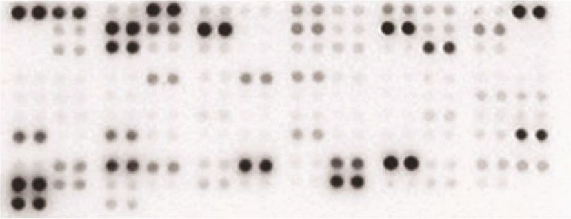
Oxidative Medicine and Cellular Longevity 3

oligodendrocyte glycoprotein peptide 35-55 (MOG35-55) Table 1: Clinical characteristics of the patients with relapsing MS
emulsified in complete Freund’s adjuvant containing and noninflammatory disorders.
2 mg/ml M. tuberculosis H37 RA on day 0, and 100 ng of per-
tussis toxin (PT) was administered intraperitoneally (i.p.) on MS (n = 7) Control (n = 7)
day 0 and day 2. Clinical scores and body weights were Gender ratio (F/M) 5/2 4/3
recorded daily starting at the beginning of immunization. Age at onset (y) 24 (15-32) 33 (20-47)
Mice were housed in accordance with the guidelines of the Disease course (m) 27.5 (7-72) —
Shaanxi Normal University Animal Care and Use Commit- EDSS at sampling 3.25 (2.5-5) —
tee and were allowed to acclimate for at least 7 days before EDSS at the last visit 1 (1-2.5) —
use.
 CSF WBC (/μl) 2 (0-20) 1 (0-4)
 According to Park et al., significant changes in lympho-
cyte activities were observed in mice treated with 20 mg/kg CSF protein (mg/L) 0.19 (0.12-0.30) 0.14 (0.07-0.34)
CAPE for 14 days [7]. Therefore, in our study, CAPE was Abnormal spinal MRI, n (%) 4/7 (57) 0/7 (0)
given at doses of 20 mg/kg or 40 mg/kg. Mice treated with Treatment, n (%) —
daily intraperitoneal injections of CAPE were further divided Azathioprine 3/7 (43)
into two groups: the prophylactic group, whose treatment Interferon-β 1/7 (14)
started on the first day of EAE induction, and the therapeutic Terflutamide 1/7 (14)
group, whose treatment started on the first day of symptom
 Oral methylprednisolone 2/7 (29)
onset. The vehicle group received daily intraperitoneal injec-
tions of PBS starting on the first day of EAE induction. Each
group contained 10 mice. At the end of the study, mononu- were incubated with Alexa Fluor-conjugated secondary anti-
clear cells were collected from spleens and the CNS after bodies (1 : 300) for 1 h at room temperature, followed by
either red blood cell lysis or Percoll separation for flow DAPI counterstaining. Images were obtained under a Leica
cytometry analysis. DM 4000 B microscope and analysed through Image J
 Clinical scores ranged from 0 to 5 as follows: 0, no signs; (National Institutes of Health, USA).
0.5, stiff tail; 1, limp tail; 2, limp tail with gait incoordination;
2.5, paralysis of one hind limb; 3, paralysis of both hind
limbs; 3.5, hind limb paralysis and weakness of one forelimb; 2.9. Statistical Analysis. Student’s t-test or the Mann–Whit-
4, moribund; and 5, death. Three areas of each lumbar ney U test was used to compare data between two groups
enlargement section (three sections/animal) were graded as (a = 0:05). Independent nonparametric data with multiple
follows: score 0, no demyelination; score 1, mild demyelin- groups, such as clinical scores, were assessed by non-
ation; score 2, severe demyelination; and score 3, massive parametric one-way ANOVA (the Kruskal-Wallis test). P <
demyelination. 0:05 was considered significant. All statistical analyses were
 performed using the GraphPad Prism 8.0 software.
2.7. Flow Cytometry. Cells were harvested and stained with an
anti-CD4 antibody for 30 min at room temperature. After
washing with PBS/1% BSA, the cells were incubated with fix- 3. Results
ation buffer at room temperature in the dark for 20 min and
washed with permeabilization (perm) buffer. After removing 3.1. Th1 Polarization Was Prominent in Patients with MS. To
the supernatant, the cells were resuspended in the perm evaluate the T cell profile in patients with MS, we measured
buffer and incubated at room temperature in the dark for the levels of several cytokines and chemokines in CSF, which
15 min. Then, the cells were centrifuged, and the pellet was can better reflect the inflammatory condition of CNS. The
resuspended in 100 μl of perm buffer. The cells were incu- clinical characteristics of the recruited patients are summa-
bated with intracellular antibodies at room temperature in rized in Table 1. No differences in the CSF protein level or
the dark for 30 min. Finally, the cells were washed twice with white cell count were found between the MS and control
PBS/1% BSA, resuspended, run on an FACS Canto II flow groups. Figure 1 shows the levels of CSF cytokines/chemo-
cytometer (BD Biosciences, San Jose, CA), and analysed by kines. The levels of proinflammatory cytokines/chemokines,
FlowJo (TreeStar, Ashland, OR). including MMP-9, IL-32a/b/c, CCL-20, CXCL-5, IL-19,
 CXCL-1, IL-16, IL-34, CCL-3, IL-15, G-CSF, CCL-5, IFN-γ,
2.8. Histopathology and Immunofluorescence. Spinal lumbar and IL-1a, as well as the level of one anti-inflammatory cyto-
enlargements were fixed in 4% paraformaldehyde, embedded kine, IL-10, were significantly elevated in the MS patients
in paraffin, and sectioned (4 μm). Some lumbar enlargement compared with the controls, and most of these cytokines
sections (3-4 mice per group; 3 sections per animal) were are associated with Th1 differentiation (e.g., IFN-γ, IL-15,
stained with HE to evaluate the degree of inflammatory cell and IL-16). Unexpectedly, we observed that the levels of
infiltration. Other sections underwent immunofluorescence some Th2-type cytokines (IL-4, IL-5, and IL-13) were also
staining as follows: sections were paraffinized, rehydrated, higher in the MS group than in the control group, which is
and blocked with 10% goat serum. Then, the sections were in accordance with limited studies [8]. No correlation was
incubated overnight at 4°C with primary antibodies against observed between significantly altered Th1 and Th2
Iba-1 (1 : 100) and MBP (1 : 200). After washing, the sections cytokines.
4 Oxidative Medicine and Cellular Longevity

 CD14
 CD30
 CD40L
 CD26
 EGF
 CXCL5
 CD178
 G-CSF
 GM-CSF
 CXCL-1
 ICAM-1
 IFN-r
 IL-1a 40
 IL-1b
 IL-1ra
 IL-2
 IL-3
 IL-4
 IL-5
 IL-6
 IL-8
 IL-10
 IL-11
 IL-12p70
 IL-13
 IL-15
 IL-16
 IL-17A
 IL-18BPa
 IL-19
 IL-22 20
 IL-23
 IL-24
 IL-27
 IL-31
 IL-32a/b/r
 IL-33
 IL-34
 IP-10
 M-CSF
 CCL3
 CCL20
 CCL19
 MMP-9
 CCL5
 CXCL12
 CCL17
 TNF-alpha
 VEGF
 MS 1

 MS 2

 MS 3

 MS 4

 MS 5

 MS 6

 MS 7

 Con 1

 Con 2

 Con 3

 Con 4

 Con 5

 Con 6

 Con 7

 (a)
 8

 6
 Fold change to Con

 4

 2

 0
 MMP-9

 IL-5

 IL-32a/b/r

 CCL20

 IL-18BPa

 TNF-alpha

 CXCL5

 IL-19

 IL-11

 CXCL-1

 IL-8

 CXCL12

 IL-16

 IL-4

 IL-34

 CCL3

 IL-33

 IL-13

 IL-10

 IL-15

 G-CSF

 CCL5

 IFN-r

 IL-1a

 (b)

Figure 1: The levels of CSF cytokines and chemokines in patients with relapsing MS (n = 7) and patients with noninflammatory disorders
(n = 7) (a). The fold changes of significantly altered cytokines/chemokines compared to those in the control group (b).

3.2. CAPE Inhibited ConA-Induced T Cell Proliferation and at a concentration of 20 μM CAPE compared to the control
Activation. As shown in Figure 2(a), cell viability was similar group (Figure 2(c)). Since CD4+ T cells play a pivotal role
among the groups treated with various concentrations of in MS, we investigated the frequency of CD4+ T cells among
CAPE for 48 h, indicating that CAPE had no toxicity to ConA-stimulated splenocytes. No differences were detected
splenocytes. To test the effect of CAPE on mitogen- among cells treated with the indicated doses of CAPE
induced cell proliferation, cells were incubated with ConA (Figure 2(d)). Upon activation, the expression of several mol-
or LPS, which mainly stimulates T cells or B cells, respec- ecules on T cells, such as CD69 (an early activation marker)
tively. CAPE significantly inhibited ConA-stimulated cell and CD25 (a middle activation marker), was increased under
proliferation in a dose-dependent manner (Figure 2(b)). In the regulation of transcription factors, such as NF-κB [9].
contrast, LPS-induced proliferation was not affected by treat- Splenocytes were stimulated with ConA for 8 h (for CD69)
ment with 0-10 μM CAPE. A significant change was observed or 24 h (for CD25). Figures 3(a) and 3(b) indicates that
Oxidative Medicine and Cellular Longevity 5

 1.5 1.5 1.5

 Cell proliferation

 Cell proliferation
 1.0 1.0 ⁎ ⁎ 1.0
Cell viability

 ⁎⁎

 ⁎⁎⁎⁎⁎⁎⁎⁎ ⁎⁎⁎⁎
 0.5 0.5 0.5

 0.0 0.0 0.0
 0 1 2.5 5 10 20 CAPE ( M) 0 0 1 2.5 5 10 20 CAPE ( M) 0 0 1 2.5 5 10 20
 CAPE ( M) ConA – + + + + + + LPS – + + + + + +
 (a) (b) (c)
 0 M 1 M 5 M
 200 200
 200
 150 150
 150
 28.4% 28.9% 29.8%
 100 100
 100

 50 50 50

 0 0 0
 0 102 103 104 105 0 102 103 104 105 0 102 103 104 105

 10 M 20 M
 200 200

 150 150
 29.3% 32.5%
 100 100

 50
 50

 0 0
 0 102 103 104 105 0 102 103 104 105
 (d)

Figure 2: Effects of CAPE on lymphocyte proliferation and CD4+ T cell expression. Cells were cultured at a concentration of 5 × 10⁶ cells/ml
per well in 96-well plates. To induce lymphocyte proliferation, cells were stimulated with ConA (5 μg/ml) or LPS (1 μg/ml) for 48 h. Cell
viability among the groups treated with various concentrations of CAPE (0-20 μM) (a). Cell proliferation of ConA- (b) and LPS-
stimulated (c) lymphocytes. The percentage of CD4+ T cells in ConA-stimulated splenocytes (d). The data (mean ± SEM) represent three
independent experiments. ∗ P ≤ 0:05, ∗∗ P ≤ 0:01, ∗∗∗∗ P ≤ 0:0001 versus the vehicle group.

10 μM CAPE pretreatment significantly suppressed not only CAPE on CD4+ T cells, we detected the levels of IFN-γ, IL-
the percentage of cells expressing CD69 and CD25 but also 17A, and Foxp3 by flow cytometry. As shown in
the percentage of CD4+CD69+ and CD4+CD25+ cells. The Figures 4(a) and 4(c), CAPE substantially inhibited the level
transcriptional activity of these activation molecule genes of IFN-γ induced after T cell stimulation with anti-CD3
has been reported to be highly dependent on NF-κB [9]. Con- and anti-CD28 antibodies for 72 h. In addition, CAPE treat-
sistent with this report, nuclear translocation of NF-κB p65 ment increased Foxp3 expression in cells at a concentration
was inhibited by 20 μM CAPE treatment after stimulation of 20 μM. No difference was observed in the percentage of
with ConA for 2 h. CD4+ T cells or CD4+IL-17A+ T cells between CAPE- and
 vehicle-treated cells. Since Th1 cells were significantly
3.3. CAPE Suppressed IFN-γ Expression and Improved Foxp3 increased in the MS patients compared to the controls, we
Expression in Naive CD4+ T Cells. To evaluate the effects of established Th1-polarizing conditions with rIL-12 and an
6 Oxidative Medicine and Cellular Longevity

 40 50

 CD69 expression rate (%)

 CD25 expression rate (%)
 40 ⁎
 30
 ⁎⁎ ⁎⁎ ⁎⁎
 30 ⁎⁎⁎
 20 ⁎⁎⁎⁎ ⁎⁎⁎⁎ ⁎⁎⁎
 20

 10
 10

 0 0
 0 1 5 10 20 0 1 5 10 20
 CAPE ( M) CAPE ( M)
 (a) (b)
 ConA – + +
 CAPE 0 M 0 M 20 M

 DAPI

 RelA

 Merge

 (c)

Figure 3: Effects of CAPE on T cell activation and NF-κB p65 nuclear translocation. Lymphocytes were cultured at a concentration of 5 × 10⁶
cells/ml per well in 96-well plates or 2 × 10⁶ cells/ml per well in 24-well plates and stimulated with ConA (5 μg/ml). The percentages of CD69
and CD25 in CD4+ T cells after ConA stimulation for 8 h or 24 h (a, b). NF-κB p65 nuclear translocation after stimulating splenocytes with
ConA for 2 h (c). The data (mean ± SEM) represent three independent experiments. ∗ P ≤ 0:05, ∗∗ P ≤ 0:01, ∗∗∗ P ≤ 0:001, ∗∗∗∗ P ≤ 0:0001
versus the vehicle group.

anti-IL-4 mAb. Figures 4(b) and 4(d) show that CAPE sig- on day 0 showed profound effects in terms of a reduced dis-
nificantly inhibited IFN-γ (P < 0:0001) but increased ease incidence and significantly reduced clinical scores from
Foxp3 (P < 0:0001) levels in CD4+ T cells. CD4+ T cells day 21 onward. More relevant to human MS management,
and CD4+IL-17A+ T cell numbers were similar between therapeutic administration of CAPE subsequent to the
CAPE- and vehicle-treated T cells. These results strongly appearance of clinical symptoms produced no benefits in
indicated that the anti-inflammatory functions of CAPE EAE. Body weights were much lower in the vehicle group
were mediated at least partly through a direct inhibitory than in the CAPE pretreatment group and were slightly
effect on the differentiation of the Th1 and Treg subsets, higher in the CAPE treatment group (Table 2; Figure 5(b)).
which may alleviate EAE in vivo. At the end of the experiment, mononuclear cells were
 collected from spleens and the CNS from mice treated with
3.4. Prophylactic CAPE Treatment Reduced EAE Severity. To CAPE and vehicle under a prophylactic regimen, and the
investigate the effect of CAPE on EAE, we administered percentages of different T cell lineages were determined.
CAPE in a prophylactic or therapeutic treatment regimen Consistent with the in vitro findings, CAPE treatment signif-
and monitored disease severity (Figure 5(a)). Prophylactic icantly reduced the level of Th1 cells in both the spleen and
CAPE groups were initially treated with 20 or 40 mg/kg. the CNS, and increased Treg numbers were found in the
We observed reductions in EAE scores at both concentra- CNS (Figure 5(e)). Compared to the vehicle group, the CAPE
tions, with no further improvement at 40 mg/kg treatment group exhibited milder inflammatory cell and
(Figure 5(b)). CAPE at 20 mg/kg was therefore used in the microglia/macrophage infiltration around the spinal cord
subsequent experiments. Administration of CAPE starting (Figures 5(c) and 5(d)). In addition, demyelination and HE
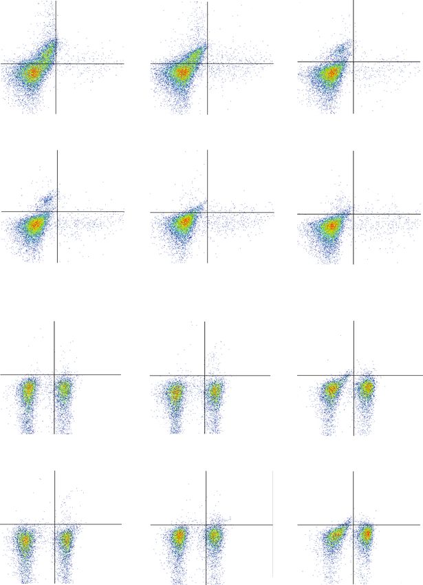
Oxidative Medicine and Cellular Longevity 7

 0 M 1 M 5 M
 14.4% 0.549% 5.58% 0.238% 5
 4.32% 0.576%
 5
 10 105 10

 104 104 104

 103 103 103

 102 102 102
 0 0 0
 82.3% 2.75% 92.8% 1.41% 91.2% 3.89%
 2 3 4
 0 10 2
 10 3
 10 4
 10 5
 0 102 103 104 105 0 10 10 10 105
 10 M 20 M 50 M
 1.86% 0.118% 1.63% 0.379% 1.15% 0.215%
 105 105 105

 104 104 104

 103 103 103
 IFN-

 102 102 102
 0 0 0
 95.2% 2.83% 94.0% 3.96% 95.0% 3.66%
 2 3 4 5 2 3 4 5 2 3 4
 0 10 10 10 10 0 10 10 10 10 0 10 10 10 105
 IL-17A

 0 M 1 M 5 M
 0.431% 0.882% Q1 Q2 0.427% 1.24%
 105 105 0.312% 0.886% 105

 104 104 104

 103 103 103

 102 102 102
 0 0 Q4 Q3 0
 54.7% 44.0% 56.7% 42.2% 57.2% 41.1%

 0 102 103 104 105 0 102 103 104 105 0 10 2
 10 3
 10 4
 105
 10 M 20 M 50 M
 1.01% 0.946% 0.651% 1.65% 2.02% 2.26%
 105 105 105

 104 104 104

 103 103 103
 Foxp3

 102 102 102
 0 0 0
 53.8% 44.3% 56.4% 41.3% 51.4% 44.3%
 2 3 4 5 2 3 4
 0 10 10 10 10 0 10 2
 103
 104
 105 0 10 10 10 105
 CD4

 (a)

 Figure 4: Continued.
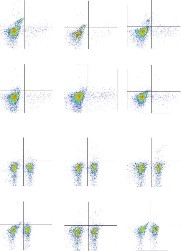
8 Oxidative Medicine and Cellular Longevity

 0 M 1 M 5 M
 30.0% 2.01% 24.5% 1.64% 5
 7.99% 0.608%
 5
 10 105 10

 104 104 104

 103 103 103

 102 102 102
 0 0 0
 67.2% 0.783% 72.4% 1.47% 89.5% 1.86%
 2 3 4
 0 10 2
 10 3
 10 4
 10 5
 0 102 103 104 105 0 10 10 10 105
 10 M 20 M 50 M
 4.86% 0.508% 4.74% 0.786% 2.23% 0.713%
 105 105 105

 104 104 104

 103 103 103
 IFN-

 102 102 102
 0 0 0
 91.9% 2.74% 91.4% 3.07% 94.8% 2.31%

 104
 2 3 4 5 2 3 4 5 2 3
 0 10 10 10 10 0 10 10 10 10 0 10 10 105
 IL-17A

 0 M 1 M 5 M
 0.277% 0.593% Q1 Q2 Q1 Q2
 105 105 0.340% 0.739% 105 0.893% 1.05%

 104 104 104

 103 103 103

 102 102 102
 0 0 Q4 Q3 0 Q4 Q3
 59.7% 39.7% 59.4% 39.6% 53.4% 44.7%

 0 102 103 104 105 0 102 103 104 105 0 102 103 104 105
 10 M 20 M 50 M
 0.412% 0.969% 0.789% 1.69% 1.57% 1.69%
 105 105 105

 104 104 104

 103 103 103
 Foxp3

 102 102 102
 0 0 0
 58.1% 40.5% 58.1% 39.4% 51.6% 45.1%
 2 3 4 5 2 3 4
 0 10 10 10 10 0 10 2
 103
 104
 105 0 10 10 10 105
 CD4

 (b)

 Figure 4: Continued.
Oxidative Medicine and Cellular Longevity 9

 The percentage of CD4+ IL-17AT cell
 The percentage of CD4+ IFN- + T cell

 The percentage of CD4+ Foxp3+ T cell
 20 2.5 ⁎⁎⁎⁎ 5

 2.0 4
 15 ⁎⁎
 1.5 3
 10
 ⁎⁎⁎ 1.0 2
 ⁎⁎⁎
 5 0.5
 ⁎⁎⁎⁎ ⁎⁎⁎⁎ ⁎⁎⁎⁎ 1

 0 0.0 0

 Vehicle

 1 M CAPE

 5 M CAPE

 10 M CAPE

 20 M CAPE

 50 M CAPE
 Vehicle

 1 M CAPE

 5 M CAPE

 10 M CAPE

 20 M CAPE

 50 M CAPE

 Vehicle

 1 M CAPE

 5 M CAPE

 10 M CAPE

 20 M CAPE

 50 M CAPE
 (c)

 The percentage of CD4+ IL-17AT cell
 The percentage of CD4+ IFN- + T cell

 The percentage of CD4+ Foxp3+ T cell

 40 5
 2.0 ⁎⁎⁎⁎
 ⁎⁎⁎⁎
 4
 30
 ⁎⁎ 1.5
 ⁎⁎
 ⁎⁎ 3
 20 1.0
 2
 ⁎⁎⁎⁎
 10 ⁎⁎⁎⁎ ⁎⁎⁎⁎ 0.5
 1
 ⁎⁎⁎⁎
 0 0.0 0
 Vehicle

 1 M CAPE

 5 M CAPE

 10 M CAPE

 20 M CAPE

 50 M CAPE

 Vehicle

 1 M CAPE

 5 M CAPE

 10 M CAPE

 20 M CAPE

 50 M CAPE

 Vehicle

 1 M CAPE

 5 M CAPE

 10 M CAPE

 20 M CAPE

 50 M CAPE
 (d)

Figure 4: Effects of CAPE on T cell differentiation in Th0- (a, c) and Th1-polarizing (b, d) conditions. Purified T cells (1 × 10⁵ cells/ml) were
stimulated with plate-coated anti-CD3 (2 μg/ml) and soluble anti-CD28 (1 μg/ml) monoclonal antibodies for 72 h in the presence or absence
of CAPE in 96-well plates. rIL-12 (10 ng/ml) and an anti-IL-4 mAb (5 μg/ml) were added along with the anti-CD28 mAb to polarize T cells
into Th1 cells. The data (mean ± SEM) represent three independent experiments. ∗ P ≤ 0:05, ∗∗ P ≤ 0:01, ∗∗∗ P ≤ 0:001, ∗∗∗∗ P ≤ 0:0001 versus
the vehicle group.

scores were significantly lower in the CAPE treatment group and nitric oxide in LPS-stimulated microglia in vitro [11],
than in the vehicle group (Figure 5(e)). suggesting that CAPE may be a potent antioxidant.
 Comparison of CSF cytokine/chemokine levels between
4. Discussion patients with relapsing MS and other noninflammatory dis-
 eases revealed that the level of IFN-γ, a typical Th1 cytokine,
Our present study showed the therapeutic potential of CAPE was significantly increased in the MS group. However, the
in alleviating disease onset and severity in an EAE model levels of Th17 cytokines, such as IL-17, IL-21, and IL-22,
through Th1 infiltration inhibition, macrophage/microglia were not increased, which is consistent with previous find-
activation, and leukocyte recruitment in the CNS. Consis- ings [12–14]. In a recent report, IFN-γ was found to be
tently, a previous study by Ilhan et al. demonstrated that increased in CSF from patients with MS, whereas increased
CAPE can protect against EAE and decrease oxidative tissue IL-17 was detected in serum [14], suggesting that the Th1
damage by blocking reactive oxygen species (ROS) produc- response plays a more critical role in orchestrating the MS
tion and NF-κB activation [10]. Activated macrophages and immunopathological cascade. Since CD4+ T cells can differ-
microglia are the main sources of ROS in the CNS, thereby entiate into various subsets with distinct immune capacities,
playing a crucial role in aggravating the immune response downregulating the frequencies of pathogenic Th1/Th17
and tissue injury. In our study, CAPE treatment efficiently populations while maximally reversing anti-inflammatory
inhibited the activation of these cells, which helps explain subsets is a reasonable strategy when treating MS [15]. Our
the therapeutic value reported by Ilhan et al. In addition, in vitro and in vivo findings indicated that CAPE was able
CAPE has been found to downregulate the secretion of to decrease the percentage of Th1 cells without altering the
inducible nitric oxide synthase (iNOS), cyclooxygenase-2, percentages of CD4+ T cells and Th17 cells. Consistently,
10 Oxidative Medicine and Cellular Longevity

 MOG35-55/CFA PTX Daily injection of CAPE
 PTX (treatment)

 Day 0 2 4 6 8 10 12 Analysis

 Daily injection of CAPE
 (pretreatment)
 (a)
 4 4

 3 3 ⁎⁎⁎⁎⁎ ⁎⁎⁎

 EAE score
 20
 ⁎
 EAE score

 Body weight (g)
 2 19
 2
 18
 1 ⁎⁎
 ⁎⁎ 1 17
 ⁎⁎⁎⁎
 16
 0 0
 9 14 19 24 29 15
 10 12 14 16 18 20 22 24

 12
 13
 14
 15
 16
 17
 18
 19
 20
 21
 22
 23
 24
 Days post immunization Days post immunization Days post immunization
 Vehicle CAPE treatment CAPE pretreatment
 CAPE 20 mg/kg CAPE pretreatment CAPE treatment
 CAPE 40 mg/kg Vehicle Vehicle
 (b)
 20 mg/kg CAPE
 Vehicle pretreatment

 HE (5X)

 HE (10X)

 (c)

 Figure 5: Continued.
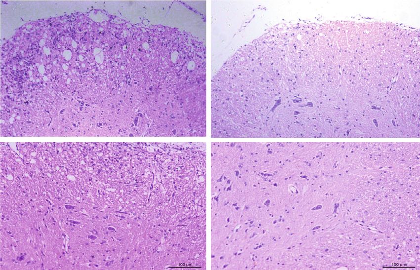
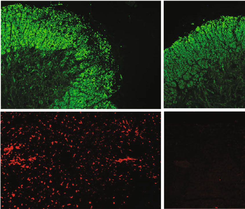
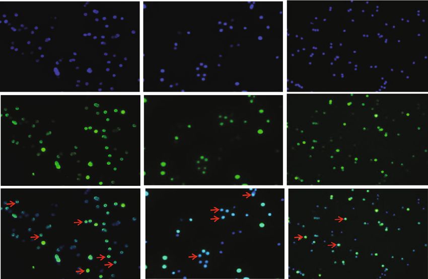
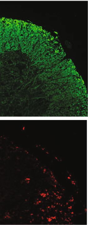
Oxidative Medicine and Cellular Longevity 11

 20 mg/kg CAPE
 Vehicle pretreatment

 MBP

 Iba-1

 (d)
 ⁎ ⁎
 The percentage of CNS CD4+ IFN- + T cell

 The percentage of spleen CD4+ Foxp3+ T cell

 ⁎
 15 3 4
 1.5

 Demyelination score
 3
 10 1.0 2
 HE score

 2

 5 0.5 1
 1

 0 0.0 0 0
 Vehicle

 CAPE

 Vehicle

 CAPE

 Vehicle

 CAPE
 Vehicle

 CAPE
 The percentage of spleen CD4+ IFN- + T cell

 ⁎ ⁎
 The percentage of CNS CD4+ Foxp3+ T cell

 ⁎⁎ ⁎⁎
 2.5 500
 4 200
 Iba-1+ cell count/picture

 2.0 400
 cell count/picture

 3 150
 1.5 300
 2 100
 1.0 200

 1 50
 0.5 100

 0.0 0 0
 0
 Vehicle

 CAPE

 Vehicle

 CAPE

 Vehicle

 CAPE

 Vehicle

 CAPE

 (e)

Figure 5: Therapeutic efficacy of CAPE in MOG35-55-induced EAE mice. Mice were immunized with MOG35-55 peptide emulsified in
complete Freund’s adjuvant. Daily CAPE administration was initiated on the day of immunization (pretreatment) or on the day of
symptom onset (treatment). Body weights and EAE scores were evaluated starting on the day of immunization. Different regimens of
CAPE administration in EAE mice (a). The impacts of different regimens and doses on the course of EAE (b). Inflammatory infiltration
assessed by HE staining (c), microglia/microphage activation assessed by Iba-1, and demyelination demonstrated by MBP (d) in EAE
mice. HE scores, demyelination scores, cell infiltration, and the proportion of T cells in EAE mice (e). Each group contained 10 mice. Data
are presented as the mean ± SEM. ∗ P ≤ 0:05, ∗∗ P ≤ 0:01, ∗∗∗ P ≤ 0:001, ∗∗∗∗ P ≤ 0:0001 versus the vehicle group.
12 Oxidative Medicine and Cellular Longevity

 Table 2: The impacts of different CAPE regimens on the EAE course.

 (a)

 Vehicle CAPE 20 mg/kg CAPE 40 mg/kg
Disease incidence 100% 60% 60%
Death 0% 0% 0%
Disease onset (d) 15:0 ± 2:0 14:5 ± 1:2 14:5 ± 1:5
EAE score at the peak time 3:1 ± 0:7 2:8 ± 0:7 2:7 ± 0:5
EAE score at sampling 2:8 ± 0:7 1:58 ± 0:8∗ 1:58 ± 0:9∗

 (b)

 Vehicle CAPE pretreatment CAPE treatment
 ∗
Disease incidence 89% 54% 88%
Death 0% 0% 0%
Disease onset (d) 14:1 ± 2:5 13:3 ± 1:3 14:4 ± 1:6
EAE score at the peak time 2:9 ± 1:4 2:4 ± 1:0 2:8 ± 0:8
 ∗
EAE score at sampling 2:8 ± 0:8 1:4 ± 1:0 2:3 ± 1:0

Choi et al. observed that CAPE significantly reduced the level promising therapeutic potential in treating EAE [21, 22].
of IFN-γ, whereas Th17 and Th2 cytokines were not affected Our data indicated that CAPE administration reduced the
in an experimental autoimmune uveoretinitis model [8]. By nuclear translocation of NF-κB p65. In addition to NF-κB,
contrast, the study by Wang et al. demonstrated that CAPE the nuclear factor of activated T cells (NFAT), which is
inhibited not only IFN-γ production in CD4+ T cells stimu- involved in regulating gene expression, including IFN-γ,
lated by CD3 and CD28 antibodies but also IL-5 in both was recently found to be a target of CAPE by Marquez
healthy subjects and asthma patients [16]. In our in vitro et al. [9].
findings, under both Th0- and Th1-polarizing conditions, Our study has several limitations. First, suppressing T cell
CAPE administration markedly improved the proportion of activity in healthy organisms may lead to negative effects
CD4+Foxp3+ T cells in vitro. In the EAE model, the fre- such as persistent infection, cancer, and autoimmunity.
quency of this T cell population, which was referred to as Therefore, whether the normal immune responses of other
Tregs, was also upregulated in the CNS. Our results highlight organs are impaired during CAPE treatment should be
the importance of CAPE in modulating T cell activation and considered at the same time. Second, although the immuno-
differentiation, which may help in the management of MS fluorescence data indicated that CAPE likely inhibits the
patients. Further efforts are required to investigate the exact nuclear translocation of NF-κB p65, Western blot will be a
mechanism by which CAPE modulates these T cell subsets. better method to confirm this conclusion. Third, the effects
 Consistent with earlier findings [9], CAPE effectively of CAPE on unstimulated lymphocytes/T cells require
inhibited ConA-stimulated T cell proliferation in vitro. Inter- further investigation.
estingly, one study reported that T cell proliferation induced In conclusion, our results indicate that CAPE exhibits
by ConA was significantly increased in mice treated with strong anti-inflammatory and immunomodulatory effects
20 mg/kg CAPE for two weeks [11]. Furthermore, an increase by suppressing NF-κB activation and T cell activity in EAE,
in the ratio of CD4+/CD8+ T cells was noted in the CAPE- implying the possibility of using CAPE as an immunomodu-
treated group compared to the vehicle group [9], which con- latory agent for MS treatment.
flicts with our findings and those of other groups [7]. We
assume that the discrepancy may be attributable to the basic Data Availability
state of the studied mice. Evaluating the impact of CAPE on
immune cells in healthy animals would be interesting. Addi- The research data used to support the findings of this study
tionally, activation of T cells induced by TCR and costimula- are included within the article (tables, figures).
tory molecules immediately promotes the activity of many
transcription factors, including the NF-κB family [17]. Conflicts of Interest
Increasing evidence has demonstrated that inhibiting NF-
κB might improve EAE by altering peripheral and CNS T cell The authors declare that they have no conflicts of interest.
infiltration, impairing T cell proliferation, and skewing T
cells towards a non-Th1/Th17 phenotype [18–20]. In addi- Authors’ Contributions
tion, many natural compounds have been identified to at
least partly target the NF-κB pathway and have exhibited YiFan Zhou and Jingqi Wang are co-first authors.
Oxidative Medicine and Cellular Longevity 13

Acknowledgments glial cells,” International Journal of Molecular Sciences,
 vol. 16, no. 12, pp. 5572–5589, 2015.
We appreciate the help of Professor Yaping Yan at the [12] K. Kaneko, D. K. Sato, I. Nakashima et al., “CSF cytokine
College of Life Sciences, Shaanxi Normal University, who profile in MOG-IgG+ neurological disease is similar to
supported the experiments. This work was supported by AQP4-IgG+ NMOSD but distinct from MS: a cross-sectional
grants from the National Natural Science Foundation of study and potential therapeutic implications,” Journal of Neu-
China (#81771300, #81971140), the Natural Science Founda- rology, Neurosurgery & Psychiatry, vol. 89, no. 9, pp. 927–936,
tion of Guangdong Province (#2017A030313853), and the 2018.
Guangzhou Science and Technology Plan Project [13] A. Mouzaki, M. Rodi, N. Dimisianos et al., “Immune Parame-
(#201904010444). ters That Distinguish Multiple Sclerosis Patients from Patients
 with Other Neurological Disorders at Presentation,” PLoS
 One, vol. 10, no. 8, article e135434, 2015.
Supplementary Materials [14] T. Khaibullin, V. Ivanova, E. Martynova et al., “Elevated levels
 of proinflammatory cytokines in cerebrospinal fluid of multi-
The chemical structure of CAPE. (Supplementary Materials) ple sclerosis patients,” Frontiers in Immunology, vol. 8,
 p. 531, 2017.
References [15] H. Kataoka, K. Sugahara, K. Shimano et al., “FTY720,
 sphingosine 1-phosphate receptor modulator, ameliorates
 [1] C. Baecher-Allan, B. J. Kaskow, and H. L. Weiner, “Multiple experimental autoimmune encephalomyelitis by inhibition
 sclerosis: mechanisms and immunotherapy,” Neuron, vol. 97, of T cell infiltration,” Cellular & Molecular Immunology,
 no. 4, pp. 742–768, 2018. vol. 2, no. 6, pp. 439–448, 2005.
 [2] B. W. van Oosten, M. Lai, S. Hodgkinson et al., “Treatment of [16] L. C. Wang, K. H. Chu, Y. C. Liang, Y. L. Lin, and B. L. Chiang,
 multiple sclerosis with the monoclonal anti-CD4 antibody “Caffeic acid phenethyl ester inhibits nuclear factor-kappaB
 cM-T412: results of a randomized, double-blind, placebo-con- and protein kinase B signalling pathways and induces
 trolled, MR-monitored phase II trial,” Neurology, vol. 49, no. 2, caspase-3 expression in primary human CD4+ T cells,”
 pp. 351–357, 1997. Clinical and Experimental Immunology, vol. 160, no. 2,
 [3] M. Franchin, I. A. Freires, J. G. Lazarini et al., “The use of pp. 223–232, 2010.
 Brazilian propolis for discovery and development of novel [17] M. Srinivasan and D. K. Lahiri, “Significance of NF-κB as a
 anti-inflammatory drugs,” European Journal of Medicinal pivotal therapeutic target in the neurodegenerative pathologies
 Chemistry, vol. 153, pp. 49–55, 2018. of Alzheimer's disease and multiple sclerosis,” Expert Opinion
 [4] K. Natarajan, S. Singh, T. J. Burke, D. Grunberger, and B. B. on Therapeutic Targets, vol. 19, no. 4, pp. 471–487, 2015.
 Aggarwal, “Caffeic acid phenethyl ester is a potent and specific [18] B. Hilliard, E. B. Samoilova, T. S. Liu, A. Rostami, and Y. Chen,
 inhibitor of activation of nuclear transcription factor NF- “Experimental autoimmune encephalomyelitis in NF-kappa
 kappa B,” Proceedings of the National Academy of Sciences, B-deficient mice: roles of NF-kappa B in the activation
 vol. 93, no. 17, pp. 9090–9095, 1996. and differentiation of autoreactive T cells,” Journal of
 [5] L. Cornara, M. Biagi, J. Xiao, and B. Burlando, “Therapeutic Immunology, vol. 163, no. 5, pp. 2937–2943, 1999.
 properties of bioactive compounds from different honeybee [19] B. A. Hilliard, N. Mason, L. Xu et al., “Critical roles of c-Rel in
 products,” Frontiers in Pharmacology, vol. 8, 2017. autoimmune inflammation and helper T cell differentiation,”
 [6] A. J. Thompson, B. L. Banwell, F. Barkhof et al., “Diagnosis of The Journal of Clinical Investigation, vol. 110, no. 6, pp. 843–
 multiple sclerosis: 2017 revisions of the McDonald criteria,” 850, 2002.
 Lancet Neurology, vol. 17, no. 2, pp. 162–173, 2018. [20] H. Zhang, J. Bi, H. Yi et al., “Silencing c-Rel in macrophages
 [7] J. H. Park, J. K. Lee, H. S. Kim et al., “Immunomodulatory dampens Th1 and Th17 immune responses and alleviates
 effect of caffeic acid phenethyl ester in Balb/c mice,” Interna- experimental autoimmune encephalomyelitis in mice,” Immu-
 tional Immunopharmacology, vol. 4, no. 3, pp. 429–436, 2004. nology and Cell Biology, vol. 95, no. 7, pp. 593–600, 2017.
 [8] J. H. Choi, K. H. Roh, H. Oh et al., “Caffeic acid phenethyl ester [21] O. Aktas, T. Prozorovski, A. Smorodchenko et al., “Green tea
 lessens disease symptoms in an experimental autoimmune epigallocatechin-3-gallate mediates T cellular NF-kappa B
 uveoretinitis mouse model,” Experimental Eye Research, inhibition and exerts neuroprotection in autoimmune enceph-
 vol. 134, pp. 53–62, 2015. alomyelitis,” Journal of Immunology, vol. 173, no. 9, pp. 5794–
 [9] N. Márquez, R. Sancho, A. Macho, M. A. Calzado, B. L. Fie- 5800, 2004.
 bich, and E. Muñoz, “Caffeic acid phenethyl ester inhibits [22] G. van Loo, M. Sze, N. Bougarne et al., “Antiinflammatory
 T-cell activation by targeting both nuclear factor of acti- properties of a plant-derived nonsteroidal, dissociated gluco-
 vated T-cells and NF-kappaB transcription factors,” The corticoid receptor modulator in experimental autoimmune
 Journal of Pharmacology and Experimental Therapeutics, encephalomyelitis,” Molecular Endocrinology, vol. 24, no. 2,
 vol. 308, no. 3, pp. 993–1001, 2004. pp. 310–322, 2010.
[10] A. Ilhan, O. Akyol, A. Gurel, F. Armutcu, M. Iraz, and
 E. Oztas, “Protective effects of caffeic acid phenethyl ester
 against experimental allergic encephalomyelitis-induced oxi-
 dative stress in rats,” Free Radical Biology and Medicine,
 vol. 37, no. 3, pp. 386–394, 2004.
[11] C. F. Tsai, Y. H. Kuo, W. L. Yeh et al., “Regulatory effects of
 caffeic acid phenethyl ester on neuroinflammation in micro-
You can also read