Cooperative interaction of Ig a and Ig b of the BCR regulates the kinetics and specificity of antigen targeting
←
→
Page content transcription
If your browser does not render page correctly, please read the page content below

International Immunology, Vol. 14, No. 10, pp. 1179±1191                                ã 2002 The Japanese Society for Immunology

Cooperative interaction of Iga and Igb of the
BCR regulates the kinetics and speci®city of
antigen targeting
Chang Li1, Karyn Siemasko2, Marcus R. Clark2 and Wenxia Song1
1Department of Cell Biology and Molecular Genetics, University of Maryland, College Park, MD 20742,

USA
2Section of Rheumatology, Department of Medicine, University of Chicago, IL 60637, USA

Keywords: antibody/antigen receptor, antigen processing, B lymphocytes, Iga/Igb heterodimer

                                                                                                                                       Downloaded from http://intimm.oxfordjournals.org/ by guest on September 29, 2015
Abstract
Following the binding of antigens, the BCR transduces signals and internalizes antigens for
processing and presentation, both of which are required for initiating an effective antibody
response. The BCR, consisting of membrane Ig and Iga/Igb heterodimer, facilitates antigen
processing by accelerating antigen targeting to the processing compartment. Previous reports
showed that Iga or Igb alone is competent for internalizing antigens. However, both Iga and Igb are
required for BCR-enhanced antigen presentation. Using chimeric proteins containing the
extracellular and transmembrane domains of human platelet-derived growth factor receptor fused
with the cytoplasmic domain of Iga or Igb, we studied the roles of the cytoplasmic tails of Iga and
Igb in BCR-mediated antigen transport. The Igb chimera rapidly moves through the endocytic
pathway to lysosomes, while the Iga chimera slows down this movement. The Iga, but not the Igb
chimera, is required for an increase in the turnover rate of the chimeras in response to stimulation.
Only when Iga and Igb chimeras are co-expressed do the chimeras rapidly and speci®cally target
antigens to the processing compartment. These ®ndings suggest that Iga and Igb play distinct
roles in BCR traf®cking, and the cooperative interaction of Iga and Igb controls and regulates the
kinetics and speci®city of antigen targeting.

Introduction
Processing and presentation of antigens by B lymphocytes to         pinocytosis or binding to surface receptors other than the BCR
T lymphocytes is the central event for the initiation of humoral    can be processed and presented, the presentation of these
responses to T cell-dependent antigens. B cells express             antigens is far less ef®cient than BCR-mediated antigen
clonally speci®c antigen receptors that sense and capture           presentation (9±11). The BCR increases the kinetics and
antigens. The binding of an antigen to the BCR initiates            speci®city of antigen targeting to facilitate antigen processing
signaling cascades (1±3) that provide the ®rst stage of signals     (5,6). The BCR captures antigens speci®cally and internalizes
for B cell activation. Subsequently, the BCR internalizes and       them from the cell surface. Upon entering the endocytic
targets the antigen to the processing compartment where             pathway, antigen±BCR complexes transiently pass through
complexes of antigenic peptides and MHC class II molecules          early endosomes and reach the MHC class II-containing
are assembled (4±6). The interaction of T cells and B cells in      compartment (MIIC) (4±6). The MIIC is located in the later part
the context of antigenic peptide±MHC class II complexes             of the endocytic system, and contains newly synthesized MHC
provides the second stage of signals for B cell activation (7,8).   class II (4,12±15), the catalyst of class II±peptide exchange,
   When the immune system initially encounters an antigen, the      DM (15,16), the DM regulator, DO (17,18) and residential
concentration of the antigen often is low, and the number of        proteins of late endosomes, like lysosomal-associated mem-
antigen-speci®c B cells and T cells is limited. The antigen-        brane glycoprotein-1 (LAMP-1). Antigen±BCR complexes are
processing ef®ciency of B cells becomes extremely critical for      degraded in the endosomes and the resulting peptides are
the induction of a rapid humoral response speci®c to the            loaded onto class II molecules in the MIIC. Although the
antigen. Although antigens internalized through ¯uid-phase          intracellular traf®cking pathway of the BCR has been well

Correspondence to: W. Song; E-mail: ws98@umail.umd.edu
Transmitting editor: L. H. Glimcher                                              Received 28 November 2002, accepted 15 July 2002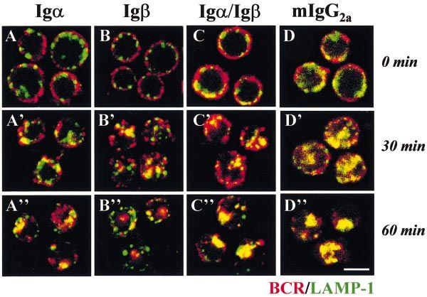
1180 Kinetics and speci®city of antigen targeting
characterized, the molecular mechanisms for the speci®c            Stimulation of the chimeras
targeting of the BCR are not well understood.                      To activate the chimeras, cells expressing different chimeras
   BCR-initiated signaling plays a major role in regulating        were incubated with PDGF-BB (100 ng/ml; Zymed, South San
antigen processing. Activating the BCR by cross-linking            Francisco, CA) in DME containing 6 mg/ml BSA and 20 mM
induces a rapid internalization of the BCR and accelerates         MOPS, pH 7.4 (DME/BSA) for 10 min, followed by mouse anti-
targeting of the BCR to the MIIC (5). Tyrosine kinase inhibitors   human PDGFRb mAb (anti-hPDGFRb) (5 mg/ml; Zymed) for 10
that block BCR signaling lower the antigen-presenting ef®-         min and then anti-mouse IgG1 (5 mg/ml; Zymed) for 20 min at
ciency of B cells and inhibit accelerated antigen transport        4°C.
(19,20). In addition, the tyrosine phosphorylation sites in the
cytoplasmic tail of the Iga chain of the BCR (21,22) and           Turnover of the surface-biotinylated chimeras
tyrosine kinase, Syk (23), have been shown to be important for
BCR-mediated antigen processing. BCR-initiated signaling           Cells were washed at 4°C with HBSS lacking phosphate and
has also been reported to induce a reorganization and              containing 20 mM Na HEPES, pH 7.4, and incubated in the
acidi®cation of late endosomes (24). These demonstrate that        same buffer containing 0.2 mg/ml sulfosuccinimidyl-6-(bioti-
there are multiple links between BCR signaling and antigen-        namido)hexanoate (Pierce, Rockford, IL) for 15 min at 4°C to
processing pathways.                                               label the surface proteins. After 15 min of incubation, a freshly
   The BCR is composed of membrane Ig (mIg) and Iga/Igb            made biotin solution was added and the incubation was

                                                                                                                                         Downloaded from http://intimm.oxfordjournals.org/ by guest on September 29, 2015
heterodimer (Iga/Igb). The Iga/Igb and mIg form a complex          extended for another 15 min at 4°C. The cells then were
through non-covalent interaction. Both Iga and Igb are             washed with DME/BSA to remove unreacted biotin, treated
essential for the development (25,26), activation (27, 28) and     with DME/BSA alone or PDGF-BB, anti-hPDGFRb and anti-
programmed cell death of B cells (29,30). These two chains         mouse IgG1 antibodies sequentially at 4°C to cross-link the
have been shown to play distinct and complementary roles in        chimeras, and chased at 37°C for varying lengths of time. The
the signal-transduction (31,32) and antigen-processing             cells were lysed with 1% NP-40 lysis buffer (1% NP-40, 50 mM
(22,33) functions of the BCR. Although either the Iga or Igb       Tris/HCl, pH 7.4, 150 mM NaCl, 5 mM EDTA and protease
chain appears to be competent for signal transduction and          inhibitors) and the cell lysates were subjected to immunopre-
antigen internalization, it has been demonstrated that both        cipitation using anti-hPDGFRb antibody. The immunoprecipi-
chains are required for an optimal level of signaling and a high   tates were analyzed by SDS±PAGE and Western blotting. The
ef®ciency of antigen presentation (22,32). Using human             biotinylated chimeras were detected using horseradish
platelet-derived growth factor receptor (hPDGFR) chimeras          peroxidase±streptavidin and analyzed by densitometry.
containing the cytoplasmic tail of either Iga or Igb, Siemasko
et al. (22) recently showed that only when Iga and Igb             Subcellular fractionation
chimeras were co-expressed did the chimeric proteins facili-       Anti-hPDGFRb antibody was iodinated to a sp. sct. of 1.0±1.5
tate antigen presentation as the BCR does, indicating that the     3 107 c.p.m./mg as previously described (35). More than 95%
interaction between Iga and Igb is crucial for the ef®ciency of    of [125I]anti-hPDGFRb antibody was precipitated by 10%
antigen presentation. Here we analyzed the intracellular           trichloroacetic acid, indicating little or no free 125I. Cells were
traf®cking of the Iga and Igb chimeras using three different       washed and incubated with 100 ng/ml PDGF-BB, 2 mg/ml
approaches, including subcellular fractionation, immuno¯uor-       [125I]anti-hPDGFRb and anti-mouse IgG1 (5 mg/ml) antibodies
escence microscopy and electron microscopy. The results            in DME/BSA at 4°C for 60 min. The cells were washed,
reported here show that Iga and Igb play distinct roles in         homogenized and subjected to the Percoll gradient centrifu-
controlling and regulating the kinetics and speci®city of          gation as previously described (13). Brie¯y, cells (~2 3 108)
antigen targeting.                                                 were homogenized in homogenization buffer (10 mM Tris, 1
                                                                   mM EDTA and 0.25 M sucrose, pH 7.4). The homogenate was
                                                                   centrifuged at 900 g for 15 min to remove nuclei and at 10,000
                                                                   g for 15 min to remove mitochondria. Then 2 ml of the
Methods
                                                                   supernatant was layered onto 9 ml Percoll (1.05 g/ml) and
Cell culture                                                       centrifuged at 34,800 gmax for 20 min. Fractions of 0.5 ml were
                                                                   collected. Radioactivity of each fraction was counted and
The mouse B cell lymphoma A20 IIA1.6 is a H-2d, IgM±, IgG2a+
                                                                   calculated as a percentage of the total cell-associated
and FcR± cell line. The A20 cells were cultured in DMEM that
                                                                   radioactivity. The total cell-associated radioactivity was
was supplemented as described (34) and contained 10%
                                                                   10,000±20,000 c.p.m.
FCS.
                                                                      To determine the distribution of molecular markers for
                                                                   different subcellular organelles on the Percoll gradient, the
Construction of the Iga and Igb chimeras                           fractions were boiled in equal volumes of the reducing sample-
Construction and expression of the chimeras containing the         loading buffer of SDS±PAGE, and centrifuged to remove
extracellular and transmembrane domains of hPDGFR and the          Percoll before subjected to SDS±PAGE and Western blotting.
cytoplasmic domain of Iga or Igb have been previously              The blots were probed for transferrin (Tf) receptor (TIB219),
described (22,32). The stably transfected A20 cells were           LAMP-1 (1D4B), mIgG2a or invariant chain (Ii; IN-1) (generous
thawed from the stocks every month and the expression levels       gifts from Dr S. K. Pierce, NIAID). MHC class II molecules were
of the chimeras were periodically examined using ¯ow               isolated from each fraction by immunoprecipitation using mAb
cytometry as previously described (32).                            MDK6 or M5/114.15.2 (PharMingen, Franklin Lakes, NJ). The
Kinetics and speci®city of antigen targeting 1181
mouse IgG1 antibodies. Background labeling using the
secondary antibody FITC±goat anti-rat IgG was negligible.
Conventional electron microscopy
Gold-conjugated goat anti-mouse IgG1 (gold±anti-mouse
IgG1) and BSA (gold±BSA) were prepared as previously
described (36). Cells were incubated sequentially with PDGF-
BB (100 ng/ml), anti-hPDGFRb (5 mg/ml) and gold±anti-mouse
IgG1 (15 nm) antibodies at 4°C for 40 min, pulsed at 37°C for
15 min, washed at 4°C and chased at 37°C for 15 or 45 min.
Gold±BSA (10 nm) was included in the pulse medium. The
cells were ®xed with 2% glutaraldehyde, post-®xed with 1%
osmium tetroxide, dehydrated, in®ltrated and embedded in
Fig. 1. The structures of the chimera proteins. The chimeric proteins epoxy resin (EM Science, Ft Washington, PA). Thin sections
contain the extracellular and transmembrane domains of human (60±90 nm) of the cells were contrasted with uranyl acetate
PDGFRa or PDGFRb fused to the cytoplasmic domain of Iga or Igb. and lead citrate, and examined in a Zeiss EM10CA electron
Since both PDGFRa and PDGFRb have the same af®nity for PDGF- microscope. To evaluate the results quantitatively, 20 cell
Downloaded from http://intimm.oxfordjournals.org/ by guest on September 29, 2015
BB, homodimers can be formed on singly transfected cells (Iga or
Igb chimeras) and heterodimers can be formed on doubly pro®les were randomly selected from sections generated from
transfected cells (Iga/Igb chimeras). The chimeras can be further three individual experiments. The numbers of different sizes of
aggregated by anti-hPDGFRb antibodies. gold particles in each cell pro®le were counted. For gold±anti-
mouse IgG1 or gold±BSA in each type of structures, percent-
ages of the total cell-associated immunogold particles were
immunoprecipitates were subjected to SDS±PAGE and calculated.
Western blotting. The blots were probed with mAb MDK6 or
M5/114.15.2. The enzymatic activities of a-mannosidase II Immunoelectron microscopy
and b-hexosaminidase were measured as previously de- Cells were incubated with PDGF-BB (100 ng/ml), anti-
scribed (39,40). Tf receptor was used to mark the plasma hPDGFR-antibody (5 mg/ml) and gold±anti-mouse IgG1 (15
membrane (PM) and early/recycling endosomes. The activity nm) sequentially at 4°C for 40 min, pulsed at 37°C for 15 min,
of a-mannosidase II marked the Golgi. The dense fractions washed at 4°C and chased at 37°C for 15 min. The cells were
containing LAMP-1 and b-hexosaminidase activity were iden- ®xed by 2% paraformaldehyde in 0.2 M phosphate buffer (pH
ti®ed as dense late endosomes and lysosomes. The dense 7.4) for 2 h at room temperature and embedded in 7.5%
fractions where MHC class II molecules and LAMP-1, but not gelatin. The gelatin blocks containing cells were immersed in
Ii, were detected were characterized as MIIC-like compart- 2.3 M sucrose in phosphate buffer for 2 h at 4°C and snap-
ments. frozen in liquid nitrogen. Ultra-thin cryosections (60±90 nm)
were collected on a mixture of sucrose and methylcellulose,
and labeled with mouse I-A/I-E (M5/114.15.2)-, Tf receptor
Immuno¯uorescence microscopy (TIB219)- and LAMP-1 (1D4B)-speci®c mAb, and gold-conju-
To label the chimeras, cells were incubated sequentially with gated rat IgG-speci®c antibody (6 nm) (Sigma), and examined
PDGF-BB (100 ng/ml), anti-hPDGFRb (5 mg/ml) and TRITC± in a Zeiss EM10CA electron microscope. Background labeling
anti-mouse IgG1 (5 mg/ml) antibodies (Zymed) for 40 min at using irrelevant antibodies was negligible.
4°C on polylysine-coated slides (Sigma, St Louis, MO). To
label the endogenous BCR, cells were incubated with 10 mg/
ml TRITC±goat anti-mouse IgG2a antibody (Southern Results
Biotechnology Associates, Birmingham, AL) at 4°C for 40
min. The cells were washed at 4°C and chased at 37°C for The chimeras containing different cytoplasmic tails have
varying lengths of time. After the chase, the cells were ®xed different turnover rates
with 4% paraformaldehyde and permeabilized by incubation Internalized antigens are proteolytically degraded in the
with a permeabilization buffer (1% gelatin, 0.05% saponin, 10 endocytic system before being loaded onto MHC class II
mM glycine and 10 mM HEPES, pH 7.4). They were then molecules. To understand how Iga and Igb function together
incubated with either LAMP-1 (1D4B)- or Tf receptor (TIB219)- to facilitate antigen processing, we determined the turnover
speci®c mAb in the permeabilization buffer, followed by FITC± rates of chimeric proteins that contain the extracellular and
goat anti-rat IgG (Jackson ImmunoResearch, West Grove, PA) transmembrane domains of hPDGFRa or b fused with the
as the secondary antibody. The cells were washed, post-®xed, cytoplasmic tail of either Iga (Iga chimera) or Igb (Igb chimera)
mounted with Gel/Mount (Biomeda, Foster City, CA) and (Fig. 1). The surfaces of A20 cells expressing Iga or Igb
analyzed using a scanning laser confocal microscope (Zeiss chimera alone or expressing both Iga and Igb chimeras (Iga/
LSM 510). Images were acquired using a 3100 oil immersion Igb chimeras) were biotinylated. The surface-biotinylated cells
objective and cropped using Photoshop (Adobe, Mountain were treated with either medium alone or PDGF-BB, anti-
View, CA). Optical sections from the middle of cells were hPDGFRb and anti-mouse IgG1 antibodies sequentially at 4°C
selected. No labeling was detectable when untransfected A20 and then chased at 37°C for up to 4 h. PDGF-BB is a dimer that
cells were incubated with anti-hPDGFRb and TRITC±anti- has an equal af®nity to PDGFRa and PDGFRb, and dimerizes1182 Kinetics and speci®city of antigen targeting
the chimeras. Anti-hPDGFRb and anti-mouse IgG1 antibodies
further cross-link the dimerized chimeras (Fig. 1). Cells were
lysed and the total chimeras in the cell lysates were
immunoprecipitated with anti-hPDGFRb antibody. For cells
that co-express Iga and Igb chimeras, only Iga chimeras that
contain the extracellular and transmembrane domains of
hPDGFRb were isolated by immunoprecipitation. Membrane
IgG2a co-puri®ed by Protein A±Sepharose beads served as
internal controls. The immunoprecipitates were analyzed
using SDS±PAGE and Western blotting. The biotinylated
proteins were detected by horseradish peroxidase±streptavi-
din (Fig. 2A) and quanti®ed by densitometry (Fig. 2B). In the
absence of cross-linking, the surface-biotinylated mIgG2a
disappeared in a time-dependent manner (Fig. 2). The
surface-biotinylated Iga/Igb chimeras in untreated cells dis-
appeared at a rate similar to the endogenous mIgG2a. By 2 h,
50% of the biotinylated Iga/Igb chimeras remained, compar-
Downloaded from http://intimm.oxfordjournals.org/ by guest on September 29, 2015
able to that of mIgG2a (Fig. 2). The reduction of surface-
biotinylated Igb chimeras was detected as early as 1 h and
faster than that of Iga/Igb chimeras. By 2 h, only 35% of the
surface-biotinylated Igb remained. In contrast, the biotinylated
Iga chimeras disappeared much slower than Iga/Igb chi-
meras. No reduction of the biotinylated Iga chimeras was
detectable until 4 h (Fig. 2). The endogenous mIgG2a in cells
expressing different chimeras disappeared at a similar rate
(Fig. 2), indicating that the differential turnover rates of the
chimeras were not the result of clonal variation. Stimulating the
chimeras by dimerization with PDGF-BB, and cross-linking
with anti-hPDGFRb and anti-mouse IgG1 antibodies (Fig. 1),
which induces the phosphorylation of the chimeras (32),
speeded up the disappearance of surface-biotinylated Iga/
Igb chimeras and Iga chimeras. With the stimulation, the Iga
chimera still disappeared slower than the Iga/Igb chimera. In
contrast, the stimulation did not affect the disappearance rate
of surface-biotinylated Igb chimeras (Fig. 2).
These data suggest that the different surface chimeras are
degraded at different rates and respond differently to stimu-
lation. Igb chimeras appear to promote the turnover of co-
expressed Iga chimeras and Iga chimeras are required for the
increase of the turnover rate in response to stimulation.
The chimeras containing different cytoplasmic tails travel
through the endocytic pathway at different kinetics
The different turnover rates of the chimeras probably are the
Fig. 2. Turnover of the surface biotinylated chimeras. The cell result of their different intracellular traf®cking routes or
surface was biotinylated at 4°C. Then the cells were treated with traf®cking kinetics. To follow the intracellular traf®cking of the
medium alone (±XL) or PDGF-BB (100 ng), anti-hPDGFRb (5 mg/ml)
chimeras, the chimeras on the cell surface were dimerized by
and anti-mouse IgG1 (5 mg/ml) antibodies sequentially (+XL) to
dimerize and cross-link the chimeras at 4°C, and chased at 37°C for PDGF-BB, labeled, and cross-linked by [125I]anti-hPDGFRb
times indicated. An equal number of cells from each chase time and anti-mouse IgG1 antibodies at 4°C. Then the cells were
point were lysed and the total chimeras in the cell lysates were washed and chased at 37°C for up to 60 min to allow the
immunoprecipitated with anti-hPDGFRb antibody. In cells chimeras to enter cells. To minimize the effect of degradation
expressing both Iga and Igb chimeras, only Iga chimeras were
immunoprecipitated by anti-hPDGFRb antibody. The heavy chain of of the chimeras on the analyses, the cellular distribution of the
endogenously expressed mIgG2a [mIgG2a (H)] was co-puri®ed by chimeras was followed in the ®rst hour of chase. The
Protein A-conjugated Sepharose beads. The immunoprecipitates subcellular locations of the chimeras were determined by
were subjected to SDS±PAGE and Western blotting. The biotinylated subcellular fractionation.
proteins were detected by horseradish peroxidase±streptavidin. (A)
The distribution of various subcellular organelles of A20
Representative blots for the surface-biotinylated chimeras and
mIgG2a (H) are shown. (B) Data from densitometry analyses were cells on the Percoll gradient was determined by a set of
plotted as percentages of total biotinylated chimeras before enzymatic markers and serological reagents. The activity of a-
warming. Averages (6SE) of the results of three independent mannosidase II, as a marker for the Golgi, was detected in
experiments are shown. light fractions 1±10 (Fig. 3B), and Tf receptor, as an early andKinetics and speci®city of antigen targeting 1183
Downloaded from http://intimm.oxfordjournals.org/ by guest on September 29, 2015
Fig. 3. Distribution of the subcellular organelles of A20 cells on the
Percoll gradient. The distribution of subcellular organelles of A20
cells on the Percoll density gradient was determined using a series Fig. 4. Subcellular distribution of the chimeras. Cells were incubated
of enzymatic markers and serological reagents. The activities of a- sequentially with PDGF-BB (100 ng/ml), [125I]anti-hPDGFRb (2 mg/ml)
mannosidase II and b-hexosaminidase were measured as previously and anti-mouse IgG1 (5 mg/ml) antibodies at 4°C, washed, and
described (39,40), and plotted as percentages of total cell- chased at 37°C for the times indicated to allow the radiolabeled
associated activity. Tf receptor, LAMP-1, mIgG2a, MHC class II a chimeras to internalize and enter different subcellular organelles.
and b chains, and Ii were detected by SDS±PAGE and Western The cells were then cooled to 4°C and subjected to subcellular
blotting. a-Mannosidase II marks the Golgi and b-hexosaminidase fractionation (13). Fractions (0.5 ml) were collected. The amount of
marks the lysosomes. Late endosomes/lysosomes are marked by radioactivity in each fraction was measured. The radioactivity in the
and LAMP-1. Dense fractions where MHC class II, LAMP-1 but not Ii fractions 1±10 was summed as the light fractions and the
were detected were identi®ed as the MIIC-like compartment. radioactivity in the fractions 17±21 was summed as the dense
fractions. The radioactivity of the light and dense fractions was
plotted as a percentage of total cell-associated radioactivity. The
total cell-associated radioactivity for each sample was 10,000±
recycling endosomal marker, was located in fractions 3±8 20,000 c.p.m. Shown are the average results (6SD) of three
(Fig. 3A). The activity of b-hexosaminidase, marking lyso- independent experiments.
somes, was detected in dense fractions 17±21 (Fig. 3B).
LAMP-1, a marker for late endosomes/lysosomes, was pri-
Using subcellular fractionation, the traf®cking of the chi-
marily found in dense fractions 17±21. Ii that is mainly located
meras from the PM to the dense late endosomes/lysosomes
in the endoplasmic reticulum (ER) and Golgi was detected in
was determined by movement of the surface-labeled chimeras
the fractions 3±10 (Fig. 3A). Thus, the PM, early endosomes, from the light (1±10) to dense fractions (17±21) (Fig. 4). After
ER and Golgi are distributed in the light fractions 1±10. LAMP- being chased at 37°C for 15 min, the majority of the chimeras
1+ vesicles in the light fractions probably are immature late were in the light fractions containing the PM and early
endosomes and transport vesicles traveling between the Golgi endosomes, and a small portion of the chimeras was detected
and endosomes. The dense fractions 17±21 contain the dense in the dense fractions. The relative amount of Igb chimeras
late endosomes and lysosomes. MHC class II molecules and (16%) in the dense fractions was higher than those of Iga (6%)
the endogenous mIgG2a BCR were detected in fractions 3±10 or Iga/Igb (10%) chimeras. By 30 min, the amount of all
and 15±21. The dense fractions where MHC class II molecules chimeras in the dense fractions was increased (Fig. 4). By 60
and mIgG2a, but not Ii, were detected are likely to contain the min, ~20% of the chimeras was detected in the dense
MIIC (13,37). As shown previously in B cell lymphoma CH27 fractions and there was no signi®cant difference among
cells (5), cross-linking the chimeras did not signi®cantly alter different chimeras (Fig. 4). When cells were chased at 37°C
the distribution of these markers on the Percoll gradient (data for a time longer than 60 min, the total cell-associated
not shown). radioactivity was dramatically decreased, especially in Igb1184 Kinetics and speci®city of antigen targeting
chimera-expressing cells, but the percentage of radioactivity ments, suggesting that different cytoplasmic tails target the
in the dense fractions did not increase (data not shown), chimeras to different compartments. To characterize sub-
suggesting the degradation of the chimeras in the dense cellular structures that the different chimeras were targeted to,
fractions. The result from the subcellular fractionation study we analyzed the ultrastructures of the endocytic compart-
shows that Igb chimeras move from the light fractions to the ments containing pulse-labeled chimeras using conventional
dense fractions slightly faster than Iga and Iga/Igb chimeras. electron microscopy. The chimeras on the cell surface were
To further analyze the traf®cking kinetics and characterize dimerized by PDGF-BB, cross-linked by anti-hPDGFRb anti-
the traf®cking pathways of different chimeras, we followed the body and labeled by gold±anti-mouse IgG1 (15 nm) at 4°C.
intracellular traf®cking of the chimeras using immuno¯uores- Cells were pulsed for 15 min at 37°C, washed at 4°C and
cence microscopy. The chimeras on the cell surface were chased at 37°C for 15 or 45 min. To mark the endocytic
labeled with anti-hPDGFRb and TRITC-conjugated anti-mouse pathway, gold±BSA (10 nm) was simultaneously taken up
IgG1 antibodies in the presence of PDGF-BB at 4°C. The during the pulse. Previously, using immunoelectron micro-
endogenous BCR was labeled with TRITC±anti-mouse IgG2a scopy, Kleijmeer et al. (15) characterized the endocytic
as a control. The cells were washed and chased at 37°C for compartments of A20 cells in great detail. Based on their
varying lengths of time. Early and recycling endosomes were analysis, ®ve types of morphologically distinct structures in
labeled with a Tf receptor-speci®c mAb (Fig. 5) and late transfected A20 cells that contained gold±BSA were identi-
endosomes/lysosomes were labeled with a LAMP-1-speci®c ®ed. The numbers of gold±anti-mouse IgG1 for the chimeras
Downloaded from http://intimm.oxfordjournals.org/ by guest on September 29, 2015
mAb (Fig. 6). Before chase, the surface-labeled BCR and or gold±BSA in each type of structures were counted and
chimeras were all found at the outer rim of cells, where they calculated as percentages of the total cell-associated im-
partially co-localized with Tf receptor (Fig. 5A±D) but not with munogold particles. Type I structures were small or tubular
LAMP-1 (Fig. 6A±D). Upon warming to 37°C, the endogenous vesicles that were located in the cell periphery and contained
BCR started to move into the cells. By 30 min, the BCR 22% of cell-associated gold±BSA after 15 min of chase (Fig. 7A
appeared as punctate staining scattered through cells and and B, and Table 1). The morphology and the presence of the
partially co-localized with Tf receptor (Fig. 5D¢) and LAMP-1 pulsed BSA after a 15-min chase suggest that the type I
(Fig. 6D¢), suggesting that the BCR moves from the PM to early structures are early endosomes. Type II structures were
endosomes and is on its way to late endosomes. After 60 min relatively bigger, electron-lucent vesicles containing a few
of chase, the majority of the BCR staining clustered in the internal vesicles (Fig. 7B). A portion of the pulsed gold±BSA
perinuclear area and co-localized with LAMP-1 extensively, an (18%) reached the type II structures after 15 min of chase
indication of its late endosomal and lysosomal location (Fig. 7B and Table 1). The type II structures appeared to be
(Fig. 6D¢¢). The staining patterns of Iga chimeras that were the intermediates between early endosomes and multi-
co-expressed with Igb chimeras at all chase times analyzed vesicular bodies (MVB) or early MVB. Type III structures had
were similar to those of the endogenous BCR (Figs 5C±C¢¢ and the typical morphology of the MVB (Fig. 7C). Type IV
6C±C¢¢). Interestingly, while their staining pattern was similar to structures were morphologically similar to the type III struc-
the endogenous BCR, Igb chimeras only brie¯y co-localized tures, but they were in close proximity to the electron-dense
with LAMP-1 around 30 min (Fig. 6B¢), and by 60 min no structures, type V structures (Fig. 7D and E). The type V
signi®cant co-localization between Igb chimeras and LAMP-1 structures were the densest organelles detected in A20 cells
was detected (Fig. 6B¢¢). No signi®cant amount of Iga chimera and contained tightly stacked membrane lamellas (Fig. 7D±F),
staining was found inside cells until 60 min (Figs 5A±A¢¢ and which are the characteristic of lysosomes. After 45 min of
6A±A¢¢). After 60 min of chase, Iga chimeras partially co- chase, gold±BSA primarily located in type III (28%), type IV
localized with LAMP-1 (Fig. 6A¢¢), but a large portion of Iga (25%) and type V (29%) structures (Table 1), suggesting that
chimeras remained co-localized with Tf receptor (Fig. 5A¢¢). they are the late endosomes and lysosomes.
In addition, the cellular distribution of LAMP-1 varied among Next we determined which type of structures different
cells expressing different chimeras. Cross-linking the chi- chimeras were targeted to. By 15 min, Iga/Igb chimeras
meras in cells expressing both Iga and Igb chimeras induced concentrated in the type II (43%) structures, the early MVB,
the movement of the LAMP-1+ vesicles from the cell periphery and the type III (23%) structures, the MVB (Fig. 7C and
to the perinuclear area (Fig. 6C¢¢) as seen in cells treated with Table 1), while Iga chimeras (51%) were primarily located in
anti-mouse IgG2a antibody (Fig. 6D¢¢). However, cross-linking the type I structures (Fig. 7A and Table 1). Meanwhile Igb
the chimeras in cells expressing either Iga or Igb chimeras chimeras (57%) had already accumulated in the type V
alone failed to induce the redistribution of the LAMP-1+ structures (Fig. 7F and Table 1). After 45 min of chase, Igb
compartment (Fig. 6A¢¢ and B¢¢), which is similar to what chimeras continued accumulating in the type V structures,
Siemasko et al. showed previously (24). reaching 65%. The majority of Iga/Igb chimeras were found in
Taken together, all the chimeras appear to travel through a the type III (54%) and type IV (26%) structures, the MVB. By
similar pathway, but at different kinetics. Iga chimeras move now, Iga chimeras had entered the type III structures (50%).
from the PM to late endosomes and lysosomes slower than Igb However, a major portion of Iga chimeras (29%) still remained
and Iga/Igb chimeras. in the type II structures, the early MVB, and no signi®cant
amount of Iga chimera was detected in the type IV and V
Ultrastructural analysis of the subcellular compartments structures (Table 1). Thus, Igb chimeras are rapidly targeted to
containing the chimeras the type V structure and Iga/Igb chimeras are accumulated in
The Igb chimera transiently passed though the LAMP-1+ MVB. The rate of Iga chimeras moving to the MVB is
compartment and was accumulated in LAMP-1± compart- signi®cantly reduced.Kinetics and speci®city of antigen targeting 1185
Downloaded from http://intimm.oxfordjournals.org/ by guest on September 29, 2015
Fig. 5. Co-localization of the chimeras with Tf receptor. Cells were incubated sequentially on ice with 100 ng/ml PDGF±BB, 5 mg/ml anti-
hPDGFRb and 5 mg/ml TRITC±anti-mouse IgG1 antibodies without pre-starvation. For the endogenous BCR, cells were incubated with TRITC±
goat anti-mouse IgG2a antibody at 4°C for 30 min. After washes, cells were chased at 37°C for varying lengths of time, washed, ®xed and
permeabilized. Then the cells were stained with a mAb speci®c for Tf receptor (TIB219) and FITC±goat anti-rat IgG as the secondary
antibody. Single optical sections from the middle of cells were acquired using a scanning laser confocal microscope. Shown are the
representative images from three or four independent experiments. Bar, 10 mm.
Fig. 6. Co-localization of the chimeras with LAMP-1. The chimeras and the endogenous BCR were labeled as described in Fig. 5. After ®xation
and permeabilization cells were stained with LAMP-1-speci®c mAb (1D4B) and FITC±goat anti-rat IgG as the secondary antibody. Single
optical sections from the middle of the cells were acquired using a scanning laser confocal microscope. Shown are the representative images
from three or four independent experiments. Bar, 10 mm.1186 Kinetics and speci®city of antigen targeting
Downloaded from http://intimm.oxfordjournals.org/ by guest on September 29, 2015
Fig. 7. Ultrastructural analysis of the endocytic compartments accessed by the chimeras. Cells were incubated sequentially on ice with PDGF-
BB (100 ng/ml), anti-hPDGFRb (5 mg/ml) and gold±anti-mouse IgG1 (15 nm) antibodies with gold±BSA (10 nm). The cells were pulsed at 37°C
for 15 min, washed at 4°C, and chased at 37°C for 15 and 45 min. The cells were then processed for electron microscopy. (A and B) Iga
chimeras. (C and D) Iga/Igb chimeras. (E and F) Igb chimeras. Roman letters indicate the structural types. G, Golgi; N, nuclei; arrows,
chimeras. Bar, 100 nm.
The type IV structures have drawn our special attention. The Moreover, at both chase times examined, the amount of either
type IV structures were MVB closely tethered to the type V Iga (1 and 4%) or Igb (2 and 16%) chimera in the type IV
structures. The close proximity of these two structures implies compartments was much less than the amount of Iga/Igb
that they undergo tethering, fusion and/or content exchanges. chimera (10 and 26%). Signi®cantly, in pairs of the attachedKinetics and speci®city of antigen targeting 1187
Table 1. Quantitative analysis of the subcellular distribution of chimeras and BSAa
Type of subcellular structures PM I II III IV V
15 min
BSA 29 22 18 20 6 5
Iga chimeras 2 51 32 14 1 0
Igb chimeras 1 2 10 28 2 57
Iga/Igb chimeras 2 15 43 23 10 7
45 min
BSA 5 3 10 28 25 29
Iga chimeras 0 16 29 50 4 1
Igb chimeras 0 0 0 19 16 65
Iga/Igb chimeras 0 0 0 54 26 20
aThe experiments were carried out as described in Fig. 7. For each condition, 20 cell pro®les were randomly selected from sections
collected from three independent experiments. Five types of intracellular structures containing gold±BSA were distinguished and their
morphological characteristics are shown. The numbers of different-sized gold particles in each type of structure were counted. Percentages of
the total cell-associated gold±anti-mouse IgG1 labeling the chimeras or gold±BSA are shown. The total number of gold particles counted for
Downloaded from http://intimm.oxfordjournals.org/ by guest on September 29, 2015
each condition is >200.
type IV and V structures, Iga/Igb chimeras were preferentially and presentation by speci®cally targeting antigens to the
located in the MVB side (Fig. 7D) and Igb chimeras were in the processing compartment (5,6). Here, using a chimeric protein
side of the type V structure, lysosomes (Fig. 7E). This suggests system, we found that the chimeras containing the cytoplas-
that Iga/Igb chimeras, but not Igb chimeras, have the ability to mic tail of Iga or Igb moved through the endocytic pathway
remain in the MVB to delay the movement to lysosomes. and were degraded at different rates. Only when Iga and Igb
In addition, the cellular distribution of the type III, IV and V chimeras were co-expressed were the chimeras targeted to
structures in cells expressing both Iga and Igb chimeras was the MIIC-like compartment. Our ®ndings suggest that the
pronouncedly different from that seen in cells expressing cytoplasmic domains of Iga and Igb play distinct roles in the
either Iga or Igb chimeras alone. In cells expressing Iga/Igb intracellular traf®cking of the BCR, and the interaction of Iga
chimeras, these structures clustered in a greater number and Igb determines the kinetics of BCR traveling through the
(Fig. 8A) than those in cells expressing Iga or Igb chimeras endocytic pathway and the endocytic compartment that the
alone (Fig. 8B). This is consistent with our immuno¯uores- BCR is targeted to.
cence microscopy studies that showed that cross-linking the The antigen-processing compartment, MIIC, has been
endogenous BCR or Iga/Igb chimeras, but not Iga chimeras or characterized as a conventional endocytic compartment that
Igb chimeras, induced the clustering of the LAMP-1+ vesicles. is located in the late part of the endocytic pathway (15). One of
To con®rm that the type III and IV structures where Iga/Igb the important roles of the BCR in antigen processing is to
chimeras are targeted to are the MIIC or MIIC-like compart- accelerate the intracellular movement of antigens to the MIIC.
ment, the co-localization of the chimeras with MHC class II Our results generated from three different approaches,
molecules, LAMP-1 or Tf receptor was analyzed using including subcellular fractionation, immuno¯uorescence and
immunoelectron microscopy. In addition to the multi-vesicular electron microscopy, consistently showed that the chimeras
or multi-lamellar morphology, the MIIC contains MHC class II containing the different cytoplasmic tails of the BCR move
molecules and LAMP-1, but not Tf receptor. Cells were pulsed through the endocytic pathway at different speeds. The
with PDGF-BB, anti-hPDGFRb and gold±anti-mouse IgG1 (10 cytoplasmic tail of Iga slows down, and the cytoplasmic tail
nm) antibodies, and chased at 37°C for 15 min as described of Igb speeds up, the movement of the chimeras through the
above. Then cells were ®xed, embedded, in®ltrated and snap- endocytic pathway, indicating that the cytoplasmic tails of Iga
frozen in liquid nitrogen. The cryo-thin sections of cells were and Igb play distinct roles in controlling the traf®cking kinetics
labeled with either MHC class II-, LAMP-1- or Tf receptor- of the BCR. Iga chimeras co-expressed with Igb chimeras
speci®c mAb and gold-conjugated secondary antibodies (6 move through the endocytic pathway at a speed faster than
nm). After 15 min of chase, Iga/Igb chimeras co-localized with that of Iga chimeras alone and slower than that of Igb chimeras
class II molecules (Fig. 9A) and LAMP-1 (Fig. 9B) in the MVB, alone, suggesting that Iga and Igb cooperatively control the
but not with Tf receptor (data not shown). In contrast, no traf®cking kinetics of the BCR. The cytoplasmic tails of Iga/Igb
signi®cant co-localization of Igb chimeras with class II (Fig. 9C) potentially carry the targeting signals for the BCR. The
or LAMP-1 (data no shown) was detected. Thus, Iga/Igb interaction between Iga and Igb chains may control the
chimeras, but not Igb chimeras, are targeted to the MIIC-like exposure of right targeting signals in response to BCR
compartment. activation. At present, the targeting signals carried by Iga/
Igb have not been completely identi®ed.
The cytoplasmic tails of Iga and Igb appear to be one of the
Discussion
important factors that control the turnover rate of the BCR.
The binding of antigens to the BCR induces signaling Surface-labeled chimeras with different cytoplasmic tails are
cascades and rapid internalization, processing and presen- degraded at different rates. Compared to that of Iga/Igb
tation of the antigens. The BCR facilitates antigen processing chimeras, the reduction of the surface-biotinylated Igb1188 Kinetics and speci®city of antigen targeting
Downloaded from http://intimm.oxfordjournals.org/ by guest on September 29, 2015
Fig. 8. Aggregation of Iga/Igb chimeras induces the clustering of late endosomes and lysosomes. Cells were treated and processed as
described in Fig. 7. (A) Cell expressing both Iga and Igb. (B) Cell expressing Igb chimeras only. Bar, 100 nm.
chimeras was detected at least 30 min earlier and the degradation of antigens do not necessarily lead to a higher
reduction of the surface-biotinylated Iga chimeras was ef®ciency of antigen processing and presentation.
detected until 2 h later. The traf®cking kinetics of the chimeras The ®nding here raises an interesting question why the fast
is apparently correlated to their turnover rate. The faster the runner, the Igb chimera, is not able to facilitate antigen
chimeras reach late endosomes and lysosomes, the more processing and presentation. Our ultrastructural analyses
rapidly they are degraded. Among the three chimeras tested provide an explanation for this question. Iga/Igb chimeras, but
here, the Igb chimera has the highest and the Iga the lowest not Igb chimeras, are targeted to the MIIC-like compartment
traf®cking kinetics and turnover rate. However, the Igb that contains MHC class II and LAMP-1, and has multi-
chimera is not able to facilitate antigen processing and vesicular morphology. The movement of Iga chimeras to the
presentation to a level similar to Iga/Igb chimeras (22), MIIC-like compartment is delayed by their slow kinetics. Igb
suggesting that accelerated intracellular movement and chimeras transiently pass through the LAMP-1+ compartmentKinetics and speci®city of antigen targeting 1189
Downloaded from http://intimm.oxfordjournals.org/ by guest on September 29, 2015
Fig. 9. Co-localization of the chimeras with MHC class II molecules. Cells were incubated sequentially on ice with PDGF-BB (100 ng/ml), anti-
hPDGFRb (5 mg/ml) and gold±anti-mouse IgG1 (15 nm) antibodies. The cells were pulsed at 37°C for 15 min, washed at 4°C and chased at
37°C for 15 min. The cells were ®xed by 2% paraformaldehyde, embedded in 7.5% gelatin and snap-frozen in liquid nitrogen. Ultra-thin
cryosections were collected and labeled with anti-mouse I-A/I-E (M5/114.15.2) (A and C) or LAMP-1 (1D4B) (B) antibodies, and gold±anti-rat
IgG antibody (6 nm). (A and B) Cell expressing both the Iga and Igb chimeras. (C) Cell expressing Igb chimeras. Arrows, MHC class II (A) or
LAMP-1 (B). Bar, 100 nm.
and MVB, and are targeted to electron-dense vesicles that do contrast, Igb chimeras quickly move through the MVB down to
not contain MHC class II, LAMP-1 or Tf receptor. Their the dense vesicles. This suggests that Iga/Igb chimeras not
morphology and degradative properties suggest that these only move to the MIIC-like compartments quickly, but also are
dense vesicles are the mature lysosomes. It is unclear why capable of remaining there, which probably provides a
LAMP-1 is not located in these mature lysosomes. suf®cient amount of time for antigen fragmentation and
Signi®cantly, while Iga/Igb chimeras reach the MVB as fast peptide loading.
as Igb chimeras, Iga/Igb chimeras are able to remain in the Signals transduced through Iga/Igb are important regulat-
MVB side of the clusters of the MVB and dense vesicles. In ing factors for BCR traf®cking. Iga and Igb chains play1190 Kinetics and speci®city of antigen targeting
different roles in signal transduction. Using the same set of Abbreviations
chimeric proteins, Luisiri et al. (32) showed that when ER endoplasmic reticulum
expressed individually, Iga and Igb chimeras all induce a Iga/Igb Iga/Igb heterodimer
low level of protein tyrosine phosphorylation in response to Ii invariant chain
stimulation; however, the phosphorylation of the Igb chimera is LAMP-1 lysosomal associated membrane glycoprotein-1
MIIC MHC class II-containing compartment
very transient. When co-expressed, Iga/Igb chimeras induce a mIg membrane Ig
much higher level of protein phosphorylation than Iga or Igb hPDGFR human platelet-derived growth factor receptor
chimeras alone, and the Iga tails of Iga/Igb chimeras are MVB multi-vesicular body
dominantly phosphorylated. Here we found that Iga and Iga/ PM plasma membrane
Tf transferrin
Igb chimeras, but not Igb chimeras, increase their turnover XL cross-linking
rate in response to stimulation, suggesting that the signal-
transducing ability of the chimeras correlates with their
turnover rate, and that signaling induced by Iga chimeras or References
Iga/Igb chimeras is suf®cient to promote the degradation of 1 Cambier, J. C., Pleiman, C. M. and Clark, M. R. 1994. Signal
the chimeras. Since the Iga tail is dominantly phosphorylated transduction by the B cell antigen receptor and its coreceptors.
upon activation, it appears that Iga, but not Igb, plays a major Annu. Rev. Immunol. 12:457.
2 Reth, M. and Wienands, J. 1997. Initiation and processing of
Downloaded from http://intimm.oxfordjournals.org/ by guest on September 29, 2015
role in coordinating the signaling and antigen-targeting func-
signals from the B cell antigen receptor. Annu. Rev. Immunol.
tions of the BCR. 15:453.
Various chimeras have been used for studies of functions of 3 Kurosaki, T. 1999. Genetic analysis of B cell antigen receptor
Iga and Igb, and these studies have shown that Iga and Igb signaling. Annu. Rev. Immunol. 17:555.
play important roles in BCR-mediated antigen processing 4 West, M. A., Lucocq, J. M. and Watts, C. 1994. Antigen
processing and class II MHC peptide-loading compartments in
(23,33,38). Our studies using the chimeras consisting of the
human B-lymphoblastoid cells [see Comments]. Nature 369:147.
extracellular and transmembrane domains of PDGFR fused 5 Song, W., Cho, H., Cheng, P. and Pierce, S. K. 1995. Entry of B
with the cytoplasmic tail of Iga or Igb extend the previous cell antigen receptor and antigen into class II peptide-loading
®nding by demonstrating a cooperative role of Iga and Igb compartment is independent of receptor cross-linking. J.
chains in antigen targeting. Since PDGFR functions as a Immunol. 155:4255.
6 Cheng, P. C., Steele, C. R., Gu, L., Song, W. and Pierce, S. K.
dimer, the Iga and Igb chimeras have the potential to form 1999. MHC class II antigen processing in B cells: accelerated
homodimers when they are expressed individually or to form intracellular targeting of antigens. J. Immunol. 162:7171.
heterodimers when they are co-expressed. These chimeras 7 Lanzavecchia, A. 1985. Antigen-speci®c interaction between T
may not completely represent the native BCR since they do not and B cells. Nature 314:537.
8 Watts, C. 1997. Capture and processing of exogenous antigens
interact with mIg, and the Iga and Igb chimeras may not all for presentation on MHC molecules. Annu. Rev. Immunol. 15:821.
form heterodimers when treated with PDGF-BB. In this study, 9 Casten, L. A., Lakey, E. K., Jelachich, M. L., Margoliash, E. and
the behavior of Iga chimeras in cells co-expressing Iga and Pierce, S. K. 1985. Anti-immunoglobulin augments the B-cell
Igb chimeras was followed. Therefore, the results re¯ect the antigen-presentation function independently of internalization of
receptor±antigen complex. Proc. Natl Acad. Sci. USA 82:5890.
combination of Iga/Igb chimera heterodimers and Iga chimera
10 Casten, L. A. and Pierce, S. K. 1988. Receptor-mediated B cell
homodimers. Cross-linking with an antibody speci®c for antigen processing: increased antigenicity of a globular protein
hPDGFRb brings the heterodimers and homodimers together. covalently coupled to antibodies speci®c for B cell surface
The exact percentage of chimeras that form heterodimers is structures. J. Immunol. 140:404.
11 Casten, L. A., Kaumaya, P. and Pierce, S. K. 1988. Enhanced T
unknown. However, our results showed that the co-expression
cell responses to antigenic peptides targeted to B cell surface Ig,
of Igb chimeras with Iga chimeras increases the traf®cking Ia, or class I molecules. J. Exp. Med. 168:171.
kinetics and turnover rate of Iga chimeras, and allows Iga 12 Harding, C. V. and Geuze, H. J. 1993. Immunogenic peptides
chimeras to enter and remain in the MIIC. This shows that Iga bind to class II MHC molecules in an early lysosomal
and Igb chimeras do interact with each other when they are co- compartment. J. Immunol. 151:3988.
13 Qiu, Y., Xu, X., Wandinger-Ness, A., Dalke, D. P. and Pierce, S. K.
expressed, and suggests that the interaction of Iga and Igb 1994. Separation of subcellular compartments containing distinct
generates synergetic and cooperative actions that control and functional forms of MHC class II. J. Cell Biol. 125:595.
regulate the traf®cking kinetics, targeting speci®city and 14 Rudensky, A. Y., Maric, M., Eastman, S., Shoemaker, L., DeRoos,
turnover rate of the BCR. The fact that the Iga/Igb chimeras P. C. and Blum, J. S. 1994. Intracellular assembly and transport of
endogenous peptide±MHC class II complexes. Immunity 1:585.
are functionally similar to the BCR supports the notion that Iga/ 15 Kleijmeer, M. J., Morkowski, S., Grif®th, J. M., Rudensky, A. Y. and
Igb and their interaction are required and suf®cient for Geuze, H. J. 1997. Major histocompatibility complex class II
accelerated and speci®c antigen targeting. compartments in human and mouse B lymphoblasts represent
conventional endocytic compartments. J. Cell Biol. 139:639.
16 Schafer, P. H., Green, J. M., Malapati, S., Gu, L. and Pierce, S. K.
1996. HLA-DM is present in one-®fth the amount of HLA-DR in the
Acknowledgements class II peptide-loading compartment where it associates with
leupeptin-induced peptide (LIP)±HLA-DR complexes. J. Immunol.
This work was supported by National Institute of Health Grants 157:5487.
AI42093 (to W. S.) and GM52736 (to M. R. C.). K. S. was supported by 17 Liljedahl, M., Kuwana, T., Fung-Leung, W. P., Jackson, M. R.,
a Postdoctoral Fellowship from the Cancer Research Institute. We Peterson, P. A. and Karlsson, L. 1996. HLA-DO is a lysosomal
thank Dr Susan K. Pierce for providing antibodies and the Laboratory resident which requires association with HLA-DM for ef®cient
for Biological Ultrastructure at University of Maryland for technical intracellular transport. EMBO J. 15:4817.
support (Contribution no. 90). 18 van Lith, M., van Ham, M., Griekspoor, A., Tjin, E., Verwoerd, D.,Kinetics and speci®city of antigen targeting 1191
Calafat, J., Janssen, H., Reits, E., Pastoors, L. and Neefjes, J. 29 Yao, X. R., Flaswinkel, H., Reth, M. and Scott, D. W. 1995.
2001. Regulation of MHC class II antigen presentation by sorting Immunoreceptor tyrosine-based activation motif is required to
of recycling HLA-DM/DO and class II within the multivesicular signal pathways of receptor-mediated growth arrest and
body. J. Immunol. 167:884. apoptosis in murine B lymphoma cells. J. Immunol. 155:652.
19 Pure, E. and Tardelli, L. 1992. Tyrosine phosphorylation is 30 Tseng, J., Eisfelder, B. J. and Clark, M. R. 1997. B-cell antigen
required for ligand-induced internalization of the antigen receptor-induced apoptosis requires both Ig alpha and Ig beta.
receptor on B lymphocytes. Proc. Natl Acad. Sci. USA 89:114. Blood 89:1513.
20 Wagle, N. M., Kim, J. H. and Pierce, S. K. 1998. Signaling through 31 Kim, K. M., Alber, G., Weiser, P. and Reth, M. 1993. Differential
the B cell antigen receptor regulates discrete steps in the antigen signaling through the Ig-alpha and Ig-beta components of the B
processing pathway. Cell. Immunol. 184:1. cell antigen receptor. Eur. J. Immunol. 23:911.
21 Cassard, S., Salamero, J., Hanau, D., Spehner, D., Davoust, J., 32 Luisiri, P., Lee, Y. J., Eisfelder, B. J. and Clark, M. R. 1996.
Fridman, W. H. and Bonnerot, C. 1998. A tyrosine-based signal Cooperativity and segregation of function within the Ig-alpha/beta
present in Ig alpha mediates B cell receptor constitutive heterodimer of the B cell antigen receptor complex. J. Biol. Chem.
internalization. J. Immunol. 160:1767. 271:5158.
22 Siemasko, K., Eisfelder, B. J., Stebbins, C., Kabak, S., Sant, A. J., 33 Bonnerot, C., Lankar, D., Hanau, D., Spehner, D., Davoust, J.,
Song, W. and Clark, M. R. 1999. Ig alpha and Ig beta are required Salamero, J. and Fridman, W. H. 1995. Role of B cell receptor Ig
for ef®cient traf®cking to late endosomes and to enhance antigen alpha and Ig beta subunits in MHC class II-restricted antigen
presentation. J. Immunol. 162:6518. presentation. Immunity 3:335.
23 Lankar, D., Briken, V., Adler, K., Weiser, P., Cassard, S., Blank, U., 34 Jelachich, M. L., Grusby, M. J., Clark, D., Tasch, D., Margoliash,
Viguier, M. and Bonnerot, C. 1998. Syk tyrosine kinase and B cell E. and Pierce, S. K. 1984. Synergistic effects of antigen and
Downloaded from http://intimm.oxfordjournals.org/ by guest on September 29, 2015
antigen receptor (BCR) immunoglobulin-alpha subunit determine soluble T-cell factors in B- lymphocyte activation. Proc. Natl Acad.
BCR-mediated major histocompatibility complex class II- Sci. USA 81:5537.
restricted antigen presentation. J. Exp. Med. 188:819. 35 Goldstein, J. L., Basu, S. K. and Brown, M. S. 1983. Receptor-
24 Siemasko, K., Eisfelder, B. J., Williamson, E., Kabak, S. and Clark, mediated endocytosis of low-density lipoprotein in cultured cells.
M. R. 1998. Cutting edge: signals from the B lymphocyte antigen Methods Enzymol. 98:241.
receptor regulate MHC class II containing late endosomes. J. 36 Slot, J. W. and Geuze, H. J. 1985. A new method of preparing
Immunol. 160:5203. gold probes for multiple-labeling cytochemistry. Eur. J. Cell Biol.
25 Reichlin, A., Hu, Y., Meffre, E., Nagaoka, H., Gong, S., Kraus, M., 38:87.
Rajewsky, K. and Nussenzweig, M. C. 2001. B cell development is 37 Kleijmeer, M. J., Ossevoort, M. A., van Veen, C. J., van
arrested at the immature B cell stage in mice carrying a mutation Hellemond, J. J., Neefjes, J. J., Kast, W. M., Melief, C. J. and
in the cytoplasmic domain of immunoglobulin beta. J. Exp. Med. Geuze, H. J. 1985. MHC class II compartments and the kinetics of
193:13. antigen presentation in activated mouse spleen dendritic cells. J.
26 Gong, S. and Nussenzweig, M. C. 1996. Regulation of an early Immunol. 154:5715.
developmental checkpoint in the B cell pathway by Ig beta. 38 Patel, K. J. and Neuberger, M. S. 1993. Antigen presentation by
Science 272:411. the B cell antigen receptor is driven by the alpha/beta sheath and
27 Sanchez, M., Misulovin, Z., Burkhardt, A. L., Mahajan, S., Costa, occurs independently of its cytoplasmic tyrosines. Cell 74:939.
T., Franke, R., Bolen, J. B. and Nussenzweig, M. 1993. Signal 39 Tulsiani, D. R., Hubbard, S. C., Robbins, P. W. and Touster, O.
transduction by immunoglobulin is mediated through Ig alpha and 1982. alpha-D-Mannosidases of rat liver Golgi membranes.
Ig beta. J. Exp. Med. 178:1049. Mannosidase II is the GlcNacMan5-cleaving enzyme in
28 Williams, G. T., Peaker, C. J., Patel, K. J. and Neuberger, M. S. glycoprotein biosynthesis and mannosidases Ia and Ib are the
1994. The alpha/beta sheath and its cytoplasmic tyrosines are enzymes converting Man9 precursors to Man5 intermediates.
required for signaling by the B-cell antigen receptor but not for J. Biol. Chem. 257:3660.
capping or for serine/threonine-kinase recruitment. Proc. Natl 40 Suzuki, K. 1987. Enzymatic diagnosis of sphingolipidoses.
Acad. Sci. USA 91:474. Methods Enzymol. 138:727.You can also read