CircRNA circ_0004370 promotes cell proliferation, migration, and invasion and inhibits cell apoptosis of esophageal cancer via miR-1301-3p/COL1A1 axis
←
→
Page content transcription
If your browser does not render page correctly, please read the page content below
Open Medicine 2021; 16: 104–116
Research Article
Xiaobo Chen, Hongwen Sun*, Yunping Zhao, Jing Zhang, Guosheng Xiong, Yue Cui,
Changcheng Lei
CircRNA circ_0004370 promotes cell proliferation, migration,
and invasion and inhibits cell apoptosis of esophageal cancer
via miR-1301-3p/COL1A1 axis
https://doi.org/10.1515/med-2021-0001 3p/COL1A1 axis played the critical role in EC to regulate the
received July 14, 2020; accepted October 30, 2020 cell activities.
Abstract Conclusion ‒ Circ_0004370 promotes EC proliferation,
Background ‒ The aim of this study was to investigate migration and invasion, and EMT process and suppresses
the circ_0004370 expression in EC, its effects on cell pro- apoptosis by regulating the miR-1301-3p/COL1A1 axis,
liferation, apoptosis, migration, invasion, and epithelial– indicating that circ_0004370 may be used as a potential
mesenchymal transition (EMT) process, and the underlying therapeutic target for EC.
regulatory mechanisms in EC. Keywords: esophageal cancer, circ_0004370, miR-1301-
Methods ‒ The protein levels of COL1A1 and EMT-related 3p, COL1A1
proteins were detected by western blot. The role of
circ_0004370 on cell viability, proliferation, and apoptosis
was analyzed by Cell Counting Kit-8 (CCK-8) assay, colony
formation assay, and flow cytometry, respectively. The 1 Introduction
transwell assay was used to examine cell migration and
invasion. The binding sites between miR-1301-3p and circ_ Esophageal cancer (EC) is the seventh most common
0004370 or COL1A1 were predicted by starbase software diagnosed cancer and has the sixth highest mortality
and confirmed by dual-luciferase reporter assay and RNA rate among cancer diseases [1]. There are two subtypes
pull-down assay. of this disease: esophageal squamous-cell carcinoma
Results ‒ We discovered that circ_0004370 was remark- (ESCC) and esophageal adenocarcinoma (EAC) [2].
ably upregulated in EC tissues and cells. Knockdown of Although a few effective methods such like surgery, che-
circ_0004370 inhibited cell proliferation, migration as motherapy, and radiotherapy are used for EC treatment,
well as invasion, and promoted apoptosis in vitro, while the survival rate of advanced patients is still less than
its effect was rescued by miR-1301-3p inhibition. And 20% [3]. Therefore, further exploring the underlying
circ_0004370 mediated the EMT process in EC cells. molecular mechanism of EC pathogenesis is quite urgent
Moreover, we explored its regulatory mechanism and for researchers to develop novel therapeutic targets for EC
found that circ_0004370 directly bound to miR-1301-3p patients.
and COL1A1 was verified as a target of miR-1301-3p. Noncoding RNAs are the type of RNAs that cannot be
COL1A1 was highly expressed in EC cells and upregulation translated into protein. There are many functional non-
of COL1A1 reversed the effects of miR-1301-3p on cell pro- coding RNAs such as long noncoding RNAs (lncRNAs),
liferation, migration, invasion, and apoptosis. In addition, microRNAs (miRNAs), and circular RNAs (circRNAs) [4].
silencing of circ_0004370 reduced tumor volumes and Recently, emerging evidence proved that circRNA might
weights in vivo. We showed that circ_0004370/miR-1301- play a critical role in cell biology. CircRNA is formed from
the continuous closed-loop structure which means the
structure is more stable [5]. Due to its special structure,
* Corresponding author: Hongwen Sun, Department of Thoracic CircRNA is not easy to degrade compared with normal
Surgery, The First Affiliated Hospital of Kunming Medical University, RNA under the treatment of Actinomycin D or RNase R
No 295 Xichang Road, Kunming 650032, Yunnan, China, [6]. In addition, recent publication showed that the cir-
e-mail: ubr9css@163.com, tel: +86 0871 65324888
cRNA might function as a sponge of miRNA to inhibit the
Xiaobo Chen, Yunping Zhao, Jing Zhang, Guosheng Xiong, Yue Cui,
Changcheng Lei: Department of Thoracic Surgery, The First Affiliated
function of miRNA and affect the miRNA target gene [7].
Hospital of Kunming Medical University, No 295 Xichang Road, Nowadays, the role of circRNA in EC remains to be
Kunming 650032, Yunnan, China elucidated. Research reported that hsa_circ_0004370
Open Access. © 2021 Xiaobo Chen et al., published by De Gruyter. This work is licensed under the Creative Commons Attribution 4.0
International License.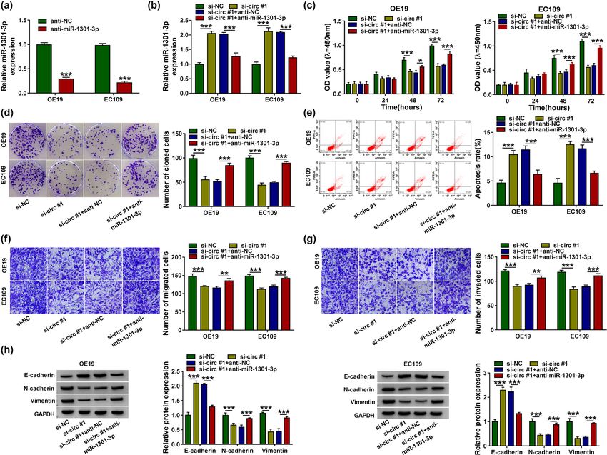
Circ_0004370/miR-1301-3p/COL1A1 axis modulates EC cells function 105
expression in EC was dramatically increased than adja- 2 Methods and materials
cent normal tissues [8]. Nevertheless, the regulatory
mechanism and specific function of circ_0004370 in
2.1 Patients and specimens
EC cells are not fully clear.
miRNAs are the short noncoding RNA of about
Fifty pairs of EC tissues and nearby healthy esophageal
18-24nt. MiRNAs play pivotal roles in gene regulation
tissues were obtained from EC patients diagnosed at the
by binding to mRNAs of targeted protein-coding genes,
First Affiliated Hospital of Kunming Medical University
then inhibits or promotes gene expression in transla-
from April 2018 to January 2019. Detailed clinicopatholo-
tional level and posttranscriptional level [9]. Thus,
gical features of all patients are shown in the Table 1. All
miRNA is the key factor for many biological procedures.
patients wrote informed consents and had not undergone
Many existing studies confirmed that miRNA aberrant
any other treatment. This experiment received the approval
expression happened in various diseases [10]. It has
from the human ethics committee of the First Affiliated
been shown that miRNAs are related to the human cancer
Hospital of Kunming Medical University.
development and act as tumor promoters or suppressors.
The first research found that miR-1301-3p directly bound
to oncogene neuroblastoma Ras viral homolog (N-Ras)
and acted as a tumor inhibitor in glioma [11]. Besides, 2.2 Cell culture and transfection
miR-1301-3p was suggested to be an effective biomarker
for colorectal cancer [12]. Furthermore, a recent article The human esophageal adenocarcinoma cell line (OE19)
showed that miR-1301-3p repressed cell viability of and esophageal squamous-cell carcinoma cell line (KYSE410,
human breast cancer by directly targeting the immature EC109 and TE11) were bought from European Collection
colon carcinoma transcript 1 (ICT1) [13]. Zhang et al. of Authenticated Cell Cultures (ECACC, Salisbury, UK).
proved that miR-1301-3p/INCENP axis played the Esophageal epithelial cell line of human (HEEC) was
crucial role in the development of ESCC [14], which obtained from ScienCell Company (San Diego, CA, USA).
provided us new insights into the mechanism of miR- For the cell culture, all cells were cultured in basal DMEM
1301-3p in EC.
Collagen type I alpha 1 (COL1A1) is a type of collagen.
Researchers have discovered that collagen is the impor- Table 1: The correlation between circ_0004370 expression and
tant protein component in teeth, bones, the adhesion of clinicopathological features of patients with ESCC
tumor cell, and extracellular matrix (ECM) [15–17]. In
previous studies, COL1A1 was upregulated in cervical Parameters Low- High- P value
cancer cells and restrained cell apoptosis [18]. In addi- circ_0004370 circ_0004370
tion, a recent study demonstrated that COL1A1 partici- (n = 25) (n = 25)
pated in epithelial-to-mesenchymal transition (EMT) Gender
process in breast cancer [19]. Furthermore, Yin et al. Male 16 12 0.254
manifested that COL1A1 enhanced cell proliferation, Female 9 13
Age (years)
migration, and invasion in ESCC [20], which implied
≤60 16 13 0.390
that COL1A1 was a crucial factor in cancer development >60 9 12
and progression. Smoking status
In this study, we uncovered that knockdown of Yes 13 7 0.083
circ_0004370 in EC was linked to restrain EC cell viabi- No 12 18
lity, proliferation, apoptosis, migration, invasion, and Histological grade
Low or undiffer 11 17 0.087
in vivo tumor formation. In the study of its regulatory
Middle or high 14 8
mechanism, we found that circ_0004370 bound to miR- TNM stages
1301-3p and inhibited its expression in EC cells. In I and II 8 19 0.002**
addition, miR-1301-3p directly targeted COL1A1 and miR- III 17 6
1301-3p overexpression reduced the expression of COL1A1. Size
≤4 cm 10 18 0.023*
Thus, a novel regulatory mechanism of circ_0004370/miR-
>4 cm 15 7
1301-3p/COL1A1 axis could be potential targets for EC
treatment and diagnosis. *P < 0.05, **P < 0.01.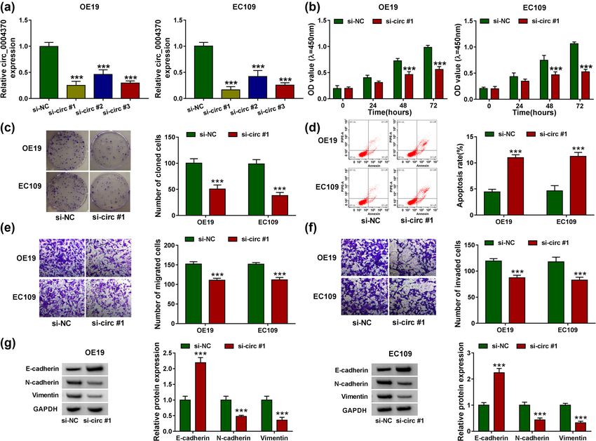
106 Xiaobo Chen et al.
(Weike Biotechnology, Shanghai, China) containing 10% 2.5 Localization of nucleus and cytoplasm
fetal bovine serum (FBS) at 37°C. OE19 and EC109 cells
were used for transfection due to their highest circ_0004370 In order to study the location of circ_0004370 in EC cell
expression level. MiR-1301-3p mimic and inhibitor, small lines, we used the NE-PER™ Nuclear and Cytoplasmic
interfering RNA against circ_0004370 (si-circ #1, si-circ Extraction Reagents Kit (Thermo Scientific). With the
#2 and si-circ #3), their control (miR-NC, anti-NC, and instructions on the manufacturer, the EC cells cytoplasm
si-NC), and the transfection plasmid vectors pcDNA and nuclear components were separated and collected.
and COL1A1 were bought from GenePharma Company RT-qPCR was utilized to examine circ_0004370 expres-
(Shanghai, China). The procedure of cell transfection sion in cell cytoplasm and nucleus. GAPDH is cytoplasm
obeyed the instructions of Lipofectamine 3000 (Invitrogen, positioning control; U6 is the nucleus positioning
USA). Successful transfected cells prepared in advance were control.
used in the following experiments; si-circ #1 sequence was
5′-GCGUCUCCGUACAGAUGACCATT-3′, si-circ #2 sequence
was 5′-GCAGCGAAGGAATAGGACA-3′, si-circ #3 sequence
was 5′-GAAGGAATAGGACAACCTT-3′, si-NC sequence was 2.6 Western blotting assay
5′-UUCUCCGAACGUGUCACGUTT-3′.
The RIPA lysis and extraction buffer were the protein
extraction buffer used in EC cells. The concentrations of
protein were measured with BCA Protein Assay Kit
2.3 Actinomycin D assay (Beyotime, Shanghai, China). Proteins were separated
by sodium dodecyl sulfate polyacrylamide gel electro-
To measure the stability of RNA, cells were treated with phoresis (SDS-PAGE). After 120 minutes, the proteins
2 mg/mL of Actinomycin D (Sigma-Aldrich, St. Louis, MO) were transferred to the polyvinylidene fluoride (PVDF)
for 0, 6, 12, 18, and 24 h. After treated with Actinomycin membrane and blocked with 5% milk. Primary antibodies
D, the circ_0004370 and PRRX1 mRNA levels were respec- anti-GAPDH (1:1,000; Cell Signaling Technology, Danvers,
tively detected by RT-qPCR assay. MA, USA), anti-COL1A1 (1:1,000; Abcam, Cambridge,
United Kingdom), anti-E-cadherin (1:1,000; Abcam),
anti-N-cadherin (1:1,000; Abcam), and anti-Vimentin
(1:1,000; Abcam) seeded into membrane at 4°C over-
2.4 RNA isolation and quantitative real-time night. And then the complexes were incubated with sec-
reverse transcription-PCR (RT-qPCR) ondary antibodies (HRP-conjugated, 1:1,000; Abcam).
Finally, the ECL method (Thermo Scientific, Waltham,
TRIzol reagent (Invitrogen, Carlsbad, CA, USA) was used MA, USA) was used for observation and detection.
to extract total RNA on the basis of the user guide and
reverse-transcribed into cDNAs used Transcriptor First
Strand cDNA Synthesis Kit (Roche, Indianapolis, IN).
RT-qPCR was performed in a 384-well plate containing 2.7 Cell viability assay
synthesized cDNA. The results of the expression were
presented using 2−ΔΔCt method. GAPDH and U6 acted as To analyze the cell viability, we used Cell Counting Kit-8
controls. We designed the primers for circ_0004370 (for- (Beyotime, Shanghai, China) assay. The cells need to be
ward: 5′-ACCCACCGATTATCTCTCCTG-3′; reverse: 5′-TCC incubated into 96-well plates and then added with CCK-8
TATTCCTTCGCTGCTTTC-3′), PRRX1 mRNA (forward: 5′- solution. The optical density (OD) values were detected at
ACGCTTCCCTCCTCAAATCC-3′; reverse: 5′-AGTAGCCATG 450 nm in 0, 24, 48, and 72 h.
GCGCTGTACG-3′), miR-1301-3p (forward: 5′-GCCCGCTTG
CAGCTGCCTGGGAG-3′; reverse: 5′-GTGCAGGGTCCGA
GGT-3′), COL1A1 (forward: 5′-CGATGGATTCCAGTTCG
AGT-3′; reverse: 5′-TTTTGAGGGGTTCAGTTTG-3′), U6 2.8 Cloning formation assay
(forward: 5′-CTCGCTTCGGCAGCACATATACT-3′; reverse:
5′-ACGCTTCACGAATTT-GCGTGTC-3′), GAPDH (forward: More than 200 cells were added to the 6-well plate, and
5′-TGTTCGTCATGGGTGTGAAC-3′; reverse: 5′-ATGGCATG the medium contained 10% FBS. First, the cells were
GACTGTGGTCAT-3′). fixed with 4% paraformaldehyde (PFA) and stained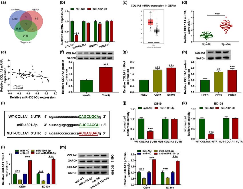
Circ_0004370/miR-1301-3p/COL1A1 axis modulates EC cells function 107
with crystal violet after two weeks. The number of colo- 2.12 RNA pull-down assay
nies was counted when the diameters of visible colonies
were greater than 1 mm. RNA pull-down assay was utilized to detect potential
target relationship between circ_0004370 and miR-1301-
3p. The biotinylated miR-1301-3p (bio-miR-1301-3p) was
purchased from GenScript Biotech Co., Ltd. (Nanjing,
2.9 Cell apoptosis assay Jiangsu, China). 1 × 10⁶ EC109 or OE19 cells were seeded
into 6-well plates and then treated with Bio-miR-1301-3p
The cells were first washed with PBS and then resus- or Bio-NC at a final concentration of 50 nM. After 48 h, EC
pended in binding buffer. FITC Annexin V/propidium cells were harvested and lysed. The streptavidin-coupled
iodide (PI) (BD Pharmingen, San Diego, CA, USA) was Dynabeads (Invitrogen, Carlsbad, CA, USA) was used to
used to double stain the cells according to the manufac- pull down the biotin–miRNA–circRNA complexes in cell
turer’s manual. The cells were detected by the flow cyt- lysates. After washing, the RNA complexes bound to the
ometer (Cytek Biosciences, model: DxP 10). beads were extracted and subjected to RT-qPCR assay.
2.10 Transwell migration and transwell 2.13 Xenograft mouse model
invasion assay
Animal experiments were followed by National Institutes
of Health guidelines. The female nude mice (4-week-old)
Cell migration was determined by transwell. First, cells
with no specific pathogen-free (SPF) were subcutaneously
were cultured in serum-free medium for 24 h. Then
injected with EC cells transfected with sh-NC or sh-
100 µL cells washed with sterile PBS were added to the
circ_0004370 at a concentration of 5 × 10⁶ cells/200 µL in
upper chamber. 600 µL DMEM containing FBS was added
sterile saline. To analyze the tumor size, the tumor volumes
to the lower chamber. After cultured for 24 h, the cells
were detected every 7 days and tumor weights were deter-
were washed twice with sterile PBS and stained with
mined after the animals were sacrificed. All the animal stu-
0.1% crystal violet. The number of cells represented
dies were approved by the Animal Care committee of the
the ability of cell migration. The matrigel (CorningLife
First Affiliated Hospital of Kunming Medical University.
Sciences, Corning, NY, USA) was added to the upper
chamber in invasion experiment, and all other steps
were similar with transwell migration assay. We observed
2.14 Statistical analysis
the migration and invasion of cells using a 100× micro-
scopic field.
GraphPad Prism 7.0 and SPSS 20.0 software were used to
fulfil a statistical analysis. The difference between two
groups was analyzed by the Student’s t-test. In addition,
the difference among multiple groups was detected by
2.11 Dual-luciferase reporter assay
one-way Analysis of Variance (ANOVA). Data were showed
as mean ± standard deviation. P value < 0.5 was considered
The binding sites between circ_0004370 and miR-1301-
significant.
3p and miR-1301-3p and COL1A1 were predicted by
online software. The sequences of wild type circ_
0004370 (WT-circ_0004370), mutant circ_0004370 (MUT-
circ_0004370), wild type COL1A1 3′UTR, and mutant 3 Results
COL1A1 3′UTR involving the putative-binding sites of
miR-1301-3p were cloned into the pMIR-REPORT luci-
ferase vector (OBio Biology, Shanghai, China). The 3.1 The expression level of circ_0004370
firefly and rellina luciferase reporter plasmids and was increased in EC
miR-1301-3p or miR-NC were cotransfected into EC cells
using Lipofectamine 3000 (Invitrogen). The correlation In order to know the function of circ_0004370 in EC,
results were detected by the dual-Luciferase reporter the RT-qPCR was used to analyze the expression of
system. circ_0004370 in EC tissues and cells. The expression level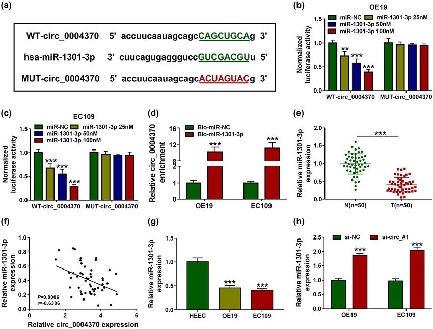
108 Xiaobo Chen et al.
Figure 1: The expression levels of circ_0004370 in EC tissues and cells. (a) Relative expression of circ_0004370 in EC tissues and adjacent
normal tissues was detected by RT-qPCR, n = 50. (b) Relative expression of circ_0004370 in OE19, TE11, KYSE410, and EC109 and normal cell
HEEC was detected by RT-qPCR. (c) Relative RNA expression of circ_0004370 and PRRX1 mRNA in OE19 cells was detected by RT-qPCR.
(d) Relative RNA expression of circ_0004370 and PRRX1 mRNA in EC109 cells was detected by RT-qPCR in different times after treatment of
Actinomycin D. (e) Relative RNA expression of circ_0004370 in OE19 cell nucleus and cytoplasm was detected by RT-qPCR. (f) Circ_0004370
distribution in EC109 cells was detected by RT-qPCR. **P < 0.01, ***P < 0.001.
of circ_0004370 in EC tissues was significantly higher After transfection with si-NC, si-circ #1, si-circ #2, or si-
than that in the adjacent normal controls (Figure 1a). circ #3 in EC cells, it was found that the expression level of
There was the correlation between circ_0004370 expres- circ_0004370 was significantly decreased in OE19 and
sion and clinicopathological features of ESCC patients EC109 cells, and because of the better efficiency of si-circ
(Table 1). The expression of circ_0004370 was increased #1, it was used in the subsequent experiments (Figure 2a).
in OE19, TE11, KYSE410, and EC109 cells compared with CCK-8 assay determined that knocking down circ_0004370
the HEEC cells, and the increase of expression was most significantly decreased OE19 and EC109 cell viability
obvious in OE19 and EC109 cells, so these two cell lines (Figure 2b). In addition, colony formation assay revealed
were selected for future experiments (Figure 1b). Further- that downregulation of circ_0004370 significantly inhib-
more, the circ_0004370 circular structure was more stable ited cell proliferation (Figure 2c). Furthermore, flow cyto-
than the linear structure of the PRRX1 mRNA in OE19 and metry indicated that the cell apoptosis in circ_0004370
EC109 cells (Figure 1c and d). We found that circ_0004370 knockdown group was markedly increased in OE19 and
mainly existed in the cytoplasm in OE19 and EC109 cells EC109 cells (Figure 2d). Then, the results of transwell
(Figure 1e and f). These data suggested that circ_0004370 assay indicated that cell migration and invasion were
might play a vital role in EC progression. reduced in the transfecting with si-circ #1 group in OE19
and EC109 cells (Figure 2e and f). Moreover, the EMT-
related proteins were analyzed by western blot assay. Wes-
tern blot analysis indicated that the protein level of E-cad-
herin was markedly upregulated after knocking down
3.2 Circ_0004370 knockdown suppressed circ_0004370 in OE19 and EC109 cells, whereas the protein
EC cell proliferation, migration, and levels of N-cadherin and Vimentin were downregulated
invasion and induced cell apoptosis after knocking down circ_0004370 in OE19 and EC109 cells
(Figure 2g). Thus, these results confirmed that downregu-
Loss-of-function experiments were performed to observe lated circ_0004370 suppressed biological activities and
whether circ_0004370 affected the behavior of EC cells. EMT process in EC cells.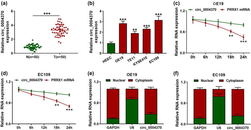
Circ_0004370/miR-1301-3p/COL1A1 axis modulates EC cells function 109
Figure 2: Circ_0004370 regulated EC progression in vitro. (a)The relative expression of circ_0004370 in cells was measured by RT-qPCR
after transfected with si-NC, si-circ #1, si-circ #2, and si_circ #3 individually. (b) Cell proliferation of OE19 and EC109 cell lines was evaluated
by CCK-8 assay. (c) Cell cloning of OE19 and EC109 cell lines was evaluated by Colony formation assay. (d) Cell apoptosis of OE19 and EC109
cell lines was evaluated by flow cytometry assay. (e and f) Cell migration and invasion of OE19 and EC109 cell lines were analyzed by
transwell assay. (g) Relative Protein expression of the EMT marker E-cadherin, N-cadherin, and Vimentin was evaluated by western blot
analysis in OE19 and EC109 cells. ***P < 0.001.
3.3 MiR-1301-3p was a direct target of RNA pull-down assay was utilized to further verify the
circ_0004370 correlation between circ_0004370 and miR-1301-3p. The
results presented that circ_0004370 was more enriched
Circ_0004370 was predicted to contain the binding sites in bio-miR-1301-3p-transfected EC cells when compared
with miR-1301-3p using starBase v2.0 software (Figure 3a). with bio-NC-transfected EC cells (Figure 3d). Moreover,
To further understand the relationship between circ_0004370 miR-1301-3p expression level was significantly reduced
and miR-1301-3p, the dual-luciferase reporter assay was
in EC tissues and cells (Figure 3e and g). The expression
performed. After overexpression of miR-1301-3p with dif-
of miR-1301-3p had a negative correlation with the expres-
ferent concentrations of miR-1301-3p mimics (25, 50, and
sion of circ_0004370 in EC tissues (Figure 3f). Also, miR-
100 nM, respectively), the luciferase activity in OE19
and EC109 cells containing the WT-circ_0004370 was 1301-3p was upregulated by circ_0004370 knockdown
decreased in a dose-dependent manner, while the luci- in OE19 and EC109 cells (Figure 3h). From these data,
ferase activity in MUT-circ0004370 group was not changed it was indicated that miR-1301-3p was a direct target of
in OE19 and EC109 cell lines (Figure 3b and c). Then the circ_0004370.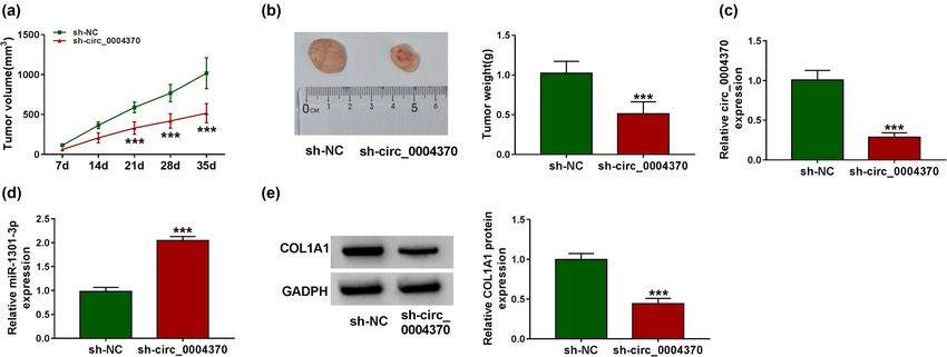
110 Xiaobo Chen et al.
Figure 3: MiR-1301-3p was a direct target of circ_0004370. (a)The binding site of circ_0004370 and miR-145-5p was measured by starBase
v2.0. (b–d) The interaction between miR-1301-3p and circ_0004370 in OE19 and EC109 cells was determined by dual-luciferase reporter
assay and RNA pull-down assays in OE19 and EC109 cells. (e) Relative miR-1301-3p expression in EC tissues and in adjacent normal tissues
was measured by RT-qPCR. (f) The correlation between miR-1301-3p and circ_0004370 levels was measured. (g) Relative miR-1301-3p
expression in OE19, EC109, and HEEC cells was detected by RT-qPCR. (h) After transfecting si-NC or si-circ_#1, the expression level of miR-
1301-3p was detected by RT-qPCR. ***P < 0.001.
3.4 MiR-1301-3p inhibitor partially rescued 1301-3p inhibitor (Figure 4c). The experiment of cell
the functions of circ_0004370 cloning proved that when the expression level of
knockdown circ_0004370 was downregulated, cell cloning in EC cells
was significantly decreased, while the number of cell
To confirm whether the interaction between miR-1301-3p cloning was recovered after the addition of miR-1301-3p
and circ_0004370 affects the cells function, we first inhibitor (Figure 4d). In cell apoptosis experiments,
used RT-qPCR to detect the miR-1301-3p expression. the number of apoptosis was upregulated by the
As expected, the miR-1301-3p expression was greatly circ_0004370 knockdown, but it was decreased by addi-
reduced after the transfection of anti-miR-1301-3p tion of anti-miR-1301-3p (Figure 4e). The transwell assay
(Figure 4a). Interestingly, the expression level of miR- showed that knockdown of circ_0004370 significantly
1301-3p was upregulated by circ_0004370 knockdown decreased the cell migration and invasion, while inhibi-
and restored after the addition of anti-miR-1301-3p tion of miR-1301-3p rescued the function of circ_0004370
(Figure 4b). The cell viability assay showed that the effect knockdown (Figure 4f and g). Besides, the western blot
of circ_0004370 knockdown was reversed by the miR- analysis showed that the addition of anti-miR-1301-3pCirc_0004370/miR-1301-3p/COL1A1 axis modulates EC cells function 111
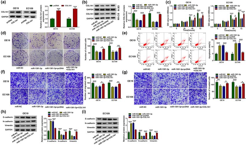
Figure 4: Anti-miR-1301-3p partially rescued the function of circ_0004370 inhibitor. (a) The expression level of miR-1301-3p in cells
significantly decreased after inhibition of miR-1301-3p. (b) In OE19 and EC109 cells transfected with si-NC, si-circ #1, si-circ #1 + anti-NC,
and si-circ #1 + anti-miR-1301-3p, relative miR-1301-3p expression was detected by RT-qPCR. (c–e) The cell proliferation, cloning, and
apoptosis were analyzed by CCK-8, colony formation assay, and flow cytometry. (f and g) The cell migration and invasion were detected by
Transwell assay. (h) EMT-related proteins were detected by western blot assay. *P < 0.05, **P < 0.01, ***P < 0.001.
rescued the EMT process changes caused by knockdown significantly reduced among the four genes, so COL1A1
of circ_0004370 in OE19 cells and EC109 cells (Figure 4h). was selected for subsequent experiments (Figure 5b).
Taken together, these data determined that miR-1301-3p In the GEPIA database, COL1A1 was highly expressed in
inhibitor partially rescued the effects of circ_0004370 EC tissues (Figure 5c). To further verify the COL1A1
knockdown on EC cell development. expression in EC, we used RT-qPCR to detect the mRNA
expression level of COL1A1 and found that the expression
level of COL1A1 in EC tissues was increased dramatically
3.5 COL1A1 was a target of miR-1301-3p (Figure 5d). The expression levels of COL1A1 and miR-
1301-3p were negatively correlated in EC tissues (Figure 5e).
Previous reports showed that COL1A1 was upregulated In western blot assay, the protein expression of COL1A1
and enhanced oncogenicity on EC cells [19]. In order to in EC tissues was significantly increased compared to
find the target genes of miR-1301-3p, three independent the adjacent normal tissues (Figure 5f). Next, RT-qPCR
databases, Starbase, targetscan, and GEPIA, were used to and western blot assay indicated that the expression
predict genes. Four genes were found in all three data- level of COL1A1 was increased in OE19 and EC109 cells
bases, namely COL1A1, MARCKSL1, MMP11, and PMEPA1 (Figure 5g and h). Besides, results from starBase v2.0
(Figure 5a). Meanwhile, COL1A1 expression was most software predicted that there was a binding site between112 Xiaobo Chen et al. Figure 5: The targeted relationship between miR-1301-3p and COL1A1. (a) Starbase, targetscan, and GEPIA databases were used to predict genes. (b) The mRNA expression level of COL1A1, MARCKSL1, MMP11, and PMEPA1 was detected by RT-qPCR through overexpression of mir- 1301-3p. (c) COL1A1 mRNA expression was predicted by GEPIA database. (d) The expression level of COL1A1 was detected by RT-qPCR in EC tissues and adjacent normal tissues. (e) The correlation level between miR-1301-3p and COL1A1 was measured. (f) The COL1A1 expression was detected by western blot analysis. (g and h) The mRNA and protein expression of COL1A1 in EC cells were tested by RT-qPCR and western blot analysis. (i) The binding site of miR-1301-3p and COL1A1 was measured by starBase v2.0. (j and k) The interaction between miR-1301-3p and COL1A1 in OE19 and EC109 cells was determined by dual-luciferase reporter assay. (l and m) The COL1A1 mRNA and protein expression were detected by RT-qPCR and western blot analysis after transfecting with miR-NC, miR-1301-3p, anti-NC, and anti-miR-1301-3p. **P < 0.01, ***P < 0.001. COL1A1 and miR-1301-3p (Figure 5i). In the dual-luci- 3.6 COL1A1 partially rescued the function of ferase reporter experiment, it was further verified that miR-1301-3p miR-1301-3p could directly bind to 3′UTR of COL1A1 in OE19 and EC109 cells (Figure 5j and k). MiR-1301-3p The western blot analysis was used to measure the downregulated the expression of COL1A1 mRNA and pro- COL1A1 protein expression in EC cells. The results tein, and miR-1301-3p inhibitor upregulated the expres- showed that COL1A1 protein expression was significantly sion of COL1A1 mRNA and protein (Figure 5l and m). increased in OE19 and EC109 cells (Figure 6a). Then, we Together, it was demonstrated that miR-1301-3p could examined the protein expression of COL1A1 after trans- regulate the expression of COL1A1. fecting with miR-NC, miR-1301-3p, miR-1301-3p + pcDNA,
Circ_0004370/miR-1301-3p/COL1A1 axis modulates EC cells function 113 Figure 6: COL1A1 partially rescued the function of miR-1301-3p. (a) The protein expression level of COL1A1 was measured by western blot analysis in OE19 and EC109 cells. (b) OE19 and EC109 cells were transfected with miR-NC, miR-1301-3p, miR-1301-3p + pcDNA, or miR-1301- 3p + COL1A1, individually. And relative COL1A1 protein level was detected by western blot analysis. (c–e) The cell proliferation, cloning, and apoptosis were analyzed by CCK-8, colony formation assay, and flow cytometry. (f and g) The cell migration and invasion were measured by Transwell assay after transfecting with miR-NC, miR-1301-3p, miR-1301-3p + pcDNA, or miR-1301-3p + COL1A1. (h and i) EMT marker proteins E-cadherin, N-cadherin, and Vimentin in OE19 and EC109 cells were detected by western blot analysis. *P < 0.05, **P < 0.01, ***P < 0.001. or miR-1301-3p + COL1A1. The results showed that the western blot assay showed that addition of COL1A1 res- expression level of COL1A1 was decreased after trans- cued the effect of miR-1301-3p on EMT process in OE19 fecting miR-1301-3p, whereas the protein expression cells and EC109 cells (Figure 6h and i). We concluded that was increased after transfecting with miR-1301-3p + COL1A1 protein could reverse the effect of miR-1301-3p on COL1A1 compared with transfection of miR-1301-3p + EC cells. pcDNA (Figure 6b, P < 0.001). We subsequently tested the cell viability using CCK-8 assay. The results revealed that upregulation of miR-1301-3p markedly reduced cell viability in OE19 and EC109 cells, whereas transfection 3.7 Knockdown of circ_0004370 inhibited EC with COL1A1 rescued the cell viability in EC cells (Figure 6c). growth in vivo via miR-1301-3p/ Then the cell cloning assay proved that cell cloning COL1A1 axis was significantly reduced in EC cells transfected with miR-1301-3p; however, the number of cell cloning was We wondered whether downregulated circ_0004370 recovered after transfection of COL1A1 (Figure 6d). The reduced the EC tumor growth. We used a xenograft flow cytometry was used to detect apoptosis. The results nude mouse model and found that knockdown of indicated that transfection of COL1A1 could partially circ_0004370 broadly suppressed the tumor volumes reverse the effects of miR-1301-3p on cell apoptosis and weights (Figure 7a and b). Moreover, the RT-qPCR (Figure 6e). The results of transwell assay showed that showed that circ_0004370 was significantly decreased miR-1301-3p significantly decreased the cell migration and miR-1301-3p expression was remarkably increased and invasion, while addition of COL1A1 rescued the func- with downregulation of circ_0004370 (Figure 7c and d). tion of miR-1301-3p in EC cells (Figure 6f and g). The Knockdown of circ_0004370 also decreased COL1A1
114 Xiaobo Chen et al.
Figure 7: Silencing of circ_0004370 reduced tumor volumes and weights in vivo. (a) The tumor volume in the tumor xenograft model after
transfecting with the sh-circ_0004370 was measured. (b) The tumor weight in the tumor xenograft model after transfecting with the sh-
circ_0004370 was measured. (c) The expression level of circ_0004370 was detected by RT-qPCR when EC tissues were transfected by sh-NC
or sh-circ_0004370. (d) The expression level of miR-1301-3p was measured after transfecting with the sh-circ_0004370. (e) The COL1A1
protein expression level was detected by western blot analysis in sh-circ_0004370 group and sh-NC group. ***P < 0.001.
protein level in tissues (Figure 7e). In conclusion, Downregulation of circ_0004370 notably affected EC cel-
circ_0004370 promoted EC growth by regulating miR- lular activities such as cell viability, cloning, apoptosis,
1301-3p/COL1A1 axis. migration and invasion, and EMT process. Furthermore,
knockdown of circ_0004370 in vivo showed that tumor
volumes and weights were significantly decreased in
xenograft mouse model.
4 Discussion CircRNAs take part in biological processes through
multiple regulatory mechanism. Specifically, circRNAs
EC is a type of high mortality serious tumors worldwide. are identified as competitive endogenous RNAs (ceRNAs)
This is a malignant lesion caused by an abnormality of to sponge miRNAs, thereby attenuating the inhibitory
esophageal squamous epithelial cells or adenocytes. effects of miRNAs and promoting the expression of
Recently, a growing number of studies have confirmed miRNAs target genes [28]. Alternatively, circRNAs have
that noncoding RNAs participate in the pathological pro- the role of regulating gene transcription [29]. In addition,
cess for various cancer [21–23]. Circular RNA (circRNA) is the function of circRNAs is interacting with RNA-binding
a novel noncoding RNA that attracts concerns. CircRNAs proteins (RBPs) [30]. We speculated that circ_0004370
are stable structures composed of precursor mRNA back- might be a ceRNA, with the function of sponging miR-
splicing. At present, many circRNAs have been identified 1301-3p. To verify this speculation, we first detected that
as tumor promoters or inhibitors, which have aberrant in the cytoplasmic distribution results, circ_0004370 was
expression levels in bladder cancer [24], papillary thyroid highly expressed in cytoplasm. Besides, we predicted
cancer [25], oral cancer [26], and colorectal cancer [27]. that circ_0004370 bound to the miR-1301-3p using online
However, the function of circRNA and the underlying software. Further dual-luciferase reporter analyzed the
regulatory mechanism remain unclear. In the current direct targeting relationship between circ_0004370 and
research, we concluded that circ_0004370 served as a miR-1301-3p. Previous study reported the negative corre-
tumor promoter to activate cell viability, cloning, migra- lation between circTNFRSF21 and miR-1227 in endome-
tion and invasion, and EMT process and restrain cell trial carcinoma tissues and cells [31]. Consistent with
apoptosis via miR-1301-3p/COL1A1 axis. this, in this study we found the markedly inverse correla-
Consistent with a previous report which reported the tion between circ_0004370 and miR-1301-3p in EC.
abnormal circ_0004370 expression in EC [8], our study Knockdown of circ_0004370 could affect the functions
first detected that circ_0004370 was upregulated in EC of EC cells, such as cell proliferation, cell cloning, migra-
tissues and cells (OE19, TE11, KYSE410, and EC109). tion, and invasion, whereas miR-1301-3p inhibitor rescuedCirc_0004370/miR-1301-3p/COL1A1 axis modulates EC cells function 115
the functions of circ_0004370 knockdown. Our results target in future, which may provide novel direction for
provided a fresh evidence for the role of circ_0004370 the further clinical trials.
in EC to downregulate miR-1301-3p.
COL1A1 as a type of group I collagen plays a signifi- Conflict of interest: The authors declare that they have no
cant role in the development of multiple cancers [32]. The conflicts of interest.
previous study indicated that COL1A1 was associated
with the gastric cancer and promoted cell migration Data availability statement: The datasets generated dur-
and metastasis [33]. Additionally, COL1A1 knockdown ing and/or analysed during the current study are avail-
suppressed the metastasis of breast cancer cells [19]. In able from the corresponding author on reasonable
addition, Yin et al. demonstrated that COL1A1 played a request.
crucial role in EC [20]. Recently, with the attention paid to
COL1A1, a large number of evidence showed that miRNA
had a targeting relationship with COL1A1. For example, it
was reported that miR-129-5p inhibited cell viability of References
gastric cancer by downregulating COL1A1 [34]. In our
findings, starbase software predicted that miR-1301-3p [1] Bray F, Ferlay J, Soerjomataram I, Siegel RL, Torre LA, Jemal A.
directly targeted COL1A1, and further experiments proved Global cancer statistics 2018: GLOBOCAN estimates of inci-
that there was a negative correlation between them. dence and mortality worldwide for 36 cancers in 185 countries.
Moreover, cell proliferation, apoptosis, migration, and CA Cancer J Clin. 2018;68(6):394–424.
[2] Pennathur A, Gibson MK, Jobe BA, Luketich JD. Oesophageal
other experiments demonstrated that COL1A1 could
carcinoma. Lancet. 2013;381(9864):400–12.
restore the effect of miR-1301-3p. Finally, we found that [3] Codipilly DC, Qin Y, Dawsey SM, Kisiel J, Topazian M,
knockdown of circ_0004370 upregulated miR-1301-3p Ahlquist D, et al. Screening for esophageal squamous cell
and further downregulated COL1A1 expression. Previous carcinoma: recent advances. Gastrointest Endosc.
studies exhibited that circNEK6 promoted thyroid cancer 2018;88(3):413–26.
[4] Ning S, Li X. Non-coding RNA resources. Adv Exp Med Biol.
progression through Wnt signaling pathway [35]. In addi-
2018;1094:1–7.
tion, circ_100290 played the critical role in colorectal [5] Zhang J, Liu H, Hou L, Wang G, Zhang R, Huang Y, et al. Circular
cancer initiation via Wnt/β-catenin signaling pathway RNA_LARP4 inhibits cell proliferation and invasion of gastric
[36]. However, it is not clear whether circ_0004370 cancer by sponging miR-424-5p and regulating LATS1 expres-
affects the development of EC by the Wnt signaling sion. Mol Cancer. 2017;16(1):151.
[6] Li XN, Wang ZJ, Ye CX, Zhao BC, Huang XX, Yang L. Circular RNA
pathway, which will be the focus of our future study.
circVAPA is up-regulated and exerts oncogenic properties by
In this study, there are some limitations for in vivo
sponging miR-101 in colorectal cancer. Biomed Pharmacother.
experiments. We have detected the effect of circ_0004370 2019;112:108611.
depletion on tumor growth in vivo. However, the effects of [7] Sun H, Tang W, Rong D, Jin H, Fu K, Zhang W, et al.
circ_0004370/miR-1301 3p/COL1A1 axis on tumor meta- Hsa_circ_0000520, a potential new circular RNA biomarker, is
stasis cannot be done currently due to the laboratory con- involved in gastric carcinoma. Cancer Biomark.
2018;21(2):299–306.
ditions. Future works are expected to refine this
[8] Zhang Z, Lin W, Gao L, Chen K, Yang C, Zhuang L, et al.
mechanism in EC using mouse model. Hsa_circ_0004370 promotes esophageal cancer progression
through miR-1294/LASP1 pathway. Biosci Rep.
2019;39(5):BSR20182377.
[9] Bartel DP. MicroRNAs: Target recognition and regulatory
5 Conclusions [10]
functions. Cell. 2009;136(2):215–33.
Backes C, Meese E, Keller A. Specific miRNA disease biomar-
kers in blood, serum and plasma: Challenges and prospects.
In conclusion, we discovered circ_0004370 was upregu- Mol Diagn Ther. 2016;20(6):509–18.
lated in EC cells and tissues. Moreover, as a tumor pro- [11] Zhi T, Jiang K, Zhang C, Xu X, Wu W, Nie E, et al. MicroRNA-1301
moter in EC, circ_0004370 could greatly promote the inhibits proliferation of human glioma cells by directly tar-
cell viability, cloning, migration, and invasion, remark- geting N-Ras. Am J Cancer Res. 2017;7(4):982–98.
[12] Wang L, Zhao Y, Xu M, Zhou F, Yan J. Serum miR-1301-3p, miR-
ably suppressed apoptosis, and affected EMT process
335-5p, miR-28-5p, and their target B7-H3 may serve as novel
of EC through regulation of miR-1301-3p/COL1A1 axis. biomarkers for colorectal cancer. J Buon. 2019;24(3):1120–7.
Therefore, our study suggested that circ_0004370/miR- [13] Peng X, Yan B, Shen Y. MiR-1301-3p inhibits human breast
1301-3p/COL1A1 axis might be potential therapeutic cancer cell proliferation by regulating cell cycle progression116 Xiaobo Chen et al.
and apoptosis through directly targeting ICT1. Breast Cancer. through modulating CTNNBIP1-dependent activation of
2018;25(6):742–52. β-catenin pathway. J Exp Clin Cancer Res. 2018;37(1):275.
[14] Zhang C, Xie L, Fu Y, Yang J, Cui Y. lncRNA MIAT promotes [26] Chen L, Zhang S, Wu J, Cui J, Zhong L, Zeng L, et al.
esophageal squamous cell carcinoma progression by regu- circRNA_100290 plays a role in oral cancer by functioning as
lating miR-1301-3p/INCENP axis and interacting with SOX2. a sponge of the miR-29 family. Oncogene. 2017;36(32):
J Cell Physiol. 2020;235(11):7933–44. 4551–61.
[15] Marini JC, Forlino A, Cabral WA, Barnes AM, San Antonio JD, [27] Li XN, Wang ZJ, Ye CX, Zhao BC, Li ZL, Yang Y. RNA sequencing
Milgrom S, et al. Consortium for osteogenesis imperfecta reveals the expression profiles of circRNA and indicates that
mutations in the helical domain of type I collagen: regions circDDX17 acts as a tumor suppressor in colorectal cancer.
rich in lethal mutations align with collagen binding sites J Exp Clin Cancer Res. 2018;37(1):325.
for integrins and proteoglycans. Hum Mutat. [28] Cui M, Shen W, Qin W, Wang X, Li Y, Xu F, et al. Circular RNA
2007;28(3):209–21. ciRS-7 promotes tube formation in microvascular endothelial
[16] Müller WEG, Ackermann M, Neufurth M, Tolba E, Wang S, cells through downregulation of miR-26a-5p. J Biochem Mol
Feng Q, et al. A novel biomimetic approach to repair enamel Toxicol. 2020;34(5):e22468.
cracks/carious damages and to reseal dentinal tubules by [29] Chen LL. The biogenesis and emerging roles of circular RNAs.
amorphous polyphosphate. Polymers. 2017;9(4):120. Nat Rev Mol Cell Biol. 2016;17(4):205–11.
[17] He X, Lee B, Jiang Y. Cell-ECM interactions in tumor invasion. [30] Holdt LM, Stahringer A, Sass K, Pichler G, Kulak NA, Wilfert W,
Adv Exp Med Biol. 2016;936:73–91. et al. Circular non-coding RNA ANRIL modulates ribosomal RNA
[18] Liu S, Liao G, Li G. Regulatory effects of COL1A1 on apoptosis maturation and atherosclerosis in humans. Nat Commun.
induced by radiation in cervical cancer cells. Cancer Cell Int. 2016;7:12429.
2017;17:73. [31] Liu Y, Chang Y, Cai Y. circTNFRSF21, a newly identified circular
[19] Liu J, Shen JX, Wu HT, Li XL, Wen XF, Du CW, et al. Collagen 1A1 RNA promotes endometrial carcinoma pathogenesis through
(COL1A1) promotes metastasis of breast cancer and is a regulating miR-1227-MAPK13/ATF2 axis. Aging.
potential therapeutic target. Discov Med. 2018;25(139):211–23. 2020;12:6774–992.
[20] Yin Y, Du L, Li X, Zhang X, Gao Y. miR-133a-3p suppresses cell [32] Li J, Ding Y, Li A. Identification of COL1A1 and COL1A2 as can-
proliferation, migration, and invasion and promotes apoptosis didate prognostic factors in gastric cancer. World J Surg Oncol.
in esophageal squamous cell carcinoma. J Cell Physiol. 2016;14(1):297.
2019;234(8):12757–70. [33] Wang F, Xue Q, Xu D, Jiang Y, Tang C, Liu X. Identifying the hub
[21] Rupaimoole R, Slack FJ. MicroRNA therapeutics: Towards a new gene in gastric cancer by bioinformatics analysis and in vitro
era for the management of cancer and other diseases. Nat Rev experiments. Cell Cycle. 2020;19:1326–37.
Drug Discov. 2017;16(3):203–22. [34] Wang Q, Yu J. MiR-129-5p suppresses gastric cancer cell
[22] Peng WX, Koirala P, Mo YY. LncRNA-mediated regulation of cell invasion and proliferation by inhibiting COL1A1. Biochem Cell
signaling in cancer. Oncogene. 2017;36(41):5661–7. Biol. 2018;96(1):19–25.
[23] Zhang HD, Jiang LH, Sun DW, Hou JC, Ji ZL. CircRNA: [35] Chen F, Feng Z, Zhu J, Liu P, Yang C, Huang R, et al. Emerging
a novel type of biomarker for cancer. Breast Cancer. roles of circRNA_NEK6 targeting miR-370-3p in the prolifera-
2018;25(1):1–7. tion and invasion of thyroid cancer via Wnt signaling pathway.
[24] Li P, Yang X, Yuan W, Yang C, Zhang X, Han J, et al. CircRNA- Cancer Biol Ther. 2018;19(12):1139–52.
Cdr1as exerts anti-oncogenic functions in bladder cancer by [36] Fang G, Ye BL, Hu BR, Ruan XJ, Shi YX. CircRNA_100290
sponging microRNA-135a. Cell Physiol Biochem. promotes colorectal cancer progression through miR-516b-
2018;46(4):1606–16. induced downregulation of FZD4 expression and Wnt/
[25] Bi W, Huang J, Nie C, Liu B, He G, Han J, et al. CircRNA β-catenin signaling. Biochem Biophys Res Commun.
circRNA_102171 promotes papillary thyroid cancer progression 2018;504(1):184–9.You can also read