Bioinformatics and immunohistochemistry analyses of expression levels and clinical significance of CXCL2 and TANs in an oral squamous cell ...
←
→
Page content transcription
If your browser does not render page correctly, please read the page content below
ONCOLOGY LETTERS 21: 189, 2021

          Bioinformatics and immunohistochemistry analyses of
           expression levels and clinical significance of CXCL2
           and TANs in an oral squamous cell carcinoma tumor
          microenvironment of Prophyromonas gingivalis infection
ZHI‑CHEN GUO1, SAKENDEKE JUMATAI1, SI‑LI JING2, LU‑LU HU1, XIN‑YU JIA1 and ZHONG‑CHENG GONG1,3

              1
               Oncological Department of Oral and Maxillofacial Surgery, The Affiliated Stomatology Hospital of
                                The First Affiliated Hospital of Xinjiang Medical University;
                2
                 Department of Ophthalmology, The First Affiliated Hospital of Xinjiang Medical University;
                           3
                            Xinjiang Uygur Autonomous Region Institute of Stomatology, Urumqi,
                                  Xinjiang Uyghur Autonomous Region 830054, P.R. China

                                    Received February 25, 2020; Accepted December 7, 2020

                                                    DOI: 10.3892/ol.2021.12450

Abstract. The present study aimed to detect the immunoex‑            gene in the two datasets. Immunoexpression of P. gingivalis
pression and clinical significance of Porphyromonas gingivalis       was positively associated with CXCL2 and TANs expression.
(P. gingivalis) in the tumor microenvironment (TME) of oral          Furthermore, P. gingivalis was associated with survival status
squamous cell carcinoma (OSCC). The immunoexpression                 (P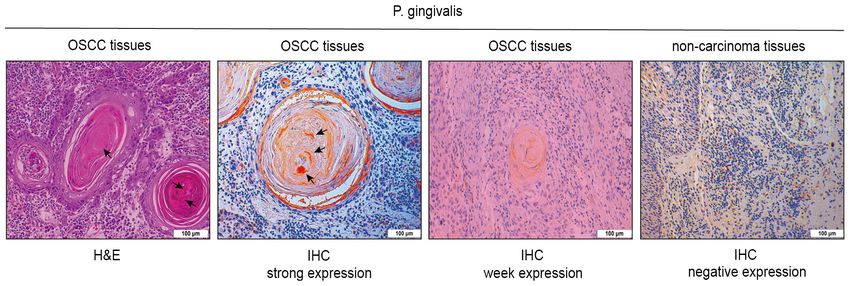
2           GUO et al: IMMUNOEXPRESSION AND CLINICAL SIGNIFICANCE OF THE TME OF OSCC OF P. gingivalis INFECTION

with chemotherapy is an effective treatment for OSCC (1).          Materials and methods
However, chemotherapy is not a first‑line therapy for OSCC,
since most OSCC cases develop resistance to chemothera‑            Patient selection. The present study included 205 surgical
peutic reagents (7). Periodontitis has been suggested to be        specimens from patients [age range, 36‑90 years; mean age,
associated with the TME of OSCC, which could be involved           63 years; 103 men (50.2%); 102 women (49.8%); 76 patients
in the development of chemotherapy resistance in OSCC (7).
ONCOLOGY LETTERS 21: 189, 2021                                                     3

conditions. Hierarchical clustering of hub genes was performed     were classified into three groups: 0, absent immunostaining;
using UCSU Cancer Genomics Browser (http://genome‑cancer.          1, weak immunostaining; and 2, strong immunostaining. The
ucsc.edu) (24). The adjusted P‑values (adj. P) and Benjamini       proportion of positive cells was classified as follows: 0, 0;
and Hochberg false discovery rate were applied to provide a        1, 1‑25; 2, 26‑75; 3, 76‑100%. The final staining score was
balance between the discovery of statistically significant genes   calculated by multiplying the staining intensity score by extent
and limitations of false‑positives. The probe sets without         of staining score. A final staining score of ≥3 was considered
corresponding gene symbols or genes with >1 probe set were         positive, and others were classified as low expression.
removed or averaged, respectively. Log fold‑change >1 and
adj. P
4 GUO et al: IMMUNOEXPRESSION AND CLINICAL SIGNIFICANCE OF THE TME OF OSCC OF P. gingivalis INFECTION
Table I. General information of 205 patients with oral squa‑ Table II. Immunohistochemical expression of P. gingivalis in
mous cell carcinoma. samples from 205 patients with oral squamous cell carcinoma
according to clinical data and follow‑up.
Variable No. of patients (%)
P. gingivalis
Sex ‑‑‑‑‑‑‑‑‑‑‑‑‑‑‑‑‑‑‑‑‑‑‑‑‑‑‑‑‑‑‑‑‑‑‑‑‑‑‑‑‑‑‑
Male 103 (50.2) Weak, Strong,
Female 102 (49.8) Variable n (%) n (%) P‑value
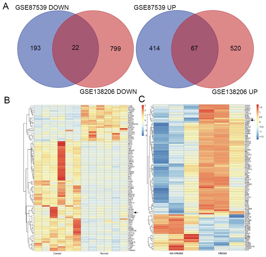
Age, years Sex 0.233ONCOLOGY LETTERS 21: 189, 2021 5 Figure 1. H&E staining of P. gingivalis in OSCC tissues, strong P. gingivalis expression in OSCC tissues, weak P. gingivalis expression in OSCC tissues and negative P. gingivalis expression in non‑carcinoma tissues. Scale bars, 100 µm. P. gingivalis, Porphyromonas gingivalis; OSCC, oral squamous cell carcinoma; IHC, immunohistochemistry. Figure 2. Venn diagrams and heat map of DEGs. CXCL2 was one of the DEGs included in the two datasets. (A) DEGs with a fold‑change >2 and P
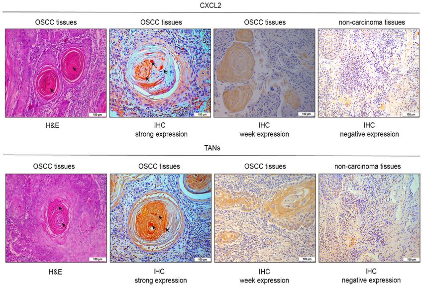
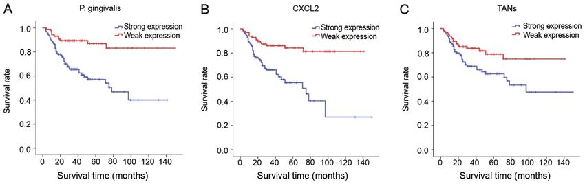
6            GUO et al: IMMUNOEXPRESSION AND CLINICAL SIGNIFICANCE OF THE TME OF OSCC OF P. gingivalis INFECTION

Figure 3. H&E staining of CXCL2 and TANs in OSCC tissues, strong CXCL2 and TANs expression in OSCC tissues, weak CXCL2 and TANs expres‑
sion in OSCC tissues, and negative CXCL2 and TANs expression in non‑carcinoma tissues. Scale bars, 100 µm. OSCC, oral squamous cell carcinoma;
CXCL2, C‑X‑C motif chemokine ligand 2; TANs, tumor‑associated neutrophils; IHC, immunohistochemistry.

IHC expression of CXCL2 and TANs in P. gingivalis‑infected              dissection. Furthermore, the immunoexpression levels of
TME of OSCC. A total of 119 samples with high expression                P. gingivalis, CXCL2 and TANs were significantly associated
levels of P. gingivalis were selected for IHC examination of            with the 10‑year CSR. Patients with a high immunoexpression
CXCL2 and TANs. In 119 samples with high P. gingivalis                  of P. gingivalis had an increased risk with a lower 10‑year CSR
expression, CXCL2 and TANs were weakly positively                       [hazard ratio (HR), 0.260; 95% CI, 0.135‑0.503; P
ONCOLOGY LETTERS 21: 189, 2021                                                                                          7

Table III. Immunohistochemical expression of CXCL2 and TANs in 119 samples with strong staining for Porphyromonas
gingivalis from patients with oral squamous cell carcinoma according to clinical data and follow‑up.

                                                           CXCL2                                                                       TANs
                                     ‑‑‑‑‑‑‑‑‑‑‑‑‑‑‑‑‑‑‑‑‑‑‑‑‑‑‑‑‑‑‑‑‑‑‑‑‑‑‑‑‑‑‑‑‑‑‑‑‑‑‑‑‑‑‑‑‑‑‑               ‑‑‑‑‑‑‑‑‑‑‑‑‑‑‑‑‑‑‑‑‑‑‑‑‑‑‑‑‑‑‑‑‑‑‑‑‑‑‑‑‑‑‑‑‑‑‑‑‑‑‑‑‑‑‑‑‑‑‑
Variable                             Weak, n (%)                      Strong, n (%)                P‑value     Weak, n (%)                      Strong, n (%)                P‑value

Sex			                                            0.631			                    0.824
 Male                        15 (50.0) 49 (55.1)		        21 (55.3) 43 (53.1)
 Female                      15 (50.0) 40 (44.9)		        17 (44.7) 38 (46.9)
Age, years			                                     0.038a     		0.229
8 GUO et al: IMMUNOEXPRESSION AND CLINICAL SIGNIFICANCE OF THE TME OF OSCC OF P. gingivalis INFECTION
Table IV. Association between P. gingivalis immunohistochemical expression and CXCL2 and TANs in 205 patients.
CXCL2 TANs
‑‑‑‑‑‑‑‑‑‑‑‑‑‑‑‑‑‑‑‑‑‑‑‑‑‑‑‑‑‑‑‑‑‑‑‑‑‑‑‑‑‑‑‑‑‑‑‑‑‑‑‑‑‑‑‑‑‑‑‑‑‑‑‑‑ ‑‑‑‑‑‑‑‑‑‑‑‑‑‑‑‑‑‑‑‑‑‑‑‑‑‑‑‑‑‑‑‑‑‑‑‑‑‑‑‑‑‑‑‑‑‑‑‑‑‑‑‑‑‑‑‑‑‑‑‑‑‑‑‑‑
P. gingivalis Weak, n (%) Strong, n (%) P‑value Weak, n (%) Strong, n (%) P‑value
Weak 69 (80.2) 17 (19.8)ONCOLOGY LETTERS 21: 189, 2021 9 Table V. Univariate analysis of Cox risk ratio model regression in 205 patients with oral squamous cell carcinoma. Variable No. of patients (%) P‑value HR HR (95% CI) Sex 0.684 0.898 0.535‑1.507 Male 103 (50.2) Female 102 (49.8) Age, years 0.023a 0.503 0.277‑0.911
10          GUO et al: IMMUNOEXPRESSION AND CLINICAL SIGNIFICANCE OF THE TME OF OSCC OF P. gingivalis INFECTION

Table VI. Multivariate analysis using Cox risk ratio regression model in 205 patients with oral squamous cell carcinoma.

                                                                                                                       HR (95% CI)
                                                                                                                ‑‑‑‑‑‑‑‑‑‑‑‑‑‑‑‑‑‑‑‑‑‑‑‑‑‑‑‑‑‑‑‑‑‑‑‑‑‑
Variable                                     B            SE          Wald          P‑value         HR          Upper                     Lower

Age (
ONCOLOGY LETTERS 21: 189, 2021 11
Funding 9. Inaba H, Sugita H, Kuboniwa M, Iwai S, Hamada M, Noda T,
Morisaki I, Lamont RJ and Amano A: Porphyromonas gingi‑
valis promotes invasion of oral squamous cell carcinoma through
No funding was received. induction of proMMP9 and its activation. Cell Microbiol 16:
131‑145, 2014.
10. Lanzós I, Herrera D, Santos S, O'Connor A, Peña C, Lanzós E
Availability of data and materials and Sanz M: Microbiological effects of an antiseptic mouth rinse
in irradiated cancer patients. Med Oral Patol Oral Cir Bucal 16:
The datasets used and/or analyzed during the current study are e1036‑e1042, 2011.
11. Wen L, Mu W, Lu H, Wang X, Fang J, Jia Y, Li Q, Wang D,
available from the corresponding author on reasonable request. Wen S, Guo J, et al: Porphyromonas gingivalis Promotes oral
squamous cell carcinoma progression in an immune microenvi‑
Authors' contributions ronment. J Dent Res 99: 666‑675, 2020.
12. da Silva JM, Soave DF, Moreira Dos Santos TP, Batista AC,
Russo RC, Teixeira MM and da Silva TA: Significance of chemo‑
ZCGo conceived and designed the study. ZCGu conducted the kine and chemokine receptors in head and neck squamous cell
experiments. SJ, SLJ, XYJ and LLH performed the statistical carcinoma: A critical review. Oral Oncol 56: 8‑16, 2016.
13. Vashishta A, Jimenez‑Flores E, Klaes CK, Tian S, Miralda I,
analysis. ZCGu wrote the manuscript. ZCGu reviewed and Lamont RJ and Uriarte SM: Putative periodontal pathogens,
edited the manuscript. All authors agree to be accountable Filifactor alocis and Peptoanaerobacter Stomatis, induce
for all aspects of the research in ensuring that the accuracy differential cytokine and chemokine production by human
neutrophils. Pathogens 8: 59, 2019.
or integrity of any part of the work are appropriately investi‑ 14. Hardaway AL, Herroon MK, Rajagurubandara E and Podgorski I:
gated and resolved. ZCGo and ZCGu confirm the authenticity Marrow adipocyte‑derived CXCL1 and CXCL2 contribute to
of all the raw data. All authors read and approved the final osteolysis in metastatic prostate cancer. Clin Exp Metastasis 32:
353‑368, 2015.
manuscript. 15. Zhang H, Ye YL, Li MX, Ye SB, Huang WR, Cai TT, He J,
Peng JY, Duan TH, Cui J, et al: CXCL2/MIF‑CXCR2 signaling
Ethics approval and consent to participate promotes the recruitment of myeloid‑derived suppressor cells
and is correlated with prognosis in bladder cancer. Oncogene 36:
2095‑2104, 2017.
The present study was approved by the Ethics Committee of 16. Fridlender ZG, Sun J, Mishalian I, Singhal S, Cheng G, Kapoor V,
the Affiliated Stomatological Hospital of Xinjiang Medical Horng W, Fridlender G, Bayuh R, Worthen GS and Albelda SM:
Transcriptomic analysis comparing tumor‑associated neutrophils
University (approval no. IACUC20180411‑13; Urumqi, China). with granulocytic myeloid‑derived suppressor cells and normal
Written consent was obtained at the time of the initial data neutrophils. PLoS One 7: e31524, 2012.
collection. 17. Hagerling C and Werb Z: Neutrophils: Critical components in
experimental animal models of cancer. Semin Immunol 28:
197‑204, 2016.
Patient consent for publication 18. Powell DR and Huttenlocher A: Neutrophils in the tumor
microenvironment. Trends Immunol 37: 41‑52, 2016.
19. Li S, Cong X, Gao H, Lan X, Li Z, Wang W, Song S, Wang Y,
Not applicable. Li C, Zhang H, et al: Tumor‑associated neutrophils induce EMT
by IL‑17a to promote migration and invasion in gastric cancer
Competing interests cells. J Exp Clin Cancer Res 38: 6, 2019.
20. Sahingur SE and Yeudall WA: Chemokine function in
periodontal disease and oral cavity cancer. Front Immunol 6:
The authors declare that they have no competing interests. 214, 2015.
21. Carey LA, Metzger R, Dees EC, Collichio F, Sartor CI,
Ollila DW, Klauber‑DeMore N, Halle J, Sawyer L, Moore DT
References and Graham ML: American Joint Committee on cancer
tumor‑node‑metastasis stage after neoadjuvant chemotherapy and
breast cancer outcome. J Natl Cancer Inst 97: 1137‑1142, 2005.
1. Chi AC, Day TA and Neville BW: Oral cavity and oropharyn‑ 22. Geng F, Liu J, Guo Y, Li C, Wang H, Wang H, Zhao H and
geal squamous cell carcinoma‑an update. CA Cancer J Clin 65: Pan Y: Persistent exposure to Porphyromonas gingivalis
401‑421, 2015. promotes proliferative and invasion capabilities, and tumorigenic
2. Siegel R, Naishadham D and Jemal A: Cancer statistics, 2013. properties of human immortalized oral epithelial cells. Front
CA: A Cancer J Clin 63: 11‑30, 2013. Cell Infect Microbiol 7: 57‑61, 2017.
3. Li P, Cao Q, Shao P, Cai H, Zhou H, Chen J, Qin C, Zhang Z, Ju X 23. Qiu X, Lei Z, Wang Z, Xu Y, Liu C, Li P, Wu H and Gong Z:
and Yin C: Genetic polymorphisms in HIF1A are associated with Knockdown of LncRNARHPN1‑AS1 inhibits cell migration,
prostate cancer risk in a Chinese population. Asian J Androl 14: invasion and proliferation in head and neck squamous cell
864‑869, 2012. carcinoma. J Cancer 10: 4000‑4008, 2019.
4. Kim SS, Ruiz VE, Carroll JD and Moss SF: Helicobacter pylori 24. Li L, Lei Q, Zhang S, Kong L and Qin B: Screening and
in the pathogenesis of gastric cancer and gastric lymphoma. identification of key biomarkers in hepatocellular carcinoma:
Cancer Lett 305: 228‑238, 2011. Evidence from bioinformatic analysis. Oncol Rep 38: 2607‑2618,
5. Whitmore SE and Lamont RJ: Oral bacteria and cancer. PLos 2017.
Pathog 10: e1003933, 2014. 25. Bryne M, Koppang HS, Lilleng R, Stene T, Bang G and
6. Gershkovitz M, Fainsod‑Levi T, Zelter T, Sionov RV and Dabelsteen E: New malignancy grading is a better prognostic
Granot Z: TRPM2 modulates neutrophil attraction to murine indicator than Broders' grading in oral squamous cell carci‑
tumor cells by regulating CXCL2 expression. Cancer Immunol nomas. J Oral Pathol Med 18: 432‑437, 1989.
Immunother 68: 33‑43, 2019. 26. Carus A, Ladekarl M, Hager H, Nedergaard BS and Donskov F:
7. Song JM, Woo BH, Lee JH, Yoon S, Cho Y, Kim YD and Tumour‑associated CD66b+ neutrophil count is an independent
Park HR: Oral Administration of Porphyromonas gingivalis, a prognostic factor for recurrence in localised cervical cancer.
major pathogen of chronic periodontitis, promotes resistance to Br J Cancer 108: 2116‑2122, 2013.
paclitaxel in mouse xenografts of oral squamous cell carcinoma. 27. Akinkugbe AA, Garcia DT, Brickhouse TH and Mosavel M:
Int J Mol Sci 20: 2494, 2019. Lifestyle risk factor related disparities in oral cancer examina‑
8. Gao JL, Kwan AH, Yammine A, Zhou X, Trewhella J, tion in the U.S: A population‑based cross‑sectional study. BMC
Hugrass BM, Collins D, Horne J, Ye P, Harty D, et al: Structural Public Health 20: 153‑163, 2020.
properties of a haemophore facilitate targeted elimination of the 28. Danaei G: Global burden of infection‑related cancer revisited.
pathogen Porphyromonas gingivalis. Nat Commun 9: 4097, 2018. Lancet Oncol 13: 564‑565, 2012.12 GUO et al: IMMUNOEXPRESSION AND CLINICAL SIGNIFICANCE OF THE TME OF OSCC OF P. gingivalis INFECTION
29. Iqbal J, McRae S, Banaudha K, Mai T and Waris G: Mechanism 43. Ban Y, Mai J, Li X, Mitchell‑Flack M, Zhang T, Zhang L,
of hepatitis C virus (HCV)‑induced osteopontin and its role Chouchane L, Ferrari M, Shen H and Ma X: Targeting autocrine
in epithelial to mesenchymal transition of hepatocytes. J Biol CCL5‑CCR5 axis reprograms immunosuppressive myeloid
Chem 293: 20010, 2018. cells and reinvigorates antitumor immunity. Cancer Res 77:
30. Horikawa T, Yang J, Kondo S, Yoshizaki T, Joab I, Furukawa M 2857‑2868, 2017.
and Pagano JS: Twist and epithelial‑mesenchymal transition 44. Lafuente Ibáñez de Mendoza I, Maritxalar Mendia X,
are induced by the EBV oncoprotein latent membrane protein 1 García de la Fuente AM, Quindós Andrés G and Aguirre Urizar JM:
and are associated with metastatic nasopharyngeal carcinoma. Role of Porphyromonas gingivalis in oral squamous cell carcinoma
Cancer Res 67: 1970‑1978, 2007. development: A systematic review. J Periodontal Res 55: 13‑22, 2020.
31. Chandrakesan P, Roy B, Jakkula LU, Ahmed I, Ramamoorthy P, 45. Ohms M, Moller S and Laskay T: An attempt to polarize human
Tawfik O, Papineni R, Houchen C, Anant S and Umar S: Utility neutrophils toward N1 and N2 phenotypes in vitro. Front
of a bacterial infection model to study epithelial‑mesenchymal Immunol 11: 532, 2020.
transition, mesenchymal‑epithelial transition or tumorigenesis. 46. Feliciano P: CXCL1 and CXCL2 link metastasis and chemoresis‑
Oncogene 33: 2639‑2654, 2014. tance. Nat Genet 44: 840, 2012.
32. Parkin DM: The global health burden of infection‑associated 47. Eckert AW, Wickenhauser C, Salins PC, Kappler M, Bukur J
cancers in the year 2002. Int J Cancer 118: 3030‑3044, 2006. and Seliger B: Correction to: Clinical relevance of the tumor
33. Lee J, Roberts JS, Atanasova KR, Chowdhury N, Han K and microenvironment and immune escape of oral squamous cell
Yilmaz Ö: Human primary epithelial cells acquire an epithe‑ carcinoma. J Transl Med 16: 40, 2018.
lial‑mesenchymal‑transition phenotype during long‑term infection 48. Eruslanov EB, Bhojnagarwala PS, Quatromoni JG, Stephen TL,
by the oral opportunistic pathogen, Porphyromonas gingivalis. Ranganathan A, Deshpande C, Akimova T, Vachani A, Litzky L,
Front Cell Infect Microbiol 7: 493, 2017. Hancock WW, et al: Tumor‑associated neutrophils stimulate
34. Woo BH, Kim DJ, Choi JI, Kim SJ, Park BS, Song JM, Lee JH T cell responses in early‑stage human lung cancer. J Clin
and Park HR: Oral cancer cells sustainedly infected with Invest 124: 5466‑5480, 2014.
Porphyromonas gingivalis exhibit resistance to Taxol and have 49. Wu P, Wu D, Ni C, Ye J, Chen W, Hu G, Wang Z, Wang C,
higher metastatic potential. Oncotarget 8: 46981‑46992, 2017. Zhang Z, Xia W, et al: γδT17 cells promote the accumulation
35. Wen L, Mu W, Lu H, Wang X, Fang J, Jia Y, Li Q, Wang D, and expansion of myeloid‑derived suppressor cells in human
Wen S, Guo J, et al: Porphyromonas gingivalis Promotes oral colorectal cancer. Immunity 40: 785‑800, 2014.
squamous cell carcinoma progression in an immune microenvi‑ 50. Peltanova B, Raudenska M and Masarik M: Effect of tumor micro‑
ronment. J Dent Res 99: 666‑675, 2020. environment on pathogenesis of the head and neck squamous cell
36. Peres MA, Macpherson LMD, Weyant RJ, Daly B, Venturelli R, carcinoma: A systematic review. Mol Cancer 18: 63, 2019.
Mathur MR, Listl S, Celeste RK, Guarnizo‑Herreño CC, 51. Yasunaga JI and Matsuoka M: Oncogenic spiral by infectious
Kearns C, et al: Oral diseases: A global public health challenge. pathogens: Cooperation of multiple factors in cancer develop‑
Lancet 394: 249‑260, 2019. ment. Cancer Sci 109: 24‑32, 2018.
37. Yee M, Kim S, Sethi P, Duzgunes N and Konopka K: 52. Wen J, Wang Y, Gao C, Zhang G, You Q, Zhang W, Zhang Z,
Porphyromonas gingivalis stimulates IL‑6 and IL‑8 secretion in Wang S, Peng G and Shen L: Helicobacter pylori infection promotes
GMSM‑K, HSC‑3 and H413 oral epithelial cells. Anaerobe 28: Aquaporin 3 expression via the ROS‑HIF‑1α‑AQP3‑ROS loop
62‑67, 2014. in stomach mucosa: A potential novel mechanism for cancer
38. Geng F, Wang Q, Li C, Liu J, Zhang D, Zhang S and Pan Y: pathogenesis. Oncogene 37: 3549‑3561, 2018.
Identification of potential candidate genes of oral cancer in 53. Nagarsheth N, Wicha MS and Zou W: Chemokines in the cancer
response to chronic infection with Porphyromonas gingivalis microenvironment and their relevance in cancer immunotherapy.
using bioinformatical analyses. Front Oncol 9: 91, 2019. Nat Rev Immunol 17: 559‑572, 2017.
39. Bunt SK, Yang L, Sinha P, Clements VK, Leips J and 54. Damgaard C, Kantarci A, Holmstrup P, Hasturk H, Nielsen CH
Ostrand‑Rosenberg S: Reduced inflammation in the tumor and Van Dyke TE: Porphyromonas gingivalis‑induced production
microenvironment delays the accumulation of myeloid‑derived of reactive oxygen species, tumor necrosis factor‑α, interleukin‑6,
suppressor cells and limits tumor progression. Cancer Res 67: CXCL8 and CCL2 by neutrophils from localized aggressive
10019‑10026, 2007. periodontitis and healthy donors: Modulating actions of red blood
40. Zenobia C and Hajishengallis G: Porphyromonas gingivalis cells and resolvin E1. J Periodontal Res 52: 246‑254, 2017.
virulence factors involved in subversion of leukocytes and
microbial dysbiosis. Virulence 6: 236‑243, 2015. This work is licensed under a Creative Commons
41. Zhang Y and Li X: Lipopolysaccharide‑regulated production Attribution-NonCommercial-NoDerivatives 4.0
of bone sialoprotein and interleukin‑8 in human periodontal International (CC BY-NC-ND 4.0) License.
ligament fibroblasts: The role of toll‑like receptors 2 and 4 and
the MAPK pathway. J Periodontal Res 50: 141‑151, 2015.
42. Shi H, Han X, Sun Y, Shang C, Wei M, Ba X and Zeng X:
Chemokine (C‑X‑C motif) ligand 1 and CXCL2 produced by
tumor promote the generation of monocytic myeloid‑derived
suppressor cells. Cancer Sci 109: 3826‑3839, 2018.You can also read