Propofol postpones colorectal cancer development through circ_0026344/miR-645/ Akt/mTOR signal pathway - De Gruyter
←
→
Page content transcription
If your browser does not render page correctly, please read the page content below
Open Medicine 2021; 16: 570–580
Research Article
Xiaomin Cui, Jiying Feng, Jian Wu, Xiaobao Zhang, Mengyao Ding*
Propofol postpones colorectal cancer
development through circ_0026344/miR-645/
Akt/mTOR signal pathway
https://doi.org/10.1515/med-2021-0254 apoptosis in CRC cells. Propofol treatment induced the
received August 7, 2020; accepted February 26, 2021 restraint in proliferation and metastasis and stimulation
Abstract: Colorectal cancer (CRC) is responsible for thou- in apoptosis, which were allayed by si-circ#1; meanwhile,
sands of slow and painful annual deaths. Propofol, an anes- this alleviation could further be abolished by anti-miR-645 in
thetic, is commonly used in CRC surgery. The role of CRC cells. Furthermore, circ_0026344 sponged miR-645 to
circularRNA0026344 (circ_0026344) in propofol-treated inhibited Akt/mTOR signal pathway in propofol-treated CRC
CRC remains unclear, which was further explored in this cells. Propofol postponed CRC process by circ_0026344/miR-
study. Real-time polymerase chain reaction (qPCR) was 645/Akt/mTOR axis. This finding might provide a possibility
used to detect the expression of circ_0026344 and to improve the therapy of CRC with propofol.
microRNA645 (miR-645) in CRC cells and normal cells. Keywords: colorectal cancer, circ_0026344, miR-645, Akt/
Western blot was devoted to testing the protein expression mTOR, propofol
of phospho-protein kinase B (p-AKT), AKT, phospho-mam-
malian target of rapamycin (p-mTOR), and mTOR in CRC
cells. Moreover, cell counting kit-8 (CCK8), colony forma-
tion, flow cytometry, and transwell assays were employed 1 Introduction
to assess the proliferation, apoptosis, and metastasis in CRC
cells. Circinteractome online tool was applied to predict the Colorectal cancer (CRC) deserved a seat among malignant
combination between circ_0026344 and miR-645, which tumors due to the third-ranking of incidence (10.2%)
was further verified by dual-luciferase reporter system. and the second-ranking mortality (9.2%) over the world
circ_0026344 was lowly expressed and miR-645 was abun- in 2018 [1]. Studies looked at the incidence characters
dantly expressed in CRC cells. The relative protein expression of CRC and exposed that CRC possessed the sporadic,
of p-AKT/AKT and p-mTOR/mTOR was strikingly elevated inherited, and familial features [2–4]. For the sporadic
by si-circ#1, which could be reversed by anti-miR-645 in CRC, lifestyle and living environment assumed the main
propofol-treated CRC cells. circ_0026344 overexpression responsibility; however, genetic factors have mastered
inhibited the proliferation and metastasis and promoted the inherited and familial CRC [5]. CRC therapy has cen-
tered on the surgery due to the unsatisfying diagnosis
that 80% CRC patients were judged as local stage [6].
* Corresponding author: Mengyao Ding, Department of Surgery tends to bring anguish to CRC patients, which
Anesthesiology, The Affiliated Hospital of Kangda College of Nanjing is readily solved by the application of anesthetic. As
Medical University (The First People’s Hospital of Lianyungang), an agent, 2,6-diisopropylphenyl (propofol) is extensively
No. 188 Jianshe East Road, Lianyungang, 222002, Jiangsu, China, used in clinic to induct and maintain anesthesia [7].
e-mail: dingmy801025@163.com, tel: +86-0518-85605496,
The efficacy of propofol was not only reflected in the
fax: +86-0518-85605496
Xiaomin Cui: Department of Postanesthesia Care Unit, The Affiliated anesthesia, but also embodied in the affection of cancer
Hospital of Kangda College of Nanjing Medical University (The First process [8]. The inhibition of HIF-1α pathway was rea-
People’s Hospital of Lianyungang), Lianyungang, Jiangsu, China lized by the application of propofol in prostate cancer [9].
Jiying Feng, Xiaobao Zhang: Department of Anesthesiology, The Propofol treatment induced the promotion of the prolife-
Affiliated Hospital of Kangda College of Nanjing Medical University
ration and inhibition in the apoptosis in natural killer cells
(The First People’s Hospital of Lianyungang), Lianyungang, Jiangsu,
China
in colon cancer [10]. In bladder cancer cells, propofol trig-
Jian Wu: Department of Emergency, The First People’s Hospital of gered the facilitation in proliferation, metastasis, and the
Lianyungang, Lianyungang, Jiangsu, China inhibition in apoptosis [11]. Propofol put off the progression
Open Access. © 2021 Xiaomin Cui et al., published by De Gruyter. This work is licensed under the Creative Commons Attribution 4.0
International License.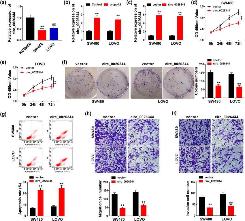
Role of circ_0026344/miR-645/Akt/mTOR axis in colorectal cancer 571
of CRC by regulating STAT3/HOTAIR/WIF-1/Wnt axis [12]. 2 Materials and methods
These documents conveyed the information about the
functional role of propofol in cancers; however, the regu-
2.1 Cells and treatment
latory mechanism of propofol in CRC is still fuzzy.
As single-stranded covalently closed RNAs, circular
Normal human colonic epithelial cells (NCM460) were
RNAs (circRNAs) have been recognized as participators in
purchased from INCELL Corporation LLC (San Antonio,
various diseases [13–15]. Has_circ_0000520 was lowly
Texas, USA). Colon adenocarcinoma cells (SW480 and
expressed in gastric cancer cells, and this downregula-
LOVO) were obtained from American Type Culture Collection
tion was negatively relevant to tumor node metastasis
(Rockville, MD, USA). Cells were seeded into Dulbecco’s
(TNM) stage [16]. The dysregulation of circ-LDLRAD3
Modified Eagle’s Medium (DMEM, Gibco, Carlsbad, CA,
was related to the metastasis in pancreatic cancer cells
USA), containing 10% fetal bovine serum (FBS, Gibco)
[17]. The interaction between circRNAs and micro RNA
and 100 U/mL penicillin (Sigma-Aldrich, St. Louis, MO,
(miRNAs)/message RNAs (mRNAs) attracted great atten-
USA) and 100 µg/mL streptomycin (Sigma-Aldrich) with
tion of researchers in exploring the regulatory mechanism
5% CO2 at 37°C. For the assessment of the effect of propofol
of circRNAs in cancers. circ-TTBK2 boosted glioma devel-
on CRC cells, 0, 5, and 10 μg/mL of propofol (Sigma-
opment by regulating miR-761/ITGB8 pathway [18]. circ-
Aldrich) were applied to treat CRC cells.
SERPINE2 facilitated the proliferation and repressed the
apoptosis by miR-375/YWHAZ axis in gastric cancer [19].
For the underlying way of circRNA in propofol-treated
CRC, the available information was limited, which was 2.2 Cell transfection
further studied in this project.
During the development of CRC, signal pathways SW480 and LOVO cells were cultured in 6-well plates for
play a crucial role. Protein kinase B (AKT)/mammalian 12 h. After that, the transient transfection was performed
target of rapamycin (mTOR) signal pathway was taking by Lipofectomine 3000 transfection reagent (Invitrogen,
charge of CRC biology including proliferation, apoptosis, Carlsbad, CA, USA) in SW480 and LOVO cells. Small
and metastasis [20,21]. As the upstream stimulating factor interfering RNA targeting circ_0026344 (si-circ_0026344
of mTOR, AKT can be activated into the phosphorylated (si-circ#1, si-circ#2, si-circ#3)), circ_0026344 overexpres-
state, which further induces the activation of mTOR [22]. sion (circ_0026344), miR-645 overexpression (miR-645),
The activation of AKT/mTOR led to repress the autophagy miR-645 inhibition (anti-miR-645), and its negative con-
in myocardial injury [23]. GHET1 facilitated gastric cancer trols (si-NC, vector, miR-NC, anti-NC) were synthesized
process by activating AKT/mTOR pathway [24]. ATL-1- and provided by Ribobio (Guangzhou, China).
induced suppression in AKT/mTOR pathway was associated
with the postponement of CRC process [25]. However,
AKT/mTOR pathway that participated in propofol-treated
CRC progress continues to be unknown. 2.3 Cell counting Kit-8 (CCK8) assay
In the present study, the downregulation of circ_0026344
was founded in CRC cells. circ_0026344 overexpression SW480 and LOVO cells (100 μL) were cultured in 96-well
suppressed the proliferation and metastasis and boosted plates at 37°C for 12 h. CCK8 assay (Beyotime, Shanghai,
the apoptosis in CRC cells. Meanwhile, circ_0026344 China) was applied to evaluate the viability of SW480
knockdown allayed propofol treatment-induced inhibition and LOVO cells following the protocols. Following that,
in proliferation, metastasis, and promotion of apoptosis in 100 μL of CCK8 was added into each well. Spectra Max
CRC cells. Moreover, circ_0026344 sponged miR-645 to 250 spectrophotometer (Molecular Devices, Sunnyvale,
regulate AKT/mTOR signal pathway in propofol-treated CA, USA) was applied to detect the absorbance at 450 nm
CRC cells. The circ_0026344/miR-645/AKT/mTOR pathway at the predetermined time of 0, 24, 48, and 72 h after the
might conduce to improve CRC treatment. incubation.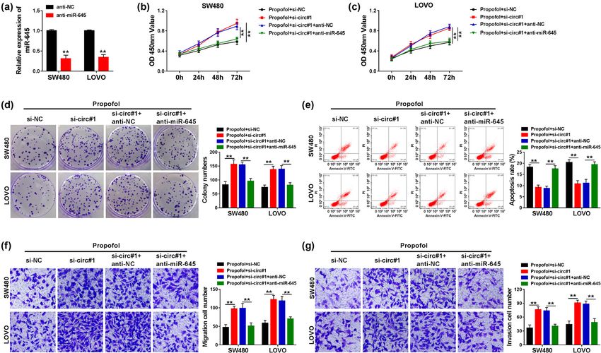
572 Xiaomin Cui et al.
2.4 Colony formation assay Foster City, CA, USA). Next, SYBR Green Master Mix kit
(Takara, Dalian, China) and ABI7500 Real-time PCR system
SW480 and LOVO cells were seeded into 6-well plates (Applied Biosystems) were employed to detect the relative
until the appearance of visible clones at 37°C with 5% expression of the interested RNAs, which was calculated by
CO2. Following that, cells were washed by phosphate buffer the method of 2−ΔΔCt. The primers used in this research were
saline (PBS, Sigma-Aldrich), then blocked by methanol provided by Beijing Genomics Institute (BGI, Shenzhen,
(Sigma-Aldrich) and stained by 0.05% crystal violet China) and listed as: circ_0026344, forward, 5′-CGTACCTG
(Sigma-Aldrich) for 30 min each. The colonies were visua- GAGACGCTGTTT-3′, reverse, 5′-GGGTTTGGGTACCAGCACT-
lized by a camera (Nikon, Tokyo, Japan). 3′; miR-645, forward, 5′-TCTAGGCTGGTACTGCT-3′, reverse,
5′-GAACATGTCTGCGTATCTC-3′; Glyceraldehyde 3-phos-
phate dehydrogenase (GAPDH), forward, 5′-CAGTCAG
CCGCATCTTCTTTT-3′, reverse, 5′-GTGACCAGGCGCCCAA
2.5 Flow cytometry assay TAC-3′; U6 small nuclear RNA (U6), forward, 5′-CTCGCTT
CGGCAGCACA-3′, reverse, 5′-AACGCTTCACGAATTTG
SW480 and LOVO cells were collected and suspended in CGT-3′. GAPDH and U6 were recognized as the reference
cold PBS (Sigma-Aldrich). Following that, Annexin V- of circ_0026344 and miR-645, respectively.
Fluorescein isothiocyanate (FITC)/Propidium lodide (PI)
Apoptosis Kit (BD Biosciences, San Jose, CA, USA) was
used to measure the apoptosis upon the guidance of the
corresponding instruction. BD FACS Canto™ II flow cyto- 2.8 Dual-luciferase reporter system
meter (BD Biosciences) was employed to detect the
apoptosis. Dual-luciferase reporter system was devoted to confirming
the prediction which was presented by Circinteractome
online database about the combination between circ_0026344
and miR-645. The complementary sequence or the corre-
2.6 Transwell assay sponding mutated sequence between circ_0026344 and
miR-645 was cloned into pmirGLO vector (Promega,
The migration and invasion of SW480 and LOVO cells were Madison, WI, USA) and named as circ_0026344 WT or
assessed by transwell assay. For the detection of invasion, circ_0026344 MUT. Then, the circ_0026344 WT or
transwell chamber (8.0 μm pore, Corning, Corning, NY, circ_0026344 MUT reporters were transfected into SW480
USA) with the application of Matrigel coating (BD and LOVO cells with miR-645 or miR-NC. Dual-Luciferase
Biosciences) was devoted. For the migration assay, trans- Reporter Assay Kit (Promega) was used to determine the
well chambers (Corning) without Matrigel coating (BD luciferase activity following the protocol.
Biosciences) were applied. Cells were seeded into the
upper chamber and the DMEM (Gibco) medium and 10%
FBS (Gibco) was added into the lower chamber. Whether
the migration assay or invasion assay, cells were cultured 2.9 Western blot
in an incubator with the setting of 37°C, 5% CO2, 24 h.
Subsequently, cells were blocked and stained by 4% para- SW480 and LOVO cells were lysed by RIPA buffer (Beyotime).
formaldehyde (Sigma-Aldrich) and 0.1% crystal violet (Sigma- Following that, proteins were loaded onto sodium dodecyl
Aldrich). Data were captured by an inverted microscope sulfate polyacrylamidegel electrophoresis (SDS-PAGE,
(magnification, ×100, CarlZeiss, Hallbergnoos, Germany). Beyotime) and polyvinylidene difluoride membrane (PVDF,
Millipore, Boston, MA, USA). After washed by PBS (Sigma-
Aldrich) for three times and blocked by nonfat milk, the
2.7 RNA isolation and real-time polymerase membranes were incubated with the primary antibodies:
chain reaction (qPCR) phospho-protein kinase B (p-AKT, 1:2,000, Sigma-Aldrich)
with the phosphorylation site in p308, AKT (1:1,000,
TRIzol reagent (Thermo Fisher Scientific, Waltham, MA, Sigma-Aldrich), phospho-mammalian target of rapamycin
USA) was applied to isolate the total RNA from SW480 (p-mTOR, 1:1,000, Sigma-Aldrich) with the phosphoryla-
and LOVO cells. Then, the RNA was reversed into cDNA tion site in S2481 and mTOR (1:1,000, Cell Signaling
by cDNA reverse transcription kit (Applied Biosystems, Technologies, Danvers, MA, USA) and GAPDH (1:1,000,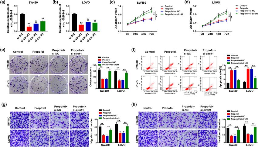
Role of circ_0026344/miR-645/Akt/mTOR axis in colorectal cancer 573
Cell Signaling Technologies), and the second antibody: circ_0026344 was conspicuously downregulated in CRC
anti-rabbit IgG (1:5,000, Ruiyingbio, Shanghai, China), sequen- cells compared with normal cells (Figure 2a). Interest-
tially. ECL Detection Reagents (Amersham Biosciences, ingly, the striking increase in circ_0026344 expression
Piscataway, NJ, USA) were applied to determine the result. was induced by propofol in CRC cells (Figure 2b). This
GAPDH served as a reference. phenomenon inspired us to further study the relationship
between circ_0026344 and propofol treatment in CRC.
CRC cells were transfected with circ_0026344 or vector,
and the successful transfection efficiency was exposed by
2.10 Data analysis
the notable increase in circ_0026344 expression in CRC
cells (Figure 2c). Moreover, circ_0026344 overexpression
GraphPad software 6.0 (GraphPad Inc., San Diego, CA,
triggered the restraint in proliferation, metastasis, and
USA) was adopted to analyze data. Each group of data
the promotion of apoptosis (Figure 2d–i). These data
was obtained from triplicate experiments and performed
exposed that circ_0026344 might serve as a suppressor
as mean ± standard deviation (SD). Between-group variance
in CRC.
was presented by Student’s t-test or one-way Analysis of
Variance (ANOVA). P < 0.05 was recognized as statistical
significance.
3.3 circ_0026344 knockdown allayed
propofol-induced restraint in
proliferation, metastasis, and promotion
3 Results of apoptosis in CRC cells
3.1 Propofol impaired the proliferation and Given the potential role of circ_0026344 in CRC, the inter-
metastasis and promoted the apoptosis action between circ_0026344 and propofol treatment
in CRC cells was further studied by loss-of-function approaches. The
knockdown efficiency of circ_0026344 was exposed by
Due to the frequent discovery in the association between the conspicuous decrease in circ_0026344 expression in
propofol and CRC, the direct evidence in the effect of CRC cells (Figure 3a and b). Due to the preeminent per-
propofol on CRC was carried out in this study. The viabi- formance, si-circ#1 was employed to the following tests.
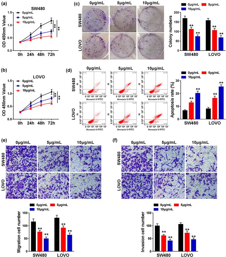
lity of SW480 and LOVO cells showed a time-dependent In terms of the proliferation, propofol treatment led to a
increase and a dose-dependent decrease in the treatment noticeable suppression, which was ameliorated by si-
of propofol (Figure 1a and b). For the clonality, propofol circ#1 in CRC cells (Figure 3c–e). However, si-circ#1
also led to a dose-dependent inhibition in SW480 and reversed propofol-mediated facilitation in apoptosis in
LOVO cells (Figure 1c). A significant promotion of apop- CRC cells (Figure 3f). For the metastasis, si-circ#1 possessed
tosis was associated with the higher concentration pro- the ability to attenuate propofol-induced restraint in the
pofol treatment in CRC cells (Figure 1d). As expected, migration and invasion in CRC cells (Figure 3g and h). These
the ability of migration and invasion presented a dose- results clarified that circ_0026344 deletion impaired the
dependent impairment in the treatment of propofol efficacy of propofol in CRC cells in vitro.
in CRC cells (Figure 1e and f). These data highlighted
that propofol treatment restrained the proliferation and
metastasis and triggered the apoptosis in CRC cells 3.4 circ_0026344 sponged miR-645
in vitro.
According to the above results, we wondered whether
circ_0026344 participated in propofol-treated CRC pro-
3.2 circ_0026344 overexpression inhibited gression by regulating the downstream factors. Thus, the
the proliferation and metastasis and underlying miRNA sponged by circ_0026344 was further
facilitated the apoptosis in CRC cells studied. The combination sequence between circ_0026344
and miR-645 was presented by Circinteractome online
Based on the role of propofol in CRC, we further explored tool (Figure 4a). To confirm the combination between
the potential regulatory factors in propofol-treated CRC. circ_0026344 and miR-645, miR-645 was successfully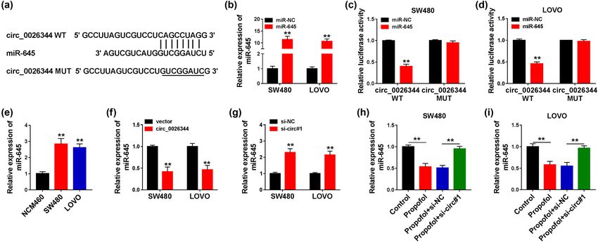
574 Xiaomin Cui et al. Figure 1: Propofol repressed cell proliferation, migration, and invasion, whereas induced cell apoptosis in SW480 and LOVO cells. SW480 and LOVO cells were treated with various doses of propofol (0, 5, and 10 μg/mL). (a–c) Cell proliferation was determined by CCK8 and colony formation assays. (d) Cell apoptosis was demonstrated by flow cytometry assay. (e and f) Cell migration and invasion were detected by transwell assay. Significant differences were compared with ANOVA. **P < 0.01. transfected into CRC cells (Figure 4b), and the noticeable (Figure 4c and d). Meanwhile, miR-645 was upregulated decrease in the luciferase activity in circ_0026344 WT in CRC cells compared with normal cells (Figure 4e). As further exposed the existence of combination in CRC cells expected, the deletion or overexpression of circ_0026344
Role of circ_0026344/miR-645/Akt/mTOR axis in colorectal cancer 575 Figure 2: circ_0026344 overexpression inhibited CRC progression. (a) circ_0026344 expression was detected by qPCR in NCM460, SW480, and LOVO cells. (b) The effect of propofol on circ_0026344 expression was determined by qPCR in SW480 and LOVO cells. (c) The transfection efficiency of circ_0026344 was determined by qPCR in SW480 and LOVO cells. (d–f) The impact of circ_0026344 over- expression on cell proliferation was revealed by CCK8 and colony formation assays. (g) The impact of ectopic circ_0026344 expression on cell apoptosis was presented by flow cytometry assay. (h and i) The effects of circ_0026344 overexpression on the migration and invasion of SW480 and LOVO cells were disclosed by transwell assay. Significant differences were compared with Student’s t-test (b–i) or ANOVA (A). **P < 0.01. led to a promotion or an inhibition in miR-645 expression 3.5 Propofol inhibited the proliferation and in CRC cells (Figure 4f and g). Moreover, si-circ#1 could metastasis and promoted apoptosis by mitigate the curb which was triggered by propofol in miR- circ_0026344/miR-645 axis in CRC cells 645 expression in CRC cells (Figure 4h and i). These data elucidated that circ_0026344 harbored miR-645 and nega- Based on the relationship between circ_0026344 and tively regulated miR-645 expression in CRC cells. miR-645, the functional regulation of circ_0026344 to
576 Xiaomin Cui et al. Figure 3: circ_0026344 silencing attenuated propofol-mediated effects on CRC progression. (a and b) The interfering efficiency of si-circ#1, si-circ#2, and si-circ#3 was determined by qPCR in SW480 and LOVO cells. (c–e) The impacts between propofol treatment and circ_0026344 silencing on cell proliferation were explained by CCK8 and colony formation assays. (f) The influences between propofol treatment and circ_0026344 knockdown on the apoptosis of SW480 and LOVO cells were presented by flow cytometry analysis. (g and h) The influences between propofol treatment and circ_0026344 absence on the migration and invasion of SW480 and LOVO cells were revealed by transwell assay. Significant differences were compared with ANOVA. **P < 0.01. Figure 4: circ_0026344 was associated with miR-645 in SW480 and LOVO cells. (a) Circinteractome online database was employed to predict the putative binding sites of circ_0026344 in miR-645. (b) The transfection efficiency of miR-645 was determined by qPCR in SW480 and LOVO cells. (c and d) Luciferase activities were detected by dual-luciferase reporter assay in SW480 and LOVO cells. (e) miR-645 expression was determined by qPCR in NCM460, SW480, and LOVO cells. (f and g) The effects of circ_0026344 overexpression and silencing on miR-645 expression were revealed by qPCR in SW480 and LOVO cells. (h and i) The impacts between propofol treatment and circ_0026344 silencing on miR-645 expression were demonstrated by qPCR in SW480 and LOVO cells. Significant differences were compared with Student’s t-test (b, c, d, f, and g) or ANOVA (e, h, and i). **P < 0.01.
Role of circ_0026344/miR-645/Akt/mTOR axis in colorectal cancer 577
miR-645 was further explored. The significant decrease was strikingly impaired by propofol, which was amelio-
in miR-645 expression indicated a successful transfec- rated by si-circ#1; however, anti-miR-645 could abrogate
tion efficiency of anti-miR-645 in CRC cells (Figure 5a). the amelioration induced by circ_0026344 deletion in CRC
Furthermore, anti-miR-645 abrogated si-circ#1-mediated cells (Figure 6a and b). These data disclosed that propofol
augmentation in proliferation and metastasis in propofol- treatment could repress Akt/mTOR signal pathway by
treated CRC cells (Figure 5b–d, f and g). In addition, circ_0026344/miR-645 axis in CRC cells.
si-circ#1-induced counteraction in propofol-mediated
promotion of apoptosis could be dispelled by anti-miR-
645 in CRC cells (Figure 5e). These data highlighted that
circ_0026344 affected the efficacy of propofol by regu- 4 Discussion
lating miR-645 in CRC cells.
Propofol was recognized as an antitumor in the treatment
of cancers. Propofol possessed the free radical scaven-
ging ability, which was instantiated in the amelioration
3.6 Propofol participated in CRC of ischemic brain injury by activating Akt/mTOR signal
progression by circ_0026344/miR-645/ pathway [26]. In CRC, propofol interacted with NMDAR/
Akt/mTOR signal pathway CAMKII/ERK signal pathway to inhibit the glycolysis,
thus hindered the development of CRC [27]. Meanwhile,
The downstream signal pathway of circ_0026344/miR-645 propofol regulated miR-124-3p/AKT3 axis to restraint the
was further explored in this study. Interestingly, the rela- proliferation in CRC cells [11]. The treatment of propofol
tive protein expression of p-AKT/AKT and p-mTOR/mTOR in CRC cells led to elevate the apoptosis [28]. In this
Figure 5: circ_0026344 regulated CRC development by binding to miR-645 in propofol-treated SW480 and LOVO cells. (a) miR-645
expression was detected by qPCR in SW480 and LOVO cells transfected with anti-NC or anti-miR-645. SW480 and LOVO cells were treated
with propofol + si-NC, propofol + si-circ#1, propofol + si-circ#1 + anti-NC, and propofol + si-circ#1 + anti-miR-645, respectively. (b–d) Cell
proliferation was determined by CCK8 and colony formation assays. (e) Cell apoptosis was detected by flow cytometry assay. (f and g) Cell
migration and invasion were detected by transwell assay. Significant differences were compared with Student’s t-test (a) or ANOVA (b–g).
**P < 0.01.578 Xiaomin Cui et al.
Figure 6: circ_0026344 silencing restrained propofol-inactivated Akt/mTOR pathway by interacting with miR-645. (a and b) The effects
among propofol, circ_0026344 silencing, and miR-645 inhibitor on Akt/mTOR pathway were presented by detecting the protein levels of
p-AKT, AKT, p-mTOR, and mTOR by western blot in SW480 and LOVO cells. Significant differences were compared with ANOVA. **P < 0.01.
study, a dose-dependent abatement in proliferation and inflammation, and promotion of proliferation, which were
metastasis and a dose-dependent augment in apoptosis induced by propofol in neuroinflammation [31]. In addi-
were presented in CRC cells treated by propofol. These tion, circ_0026344 sponged miR-645 to regulate the prolif-
findings were in accordance with the above reports. eration, metastasis, and apoptosis. circ_0026344 sponged
circRNAs were frequently recognized as biomarkers miR-21/miR-31 to restraint CRC development [29].
of cancers. circ_0026344 was downregulated in CRC cells Increasing evidences exposed the role of Akt/mTOR
and has been suggested to be a biomarker of CRC on signal pathway in regulating diseases’ development. Fan
account of the correlation between circ_0026344 expres- et al. illustrated that Akt/mTOR activation hampered
sion and the prognosis [29]. Moreover, circ_0026344 the immoderate autophagy to ameliorate the myocardial
downregulation suppressed the migration and invasion damage [23]. Akt/mTOR inhibition restrained the prolif-
of CRC cells [30]. Consistently, circ_0026344 was lowly eration of CRC [25]. In this study, propofol treatment-
expressed in CRC cells compared with normal cells; induced suppression in Akt/mTOR was reversed through
meanwhile, circ_0026344 overexpression inhibited the si-circ_0026344; however, anti-miR-645 restored the sup-
proliferation and metastasis and boosted apoptosis in pression in CRC cells. FOXD2-AS1 harbored miR-195 to
CRC cells. Interestingly, si-circ_0026344 allayed propofol activate Akt/mTOR pathway to activate esophageal squa-
treatment-induced impediment in proliferation, metas- mous cell carcinoma progression [32]. PRMT6/miR-372-3p/
tasis, and augmentation in apoptosis in CRC cells. circRNA Akt/mTOR signal pathway facilitated endometrial cancer
001372 inhibition reversed the suppression in apoptosis, process [33]. These data clarified that circ_0026344Role of circ_0026344/miR-645/Akt/mTOR axis in colorectal cancer 579
sponged miR-645 to restrain Akt/mTOR pathway in pro- colorectal cancer risk by tumor LINE-1 methylation level. J Natl
pofol-treated CRC cells. Cancer Inst. 2013;105(2):130–40. doi: 10.1093/jnci/djs482.
The disadvantage of this paper is the lack of in vivo [5] Thanikachalam K, Khan G. Colorectal cancer and nutrition.
Nutrients. 2019;11(1):164. doi: 10.3390/nu11010164.
study that propofol can mediate tumorigenesis via
[6] Dekker E, Tanis PJ, Vleugels JLA, Kasi PM, Wallace MB.
circ_0026344 in vivo. And we will study that in subse- Colorectal cancer. Lancet. 2019;394(10207):1467–80.
quent study. doi: 10.1016/s0140-6736(19)32319-0.
To sum up, circ_0026344 was downregulated and [7] Deng F, Ouyang M, Wang X, Yao X, Chen Y, Tao T, et al.
circ_0026344 overexpression or propofol treatment led Differential role of intravenous anesthetics in colorectal
cancer progression: implications for clinical application.
to suppress the proliferation and metastasis and faci-
Oncotarget. 2016;7(47):77087–95. doi: 10.18632/
litate the apoptosis in CRC cells. In addition, propofol- oncotarget.12800.
induced amelioration in CRC development was assisted [8] Mammoto T, Mukai M, Mammoto A, Yamanaka Y, Hayashi Y,
by circ_0026344/miR-645/Akt/mTOR signal pathway. Mashimo T, et al. Intravenous anesthetic, propofol inhibits
Our findings not only provide a theoretical basis for invasion of cancer cells. Cancer Lett. 2002;184:165–70.
doi: 10.1016/S0304-3835(02)00210-0.
further studying CRC therapy with propofol, but also
[9] Huang H, Benzonana LL, Zhao H, Watts HR, Perry NJ, Bevan C,
present potential in treating CRC with propofol com-
et al. Prostate cancer cell malignancy via modulation of HIF-
bined with circRNA-mediated drug. 1alpha pathway with isoflurane and propofol alone and in
combination. Br J Cancer. 2014;111(7):1338–49. doi: 10.1038/
Funding information: This work was supported by Chengdu bjc.2014.426.
Municipal Health Commission Project (No. 2019107). [10] Liu D, Sun X, Du Y, Kong M. Propofol promotes activity and
tumor-killing ability of natural killer cells in peripheral blood
of patients with colon cancer. Med Sci Monit.
Authors contribution: X. C. and M. D. contributed to the 2018;24:6119–28. doi: 10.12659/MSM.911218.
conception of the study; X. C., J. F., and J. W. performed [11] Li Y, Dong W, Yang H, Xiao G. Propofol suppresses proliferation
the experiment; X. Z. and M. D. performed the data ana- and metastasis of colorectal cancer cells by regulating miR-
lyses and wrote the manuscript. All authors read and 124-3p.1/AKT3. Biotechnol Lett. 2020;42(3):493–504.
approved the final manuscript. doi: 10.1007/s10529-019-02787-y.
[12] Zhang YF, Li CS, Zhou Y, Lu XH. Effects of propofol on colon
cancer metastasis through STAT3/HOTAIR axis by activating
Conflict of interest: The authors declare that they have no WIF-1 and suppressing Wnt pathway. Cancer Med 2020
financial conflicts of interest. 9(5):1842–54. doi: 10.1002/cam4.2840.
[13] Naeli P, Pourhanifeh MH, Karimzadeh MR, Shabaninejad Z,
Data availability statements: All data generated and/or Movahedpour A, Tarrahimofrad H, et al. Circular RNAs
and gastrointestinal cancers: epigenetic regulators
analyzed during the study are available from the corre-
with a prognostic and therapeutic role. Crit Rev Oncol
sponding author by request. Hematol. 2020;145:102854. doi: 10.1016/
j.critrevonc.2019.102854.
[14] Vicens Q, Westhof E. Biogenesis of circular RNAs. Cell.
2014;159(1):13–4. doi: 10.1016/j.cell.2014.09.005.
[15] Wang Y, Mo Y, Gong Z, Yang X, Yang M, Zhang S, et al. Circular
RNAs in human cancer. Mol Cancer. 2017;16(1):25.
References doi: 10.1186/s12943-017-0598-7.
[16] Sun H, Tang W, Rong D, Jin H, Fu K, Zhang W, et al.
[1] Bray F, Ferlay J, Soerjomataram I, Siegel RL, Torre LA, Jemal A. Hsa_circ_0000520, a potential new circular RNA biomarker,
Global cancer statistics 2018: GLOBOCAN estimates of inci- is involved in gastric carcinoma. Cancer Biomark.
dence and mortality worldwide for 36 cancers in 185 countries. 2018;21(2):299–306. doi: 10.3233/CBM-170379.
CA Cancer J Clin. 2018;68(6):394–424. doi: 10.3322/ [17] Yang F, Liu DY, Guo JT, Ge N, Zhu P, Liu X, et al. Circular RNA
caac.21492. circ-LDLRAD3 as a biomarker in diagnosis of pancreatic cancer.
[2] Amersi F, Agustin M, Ko CY. Colorectal cancer: epidemiology, World J Gastroenterol. 2017;23(47):8345–54. doi: 10.3748/
risk factors, and health services. Clin Colon Rectal Surg. wjg.v23.i47.8345.
2005;18(3):133–40. doi: 10.1055/s-2005-916274. [18] Zhang HY, Zhang BW, Zhang ZB, Deng QJ. Circular RNA TTBK2
[3] Rozen P, Fireman Z, Figer A, Legum C, Ron E, Lynch HT. Family regulates cell proliferation, invasion and ferroptosis via miR-
history of colorectal cancer as a marker of potential malig- 761/ITGB8 axis in glioma. Eur Rev Med Pharmacol Sci.
nancy within a screening program. Cancer. 2020;24(5):2585–600. doi: 10.26355/eurrev_202003_20528.
1987;60(2):248–54. doi: 10.1002/1097-0142(19870715) [19] Liu J, Song S, Lin S, Zhang M, Du Y, Zhang D, et al. circ-
60:23.0.co;2-g. SERPINE2 promotes the development of gastric carcinoma by
[4] Ogino S, Nishihara R, Lochhead P, Imamura Y, Kuchiba A, sponging miR-375 and modulating YWHAZ. Cell Prolif.
Morikawa T, et al. Prospective study of family history and 2019;52(4):e12648. doi: 10.1111/cpr.12648.580 Xiaomin Cui et al.
[20] Liu T, Sun Q, Li Q, Yang H, Zhang Y, Wang R, et al. Dual PI3K/ [27] Chen X, Wu Q, Sun P, Zhao Y, Zhu M, Miao C. Propofol disrupts
mTOR inhibitors, GSK2126458 and PKI-587, suppress tumor aerobic glycolysis in colorectal cancer cells via inactivation of
progression and increase radiosensitivity in nasopharyngeal the NMDAR-CAMKII-ERK pathway. Cell Physiol Biochem.
carcinoma. Mol Cancer Ther. 2015;14(2):429–39. doi: 10.1158/ 2018;46(2):492–504. doi: 10.1159/000488617.
1535-7163.MCT-14-0548. [28] Zhang L, Wang N, Zhou S, Ye W, Jing G, Zhang M. Propofol
[21] Martini M, De Santis MC, Braccini L, Gulluni F, Hirsch E. PI3K/ induces proliferation and invasion of gallbladder cancer cells
AKT signaling pathway and cancer: an updated review. Ann through activation of Nrf2. J Exp Clin Cancer Res.
Med. 2014;46(6):372–83. doi: 10.3109/ 2012;31(1):66. doi: 10.1186/1756-9966-31-66.
07853890.2014.912836. [29] Yuan Y, Liu W, Zhang Y, Zhang Y, Sun S. circRNA circ_0026344
[22] Francipane MG, Lagasse E. mTOR pathway in colorectal cancer: as a prognostic biomarker suppresses colorectal cancer pro-
an update. Oncotarget. 2014;5(1):49–66. doi: 10.18632/ gression via microRNA-21 and microRNA-31. Biochem Biophys
oncotarget.1548. Res Commun. 2018;503(2):870–5. doi: 10.1016/
[23] Fan C, Tang X, Ye M, Zhu G, Dai Y, Yao Z, et al. Qi-Li-Qiang-Xin j.bbrc.2018.06.089.
alleviates isoproterenol-induced myocardial injury by inhi- [30] Shen T, Cheng X, Liu X, Xia C, Zhang H, Pan D, et al.
biting excessive autophagy via activating AKT/mTOR pathway. circ_0026344 restrains metastasis of human colorectal cancer
Front Pharmacol. 2019;10:1329. doi: 10.3389/ cells via miR-183. Artif Cells Nanomed Biotechnol.
fphar.2019.01329. 2019;47(1):4038–45. doi: 10.1080/21691401.2019.1669620.
[24] Liu Z, Luo S, Wu M, Huang C, Shi H, Song X. LncRNA GHET1 [31] Wang M, Suo L, Yang S, Zhang W. circRNA 001372 reduces
promotes cervical cancer progression through regulating AKT/ inflammation in propofol-induced neuroinflammation and
mTOR and Wnt/beta-catenin signaling pathways. Biosci Rep. neural apoptosis through PIK3CA/Akt/NF-kappaB by miRNA-
2020;40(1):BSR20191265. doi: 10.1042/BSR20191265. 148b-3p. J Invest Surg. 2020;1–11. doi: 10.1080/
[25] Wang K, Huang W, Sang X, Wu X, Shan Q, Tang D, et al. 08941939.2020.1771639.
Atractylenolide I inhibits colorectal cancer cell proliferation by [32] Liu H, Zhang J, Luo X, Zeng M, Xu L, Zhang Q, et al.
affecting metabolism and stemness via AKT/mTOR signaling. Overexpression of the long noncoding RNA FOXD2-AS1 pro-
Phytomedicine. 2020;68:153191. doi: 10.1016/ motes cisplatin resistance in esophageal squamous cell carci-
j.phymed.2020.153191. noma through the miR-195/Akt/mTOR axis. Oncol Res.
[26] Wang HY, Wang GL, Yu YH, Wang Y. The role of phosphoino- 2020;28(1):65–73. doi: 10.3727/096504019X15656904013079.
sitide-3-kinase/Akt pathway in propofol-induced postcondi- [33] Jiang N, Li QL, Pan W, Li J, Zhang MF, Cao T, et al. PRMT6
tioning against focal cerebral ischemia-reperfusion injury in promotes endometrial cancer via AKT/mTOR signaling and
rats. Brain Res. 2009;1297:177–84. doi: 10.1016/ indicates poor prognosis. Int J Biochem Cell Biol.
j.brainres.2009.08.054. 2020;120:105681. doi: 10.1016/j.biocel.2019.105681.You can also read