Therapeutic effects and prognostic factors of 125I brachytherapy for pelvic recurrence after early cervical cancer surgery - Nature
←
→
Page content transcription
If your browser does not render page correctly, please read the page content below
www.nature.com/scientificreports
OPEN Therapeutic effects and prognostic
factors of 125I brachytherapy
for pelvic recurrence after early
cervical cancer surgery
Rui Wang2, Jinhu Zhu1*, Shu Yang2, Xiaoqin Chen1, Cairu Gu1, Tong Liang1, Ling Li1,
Dan Liu1 & Yanqing Cao2
To investigate the efficacy of 125I seed implantation in the treatment regimen of pelvic recurrence
after early cervical cancer surgery and to analyse prognostic factors. To evaluate efficacy and analyse
prognostic factors of 125I seed implantation for pelvic recurrence after early cervical cancer surgery. A
prospective study was conducted on 62 patients who experienced pelvic recurrence after early cervical
cancer surgery between August 2005 and September 2015. The 62 patients were treated and assessed
in 2 groups (n = 30). All 62 patients were randomized into two groups that received two different
treatment regimens: the treatment group (n = 30), which received 125I particle implantation therapy,
and the control group (n = 32), which received whole-pelvic irradiation using the anteroposterior/
posteroanterior field and cisplatin-based concurrent chemoradiation therapy. The efficacy/efficiency
of 125I seed implantation and prognostic factors were analysed by logistic regression. Overall survival
was determined by Kaplan–Meier analysis. Multivariate analysis results were obtained by the Cox
proportional hazards regression model. The effective control rates at 1, 3, 6 and 12 months were
76.7%, 80.0%, 83.3%, and 86.7% in the 125I particle implantation group. The total effective control
rates at 1, 3, 6 and 12 months were 65.6%, 65.5%, 62.5%, and 71.9% in the chemoradiotherapy
group. Significant differences were observed between the two groups. The overall survival rates at 1,
2, 3, 4, and 5 years and the median overall were 96.7%, 93.3%, 86.7%, 71.9%, 65.6% and 4.34 years,
respectively, in the 125I seed implantation group and 81.3%, 71.9%, 62.5%, 56.3%, 53.1% and 3.59
years, respectively, in the control group. There were statistically significant differences in survival
rates depending on the diameter of the largest recurrent pelvic tumour (χ2 = 6.611, P = 0.010). The
multivariate analysis showed that the survival rates were related to the diameter of the largest
recurrent pelvic tumour (χ2 = 4.538, P = 0.033). 125I implantation is an effective, safe, and promising
method for the treatment of pelvic recurrence after early cervical cancer surgery. The diameter of the
recurrent pelvic tumour was identified as a significant independent prognostic factor in patients who
received 125I implantation.
Cervical cancer has the fourth highest mortality rate among gynaecologic tumours and is a great threat to
women’s health worldwide1. More than 30% of patients experience recurrence or metastasis after treatment, and
locoregional recurrence of the pelvic cavity is the predominant pattern of treatment failure in cervical cancer2.
In recent years, because of the prevalence of early screening for cervical cancer, continuous improvements
in surgical techniques and advancements in diagnosis and treatment techniques, cervical cancer has been diag-
nosed earlier and treated in a timely manner, which has substantially reduced the mortality r ate3. However, the
therapeutic results are not satisfactory, and its therapeutic effect and prognosis are affected by many f actors4–7.
Currently, the effects and prognostic factors of recurrent cervical cancer have been the area of intense clinical
research, and how to improve the prognosis of patients with recurrent cervical cancer is an urgent problem.
125
I seeds (half-life: 59.4 days) are permanently implanted in a patient’s body and emit characteristic γ-rays of
relatively low photon energy (27.4–28.4 keV) for several months. 125I seed implantation in a pelvic mass guided by
1
Department of Gynecology, GuangZhou Red Cross Hospital, Jinan University, Guangzhou 510220, Guangdong,
China. 2Department of Oncology, The First Affiliated Hospital of Guangdong Pharmaceutical University,
Guangzhou 510080, Guangdong, China. *email: zhujinhu421@163.com
Scientific Reports | (2021) 11:11356 | https://doi.org/10.1038/s41598-021-90007-x 1
Vol.:(0123456789)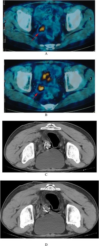
www.nature.com/scientificreports/

                                             Characteristics                              125
                                                                                                I implantation   Radiochemotherapy
                                             No. of patients                              30*                    32*
                                             Mean age (years)                             44.3 (28–65)           46.5 (31–68)
                                             Pre-operation tumor diameter (cm)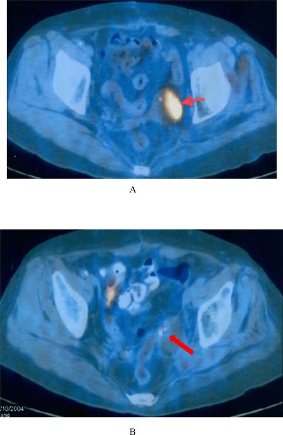
www.nature.com/scientificreports/

                                                                                                            Local control efficacy (%)
                                   Group                                    No. of patients      Date (m)   CR**         PR**        NC**       PD**       RR
                                                                                                 1          15 (50.0)*    8 (26.7)   5 (16.7)   2 (6.7)    23 (76.7)*

                                   125
                                                                                                 3          14 (46.7)*   10 (33.3)   3 (10.0)   3 (10.0)   24 (80.0)*
                                     I implantation                         30
                                                                                                 6          16 (53.3)*    9 (30.0)   3 (10.0)   2 (6.7)    25 (83.3)*
                                                                                                 12         15 (50.0)*   11 (26.7)   3 (10.0)   1 (3.3)    26 (86.7)*
                                                                                                 1           9 (28.1)    12 (37.5)   4 (12.5)   7 (21.9)   21 (65.6)
                                                                                                 3          10 (31.2)    11 (34.3)   5 (15.6)   6 (18.8)   21 (65.5)
                                   Radiochemotherapy                        32
                                                                                                 6          10 (31.2)    10 (31.2)   5 (15.6)   7 (21.9)   20 (62.5)
                                                                                                 12         12 (37.5)    11 (34.5)   4 (12.5)   5 (15.6)   23 (71.9)

                                  Table 2.  Comparison of the response rate of patients treated with 125I implantation with radiochemotherapy.
                                  Data in parentheses are percentages. **CR complete response, PR partial response, NC no change, PD
                                  progressive disease, RR indicates the percentage of patients with response, i.e. RR = PR + CR. *P < 0.05 versus
                                  radiochemotherapy group.

                                                                I implantation
                                                              125
                                                                                   Radiochemotherapy        P value
                                   Median survival (months)   4.34 ± 1.19          3.59 ± 1.67              < 0.05
                                   Overall survival
                                   1-year (%)                 96.7                 88.3                     > 0.05
                                   2-year (%)                 93.3                 71.9
                                   3-year (%)                 86.7                 62.5
                                                                                                            < 0.05
                                   4-year (%)                 71.9                 56.3
                                   5-year (%)                 65.6                 53.1

                                  Table 3.  Comparison of survival of patients treated with 125I implantation with conventional
                                  radiochemotherapy.

                                  vaginal margins, 53 cases (85.5%); moderate or low differentiation, 38 cases (61.3%); high differentiation, 25
                                  cases (40.3%); recurrent tumour volume ≤ 3 cm, 18 cases (29.0%); and recurrent tumour volume > 3 cm, 44 cases
                                  (71.0%). Specific data are shown in Table 1.
                                     All patients had mild pain at the puncture site after the operation without special treatment. After 2–3 days,
                                  the pain was relieved. Two patients experienced abdominal pain, diarrhoea and a rectal reaction after 3 months.
                                  Symptomatic diarrhoea was relieved after treatment.

                                  Tumour response. The follow-up time ranged from 7.2 to 60 months. Follow-up images showed that the
                                  local control rates the 125I implantation group after 1 month were as follows: 15 (50.0%), CR; 11 (26.7%), PR;
                                  3 (10.0%), SD; and 1 (3.3%), PD. Those for the radiochemotherapy group were as follows: 12 (37.5%), CR; 11
                                  (34.5%), PR; 4 (12.5%), SD; and 5 (15.6%), PD. After 12 months, the total effective rate in the 125I implantation
                                  group was 86.7%, which was significantly higher than that of the chemoradiotherapy group (71.9%, P < 0.05).
                                  The total effective control rates were 76.7%, 80.0%, 83.3% and 86.7% in the 125I particle implantation group, and
                                  those at 1, 3, 6 and 12 months in the combined chemoradiotherapy group were 65.6%, 65.5%, 62.5%, and 71.9%.
                                  A comparison of the above data revealed a significant difference between the two groups.
                                      At 125I particle implantation, the 1-year survival rate was 96.7%, the local total effective control rate (RR) was
                                  76.7%, and the progression-free control rate (SD) was 16.7%. In the radiotherapy group, the 12-month survival
                                  rate, local total effective control rate and progression-free control rate were 88.3%, 65.6% and 12.5%, respectively.
                                  Between the 125I implantation group and the radiochemotherapy group, the local total effective control rate was
                                  significantly different, but the 1-year survival rate was not. Response details for patients after treatment are given
                                  in Tables 2 and 3 and Figs. 1 and 2.

                                  Overall survival.       For the 125I implantation group, the 1-, 2-, 3-, 4- and 5-year overall survival rates and the
                                  median overall were 96.7%, 93.3%, 86.7%, 71.9%, 65.6% and 4.34 years, respectively, and those for the radio-
                                  chemotherapy group were 81.3%, 71.9%, 62.5%, 56.3%, 53.1% and 3.59 years, respectively. Mantel–Haenszel
                                  tests revealed that the 2-year, 3-year, 4-year, and 5-year overall survival rates for the 125I implantation group were
                                  significantly better than those for the radiochemotherapy group. The K–M survival curve for the 125I implanta-
                                  tion group was significantly better than that for the chemoradiotherapy group. The above data are presented in
                                  Table 3 and Fig. 3.

                                  Univariate analysis. The influence of patient- and tumour-related factors on overall survival is shown in
                                  Tables 4 and 5. The results showed that the diameter of the largest tumour of the recurrent pelvic tumour was

Scientific Reports |   (2021) 11:11356 |                  https://doi.org/10.1038/s41598-021-90007-x                                                                         3

                                                                                                                                                                        Vol.:(0123456789)
www.nature.com/scientificreports/
Figure 1. A 35-year-old patient with a tumor of pelvic recurrence after cervical cancer surgery. Tumor
dimensions: 20 mm × 2.5 mm × 3.0 mm before treatment. (A,B) The imaging of PET-CT before multimodality
minimally invasive treatment showed abnormal radioactivity concentration in the pelvic. (C,D) The imaging of
CT after the 125I implantation treatment.
significantly associated with the survival rate (χ2 = 6.611, P = 0.010). Statistical results also showed no signifi-
cant differences in overall survival rates attributable to age at the first operation (χ2 = 0.001, P = 0.969), diam-
eter of the largest tumour (χ2 = 1.060, P = 0.303), first procedure (χ2 = 1.190, P = 0.552), pattern of organization
Scientific Reports | (2021) 11:11356 | https://doi.org/10.1038/s41598-021-90007-x 4
Vol:.(1234567890)www.nature.com/scientificreports/
Figure 2. A 47-year-old patient with a tumor of pelvic recurrence after cervical cancer surgery. Tumor
dimensions: 20 mm × 1.5 mm × 1.0 mm before treatment. (A) The imaging of PET-CT before multimodality
minimally invasive treatment showed abnormal radioactivity concentration. (B) The imaging of PET-CT after
the treatment revealed the absence of abnormal radioactivity concentration and came into a cold lesion.
(χ2 = 0.388, P = 0.824), FIGO stage (χ2 = 0.025, P = 0.988), vaginal incision (χ2 = 0.002, P = 0.962), lymph node
invasion (χ2 = 0.389, P = 0.533), degree of differentiation (χ2 = 0.645, P = 0.422), or 125I particle usage (χ2 = 0.615,
P = 0.433).
Multivariate analysis. Multivariate analysis was performed by using the variables deemed significant in
the univariate analysis as covariates. The multivariate analysis (Table 5; Fig. 4) showed that the survival rates
were related to the diameter of the largest tumour of the recurrent pelvic tumour (χ2 = 4.538, P = 0.033).
Discussion
Permanently implanted 125I particle tissue is a close-range radiotherapy method in which radioactive particles
are implanted into tumours or invaded tissues, killing tumour cells in the target area by continuously releasing γ
rays. This method has the biophysical characteristics of a high dose, high conformability and a steep decrease in
the dose to surrounding tissue. The pathological damage is relatively minimal, and the damage range is limited
to 5 mm from the s ource8. By adjusting the spacing and activity of the particle source, the dose to the tumour
target area can be increased exponentially, which can ensure that the target area is exposed to high intensity and
protect the surrounding normal organs to the greatest e xtent9. At the same time, the radiation-related toxic side
Scientific Reports | (2021) 11:11356 | https://doi.org/10.1038/s41598-021-90007-x 5
Vol.:(0123456789)www.nature.com/scientificreports/
Figure 3. Kaplan–Meier curve shows overall survival rates for patients who received 125I implantation (A) and
for those who underwent radiochemotherapy (B).
effects do not increase10. Particle implantation has been increasingly considered for prostate cancer. With the
wide application of radioactive 125I particles, there are many reports of permanent inter-tissue irradiation in the
treatment of diverse solid tumours, and the particles have achieved a good curative e ffect11. Thus, they can be
used as a supplementary treatment for recurrent pelvic malignant tumours.
Permanent inter-organizational implantation of radioactive 125I particles is a safe and effective treatment for
brachytherapy, and it has the advantages of minimal invasiveness, a high local dose, small injury to surrounding
normal tissue and a low incidence of complications. Our centre uses CT-guided radioactive particle implanta-
tion to treat recurrent cervical cancer, but because of the complex anatomical structure and bone structure of
the pelvic cavity, it is difficult to maintain a parallel arrangement of 125I particles12. With the emergence of 3D
printing technology and 3D⁃PNCT combined with CT guidance technology, this problem has been solved13.
This study describes the use of 125I particle implantation for pelvic recurrence after early cervical cancer sur-
gery. Clinical and pathological characteristics were used to perform a detailed statistical analysis to determine
the efficacy of radiotherapy on pelvic recurrent tumours and prognostic factors. The results of this study can
provide a reference for the clinical treatment and prognosis of patients with pelvic recurrence after cervical cancer
surgery to formulate more effective treatment plans. It is very important to improve the survival of patients with
pelvic recurrence after cervical cancer surgery and to improve the long-term curative effect14.
Many studies have shown that the size of the recurrent tumour is an important factor affecting the curative
effect and prognosis of patients with pelvic recurrence after cervical cancer surgery. Hisao lto and others reported
90 cases of vaginal stump recurrence after cervical cancer surgery according to the size of the recurrent tumour
(divided into three groups). The 5-year survival rate was 0% in zero patients with a recurrent tumour ≥ 3 cm,
56% in 18 patients with a recurrent tumour < 3 cm, and 87% in 37 patients with non-obvious tumour nodules.
The difference was statistically significant (P < 0.05). The results showed that the efficacy of the non-obvious
lesion group was better than that of the other two groups, the size of the recurrent tumour was significantly cor-
related with prognosis, and the size of the recurrent tumour was an important prognostic factor (P = 0.01)15. 125I
seed implantation can be used to treat pelvic recurrence from cervical cancer. Qu et al.16 evaluated the efficacy
and dosimetry of 125I seed implantation for pelvic recurrence from cervical cancer by examining 36 patients
between July 2005 and October 2015. Nineteen (52.7%) achieved partial response (PR), 13 (36.1%) had no
change (NC), and 4 (11.2%) developed progressive disease (PD). The total effective rate (CR + PR) was 88.9%,
and the progression-free survival (PFS) time of 4 patients was no longer than 12 months. The median survival
time was 7.5 months. The 1- and 2-year local progression-free survival rates were 34.9% and 20%, r espectively16.
In this study, after 125I seed treatment, the 5-year overall survival rate of patients with a recurrent tumour ≥ 3
cm was 37.5% and that of patients with a recurrent tumour ≤ 3 cm was 81.8%. The 5-year survival rates are shown
in Table 4. Single-factor analysis in this study showed that the diameter of the locally recurrent pelvic tumours
after cervical cancer surgery influences 125I particle therapy outcomes. Multiple-factor analysis showed that the
size of the recurrent tumour was statistically significant for prognosis (P = 0.033). These findings suggest that the
size of the recurrent tumour is not only a related factor affecting the therapeutic effect and prognosis of pelvic
recurrence after cervical cancer surgery but also the only independent factor affecting its therapeutic effect and
prognosis. Therefore, the larger the recurrent tumour is, the worse the prognosis, which may be due to the large
Scientific Reports | (2021) 11:11356 | https://doi.org/10.1038/s41598-021-90007-x 6
Vol:.(1234567890)www.nature.com/scientificreports/
Factor No. of patients (%) Overall survival (%) χ2 P value
Age (years)
< 40 10 (33.3) 70.0
0.001 0.969
≥ 40 20 (66.7) 70.0
Pre-operation diameter of largest tumor (cm)www.nature.com/scientificreports/
Figure 4. Graph shows 5-year cumulative survival rate after 125I implantation therapy, compared between
patients with diameter of largest tumor ≥ 3 cm (A) and < 3 cm (B) in recurrent pelvic tumor after cervical cancer
surgery. The survival rate of patients with diameter of largest tumor < 3 cm was significantly higher than that of
patients with diameter of largest tumor ≥ 3 cm.
size of the tumour. Short-range treatment cannot be used to reach the maximum total tumour dose within the
limit of radiotherapy toxicity. Moreover, if the tumour is large, there are many oxygen-deficient cells, the sensitiv-
ity to radiotherapy decreases, and metastasis easily occurs. Therefore, for patients with large recurrent tumours
after radiotherapy and/or residual tumours, multi-modal treatment, such as combined chemotherapy, can be
considered, which may improve the curative effect17.
There are still several problems in the treatment of recurrent pelvic tumours after cervical cancer surgery.
Although previous studies used full pelvic irradiation, recurrence still occurred after treatment, and whether the
dose of radiotherapy needs to be increased and whether chemotherapy should be maintained after synchronous
radiotherapy and chemotherapy are still controversial. Siriwan Tangjitgamol and others reported that after
concurrent chemoradiotherapy, the survival of patients with locally advanced cervical cancer was improved
by maintenance chemotherapy. However, there were several potential controversies in this trial, including an
inappropriate statistical design, and there are no related reports on advanced toxicity associated with intensive
chemotherapy. Therefore, the role of maintaining chemotherapy remains to be further s tudied18.
In our study, individualized brachytherapy plans were established before treatment based on different recur-
rence sites and tumour sizes can improve survival. Given that a higher radiotherapy dose for recurrent pelvic
wall tumours is an effective measure to improve the local control rate and prognosis of patients with recurrent
pelvic tumours, it is particularly important for clinicians to develop a reasonable and scientific individualized
treatment for pelvic recurrent tumours after cervical cancer surgery19. However, our study still had limitations.
First, we did not further study the mechanism of treatment differences. Second, the competitive risks of the Fine
and Gray type were not assessed in this study, which may have resulted in competitive risk bias, but the direct
cause of patient death in this study was cancer, this study is still valuable and needed for future studies. We also
plan to perform a further study by combining this brachytherapy with systemic radiochemotherapy.
Methods
Patients. Between August 2005 and September 2015, a total of 62 patients who were diagnosed with pelvic
recurrence after cervical cancer surgery and who were admitted to two individual hospitals working collabora-
tively were included in the current study. All participating patients were approved by the Ethical Committee of
GuangZhou Red Cross Hospital. Before treatment initiation, all subjects were fully aware of the potential risks
and provided written informed consent. Our study was performed in accordance with all international, national,
and institutional policies regarding research studies involving human subjects.
Patients who developed a pelvic tumour after early cervical cancer surgery were randomized into two groups:
the experimental group, which included 30 patients who received 125I particle implantation, and the radiation and
chemotherapy group, which included 32 patients who received pelvic irradiation at the prescribed dose of ≤ 46
Gy and a dual regimen consisting of platinum concurrent chemotherapy. Randomization was achieved by using
computer-generated random numbers. Detailed information on each group is presented in Table 1. There was no
statistically significant difference between the two groups (P > 0.05). All patients had been examined by surgeons
and radiation oncologists and were deemed unsuitable for resurgery. The five patients in the treatment group with
positive lymph nodes were deemed unsuitable for EBRT, and some patients refused to undergo surgery and EBRT.
Scientific Reports | (2021) 11:11356 | https://doi.org/10.1038/s41598-021-90007-x 8
Vol:.(1234567890)www.nature.com/scientificreports/
Eligibility criteria. The strictly enforced entry criteria were as follows: (1) patient KPS ≥ 90; (2) tumour
recurrence was confirmed by pathology and immunohistochemistry or had at least two of the following positive
results: (scc-ag) level rise, CT, MRI and PET-CT indicated recurrence; and (3) the leucocyte count was above
3.5 × 10⁹/L before surgery or could be elevated to this level after supportive treatment.
Exclusion criteria. The strictly enforced exclusion criteria were as follows: (1) a history of postoperative
adjuvant therapy; (2) a surgical history of early cervical cancer or received preoperative radiotherapy and post-
operative radiotherapy; (3) para-aortic lymph node recurrence or distant metastasis; (4) only palliative treat-
ment was given after recurrence; and (5) patients with severe cardiovascular and cerebrovascular diseases, liver
and kidney diseases and other systemic diseases.
Instruments. A Picker CT-Twin Flash scanner was used to image the tumour at 120 kV and 275 mA with
a slice width of 5 mm. The treatment planning system (TPS) and radioactive seed treatment planning system
(BT-RSI) were developed by Yuanbo (Beijing, China)9. The main instruments required for seed implantation
included a turn-table implantation gun, 18-G implantation needles and 125I seeds, which were the most impor-
tant (i.e., the therapeutic part). 125I produces gamma rays (95% of 28 keV, 5% of 35 keV), has a half-life of 59.6
days, a half-value thickness of 0.025 mm of lead, a penetration depth of 17 mm, and an initial rate of 7 cGy/h20, 21.
Each seed has a diameter of 0.8 mm and a length of 4.5 mm, and the titanium capsule wall has a thickness of
0.05 mm. The 125I seeds are made from 125I absorbent silver rods and are encapsulated in a laser-welded titanium
envelope. All the 125I seeds (6711/BT-125I) were mailed to our hospital in a type-A package that had passed leak
detection and activity series tests.
125
I seed implantation planning. A detailed analysis of tumour volume was performed 1–2 weeks before
seed implantation using a 5 mm thick CT scan. On each cross-sectional image, the radiation oncologist outlined
the gross tumour volume (GTV) and the risk area for subclinical disease. The planned target volume (PTV)
included the entire GTV and a boundary of 0.5–1.0 c m22. The dose was prescribed as the minimum peripheral
dose (MPD), which included the PTV. The median MPD was 130 Gy (range 90–160 Gy)23. The distribution and
MPD of 125I seeds were calculated by using a computer processing planning system (RT-RSI, Beijing Atom and
High Technique Industries Inc., Beijing, China)24. An example is shown in Fig. 5.
CT‑guided 125I implantation and radiation protection. The patients fasted for 1.0–3.0 h and were
given sedatives and local anaesthesia before the procedure. Based on the established treatment plan, a 3-mm
incision was made on the skin, and a seed implantation applicator was inserted into each tumour under the
guidance of CT at a distance. Care was taken not to puncture large blood vessels or vital organs. For tumours less
than or equal to 1.0 cm in size, interstitial planar (surface) implants were used. After the procedure, the catheters
were retracted, and incisions were bound and compressed24.
A surgeon performed 125I implantation in a specially designated operating room. During the operation, the
medical personnel wore lead gloves, hats, ambi-necks, and other protective clothing. After the procedure, a
special technician detected the ray dosage in the surroundings in real time to detect any missing seeds or other
problems.
Chemotherapy. Thirty-two patients received cisplatin (70 mg/m2 per surface area, day 1 of each cycle)
combined with 5-fluorouracil (1000 mg/m2 per surface area, days 2–5 of each cycle), which was repeated every
4 weeks for a total of 4–6 cycles. Routine blood tests were performed 1–2 times a week during treatment, and
other haematologic tests, including liver and kidney function tests, were reviewed before and after each chemo-
therapy cycle.
Evaluation criteria. The effectiveness of 125I seed implantation was based on the Response Evaluation Cri-
teria in Solid Tumors (RECIST 1.0). The RECIST 1.0 guideline is currently recognized as the most commonly
used guideline. This guideline, updated in 2000, is an anatomic-based rather than a functional evaluation system.
(1) Complete response (CR) was defined as follows: disappearance of all target lesions. (2) Partial response (PR)
was defined as follows: a decrease of at least 30% in the sum of the LD of the target lesions, taking as a refer-
ence the baseline sum of the longest diameter. (3) Stable disease (SD) was defined as follows: neither sufficient
shrinkage to qualify for PR nor a sufficient increase to qualify for PD, taking as a reference the smallest sum of
the longest diameter since the treatment began. (4) Progressive disease (PD) was defined as follows: at least a
20% increase in the sum of the longest diameter of the target lesions, taking as a reference the smallest sum of
the longest diameter recorded since the treatment began or the appearance of one or more new lesions. The total
treatment response rate (RR) was calculated as RR = (patients who achieved CR + patients who achieved PR)/
number of patients25.
Follow‑up. Vital signs were monitored for 24 h after implantation. All changes in symptoms were recorded.
All patients were hospitalized for at least 3 days for close observations of postoperative adverse events. The aver-
age follow-up time for reexaminations and evaluations was 2–3 months. Imaging examinations, such as CT or
MRI, were used to evaluate lesion recurrence. MRI or CT results pre- and posttreatment were compared in detail
in all patients.
Scientific Reports | (2021) 11:11356 | https://doi.org/10.1038/s41598-021-90007-x 9
Vol.:(0123456789)www.nature.com/scientificreports/
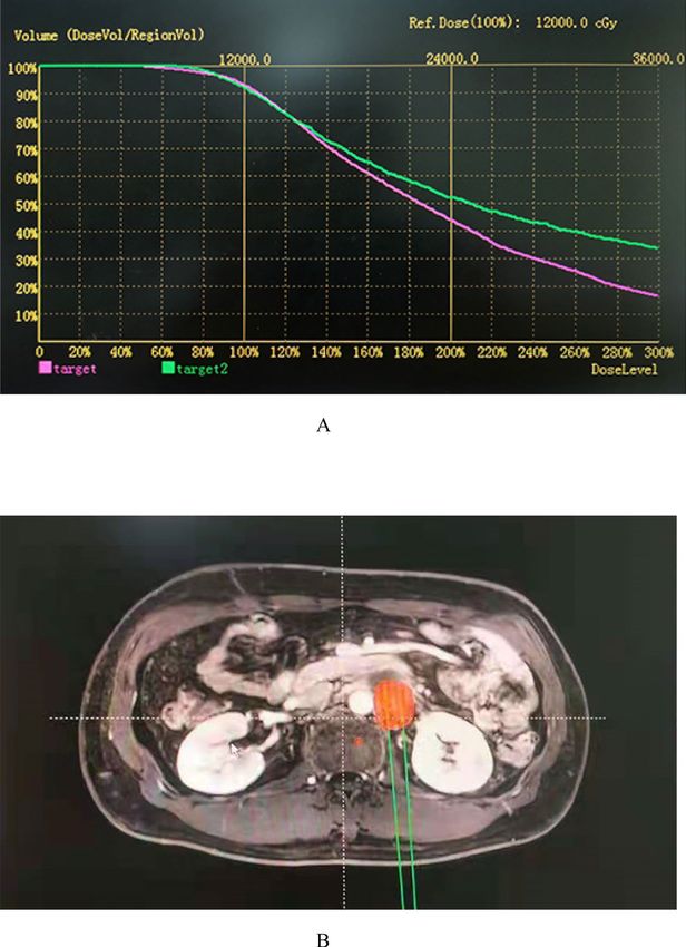
Figure 5. A 50-year-old patient with a tumor of common iliac lymph node recurrence after cervical cancer
surgery. (A) Radiotherapy specialists design dose-volume histograms on a treatment planning system based on
the patient’s tumor profile. (B) The location and dose of 125I seeds were calculated using two-dimensional images
of the tumor from the treatment planning system.
Statistical analysis. All values are expressed as the mean ± standard deviation. The overall survival curve
generated by the Kaplan–Meier method using SPSS 22.0 is shown in Fig. 3, where deaths from all causes were
classified as events. A multivariate analysis of prognostic factors (by Cox proportional hazards regression) was
performed to calculate the hazard ratios and confidence intervals. T tests were used to evaluate the significance
of differences. Models also included age (< 40 years and ≥ 40 years), preoperation diameter of the largest tumour
(< 3 cm and ≥ 3 cm), first operation, histologic type (squamous carcinoma, adenocarcinoma, other), FIGO stage
(IA, IB, IIA), surgical vaginal margin (positive, negative), differentiation (moderate, high), maximum diameter
of the recurrent pelvic tumour after surgery(< 3 cm and ≥ 3 cm), and number of I-125 particles used (< 20 grains
and ≥ 20 grains). P values less than 0.05 were considered significant.
The survival time was calculated from the date of diagnosis to the date of death or the last follow-up. Local
recurrence was defined as tumour progression within the implanted area or surrounding regions, as seen on CT.
Local recurrence and distant metastasis were scored until patient death and censored thereafter.
Scientific Reports | (2021) 11:11356 | https://doi.org/10.1038/s41598-021-90007-x 10
Vol:.(1234567890)www.nature.com/scientificreports/
Received: 28 October 2020; Accepted: 19 April 2021
References
1. Small, W. Jr. et al. Cervical cancer: A global health crisis. Cancer 123, 2404–2412 (2017).
2. Gadducci, A., Tana, R., Cosio, S. & Cionini, L. Treatment options in recurrent cervical cancer (Review). Oncol. Lett. 1, 3–11 (2010).
3. Kessler, T. A. Cervical cancer: Prevention and early detection. Semin. Oncol. Nurs. 33, 172–183 (2017).
4. Monk, B. J., Tewari, K. S. & Koh, W. J. Multimodality therapy for locally advanced cervical carcinoma: State of the art and future
directions. J. Clin. Oncol. 25, 2952–2965 (2007).
5. Tewari, K. S. et al. Improved survival with bevacizumab in advanced cervical cancer. N. Engl. J. Med. 370, 734–743 (2014).
6. Boussios, S. et al. Management of patients with recurrent/advanced cervical cancer beyond first line platinum regimens: Where
do we stand? A literature review. Crit. Rev. Oncol. Hematol. 108, 164–174 (2016).
7. Liontos, M., Kyriazoglou, A., Dimitriadis, I., Dimopoulos, M. A. & Bamias, A. Systemic therapy in cervical cancer: 30 years in
review. Crit. Rev. Oncol. Hematol. 137, 9–17 (2019).
8. Shi, F. et al. Metastatic malignant melanoma: Computed tomography-guided 125I seed implantation treatment. Melanoma. Res.
24, 137–143 (2014).
9. Li, C. et al. Feasibility of (125)I brachytherapy combined with sorafenib treatment in patients with multiple lung metastases after
liver transplantation for hepatocellular carcinoma. J. Cancer Res. Clin. Oncol. 136, 1633–1640 (2010).
10. Hu, X. et al. Recurrent gliomas: Comparison of computed tomography (CT)-guided 125I seed implantation therapy and traditional
radiochemotherapy. Cancer Biol. Ther. 13, 840–847 (2012).
11. Liu, Y., Liu, R., Wang, P., Li, S. & Shen, H. Percutaneous implantation of (125)iodine seeds for treatment of portal vein tumor
thrombosis in hepatocellular carcinoma. Med. Oncol. 32, 214 (2015).
12. Ramirez, P. T. et al. Minimally invasive versus abdominal radical hysterectomy for cervical cancer. N. Engl. J. Med. 379, 1895–1904
(2018).
13. Zhao, G. S. et al. Evaluation of radioactive (125)I seed implantation for the treatment of refractory malignant tumours based on
a CT-guided 3D template-assisted technique: Efficacy and safety. BMC Cancer 20, 718 (2020).
14. Chen, Y. et al. Dosimetry, efficacy, and safety of three-dimensional printing noncoplanar template-assisted and CT-guided (125)I
seed implantation for recurrent retroperitoneal lymphatic metastasis after external beam radiotherapy. Brachytherapy 19, 380–388
(2020).
15. Ito, H. et al. Radiotherapy for centrally recurrent cervical cancer of the vaginal stump following hysterectomy. Gynecol. Oncol. 67,
154–161 (1997).
16. Qu, A. et al. Efficacy and dosimetry analysis of image-guided radioactive (1)(2)(5)I seed implantation as salvage treatment for
pelvic recurrent cervical cancer after external beam radiotherapy. J. Gynecol. Oncol. 30, e9 (2019).
17. Blais, E. et al. Diagnosis performances and impact on therapeutic strategy of (18) F-Choline-PET/CT in biochemical relapse after
i125 brachytherapy for localized prostate cancer. Int. J. Radiat. Oncol. Biol. Phys. 99, E215 (2017).
18. Tangjitgamol, S. et al. Adjuvant chemotherapy after concurrent chemoradiation for locally advanced cervical cancer. Cochrane
Database Syst. Rev. 2014, CD010401 (2014).
19. Ghose, S. et al. A review of segmentation and deformable registration methods applied to adaptive cervical cancer radiation therapy
treatment planning. Artif. Intell. Med. 64, 75–87 (2015).
20. Tian, Y. et al. Radioactive (1)(2)(5)I seed inhibits the cell growth, migration, and invasion of nasopharyngeal carcinoma by trig-
gering DNA damage and inactivating VEGF-A/ERK signaling. PLoS One 8, e74038 (2013).
21. Wang, H. et al. Oligorecurrence non-small cell lung cancer after failure of first-line chemotherapy: Computed tomography-guided
(125)I seed implantation vs. second-line chemotherapy. Front. Oncol. 10, 470 (2020).
22. Wang, J. et al. Expert consensus statement on computed tomography-guided (125)I radioactive seeds permanent interstitial
brachytherapy. J. Cancer Res. Ther. 14, 12–17 (2018).
23. Zhang, F. et al. Refractory nasopharyngeal carcinoma: Positron emission tomography combined with computed tomography-guided
125I seed implantation therapy after repeated traditional radiochemotherapy. Otolaryngol. Head Neck Surg. 149, 417–423 (2013).
24. Shen, X., Li, Y., Zhang, Y., Kong, J. & Li, Y. An analysis of brachytherapy with computed tomography-guided permanent implanta-
tion of Iodine-125 seeds for recurrent nonkeratin nasopharyngeal carcinoma. Onco. Targets Ther. 8, 991–997 (2015).
25. Hayes, S. A. et al. Comparison of CT volumetric measurement with RECIST response in patients with lung cancer. Eur. J. Radiol.
85, 524–533 (2016).
Acknowledgements
We thank all patients who participated in this clinical research. The statistical analysis would not have been pos-
sible without the participation of the collaborating institutions that provided their trial data.
Author contributions
J.Z. conceived the original idea and guided the study design. R.W. designed the study. J.Z., R.W., and S.Y. col-
lected the data for the study, which were analysed by J.Z., R.W., S.Y., X.C. and C.G. The data interpretation and
manuscript drafting were performed by R.W., T.L., D.L., and Y.C. The manuscript was revised by R.W., X.C., and
C.G. All authors reviewed the manuscript and agreed to its submission.
Competing interests
The authors declare no competing interests.
Additional information
Correspondence and requests for materials should be addressed to J.Z.
Reprints and permissions information is available at www.nature.com/reprints.
Publisher’s note Springer Nature remains neutral with regard to jurisdictional claims in published maps and
institutional affiliations.
Scientific Reports | (2021) 11:11356 | https://doi.org/10.1038/s41598-021-90007-x 11
Vol.:(0123456789)www.nature.com/scientificreports/
Open Access This article is licensed under a Creative Commons Attribution 4.0 International
License, which permits use, sharing, adaptation, distribution and reproduction in any medium or
format, as long as you give appropriate credit to the original author(s) and the source, provide a link to the
Creative Commons licence, and indicate if changes were made. The images or other third party material in this
article are included in the article’s Creative Commons licence, unless indicated otherwise in a credit line to the
material. If material is not included in the article’s Creative Commons licence and your intended use is not
permitted by statutory regulation or exceeds the permitted use, you will need to obtain permission directly from
the copyright holder. To view a copy of this licence, visit http://creativecommons.org/licenses/by/4.0/.
© The Author(s) 2021
Scientific Reports | (2021) 11:11356 | https://doi.org/10.1038/s41598-021-90007-x 12
Vol:.(1234567890)You can also read


