Isolated Medial Subtalar Joint Dislocation during Sports Activities: A Systematic Review of the Literature with Individual Participant Data ...
←
→
Page content transcription
If your browser does not render page correctly, please read the page content below
Systematic Review
Isolated Medial Subtalar Joint Dislocation during
Sports Activities: A Systematic Review of the
Literature with Individual Participant Data Analysis
Carlo Biz 1, * , Giovanni Baldin 1 , Claudia Cappelletto 1,2 , Nicola Luigi Bragazzi 3 ,
Pietro Nicoletti 1,2 , Alberto Crimì 1 and Pietro Ruggieri 1
 1   Orthopaedic, Oncological and Traumatological Clinic, Department of Surgery, Oncology and
     Gastroenterology (DiSCOG), University of Padua, Via Giustiniani 2, 35128 Padova, Italy;
     giovannibaldin@gmail.com (G.B.); claudiacappelletto@libero.it (C.C.); pietrosea@gmail.com (P.N.);
     albe.crim@gmail.com (A.C.); pietro.ruggieri@unipd.it (P.R.)
 2   Department of Neurosciences, Institute of Human Anatomy, University of Padua, 35121 Padua, Italy
 3   Department of Mathematics and Statistics, Laboratory for Industrial and Applied Mathematics (LIAM),
     York University, Toronto, ON M3J 1P3, Canada; robertobragazzi@gmail.com
 *   Correspondence: carlo.biz@unipd.it

 Received: 10 January 2021; Accepted: 26 February 2021; Published: 5 March 2021

 Abstract: In athletes, one of the most common injuries is a sprained ankle. If the energy of the trauma
 is particularly high, this type of injury can lead to an isolated medial dislocation of the subtalar joint
 (STJ), a rare condition poorly described in the literature. The aim of this study was to verify if a
 reliable conservative treatment and a specific physiotherapy rehabilitation protocol in isolated medial
 dislocation of the STJ in athletes is described in the literature. A systematic review of the published
 literature of the last 11 years was performed by applying the Preferred Reporting Items for Systematic
 Reviews and Meta-Analyses (PRISMA) guidelines using three databases: Pubmed, Scopus, and Web
 of Science. The keywords used were “(subtalar OR talocalcaneal) AND dislocation”. We considered
 only studies that included professional or amateur athletes (athletic patients). We used the American
 Orthopedic Foot and Ankle Society (AOFAS) scale, range of motion (ROM) of the subtalar and ankle
 joint, subtalar and talonavicular joint osteoarthritis, and patient feedback to evaluate their outcomes.
 A total of 12 studies were included in our review, with a total of 26 athletic patients. Sixteen of them
 had good results with the correlation between the duration of immobilisation and the outcomes.
 Nevertheless, due to the small number of patients included in the analysed studies on this subject in
 the literature, there is not yet a univocal clinical protocol to treat the isolated medial subtalar joint
 dislocation (STJD) warranting further research in the field.

 Keywords: subtalar joint dislocation; talocalcaneal dislocation; sports injuries; ankle; athletes;
 conservative

1. Introduction
     In sports such as football and basketball, ankles injuries are quite common due to the continuous
sprinting and jumping involved [1]. Furthermore, in the case of a high energy trauma, a subtalar joint
dislocation (STJD) can occur, which consists of the displacement of two joints, namely, the talocalcaneal
and the talonavicular. This rare condition represents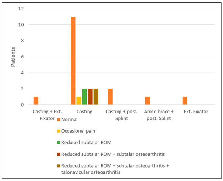
Osteology 2021, 1 49
supination in a blocked foot, the ligaments that ensure the stability of the STJ break in a specific order:
first the dorsal talonavicular ligament, then the interosseous talocalcaneal ligament and finally the
calcaneofibular ligament [5,6]. This injury can be caused by repetitive low energy traumas (repetitive
jumps and landings [1], hence its vulgar name “basketball foot” due to how prevalent those actions are
in that sport) or a single high energy trauma (like a motorcycle crash or falling from heights).
In case of suspected STJD, the first step is to obtain a thorough clinical history: what is the kind of
trauma, what is the activity that caused the trauma, timing of the injury, and history of previous ankle
trauma or surgery.
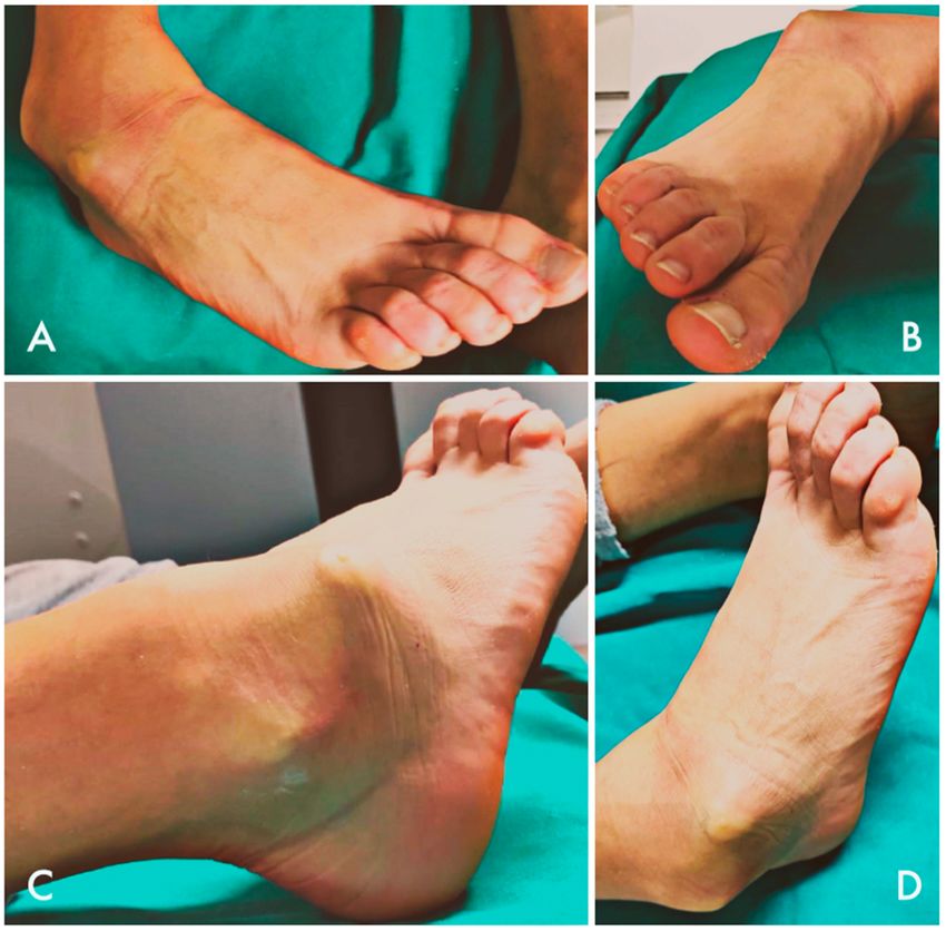
The second step is a thorough physical examination: the clinical presentation of a STJD can help
with the diagnosis, with a visible deformity of the affected ankle, where the calcaneus is displaced
medially, and the talar head is prominent dorsolaterally and associated cutaneous tension of the lateral
side of the foot, (Figure 1). Further, it is not unusual to have an open wound associated with the
dislocation. In some cases, however, the dislocation reduces spontaneously, leaving a swollen ankle
with ecchymosis [1,5–14].
Figure 1. Clinical images of an isolated medial subtalar joint dislocation (STJD) in a 25-year-old female
recreational athlete who showed deformity of her right ankle joint upon arrival at the emergency
department (ED) (A–D).
The third and last step is to obtain appropriate imaging: radiological images are important for
the diagnosis of STJD: standard antero-posterior and lateral radiographic views can show rotationalOsteology 2021, 1 50
defects or any relics of lateral displacement. An oblique radiographic projection with an internally
rotated foot in order to verify the integrity of the tibiotalar articulation is also suggested. Magnetic
resonance imaging (MRI) can be used to evaluate the condition of the ligaments. It is mandatory to
always evaluate the neurovascular state of the affected foot and the soft tissue condition [15]; to this
end, it is necessary to palpate the posterior tibial pulse and the dorsalis pedis pulse accurately and,
comparing the pulse strength between the feet, exclude vascular lesions.
Once all these aspects have been examined, if the dislocation did not reduce spontaneously,
the next action is to reduce the displacement; this can be carefully done in the ER with some local
anaesthesia, and the knee flexed 90◦ to reduce tension from the gastrocnemius [16]. It is usually
possible to perform a closed reduction in the acute dislocation; however, if the manoeuvre is impossible
to perform or there is still some instability, it can be done in an open manner. After obtaining a good
reduction, the joint is immobilised, and a CT scan is done to check for hidden fractures. As of now,
there is no “gold standard” method to immobilise the affected ankle. The most used is the classic
below-knee cast, either open or closed depending on the skin and soft tissue conditions. The duration
of the immobilisation can vary depending on the patient’s general condition, age and type of trauma.
Usually, the time is about four to six weeks; however, a study by Dr. Lasanianos [17] showed that two
to three weeks of cast immobilisation followed by partial bearing on the affected limb and specific
physiotherapy could improve the outcome of these patients. While the patient can usually obtain a
good recovery, complications must not be underestimated. They can generally be divided into acute
and chronic. The first complications are caused by damage to the neurovascular bundle and the soft
tissues, while the latter are avascular necrosis of the tarsal bone and osteoarthritis. Due to the rarity
of an isolated STJD, there is a paucity of data regarding the best way to treat patients affected by
this injury. Hence, the purpose of this systematic review was to compile the current literature about
isolated medial STJD occurring in athletes during sports activities to compare the effectiveness of the
different conservative types of treatment available and potentially find key factors that could play a
role in their outcomes.
2. Materials and Methods
2.1. Search Strategy
The present systematic review with individual participant data analysis and its related procedures
were organised, conducted and reported following the Preferred Reporting Items for Systematic Reviews
and Meta-Analyses (PRISMA) guidelines with a PRISMA checklist and algorithm [18,19]. A medical
librarian-assisted electronic search was conducted using three different databases: PubMed/MEDLINE,
Scopus and Web of Science. In each database, an advanced search was conducted using “(subtalar OR
talocalcaneal) AND dislocation” as keywords.
2.2. Selection Criteria
An 11-year time selection was used, from January 2009 to September 2020. We included only
articles written in English, and that met the following PICOS criteria:
(I) Population (P): patients of every age that suffered from an isolated medial STJD during
sports activities.
(II) Intervention and Comparison (IC): patients treated in conservative ways and their follow-up.
(III) Outcomes (OS): results were analysed using the American Orthopedic Foot and Ankle Society
(AOFAS) scale, quality and quantity of Range of Motion (ROM) of the affected joint after treatment,
evidence of osteoarthritis in the subtalar and talonavicular joints and patient feedback.
We included case reports and case series about this topic. We excluded short surveys, letters to
the editor, articles about bioengineering, studies performed on animals or cadavers, reviews of the
literature, and technical notes.Osteology 2021, 1 51
2.3. Selection Method
The selection was based on the abstract’s content; if inclusion or exclusion of the article was not
possible based on the abstract, the full-text article was read. Cross-reference research of the selected
articles was also performed to obtain other relevant articles for the study. Finally, the selected articles
and references were reviewed, assessed and discussed by the authors. If there was disagreement
among the investigators regarding the inclusion or exclusion criteria, the senior investigator made the
final decision.
2.4. Literature Search
The literature search obtained 188 papers from PubMed/MEDLINE, 258 papers from Scopus and
138 papers from ISI/Web of Science for a total of 584 papers. Of these, 279 were duplicates and so were
excluded. Of the remaining 305 manuscripts, we excluded 211 articles as their topics were not the STJ
or because they discussed fractures or congenital displacement of the STJ. Out of the remaining papers,
12 relevant reports were identified that met all of our criteria (Figure 2). We compared the outcomes of
conservative treatments of the isolated medial STJDs in the 12 selected studies from January 2009 to
September 2020 and analysed the outcomes of each study (Table 1). Level of evidence (I, II, III, or IV)
was reported for the included studies.
Figure 2. Preferred Reporting Items for Systematic Reviews and Meta-Analyses (PRISMA): flow chart
diagram for inclusion and exclusion of paper process. For this study, 94 papers articles were assessed
for eligibility after screening; among these, 12 papers fulfilled the selection criteria and were included
in the analysis.Osteology 2021, 1 52
2.5. Quality Assessment
To assess the quality of the selected studies, we used the Downs and Black scale [20]. It assesses
methodology using 27 items with a range from 0 to 28 points divided into five sections (Reporting,
External validity, Internal validity-bias, Internal validity-confounding or selection bias, and Power).
This scale evaluates methodologies and is also indicated for case series. The study methodology is
considered excellent (26–28 points), good (20–25 points), fair (15–19 points), or poor (Osteology 2021, 1 53
Table 1. Details about the type of study, evidence level, score in the Downs and Black (D&B) scale, demographic data, type of sport practiced, and type of treatment
applied for each article included in our review. AOFAS: American Orthopedic Foot and Ankle Society; ROM: range of motion (n/a: not available).
Medium Age
Authors Type of Study Evidence Level D&B Patients (M/F) Type of Sport Type of Treatment Outcome Measures
(Range)
Closed reduction, casting
Wang et al. [22] Case Report IV 9 1 (1/0) Soccer 37 and external fixation, AOFAS
assisted active exercise
Closed reduction,
casting/posterior splint,
Biz et al. [23] Case Report IV 12 3 (1/2) Volleyball 28 (16–42) AOFAS
painkillers, ankle brace
physiotherapy
Abdellatif et al. [24] Case Report IV 8 1 (1/0) Basketball 22 n/a Feedback
ROM, presence of
Closed reduction,
Giannoulis et al. [25] Case Report IV 7 1 (1/0) Climbing 36 instability, presence
casting/posterior splint
of osteoarthritis
Closed reduction, ankle
Stafford et al. [26] Case Report IV 9 1 (1/0) Soccer 23 Return to sport
brace, physiotherapy
Atik et al. [27] Case Report IV 6 1 (1/0) Tennis 22 Closed reduction, casting Feedback
Presence of
Bryant et al. [28] Case Report IV 7 1 (1/0) Baseball 26 Closed reduction, casting
osteoarthritis
Pua [29] Case Report IV 6 1 (1/0) Basketball 17 Closed reduction n/d
Closed reduction,
Pesce et al. [30] Case Report IV 10 1 (1/0) Military drills 37 n/d
casting/posterior splint
Closed reduction, casting,
Kemah et al. [31] Case Report IV 11 1 (1/0) Long Jump 20 n/d
physiotherapy
AOFAS, ROM,
Retrospective Closed reduction, casting,
Ruhlmann et al. [32] IV 15 10 (9/1) Basketball 38.4 (17–71) presence of
Cohort physiotherapy
osteoarthritis
Open/closed reduction,
Retrospective
Jungbluth et al. [33] IV 15 4 (2/2) Soccer 34.5 (27–41) external fixation/casting, AOFAS, ROM
Cohort
physiotherapyOsteology 2021, 1 54
3. Results
3.1. Patient Population
The 12 studies included reported on a total of 26 patients, all athletes (three professionals and
23 amateurs). None of them dropped out during the follow-up. Therefore, a total of 26 patients were
considered, corresponding to 26 ankles and 26 isolated medial STJDs. Of those, 20 were males and six
females. The mean age at treatment was 32.54 years, ranging from 16 to 71 years [23,32]. The total
number of patients for each study ranged between one and 10 [22,24–32]. In one paper, the affected
ankle was not specified (for a total of four patients); of the remaining 22 patients, the right ankle was
affected 15 times (68.18%) and the left ankle seven times (31.82%). In one study, the type of sports
activity practised by the patients was not specified; the isolated medial STDJ appears to be more
common in sports such as basketball, football and volleyball. The patients were assessed at a mean
follow-up time of 25.38 months, ranging from 0 to 124.8 months [25,27,29,32]. All but one patient [33]
underwent closed reduction of the displacement, and all were treated with ankle immobilisation
(except in one paper [29] where it was not specified). Noteworthy, none of the patients suffered
previously from STJD.
3.2. Type of Treatment
For the selected studies, we analysed different conservative approaches and treatment protocols
(Table 2). The first treatment performed in emergency and applied to every patient was closed reduction
under anaesthesia (except for one patient who underwent emergency open reduction). The displaced
joint was then immobilised for a mean time of 5.13 weeks, ranging from a minimum of four to a
maximum of eight weeks. To be more specific, four weeks for seven patients (26.92%), five weeks
for one patient (3.85%), six weeks for thirteen patients (50%) and eight weeks for one patient (3.85%);
however, we did not have enough data on four patients. Nineteen patients were treated with an
open cast (73.08%), three patients with the combined use of a cast and posterior splint (11.54%);
one patient was treated with the combination of external fixation, and cast (3.85%), one patient with
a posterior splint and an ankle brace (3.85%) and one patient was treated with just external fixation
(3.85%). After the immobilisation period, each patient had various treatments, mainly using painkillers
and physiotherapy.
Table 2. Details about the patient rehabilitation protocol applied in each study (n/a: not available).
Authors Patients Rehabilitation Protocol
Active assisted ROM exercises after four weeks, partial weight
Wang et al. [22] 1
bearing after six weeks and total weight bearing if tolerated.
Patient 1: painkillers; after two weeks, partial weight-bearing
supported with crutches for two weeks; after four weeks, ankle brace,
physical therapy, physiotherapy for five months.
Patient 2: painkillers; after four weeks, total weight bearing if
Biz et al. [23] 3
tolerated, ankle brace for two months; active and passive
physiotherapy, physical therapy.
Patient 3: painkillers; after four weeks, total weight bearing if
tolerated; passive physiotherapy for two months.
Abdellatif et al. [24] 1 n/d
Active and passive ROM exercises and total weight bearing if
Giannoulis et al. [25] 1
tolerated after immobilisation.
Active and passive ROM exercises and total weight bearing if
Stafford et al. [26] 1
tolerated after immobilisation.
Atik et al. [27] 1 n/a
Bryant et al. [28] 1 n/aOsteology 2021, 1 55
Table 2. Cont.
Authors Patients Rehabilitation Protocol
Pua [29] 1 n/a
Pesce et al. [30] 1 n/a
Kemah et al. [31] 1 n/a
Ruhlmann et al. [32] 10 Physiotherapy and progressive weight bearing after immobilisation.
Physiotherapy, partial weight-bearing after immobilisation; total
Jungbluth et al. [33] 4
weight-bearing after a mean of 10.6 weeks (range 10–11.5)
3.3. Outcomes
3.3.1. Overall Analysis of Clinical Scores
Considering all of the studies, 16 patients showed good clinical outcomes: one patient reported
occasional pain after the incident, two patients showed a reduction in STJ ROM, two patients showed
a reduction in ROM with associated STJ osteoarthritis, two patients showed a reduction in ROM with
associated subtalar and talonavicular joint osteoarthritis on both locations, and for three patients,
we do not have information about the outcome. The only patient who underwent open reduction
had a good outcome. No patient showed articular stiffness after treatment. The patient that reported
occasional pain was treated with closed reduction and casting for four weeks. The patients that showed
a reduction in ROM with subtalar and talonavicular arthritis were treated with casting for six weeks,
after which they started partial weight-bearing and physiotherapy.
Comparing the different techniques to immobilise the joint after reduction, casting, and external
fixation showed a good outcome in one patient; casting without any other device showed good
outcomes in 11 patients, a good outcome with occasional pain in one patient, a reduction in ROM
without associated osteoarthritis in two patients, reduction of ROM with associated STJ osteoarthritis
in two patients, reduction in ROM with associated subtalar and talonavicular joint osteoarthritis in
two patients. Casting combined with a posterior splint showed good outcomes in two patients; the use
of an ankle brace associated with a posterior splint showed good outcomes in two patients (Figure 3).
Regarding the duration of immobilisation and results, four weeks of immobilisation showed six
good results and one good result with occasional pain, five weeks showed one good result, six weeks
showed eight good results, two reductions in ROM without osteoarthritis, two reductions in ROM
with associated STJ osteoarthritis, two reductions in ROM with associated subtalar and talonavicular
joint osteoarthritis, eight weeks showed one good result (Figure 4). Considering follow-up timing,
we found one good outcome at three months, one good outcome at six months, one good outcome with
occasional pain and two good outcomes at 24 months, one reduction in ROM with associated subtalar
and talonavicular joint osteoarthritis at 24.4 months, one reduction in ROM without osteoarthritis at
25.5 months, one good outcome at 30 months, two good outcomes at 36 months, three good outcomes
at 46 months, three good outcomes at 49 months, three good outcomes at 53.5 months, one reduction in
ROM without associated osteoarthritis at 54.9 months, one good outcome at 56 months, one reduction
in ROM with STJ osteoarthritis at 57 months, two good outcomes at 60 months, two good outcomes at
73 months, one reduction of ROM with associated STJ osteoarthritis at 75.6 months, two good outcomes
at 107.4 months, two good outcomes at 123.3 months and one reduction in ROM with associated
subtalar and talonavicular joint osteoarthritis at 124.8 months.Osteology 2021, 1 56
Figure 3. Outcome depending on the type of immobilisation used.
Figure 4. Results obtained in our cohort of patients depending on immobilisation length.Osteology 2021, 1 57
3.3.2. Comparison of Clinical Scores
The AOFAS scale was used in 5 studies (41.67%) to evaluate clinical and functional results after
treatment for a total of 19 patients. This scale represents the combination of both objective and
subjective parameters with a score between 0 and 100 (the higher the score, the better the outcome).
The AOFAS scale has been validated over time, with different studies confirming its reliability.
The Italian version was recently validated [21]. The mean score was 86, with a range from 66 to
100. Good results were obtained with laser therapy and ultrasound therapy after the immobilisation
period, using an ankle-brace with crutches and partial weight bearing on the affected limb even during
the immobilisation period [23,26] and targeting physiotherapy on the muscular reinforcement of the
peroneus longus and the brevis muscles starting after the immobilisation period [22,23,25,26,31–33].
In three studies, it is specified that all of the patients were able to return to their sports activities with a
mean of 4.53 months of rest, range 4–5.67 months [22,23,31].
3.3.3. Statistical Analysis
Statistical analysis was performed considering only the studies that used the AOFAS scale
score to evaluate the outcomes. Table 3 shows descriptive statistics of continuous variables such
as age, follow-up, immobilisation duration and outcome score. For each variable, we report the
minimum and maximum value, the median and mean value, the relative standard deviation and
corresponding percentile.
Table 3. Descriptive statistics of continuous variables of the articles included in the analysis.
Standard 25–75
Minimum Maximum Mean Median
Deviation Percentiles
Age 16.0 71.0 34.9 31.0 14.3 26.0–41.8
Follow-up Time 3.0 124.8 54.2 49.0 34.1 26.6–69-8
Immobilisation length (weeks) 4.0 6.0 5.4 6.0 0.9 4.2–6.0
AOFAS score 66.0 100.0 86.0 85.0 8.9 82.0–90.0
Table 4 shows the descriptive statistics of categoric variables for the cohort of athletic patients
included in the analysis, while Table 5 shows the meta-regression results of the independent variables.
The only relevant variable is the immobilisation time: the meta-regression coefficient was –5.5580,
showing how increasing immobilisation time worsens the results.
Table 4. Descriptive statistics of categoric variables of the papers chosen for our review (sex (0 = female;
1 = male); affected side (0 = left; 1 = right); type of immobilisation used (0 = external fixator; 1 = casting;
reduction technique performed (0 = open; 1 = closed); presence of subtalar and talonavicular joint
osteoarthritis (0 = absent; 1 = present); STJ ROM quality after treatment (0 = reduced; 1 = conserved).
0 1 Total
Sex 5 (26.3%) 14 (73.7%) 19 (100%)
Affected side 4 (26.7%) 11 (73.3%) 15 (100%)
Type of immobilisation 1 (5.3%) 18 (94.7%) 19 (100%)
Type of reduction 1 (5.3%) 18 (94.7%) 19 (100%)
Subtalar osteoarthritis 14 (73.7%) 5 (26.3%) 19 (100%)
Talonavicular osteoarthritis 17 (89.5%) 2 (10.5%) 19 (100%)
STJ ROM 6 (42.9%) 8 (57.1%) 14 (100%)Osteology 2021, 1 58
Table 5. Meta-regression of the independent variables presented in the articles included in the analysis.
Coefficient Standard Error t. Statistics p-Value rpartial rsemipartial
(Constant) 117,76
Immobilisation length −5.56 2.11 −2.63 0.027 −0.66 0.4
Age −0.03 0.11 −0.28 0.789 −0.09 0.04
Follow-up −0.01 0.05 −1.18 0.106 −0.51 0.27
Affected side 6.11 3.61 1.69 0.125 0.49 0.26
Sex −0.07 3.95 −0.02 0.986 −0.01 0.003
3.4. Quality Assessment
The mean value of the Down and Black scale [20] was 9.58 points, showing that the mean quality
of the included studies was poor. Among the 12 studies considered, 10 had poor results [22–31]
and two had fair results [32,33]; more specifically, two papers had six points [27,29], two papers had
seven points [25,28], one paper had eight points [24], two papers had nine points [22,26], one paper
had 10 points [30], one paper had 11 points [31], one paper had 12 points [23], and two papers had
15 points [32,33].
4. Discussion
Isolated medial STJD is a rare and complex injury whose most common cause is a fall from
height as a result of jumping [32]. In our review, the most represented causes of STJD were basketball
(12 cases), soccer (six cases), volleyball (three cases) and one case for other causes (tennis, climbing,
baseball, military drills, and long jump). Dislocation of this joint is usually caused by the application
of a high-energy force that causes the rupture of the strong joint capsule and ligaments, such as the
deltoid complex, which plays an important role as a stabiliser of the head of the talus and the SBJ with
the tibionavicular section of the tibiocalcaneonavicular ligament. As a result, it represents a rare injury
in low- or medium-energy trauma, such as sports injuries, whereas it is more common following motor
vehicle accidents [23,34].
In the literature, the medial STJD is described as a rare condition, representingOsteology 2021, 1 59
Jungbluth et al. [33] who was treated with open reduction and external fixation. An External Fixator
was also used in the patient in the study by Wang et al. [22] after immobilisation in a short leg cast for
two weeks. The method of immobilisation that was most used in this review was a below-knee cast,
used by six authors to treat 19 patients. The posterior splint combined with the short leg cast was the
second most frequent method of immobilisation, used by three authors to treat three patients. All of
the patients (100%) treated with immobilisation with posterior splint and cast achieved good results
after the treatment, whereas a satisfactory result was achieved by 61.11% of patients immobilised in
a short leg cast only. Both methods appear to be valid, but they are not comparable due to the very
limited data available, so it was not possible to find a statistically significant difference between the two
treatments. As shown in Figure 3 of our review, the patients that complained of worsening of ROM of
the STJ or presented some degree of osteoarthritis after the injury were all treated with just casting.
Out of a total of 18 patients, 11 had good results; one complained of occasional pain; two showed
reduced ROM of the STJ; four presented subtalar or talonavicular joint osteoarthritis. This evidence is
probably linked to the greater number of patients that belong to this category, but our data suggest
that another method of immobilisation should be used.
Regarding immobilisation length, some authors [16] state that decreasing immobilisation time also
decreases the incidence of joint stiffness after treatment. This was confirmed by the data we provided in
Table 5, where we show the meta-regression of the independent variables present in the studies. As can
be seen in Table 5, we obtained a coefficient of −5.56 regarding the immobilisation length, meaning that
longer immobilisation corresponded with worse outcomes. Thus, short immobilisation (4 weeks or
less) seems to be preferred. None of the studies reviewed had an immobilisation time of fewer than
four weeks, so it is not possible to recommend immobilisation of fewer than four weeks based on this
review of the literature. Most patients in our review regained normal ROM of the STJ after six weeks
of immobilisation; that was also the most common immobilisation time, with a total of 14 patients.
However, as shown in Figure 4, out of those patients, 2 showed reduced ROM of the STJ, and four
patients showed some degree of osteoarthritis of the subtalar or talonavicular joint. Meanwhile, out of
the eight patients who were immobilised for fewer than six weeks, all had good outcomes except one
who complained of occasional pain.
This review is limited by the level of evidence of the studies included, consequently presenting
some drawbacks: firstly, the studies included are not homogenous for the outcome scores. The poor
homogeneity of clinical evaluation scores has played an important role, lowering the possibility
of obtaining a precise comparison between the different conservative treatments available. Also,
the quality of the included studies made it difficult to undertake a full and statistically reliable
comparison of the published data. Unfortunately, our selection included mainly case reports and case
series, with a small sample size, lack of control groups, and restricted statistical comparison. Hence,
all studies in the series had low levels of evidence (IV). Due to these different aspects, there is a risk of
reporting bias in the present study. Finally, additional Randomised Controlled Trials (RCT) are needed
to definitively determine the most beneficial method of immobilisation. Duration of immobilisation
has been shown to be pivotal in prognosis [16], so it deserves further study to delineate the best course
of action in professional and recreational athletes affected by isolated medial STJD.
5. Conclusions
Based on the currently available scientific literature as appraised in the present study, conservative
treatment is the first choice for isolated medial subtalar dislocation occurring during sports activities,
both for professional and recreational athletes. However, there is no “gold standard” regarding the
rehabilitation protocol for athletic patients that have suffered this kind of injury. Our review highlights
that the immobilisation length is the most impactful elements on the biological outcome and should
not exceed four weeks due to the risk of stiffness and post-traumatic osteoarthritis, especially in
athletic patients.Osteology 2021, 1 60
Author Contributions: Conceptualisation, C.B.; methodology, C.B.; software, N.L.B.; validation, C.B., A.C. and
G.B.; formal analysis, N.L.B.; investigation, C.C.; resources, C.B.; data curation, C.C. and P.N.; writing—original
draft preparation, G.B.; writing—review and editing, A.C.; supervision, P.R.; All authors have read and agreed to
the published version of the manuscript.
Funding: This research received no external funding.
Institutional Review Board Statement: Not applicable.
Informed Consent Statement: Not applicable.
Data Availability Statement: The dataset generated during the current study is available from the corresponding
authors on reasonable request.
Conflicts of Interest: The authors declare no conflict of interest.
References
1. Smith, T.; Ballard, T.; Ramanlal, R.; Butarbutar, J.C. Foot Dislocation; StatPearls Publishing: Treasure Island,
FL, USA, 3 September 2020.
2. Rammelt, S.; Goronzy, J. Subtalar Dislocations. Foot Ankle Clin. 2015, 20, 253–264. [CrossRef] [PubMed]
3. Bryson, D.; Khan, Z.; Aujla, R.; Bromage, J.D. A near miss: An uncommon injury following a common
mechanism. BMJ Case Rep. 2011, 2011. [CrossRef]
4. Kiener, A.J.; Hanna, T.N.; Shuaib, W.; Datir, A.; Khosa, F. Osseous injuries of the foot: An imaging review.
Part 3: The hindfoot. Emerg. Med. J. 2016, 34, 337–343. [CrossRef] [PubMed]
5. Azarkane, M.; Boussakri, H.; Alayyoubi, A.; Bachiri, M.; Elibrahimi, A.; Elmrini, A. Closed medial total
subtalar joint dislocation without ankle fracture: A case report. J. Med. Case Rep. 2014, 8, 313. [CrossRef]
[PubMed]
6. Byrd, Z.O.; Ebraheim, M.; Weston, J.T.; Liu, J.; Ebraheim, N.A. Isolated Subtalar Dislocation. Orthoptics 2013,
36, 714–720. [CrossRef] [PubMed]
7. Arain, A.R.; Adams, C.T.; Haddad, S.F.; Moral, M.; Young, J.; Desai, K.; Rosenbaum, A.J. Diagnosis and
Treatment of Peritalar Injuries in the Acute Trauma Setting: A Review of the Literature. Adv. Orthop. 2020,
2020, 1852025–1852028. [CrossRef] [PubMed]
8. Hoexum, F.; Heetveld, M.J. Subtalar dislocation: Two cases requiring surgery and a literature review of the
last 25 years. Arch. Orthop. Trauma Surg. 2014, 134, 1237–1249. [CrossRef]
9. Marx, R.C.; Mizel, M.S. What’s new in foot and ankle surgery. J. Bone Jt. Surg. Ser. A 2011, 93, 405–414.
[CrossRef] [PubMed]
10. Clarke, D.; Franklin, S.; Mullings, S.; Vaughan, K.; Jones, K. Subtalar Dislocation: Case Series Inclusive of
the Rare Anterior Subtalar Dislocation and Review of the Literature. West Indian Med. J. 2017, 67, 77–83.
[CrossRef]
11. Melenevsky, Y.; Mackey, R.A.; Abrahams, R.B.; Thomson, N.B. Talar Fractures and Dislocations:
A Radiologist’s Guide to Timely Diagnosis and Classification. Radiographics 2015, 35, 765–779. [CrossRef]
12. Gantsos, A.; Giotis, D.; Giannoulis, D.; Vasiliadis, H.; Georgakopoulos, N.; Mitsionis, G. Conservative
treatment of closed subtalar dislocation: A case report and 2years follow-up. Foot 2013, 23, 107–110.
[CrossRef] [PubMed]
13. Siddiqui, Y.S.; Zahid, M.; Bin Sabir, A.; Siddiqui, H.Q. Neglected peritalar dislocation: A case report with
review of literature. J. Clin. Diagn. Res. 2011, 5, 849–852.
14. Bhagat, S.; Shah, B.M. The foot and toes. In Trauma Management in Orthopedics; Springer: London, UK, 2013;
pp. 129–165.
15. Prada-Cañizares, A.; Auñón-Martín, I.; Rico, J.V.Y.; Pretell-Mazzini, J. Subtalar dislocation: Management and
prognosis for an uncommon orthopaedic condition. Int. Orthop. 2016, 40, 999–1007. [CrossRef]
16. Horning, J.; DiPreta, J. Subtalar dislocation. Orthopedics 2009, 32, 904. [CrossRef] [PubMed]
17. Lasanianos, N.G.; Lyras, D.N.; Mouzopoulos, G.; Tsutseos, N.; Garnavos, C. Early mobilization after
uncomplicated medial subtalar dislocation provides successful functional results. J. Orthop. Traumatol. 2011,
12, 37–43. [CrossRef]
18. Moher, D.; Liberati, A.; Tetzlaff, J.A.D. PRISMA 2009 Flow Diagram. PRISMA Statement 2009, 6, 1000097.Osteology 2021, 1 61
19. Moher, D.; Liberati, A.; Tetzlaff, J.; Altman, D.G. PRISMA 2009 Checklist—Preferred Reporting Items for
Systematic Reviews and Meta-Analyses. Ann. Intern. Med. 2014, 151, 264–269. [CrossRef] [PubMed]
20. Downs, S.H.; Black, N. The feasibility of creating a checklist for the assessment of the methodological quality
both of randomised and non-randomised studies of health care interventions. J. Epidemiol. Commun. Health
1998, 52, 377–384. [CrossRef]
21. Leigheb, M.; Janicka, P.; Andorno, S.; Marcuzzi, A.; Magnani, C.; Grassi, F. Italian translation, cultural
adaptation and validation of the “American Orthopaedic Foot and Ankle Society’s (AOFAS) ankle-hindfoot
scale”. Acta Bio-Medica Atenei Parm. 2016, 87, 38–45.
22. Wang, H.-Y.; Wang, B.-B.; Huang, M.; Wu, X.-T. Treatment of closed subtalar joint dislocation: A case report
and literature review. Chin. J. Traumatol. 2020, 23, 367–371. [CrossRef] [PubMed]
23. Biz, C.; Ruaro, A.; Via, A.G.; Torrent, J.; Papa, G.; Ruggieri, P. Conservative management of isolated medial
subtalar joint dislocations in volleyball players: A report of three cases and literature review. J. Sports Med.
Phys. Fit. 2019, 59, 1739–1746. [CrossRef]
24. Benabbouha, A.; Ibou, N. Rare case of pure medial subtalar dislocation in a basketball player. Pan Afr. Med. J.
2016, 23, 106. [CrossRef] [PubMed]
25. Giannoulis, D.; Papadopoulos, D.V.; Lykissas, M.G.; Koulouvaris, P.; Gkiatas, I.; Mavrodontidis, A. Subtalar
dislocation without associated fractures: Case report and review of literature. World J. Orthop. 2015,
6, 374–379. [CrossRef] [PubMed]
26. Stafford, H.; Boggess, B.; Toth, A.; Berkoff, D. Anteromedial subtalar dislocation. BMJ Case Rep. 2013, 2013.
[CrossRef] [PubMed]
27. Atik, O.S.; Dur, H. Unusual tennis injuries: Boxer’s fracture and medial subtalar dislocation: Report of two
cases. Jt. Dis. Relat. Surg. 2011, 22, 180–182.
28. Bryant, J.; Levis, J.T. Images in Emergency Medicine: Subtalar Dislocation. West. J. Emerg. Med. Integr. Emerg.
Care Popul. Health 2009, 10, 92.
29. Pua, U. Subtalar dislocation: Rare and often forgotten. Int. J. Emerg. Med. 2009, 2, 51–52. [CrossRef]
30. Pesce, D.; Wethern, J.; Patel, P. Rare Case of Medial Subtalar Dislocation from a Low-Velocity Mechanism.
J. Emerg. Med. 2011, 41, e121–e124. [CrossRef]
31. Kemah, B.; Özkut, A.T.; Esenkaya, İ.; Akan, K.H.; Türkmen, İ. Unexpected Injury During Jumping Exercise:
Isolated Subtalar Dislocation in A National Athlete. Orthop. J. Sports Med. 2014, 2 (Suppl. 3). [CrossRef]
32. Ruhlmann, F.; Poujardieu, C.; Vernois, J.; Gayet, L.-E. Isolated Acute Traumatic Subtalar Dislocations: Review
of 13 Cases at a Mean Follow-Up of 6 Years and Literature Review. J. Foot Ankle Surg. 2017, 56, 201–207.
[CrossRef]
33. Jungbluth, P.; Wild, M.; Hakimi, M.; Gehrmann, S.; Djurisic, M.; Windolf, J.; Muhr, G.; Kälicke, T. Isolated
Subtalar Dislocation. J. Bone Jt. Surg.-Am. Vol. 2010, 92, 890–894. [CrossRef] [PubMed]
34. Camarda, L.; Abruzzese, A.; La Gattuta, A.; Lentini, R.; D’Arienzo, M. Results of closed subtalar dislocations.
Musculoskelet. Surg. 2016, 100, 63–69. [CrossRef] [PubMed]
35. Perugia, D.; Basile, A.; Massoni, C.; Gumina, S.; Rossi, F.; Ferretti, A. Conservative treatment of subtalar
dislocations. Int. Orthop. 2001, 26, 56–60. [CrossRef]
36. Bibbo, C.; Anderson, R.B.; Davis, W.H. Injury Characteristics and the Clinical Outcome of Subtalar Dislocations:
A Clinical and Radiographic Analysis of 25 Cases. Foot Ankle Int. 2003, 24, 158–163. [CrossRef] [PubMed]
37. Goldner, J.L.; Poletti, S.C.; Gates, H.S.; Richardson, W.J. Severe open subtalar dislocations. Long-term results.
J. Bone Jt. Surg.-Am. Vol. 1995, 77, 1075–1079. [CrossRef]
38. Conesa, X.; Barro, V.; Barastegui, D.; Batalla, L.; Tomás, J.; Molero, V. Lateral Subtalar Dislocation Associated
with Bimalleolar Fracture: Case Report and Literature Review. J. Foot Ankle Surg. 2011, 50, 612–615.
[CrossRef]
Publisher’s Note: MDPI stays neutral with regard to jurisdictional claims in published maps and institutional
affiliations.
© 2021 by the authors. Licensee MDPI, Basel, Switzerland. This article is an open access
article distributed under the terms and conditions of the Creative Commons Attribution
(CC BY) license (http://creativecommons.org/licenses/by/4.0/).You can also read


