Multiple Respiratory Syncytial Virus Introductions Into a Neonatal Intensive Care Unit
←
→
Page content transcription
If your browser does not render page correctly, please read the page content below
Journal of the Pediatric Infectious Diseases Society

    ORIGINAL ARTICLE

Multiple Respiratory Syncytial Virus Introductions Into a
Neonatal Intensive Care Unit
Erica Billig Rose,1,2 Erica J. Washington,3 Lijuan Wang,4 Isaac Benowitz,5 Natalie J. Thornburg,1, Susan I. Gerber,1 Teresa C. T. Peret,1 and Gayle E.
Langley1

                                                                                                                                                                                    Downloaded from https://academic.oup.com/jpids/advance-article/doi/10.1093/jpids/piaa026/5816338 by guest on 21 September 2020
1
 Division of Viral Diseases, National Center for Immunization and Respiratory Diseases, Centers for Disease Control and Prevention, Atlanta, Georgia, USA; 2Epidemic Intelligence
Service, Centers for Disease Control and Prevention, Atlanta, Georgia, USA; 3Louisiana Department of Health, Office of Public Health, Infectious Disease Epidemiology Section,
New Orleans, Louisiana, USA; 4IHRC Inc, contracting agency to the Division of Viral Diseases, National Center for Immunization and Respiratory Diseases, Centers for Disease
Control and Prevention, Atlanta, Georgia, USA; and 5Division of Healthcare Quality Promotion, National Center for Emerging and Zoonotic Infectious Diseases, Centers for Disease
Control and Prevention, Atlanta, Georgia, USA

    Background. Outbreaks of respiratory syncytial virus (RSV) in neonatal intensive care units (NICUs) are of concern because of
the risk of severe disease in young infants. We describe an outbreak of RSV in a NICU and use whole genome sequencing (WGS) to
better understand the relatedness of viruses among patients.
    Methods. An investigation was conducted to identify patients and describe their clinical course. Infection control measures
were implemented to prevent further spread. Respiratory specimens from outbreak-related patients and the community were tested
using WGS. Phylogenetic trees were constructed to understand relatedness of the viruses.
    Results. Seven patients developed respiratory symptoms within an 11-day span in December 2017 and were diagnosed with
RSV; 6 patients (86%) were preterm and 1 had chronic lung disease. Three patients required additional respiratory support after
symptom onset, and none died. Six of 7 patients were part of the same cluster based on > 99.99% nucleotide agreement with each
other and 3 unique single-nucleotide polymorphisms were identified in viruses sequenced from those patients. The seventh patient
was admitted from the community with respiratory symptoms and had a genetically distinct virus that was not related to the other
6. Implementation of enhanced infection control measures likely limited the spread.
    Conclusions. Using WGS, we found 2 distinct introductions of RSV into a NICU, highlighting the risk of healthcare-associated
infections during RSV season. Early recognition and infection control measures likely limited spread, emphasizing the importance
of considering RSV in the differential diagnosis of respiratory infections in healthcare settings.
    Key words. respiratory syncytial virus; RSV; RSV outbreak.

Respiratory syncytial virus (RSV) is a leading cause of lower                                        born prematurely, or those with congenital abnormalities or
respiratory tract infection in children worldwide. In the                                            other comorbidities.
United States, RSV causes an estimated 57 000 hospitaliza-                                               RSV is divided into 2 subgroups (A and B). RSV sub-
tions in children < 5 years of age each year [1]. Besides young                                      groups can be further divided into genotypes based on the
chronologic age, other risk factors for severe disease include                                       hypervariable region of the RSV glycoprotein (G) gene [7, 8,
preterm birth, chronic lung disease, and congenital heart dis-                                       11, 12]. The G gene alone may not be enough to understand the
ease [2]. Outbreaks of RSV in healthcare settings have been                                          relatedness of viruses among those infected.
reported globally over the last few decades, including in pedi-                                          In December 2017, the Louisiana Department of Health
atric intensive care units, adult stem cell transplant units, adult                                  (LDH) was notified of 7 RSV infections that occurred among
hematology-oncology units, and neonatal intensive care units                                         patients within a single NICU over an 11-day span [13]. We re-
(NICUs) [3–10]. NICU outbreaks are of great concern because                                          port on the epidemiological findings of the investigation and
of the risk of severe disease in young infants, particularly those                                   the use of whole genome sequencing (WGS) to understand the
                                                                                                     relatedness of the viruses found in patients.

                                                                                                     METHODS
   Received 24 October 2019; editorial decision 3 March 2020; accepted 6 March 2020; Published
online April 4, 2020.                                                                                Healthcare Setting
   Corresponding Author: E. B. Rose, PhD, Centers for Disease Control and Prevention, 1600
                                                                                                     The facility is a large (50–100 bed) level III NICU with 5 wards
Clifton Rd NE, MS A-34, Atlanta, GA 30333. E-mail: nqx4@cdc.gov.
Journal of the Pediatric Infectious Diseases Society   2020;XX(XX):1–7
                                                                                                     (A–E) that all have single-family rooms. There is no difference
Published by Oxford University Press on behalf of The Journal of the Pediatric Infectious            in the level of patient acuity across the different wards, and
Diseases Society 2020. This work is written by (a) US Government employee(s) and is in the
public domain in the US.
                                                                                                     wards have shared staff. The catchment area of the facility in-
DOI: 10.1093/jpids/piaa026                                                                           cludes a 30-mile radius, which includes 3 parishes. According

                                                                                                 Multiple RSV Introductions Into a NICU • jpids 2020:XX (XX XXXX) • 1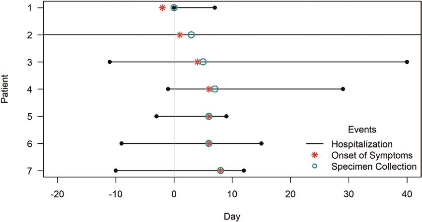
to the facility’s visitation policy, parents are invited to stay over-                                                   catchment area (we will refer to these routinely collected spe-
night with their infant, and siblings > 2 years of age are en-                                                           cimens as “community specimens”)—were then sent to the
couraged to visit during daytime hours. All family and guests                                                            Centers for Disease Control and Prevention’s (CDC) Respiratory
must wash hands before visitation. During the winter respira-                                                            Viruses Branch laboratory for further characterization [14].
tory season (that covers RSV and influenza seasons), additional
guidelines are in place that state visitors will have their temper-                                                      Laboratory Testing and Virus Sequencing at CDC
ature checked upon entrance to the unit and that any visitors                                                            Hospital and all community specimens were tested using valid-
with fever or reported respiratory symptoms will not be per-                                                             ated CDC molecular diagnostic assays to determine RSV sub-
mitted into the unit.                                                                                                    group [15]. Total nucleic acid was extracted from specimens

                                                                                                                                                                                                                  Downloaded from https://academic.oup.com/jpids/advance-article/doi/10.1093/jpids/piaa026/5816338 by guest on 21 September 2020
                                                                                                                         using a magnetic silica-based platform (NucliSens easyMAG,
Epidemiologic and Infection Control Investigation                                                                        bioMérieux, Durham, North Carolina). Nucleic acid extracts
Three days after being notified of the cluster, LDH epidemi-                                                             were then tested by an established pan-RSV real-time reverse-
ologists visited the facility to review medical records of RSV-                                                          transcription polymerase chain reaction (rRT-PCR) assay [16]
infected infants and infection control measures. The team                                                                and a duplex rRT-PCR assay for RSV subgroup identification (A
manually abstracted data on patient demographics, clinical                                                               or B) [15]. WGS was performed on a subset using a 20-amplicon
presentation, interventions and medications, preexisting condi-                                                          nested RT-PCR next-generation sequencing (NGS) method (de-
tions, and microbiology results. The team examined infection                                                             tails available upon request). In brief, RSV subgroup–specific
control policies and practices including use and availability of                                                         primers were used to amplify the RSV genomes in overlapping
personal protective equipment, infection control signage, envi-                                                          amplicons. PCR products from each sample were pooled, puri-
ronmental cleaning, and visitation policies. This investigation                                                          fied, and used for library construction using Nextera XT DNA
was determined to be a public health response by LDH and                                                                 Sample Prep Kit (Illumina, San Diego, California). Paired-end
therefore did not require institutional review board approval.                                                           sequencing was performed on the Ilumina MiSeq using 500-cycle
                                                                                                                         MiSeq Reagent Nano Kit V2 (Illumina). A tailored NGS bioin-
Specimen Collection and Laboratory Investigation at Hospital
                                                                                                                         formatics pipeline (vPipe) was used to perform read quality con-
After the first patient was identified, the facility implemented en-
                                                                                                                         trol and de novo assembly [17]. Sequences obtained from NICU
hanced surveillance for RSV. This included obtaining nasopharyn-
                                                                                                                         patients and community specimens with the same RSV B gen-
geal (NP) specimens from any NICU patient who had respiratory
                                                                                                                         otype (including 2 specimens with RSV A co-detections) were
or other RSV-related symptoms, including cyanosis, apnea, or
                                                                                                                         aligned, and phylogenetic trees were computed using Bayesian
cough, as well as from asymptomatic infants on 2 (A and B) of 3
                                                                                                                         analysis (Mr. Bayes version 3.2.6) and compared to 2 RSV B ref-
wards where the 7 patients resided. NP swabs were collected from
                                                                                                                         erence genomes (GenBank accession numbers KY924878 and
all 7 patients within 2 days of symptom onset and immediately
                                                                                                                         KY249659). Additionally, signature nucleotides were identified
placed in commercial virus transport media and frozen at –70 °C.
                                                                                                                         by single-nucleotide polymorphism (SNP) analysis.
    Specimens from the 7 NICU patients were tested initially
at the hospital laboratory using a commercial respiratory virus
                                                                                                                         RESULTS
panel. These specimens and 23 RSV specimens collected during
routine surveillance between October 2017 and January 2018                                                               Patient Identification and Characteristics of RSV-Infected Infants
from patients located in 2 parishes of Louisiana—1 where the                                                             Characteristics of patients with RSV are shown in Table 1. Patient
outbreak occurred and 1 in a parish neighboring the facility                                                             1 was admitted to the NICU at 34 days of age for acute respiratory

Table 1. Epidemiological and Clinical Characteristics of Patients (N = 7)

Patient            Gestational Age                             Day of Onset                                 Length of                                                                         Respiratory
Number               at Birth, wk              Age at Onset, d of Symptomsa                Unit          Hospitalizationb, d           Comorbidities                     Diagnosis            supportc
1                          37                          31               –2                 A                        7                  None                              RSV B                No
2                          25                         147                 1                B                     125                   Chronic lung disease; premature   RSV B, rhinovirus/   BiPAP
                                                                                                                                                                           enterovirus
3                          34                          15                 4                B                       36                  Jaundice; hemorrhagic disease     RSV B                CPAP; intubated
4                          36                            7                6                B                       23                  None                              RSV B                High-flow cannula
5                          34                            9                6                B                        3                  None                              RSV B                No
6                          34                          15                 6                B                        9                  None                              RSV B                No
7                          36                          18                 8                C                        4                  None                              RSV B                No

Abbreviations: BiPAP, bilevel positive airway pressure; CPAP, continuous positive airway pressure; RSV, respiratory syncytial virus.
a
  Relative to the hospitalization of the first identified case (day 0).
b
  From symptom onset to discharge; length of stay not necessarily due to RSV infection.
c
 Additional respiratory support required after symptom onset.

2 • jpids 2020:XX (XX XXXX) • Rose et al
to 36 weeks (median gestational age, 34 weeks; Table 1). One
                                                                                    patient had chronic lung disease. Median chronological age at
                                                                                    symptom onset was 15 days (range, 7–147 days; Table 1). One
                                                                                    patient was located on ward A, 5 on ward B, and 1 on ward C
                                                                                    (Table 1, Figure 3). The 6 patients located on wards B and C
                                                                                    were all located in adjacent single-family rooms (Figure 3).

                                                                                    Clinical Course and Outcomes
                                                                                    Patients most commonly presented with congestion (n = 7

                                                                                                                                                                    Downloaded from https://academic.oup.com/jpids/advance-article/doi/10.1093/jpids/piaa026/5816338 by guest on 21 September 2020
                                                                                    [100.0%]), cough (n = 4 [57.1%]), tachypnea (n = 4 [57.1%]),
                                                                                    and poor feeding (n = 4 [57.1%]) (Table 2). Three patients had
                                                                                    abnormal chest radiographs, including 2 with acute signs of
                                                                                    lower respiratory disease: 1 with left lower lobe infiltrates, and 1
                                                                                    with bilateral infiltrates. Three patients required additional res-
                                                                                    piratory support after onset of symptoms (Table 1): 1 required
Figure 1. Timeline of onset of symptoms of 7 neonatal intensive care unit
(NICU) patients (N = 7) relative to the hospitalization of the first reported       bilevel positive airway pressure (patient 2), 1 required high-
infected patient, defined as day 0.                                                 flow nasal cannula (patient 4), and 1 required continuous pos-
                                                                                    itive airway pressure and was intubated 5 days after symptom
                                                                                    onset (patient 3). Four patients required intravenous fluids,
distress (day 0; Figure 1). This patient had first developed respi-                 and 1 received antibiotics. No patients received palivizumab
ratory symptoms at home 2 days earlier (day –2). Patient 1 was                      either before or after becoming ill. Median length of hospital-
born at the facility and had been discharged 30 days prior to                       ization from symptom onset to hospital discharge was 9 days
readmission. RSV infection was confirmed by rRT-PCR at the                          (range, 3–125 days; Table 1). All patients recovered and were
facility’s laboratory upon admission on day 0. Between days 1                       discharged from the facility.
and 8, an additional 6 patients developed respiratory symptoms
and were diagnosed with RSV at the facility laboratory (Table 1,                    Laboratory Results
Figure 2). On day 11, the facility notified LDH of the cluster.                     At the hospital laboratory, all patient specimens were identified
   No additional RSV infections were identified after the 7                         as RSV B and were negative for influenza, coronavirus, parain-
patients originally reported. All the patients were born at the                     fluenza, and human metapneumovirus by rRT-PCR. Patient 2
facility, and no patients were on respiratory support prior to                      had a co-detection of RSV with rhinovirus/enterovirus by rRT-
onset of symptoms. Other than the first patient, no patients had                    PCR at the hospital laboratory (Table 1). CDC retested the 7
ever left the NICU before onset of symptoms. Six patients (86%)                     NICU patients and 22 community controls using RSV pan and
were preterm with gestational ages at birth that ranged from 25                     duplex rRT-PCR assays. All 7 specimens from NICU patients

Figure 2. Timeline of hospitalization duration (day of admission, or day of birth for all patients except patient 1, to day of discharge), onset of symptoms, and
day of specimen collection. Patient 2 was hospitalized for 272 days (day –146 to day 126); the entire hospitalization is not shown in the figure.

                                                                                Multiple RSV Introductions Into a NICU • jpids 2020:XX (XX XXXX) • 3
Except for 1 community specimen with very low viral load
(cycle threshold value, 37.2), complete genome sequences were
obtained from 28 specimens. Average coverage was 99 times to
171 times and genome coverage exceeded 99.1% for all samples
tested. Nucleotide alignment of the obtained G gene sequences
revealed that all RSV B strains were genotype BA and all RSV
A strains were genotype ON1.
Phylogenetic analysis of the G gene alone segregated patient
1 and patients 2–7 into 2 clusters (Figure 4A), with patients 2–7
Downloaded from https://academic.oup.com/jpids/advance-article/doi/10.1093/jpids/piaa026/5816338 by guest on 21 September 2020
clustering with some community patients. Phylogenetic analysis
of the RSV WGS further segregated NICU patients 2–7 and com-
munity patients into unique clusters (Figure 4B). NICU patients
2–6 were 100% identical, and patient 7 had 99.99% nucleotide
identity by pairwise uncorrected comparison to patients 2–6. By
WGS, patient 1 shared 99.5% nucleotide identity with patients
2–7, and the community specimens had 99.16%–100.00% nucleo-
Figure 3. Unit and room locations of the 7 respiratory syncytial virus–in- tide identity. In contrast, NICU and community specimens shared
fected patients. Blue boxes indicate patient rooms, red boxes indicate patient
room doors, and open blue lines indicate hallways between units and rooms.
97.60%–98.23% (KY924878) and 96.03%–96.97% (KY249658)
The grey box indicates a storage room. nucleotide identity when compared to reference strains of RSV B.
SNP analysis identified 3 unique signature nucleotides in
were identified as RSV B. Of the 22 community specimens, 14 patients 2–7 that were absent in patient 1 and the community
were identified as RSV B, 6 were identified as RSV A, and 2 were specimens; 2 unique SNPs were found in the M2-1 gene and 1
identified as A and B co-detections. in the polymerase (L) gene. The unique SNPs and nucleotide
Figure 4. Phylogenetic trees of respiratory syncytial virus (RSV) G gene (A) and whole genome sequencing (WGS; B) from community and cluster sequences
using Bayesian analysis (Mr. Bayes version 3.2.6), applying general time reversible (GTR) substitution model and gamma rate variation. The RSV G gene and
WGS from the patients are highlighted with red dots. Sequences from this study include GenBank accession numbers MJ929516-MJ929538.
4 • jpids 2020:XX (XX XXXX) • Rose et alTable 2. Description of Observed Case Symptoms (N = 7) by 2 distinct introductions of the virus into the NICU. One
Symptom Count (%) introduction resulted from RSV being brought into the unit
Congestion 7 (100) through readmission of a symptomatic infant who had been
Cough 4 (57) previously discharged. There was no evidence that this intro-
Poor feeding 4 (57) duction led to other illnesses among NICU patients. The other
Tachypnea 4 (57) introduction likely came from the community through a vis-
Dyspnea 2 (29)
itor or staff and resulted in 6 additional RSV infections among
Lethargy 1 (14)
Wheezing 1 (14)
NICU patients. Despite awareness of the first 2 infections, RSV
Fever 0 (0) likely spread to additional patients through shared staff and/
Downloaded from https://academic.oup.com/jpids/advance-article/doi/10.1093/jpids/piaa026/5816338 by guest on 21 September 2020
or equipment. After implementation of strict infection con-
trol measures and enhanced surveillance, no further patients
agreement among viruses found in patients 2–7 suggest a were identified among NICU patients, and all recovered from
single introduction of RSV into the NICU that resulted in in- their infections. RSV testing was not routine prior to recogni-
fections among those patients, and distinguish that introduc- tion of the outbreak, but surveillance screening was instituted
tion from that of patient 1, which did not result in additional among all patients, symptomatic or not, in the affected wards
infections. and among all patients with respiratory symptoms throughout
the facility. Rapid identification and awareness of the infections
Infection Control as well as effective infection control practices were critical to
After identification of the first patient, the NICU implemented limiting transmission of RSV.
contact precautions, in addition to standard precautions, for RSV outbreaks have been reported in NICUs and other
all symptomatic infants. All NICU healthcare workers were healthcare settings that care for newborns [3–5, 10, 12,
asked to self-report any respiratory symptoms at the start of 18–26]. Among publications describing nosocomial trans-
each shift. Additionally, all parents of NICU patients were sent mission of RSV globally since 2000 in newborn inpatient set-
a letter outlining guidelines issued during the winter respi- tings, outbreaks ranged from 2 patients in a NICU in Austria
ratory season that included routine screening of visitors for to 23 patients during a 3-month time period in South Africa
fever and disallowing visitation of anyone with respiratory [12, 23]. In 10 of the outbreaks described, palivizumab was
symptoms. LDH, in consultation with CDC, recommended given to at least 1 infant in order to contain transmission [3,
additional precautions to limit transmission, including re- 4, 10, 18–20, 22–24, 26]. The WGS results in our investiga-
striction of visitation from children < 12 years of age, use of tion led to the conclusion that the outbreak described here
surgical face masks during visitation by nonstaff, increased was likely caused by 2 separate introductions within a short
hand hygiene stations, enhanced environmental cleaning, and time span.
cohorting of staff. In the context of this cluster, droplet pre- The first patient diagnosed with RSV developed symp-
cautions were also recommended and implemented by the fa- toms after having been discharged > 30 days prior to read-
cility. Notices were placed outside the rooms of patients, as mission to the NICU. Since the incubation period for RSV is
were signs describing appropriate contact and droplet precau- 2–8 days, this patient’s exposure likely occurred outside the
tions for visitors and staff. NICU [2]. Analysis of WGS identified an RSV viral genome
The staff reported that the mother and siblings of the first from the first patient that was distinct from viruses from the
patient (patient 1) had respiratory symptoms at the time of their other 6 patients, who shared nearly identical virus genomes
NICU readmission. In addition, NICU staff observed that 2 pa- and 3 unique SNPs. These 6 infants never left the facility
tients were visited by family members with respiratory illnesses postpartum, developed symptoms within a 7-day period, and
prior to the implementation of enhanced infection control were located in adjacent rooms (Figure 3). This evidence sug-
measurements, but the specific timing of those visits relative gests that a second, distinct introduction of RSV occurred in
to the patients’ onset of symptoms was not known. No NICU the NICU and that the virus spread among these 6 patients.
healthcare workers reported any symptoms and none were Although the G gene sequence could distinguish the strain
tested for RSV. from patient 1 compared to patients 2–6, it could not dis-
criminate between outbreak and nonoutbreak strains. WGS
was able to distinguish outbreak from nonoutbreak strains,
DISCUSSION
highlighting that infections in patients 2–6 were likely caused
To our knowledge, this is the first study to use WGS to under- by a single introduction. Our findings, along with those from
stand the relatedness of viruses in an RSV outbreak within a previous studies, suggest that WGS is necessary for under-
NICU. Combining sequencing and epidemiologic data, we standing patterns of RSV transmission that occur over short
found that RSV infections among patients were likely caused time periods [27, 28].
Multiple RSV Introductions Into a NICU • jpids 2020:XX (XX XXXX) • 5During months of high community RSV circulation (typ- Notes
ically fall through spring in the United States), healthcare fa- Acknowledgments. We thank Christina Castro, Rachel Marine, and
Terry Ng for their sequencing and bioinformatics support; and Julie Hand,
cilities are at increased risk for introduction of RSV [29]. In
Andrea Salinas, Ashley Terry, Julius Tonzel, Theresa Sokol, Andrew Smith,
Louisiana, RSV circulation peaked in mid-November during and Gillian Richardson of the Louisiana Department of Health for their
the 2017–2018 season, approximately 2 weeks before the cluster support with the epidemiologic investigation.
Disclaimer. The findings and conclusions in this article are those of the
was observed. This NICU has a policy of allowing readmis-
authors and do not necessarily represent the official position of the Centers
sion of patients previously discharged to the community. Some for Disease Control and Prevention (CDC).
NICUs are “closed” to infants previously discharged to prevent Financial support. This work was made possible by federal appropri-
spread of infections. Despite a NICU policy banning ill visitors, ations to the CDC through the Advanced Molecular Detection line item.
Potential conflicts of interest. All authors: No reported conflicts of
Downloaded from https://academic.oup.com/jpids/advance-article/doi/10.1093/jpids/piaa026/5816338 by guest on 21 September 2020
staff reported that 2 patients were visited by family members interest.
with respiratory symptoms. In addition, the facility did not have All authors have submitted the ICMJE Form for Disclosure of Potential
a policy limiting visitation in the NICU by young children, who Conflicts of Interest. Conflicts that the editors consider relevant to the con-
tent of the manuscript have been disclosed.
are the most common transmitters of RSV [30–32]. While rec-
ognition of ill visitors and staff may prevent the spread of RSV,
References
RSV can also be transmitted by asymptomatic persons [33]. 1. Hall CB, Weinberg GA, Iwane MK, et al. The burden of respiratory syncytial virus
Transmission of RSV within the NICU was likely limited be- infection in young children. N Engl J Med 2009; 360:588–98.
2. American Academy of Pediatrics Committee on Infectious Diseases, American
cause of early recognition and enhanced surveillance. Infants in
Academy of Pediatrics Bronchiolitis Guidelines Committee. Updated guidance for
the NICU are often evaluated for “late-onset sepsis” but it is un- palivizumab prophylaxis among infants and young children at increased risk of hos-
clear how often viral respiratory pathogens are suspected.” Recent pitalization for respiratory syncytial virus infection. Pediatrics 2014; 134:415–20.
3. Silva Cde A, Dias L, Baltieri SR, Rodrigues TT, Takagi NB, Richtmann R.
studies have found that among patients evaluated for late-onset Respiratory syncytial virus outbreak in neonatal intensive care unit: impact of in-
sepsis, 7%–10% had respiratory viruses detected, and among fection control measures plus palivizumab use. Antimicrob Resist Infect Control
2012; 1:16.
these, RSV was a common respiratory virus isolated [34–36]. 4. Halasa NB, Williams JV, Wilson GJ, et al. Medical and economic impact of a res-
Awareness and early detection of RSV could prevent outbreaks piratory syncytial virus outbreak in a neonatal intensive care unit. Pediatr Infect
Dis J 2005; 24:1040–4.
within the NICU and prevent unnecessary use of antibiotics. 5. Kilani RA. Respiratory syncytial virus (RSV) outbreak in the NICU: description
To prevent RSV transmissions in healthcare settings, in- of eight cases. J Trop Pediatr 2002; 48:118–22.
6. Kelly SG, Metzger K, Bolon MK, et al. Respiratory syncytial virus outbreak on an
fection control measures are often multifaceted. In addition to
adult stem cell transplant unit. Am J Infect Control 2016; 44:1022–6.
standard and contact precautions, such measures may include 7. Geis S, Prifert C, Weissbrich B, et al. Molecular characterization of a respiratory
patient and staff cohorting, as well as patient and staff screening syncytial virus outbreak in a hematology unit in Heidelberg, Germany. J Clin
Microbiol 2013; 51:155–62.
[33, 37–41]. The additional benefit of using droplet precautions, 8. Nabeya D, Kinjo T, Parrott GL, et al. The clinical and phylogenetic investigation
which were implemented during this outbreak, remains unclear. for a nosocomial outbreak of respiratory syncytial virus infection in an adult
hemato-oncology unit. J Med Virol 2017; 89:1364–72.
Although palivizumab is often used in healthcare settings to con- 9. Thorburn K, Kerr S, Taylor N, van Saene HK. RSV outbreak in a paediatric inten-
trol outbreaks of RSV, the American Academy of Pediatrics does sive care unit. J Hosp Infect 2004; 57:194–201.
10. O’Connell K, Boo TW, Keady D, et al. Use of palivizumab and infection control
not recommend its use for this purpose, but rather recommends measures to control an outbreak of respiratory syncytial virus in a neonatal inten-
adherence to strict infection control policies [2, 3, 10, 19, 22, 26]. sive care unit confirmed by real-time polymerase chain reaction. J Hosp Infect
2011; 77:338–42.
There is a lack of evidence that palivizumab reduces transmission
11. Johnson PR, Spriggs MK, Olmsted RA, Collins PL. The G glycoprotein of human
of RSV or improves clinical outcomes in nosocomial outbreaks, respiratory syncytial viruses of subgroups A and B: extensive sequence divergence
and in this situation it was not given to any at-risk patients in between antigenically related proteins. Proc Natl Acad Sci U S A 1987; 84:5625–9.
12. Visser A, Delport S, Venter M. Molecular epidemiological analysis of a nosoco-
the facility. A recent review of nosocomial transmission of RSV mial outbreak of respiratory syncytial virus associated pneumonia in a kangaroo
suggested a lack of evidence regarding which control strategies mother care unit in South Africa. J Med Virol 2008; 80:724–32.
13. Washington EJ, Rose EB, Langley GE, et al. Notes from the field: respiratory syn-
are most effective and cost-effective at reducing transmission in cytial virus infections in a neonatal intensive care unit—Louisiana, December
healthcare facilities [38]. Studies evaluating the most effective in- 2017. MMWR Morb Mortal Wkly Rep 2019; 68:20–1.
14. Ronquist F, Huelsenbeck JP. MrBayes 3: Bayesian phylogenetic inference under
fection control measures would be valuable. mixed models. Bioinformatics 2003; 19:1572–4.
15. Wang L, Piedra PA, Avadhanula V, et al. Duplex real-time RT-PCR assay for de-
tection and subgroup-specific identification of human respiratory syncytial virus.
CONCLUSIONS J Virol Methods 2019; 271:113676.
16. Fry AM, Chittaganpitch M, Baggett HC, et al. The burden of hospitalized lower
Policies that ban visitation by persons with respiratory symp- respiratory tract infection due to respiratory syncytial virus in rural Thailand.
PLoS One 2010; 5:e15098.
toms and all young children when RSV is circulating may help 17. Montmayeur AM, Ng TF, Schmidt A, et al. High-throughput next-generation
prevent introduction of RSV into healthcare settings. This sequencing of polioviruses. J Clin Microbiol 2017; 55:606–15.
18. Cox R, Rao P, Brandon-Cox C. The use of palivizumab monoclonal antibody to
cluster also highlights the importance of early recognition of control an outbreak of respiratory syncytial virus infection in a special care baby
RSV, which should be considered when evaluating NICU pa- unit. J Hosp Infect 2001; 48:186–92.
19. Abadesso C, Almeida HI, Virella D, Carreiro MH, Machado MC. Use of
tients for respiratory symptoms or sepsis, particularly during palivizumab to control an outbreak of syncytial respiratory virus in a neonatal
RSV season. intensive care unit. J Hosp Infect 2004; 58:38–41.
6 • jpids 2020:XX (XX XXXX) • Rose et al20. Dizdar EA, Aydemir C, Erdeve O, et al. Respiratory syncytial virus outbreak defined 31. Shi T, Balsells E, Wastnedge E, et al. Risk factors for respiratory syncytial virus
by rapid screening in a neonatal intensive care unit. J Hosp Infect 2010; 75:292–4. associated with acute lower respiratory infection in children under five years: sys-
21. Elbadawi LI, Haupt T, Reisdorf E, Danz T, Davis JP. Use and interpretation of a tematic review and meta-analysis. J Glob Health 2015; 5:020416.
rapid respiratory syncytial virus antigen detection test among infants hospitalized 32. Peluso AM, Harnish BA, Miller NS, et al. Effect of young sibling visitation on re-
in a neonatal intensive care unit—Wisconsin, March 2015. MMWR Morb Mortal spiratory syncytial virus activity in a NICU. J Perinatol 2015; 35:627–30.
Wkly Rep 2015; 64:857. 33. Hall CB. Nosocomial respiratory syncytial virus infections: the “Cold War” has
22. Alan S, Okulu E, Kilic A, Atasay B, Arsan S. Palivizumab use during respiratory not ended. Clin Infect Dis 2000; 31:590–6.
syncytial virus outbreak in the neonatal intensive care unit. J Hosp Infect 2012; 34. Cerone JB, Santos RP, Tristram D, et al. Incidence of respiratory viral infection
81:292–3. in infants with respiratory symptoms evaluated for late-onset sepsis. J Perinatol
23. Kurz H, Herbich K, Janata O, et al. Experience with the use of palivizumab to- 2017; 37:922–6.
gether with infection control measures to prevent respiratory syncytial virus out- 35. Kidszun A, Hansmann A, Winter J, et al. Detection of respiratory viral infections
breaks in neonatal intensive care units. J Hosp Infect 2008; 70:246–52. in neonates treated for suspicion of nosocomial bacterial sepsis: a feasibility study.
24. Engle M, Cahill J, Salgado C, et al. Control of a respiratory syncytial virus outbreak in Pediatr Infect Dis J 2014; 33:102–4.
Downloaded from https://academic.oup.com/jpids/advance-article/doi/10.1093/jpids/piaa026/5816338 by guest on 21 September 2020
an academic neonatal intensive care unit. Am J Infect Control 2014; 42:S148. 36. Kidszun A, Klein L, Winter J, et al. Viral infections in neonates with suspected
25. Moreno Parejo C, Morillo García Á, Lozano Domínguez C, Carreño Ochoa C, late-onset bacterial sepsis—a prospective cohort study. Am J Perinatol 2017;
Aznar Martín J, Conde Herrera M. Respiratory syncytial virus outbreak in a tertiary 34:1–7.
hospital neonatal intensive care unit [in Spanish]. An Pediatr (Barc) 2016; 85:119–27. 37. Siegel JD, Rhinehart E, Jackson M, Chiarello L; Health Care Infection Control
26. Hammoud MS, Al-Taiar A, Raina A, et al. Use of palivizumab with other infection Practices Advisory Committee. 2007 guideline for isolation precautions:
control measures to control respiratory syncytial virus outbreaks in neonatal care preventing transmission of infectious agents in health care settings. Am J Infect
units. J Trop Pediatr 2016; 62:409–14. Control 2007; 35(10 Suppl 2):S65–164.
27. Zhu Y, Zembower TR, Metzger KE, et al. Investigation of respiratory syncytial 38. French CE, McKenzie BC, Coope C, et al. Risk of nosocomial respiratory syn-
virus outbreak on an adult stem cell transplant unit by use of whole-genome cytial virus infection and effectiveness of control measures to prevent trans-
sequencing. J Clin Microbiol 2017; 55:2956–63. mission events: a systematic review. Influenza Other Respir Viruses 2016;
28. Agoti CN, Otieno JR, Munywoki PK, et al. Local evolutionary patterns of human 10:268–90.
respiratory syncytial virus derived from whole-genome sequencing. J Virol 2015; 39. Leclair JM, Freeman J, Sullivan BF, et al. Prevention of nosocomial respiratory
89:3444–54. syncytial virus infections through compliance with glove and gown isolation pre-
29. Rose EB, Wheatley A, Langley G, Gerber S, Haynes A. Respiratory syncytial virus cautions. N Engl J Med 1987; 317:329–34.
seasonality—United States, 2014–2017. MMWR Morb Mortal Wkly Rep 2018; 40. Macartney KK, Gorelick MH, Manning ML, et al. Nosocomial respiratory syn-
67:71–6. cytial virus infections: the cost-effectiveness and cost-benefit of infection control.
30. Jacoby P, Glass K, Moore HC. Characterizing the risk of respiratory syncytial virus Pediatrics 2000; 106:520–6.
in infants with older siblings: a population-based birth cohort study. Epidemiol 41. Karanfil LV, Conlon M, Lykens K, et al. Reducing the rate of nosocomially
Infect 2017; 145:266–71. transmitted respiratory syncytial virus. Am J Infect Control 1999; 27:91–6.
Multiple RSV Introductions Into a NICU • jpids 2020:XX (XX XXXX) • 7You can also read