Performance Evaluation of Fixed Sample Entropy in Myographic Signals for Inspiratory Muscle Activity Estimation - MDPI
←
→
Page content transcription
If your browser does not render page correctly, please read the page content below
entropy
Article
Performance Evaluation of Fixed Sample Entropy in
Myographic Signals for Inspiratory Muscle
Activity Estimation
Manuel Lozano-García 1,2,3 , Luis Estrada 1,2        and Raimon Jané 1,2,3, *
 1   Biomedical Signal Processing and Interpretation group, Institute for Bioengineering of Catalonia (IBEC),
     The Barcelona Institute of Science and Technology (BIST), UPC Campus Diagonal-Besòs,
     Av. d’Eduard Maristany 10–14, 08930 Barcelona, Spain; mlozano@ibecbarcelona.eu (M.L.-G.);
     lestrada@ibecbarcelona.eu (L.E.)
 2   Biomedical Research Networking Centre in Bioengineering, Biomaterials and Nanomedicine (CIBER-BBN),
     08028 Barcelona, Spain
 3   Department of Automatic Control (ESAII), Universitat Politècnica de Catalunya (UPC)-Barcelona Tech,
     08028 Barcelona, Spain
 *   Correspondence: rjane@ibecbarcelona.eu; Tel.: +34-93-401-25-38

 Received: 15 January 2019; Accepted: 11 February 2019; Published: 15 February 2019

 Abstract: Fixed sample entropy (fSampEn) has been successfully applied to myographic signals
 for inspiratory muscle activity estimation, attenuating interference from cardiac activity. However,
 several values have been suggested for fSampEn parameters depending on the application, and there
 is no consensus standard for optimum values. This study aimed to perform a thorough evaluation
 of the performance of the most relevant fSampEn parameters in myographic respiratory signals,
 and to propose, for the first time, a set of optimal general fSampEn parameters for a proper
 estimation of inspiratory muscle activity. Different combinations of fSampEn parameters were used
 to calculate fSampEn in both non-invasive and the gold standard invasive myographic respiratory
 signals. All signals were recorded in a heterogeneous population of healthy subjects and chronic
 obstructive pulmonary disease patients during loaded breathing, thus allowing the performance of
 fSampEn to be evaluated for a variety of inspiratory muscle activation levels. The performance of
 fSampEn was assessed by means of the cross-covariance of fSampEn time-series and both mouth and
 transdiaphragmatic pressures generated by inspiratory muscles. A set of optimal general fSampEn
 parameters was proposed, allowing fSampEn of different subjects to be compared and contributing
 to improving the assessment of inspiratory muscle activity in health and disease.

 Keywords: electromyography; fixed sample entropy; mechanomyography; non-invasive physiological
 measurements; oesophageal electromyography; respiratory muscle

1. Introduction
     Measuring respiratory muscle function is a key step in the assessment of many respiratory
diseases, such as chronic obstructive pulmonary disease (COPD) [1]. Respiratory muscle function is
typically measured as pressure and lung volume changes [2]. Transdiaphragmatic pressure (Pdi ) is
the gold standard measure of the force exerted by the diaphragm, the main inspiratory muscle [3],
and depends on the neural drive to the diaphragm and the level of diaphragm electrical activation.
The electrical activity generated by the diaphragm can be accurately assessed by crural diaphragm
electromyography (oesEMGdi ), using a multipair oesophageal electrode [4]. However, invasive
measurement of Pdi and oesEMGdi is technically complex and can be uncomfortable for study
participants. Alternatively, inspiratory muscle force and activation can be measured by surface

Entropy 2019, 21, 183; doi:10.3390/e21020183                                   www.mdpi.com/journal/entropy
Entropy 2019, 21, 183 2 of 16
mechanomyography (MMG) [5,6] and electromyography (EMG) [6–10], respectively. The surface
mechanomyogram is a non-invasive measure of muscle fibre vibration during muscle contraction
and is considered to be the mechanical counterpart of motor unit electrical activity as measured by
surface electromyography. Surface mechanomyogram and electromyogram of inspiratory muscles are
typically measured using accelerometers and bipolar electrode pairs, respectively, positioned on the
chest wall over the lower intercostal spaces (sMMGlic and sEMGlic respectively) [6] or the parasternal
intercostal spaces (sMMGpara and sEMGpara respectively) [8].
Inspiratory muscle EMG and MMG signals are, however, contaminated by cardiac noise
corresponding to the electrical (ECG) and mechanical (MCG) activity of the heart, respectively.
Conventional approaches to analyse EMG and MMG signals, based on amplitude estimators, such as
the average rectified value (ARV) or the root mean square (RMS) [11,12], are greatly influenced by
cardiac activity. Therefore, an ARV- or RMS-based analysis of inspiratory muscle EMG and MMG
signals implies prior rejection of signal segments that contain cardiac noise. This is a subjective and
time-consuming task when performed manually. Furthermore, since the frequency content of cardiac
noise overlaps greatly with that of inspiratory muscle EMG and MMG signals, it is difficult to reduce
cardiac noise using conventional frequency filters. Although some automatic algorithms have been
previously proposed to remove cardiac noise from EMG signals [13,14], these algorithms involve more
complex filters or the recording of an extra ECG channel for QRS complexes detection.
As an alternative, our group has recently proposed fixed sample entropy (fSampEn) as a method
to estimate respiratory muscle function from sEMGlic [6,9] and sMMGlic [6,15] signals, attenuating
interference from cardiac activity. Based on sample entropy (SampEn) [16], fSampEn is a measure of
regularity and complexity of time-series signals, so that more regular signals are less complex and
lead to lower values of fSampEn. The following parameters must be fixed in fSampEn: the length of
the time-series analysed, N (referred as window length), the length of the sequences to be compared,
m (embedded dimension), and the tolerance for accepting that two sequences are similar, r. In SampEn,
tolerance is set for each time-series as r times the standard deviation (SD) of the time-series analysed,
so that SampEn is not influenced by differences in amplitude. However, fSampEn is intended to track
both complexity and amplitude variations, and therefore it is determined by calculating SampEn using
a fixed tolerance for different time-series. In this way, amplitude variations of a single time-series can
be tracked by calculating fSampEn within a moving window and using a fixed tolerance of r times the
SD of the whole time-series.
Therefore, fSampEn is not only sensitive to changes in signal complexity, but also to changes in
signal amplitude [15]. In myographic respiratory signals, fSampEn has proven to be less sensitive in
quantifying amplitude variations of more deterministic signal components, such as ECG and MCG,
than in quantifying amplitude variations of more complex signal components, such as inspiratory
muscle EMG and MMG [9,15]. Due to this advantageous property of fSampEn, this technique has been
used in several applications related to respiratory muscles in healthy subjects, such as estimation of
neural respiratory drive [9] and respiratory muscle activity [6,15,17] from inspiratory muscle EMG and
MMG signals acquired during incremental loaded breathing, or estimation of neural inspiratory time
onset and offset from inspiratory muscle EMG signals [18]. fSampEn has also been proposed to estimate
inspiratory muscle mechanical activation efficiency from inspiratory muscle MMG signals acquired in
COPD patients [19]. Furthermore, fSampEn has been used for the analysis of non-respiratory muscle
activity [20–24].
Despite the potential for using fSampEn to analyse respiratory muscle EMG and MMG signals,
there is no consensus standard for optimum fSampEn parameters, and several values have been
suggested for window length, m and r. Previous studies [6,9,15,17–19,25] on respiratory muscles set
m either at 1 or 2, r ranging from 0.1 to 0.5 and window length ranging from 0.25 to 1 s. A recent
study evaluated the influence of window length, m, r and the sampling frequency on the estimation
of respiratory activity from sEMGlic signals using fSampEn [26], demonstrating that window length
and r are the most critical parameters determining the shape and magnitude of fSampEn time-series.Entropy 2019, 21, 183 3 of 16
However, fSampEn time-series were calculated only for sEMGlic signals recorded from one healthy
subject and compared to non-invasive measurements of mouth pressure (Pmo ). Therefore, there is need
for further research on the performance of fSampEn in different myographic respiratory signals related
not only to the level of inspiratory muscle activation in healthy subjects, but also to that in patients
with impaired respiratory mechanics.
The principal aim of the present study is, therefore, to provide an in-depth evaluation of the
performance of fSampEn in myographic respiratory signals, which lead us to propose a set of
optimal general fSampEn parameters for inspiratory muscle activity estimation. We analysed different
combinations of window length and r parameters to ensure an optimal performance of fSampEn
in oesEMGdi , sEMGlic , sMMGlic , sEMGpara and sMMGpara signals recorded in healthy subjects and
COPD patients.
2. Materials and Methods
2.1. Data Acquisition
Measurements of inspiratory muscle force and activation were obtained from twelve healthy
subjects (six male, age 33 (30–39) years, body mass index 22.2 (20.6–24.2) kg/m2 , forced expiratory
volume in 1 second/forced vital capacity 81.9 (74.1–83.9)%), with no history of cardiorespiratory or
neuromuscular disease, and from fourteen stable COPD patients (nine male, age 68 (65–72) years,
body mass index 25.5 (19.4–28.0) kg/m2 , forced expiratory volume in 1 second/forced vital capacity
38.2 (30.2–46.5)%). This study was approved by the NHS Health Research Authority (NRES Committee
London–Dulwich 05/Q0703) and the experiments conformed to the standards of the Declaration of
Helsinki. All subjects were fully informed of any risk associated with the study and provided their
written consent before participation.
Non-invasive sEMGlic , sEMGpara , sMMGlic , sMMGpara , respiratory airflow and Pmo measurements
were obtained from all participants (Figure 1). sEMGlic was recorded bilaterally using two pairs of
disposable surface Ag/AgCl electrodes (H124SG; Covidien Kendall) placed on the skin over the
seventh or eighth intercostal spaces, between the mid-axillary and the anterior axillary lines [6,9,27].
sEMGpara was recorded using two surface electrodes positioned in the second intercostal space
bilaterally [8,28]. A ground electrode was placed on the right clavicle. The skin was appropriately
prepared prior to electrode application. sMMGlic was recorded using two triaxial accelerometers
(TSD109C2; BIOPAC Systems Inc, Goleta, CA, USA). The accelerometers were attached bilaterally
to the skin with adhesive rings as close as possible to the sEMGlic electrodes along the seventh or
eighth intercostal space, over the anterior axillary line [6,19]. sMMGpara was recorded using another
triaxial accelerometer placed on the right side in the second intercostal space, between the right
sEMGpara electrode and the right border of the sternum. In COPD patients, sEMGlic and sMMGlic were
recorded only on the right side for patients’ convenience. Respiratory airflow was measured using
a pneumotachograph (4830; Hans Rudolph Inc, Shawnee, KS, USA) connected to a differential pressure
transducer (DP45; Validyne Engineering, Northridge, CA, USA). Pmo was measured from a side port on
the pneumotachograph using a second differential pressure transducer (MP45; Validyne Engineering).
Invasive Pdi and oesEMGdi measurements were obtained from healthy subjects only (Figure 1),
since these invasive tests can be uncomfortable for patients. Pdi was measured as the difference
between gastric and oesophageal pressures obtained using a dual-pressure transducer tipped catheter
(CTO-2; Gaeltec Devices Ltd., Dunvegan, UK), as previously described [29,30]. Crural oesEMGdi was
recorded using a multipair oesophageal electrode catheter (Yinghui Medical Equipment Technology Co.
Ltd., Guangzhou, China), consisting of nine consecutive recording electrode coils, which formed five
pairs of electrodes [1,31]. The pressure transducer and electrode catheters were inserted transnasally
and once correctly positioned, taped to the nose to prevent movement during the study.
The EMG signals were amplified (gain 100), high-pass filtered at 10 Hz, and AC-coupled
before acquisition (CED 1902; Cambridge Electronic Design Limited, Cambridge, UK). All signals2.1. Data Acquisition
Measurements of inspiratory muscle force and activation were obtained from twelve healthy
subjects (six male, age 33 (30–39) years, body mass index 22.2 (20.6–24.2) kg/m2, forced expiratory
volume in 121,second/forced
Entropy 2019, 183 vital capacity 81.9 (74.1–83.9)%), with no history of cardiorespiratory
4 ofor
16
neuromuscular disease, and from fourteen stable COPD patients (nine male, age 68 (65–72) years,
body mass index 25.5 (19.4–28.0) kg/m2, forced expiratory volume in 1 second/forced vital capacity
were (30.2–46.5)%).
38.2 acquired usingThis a 16-bit
studyanalogue-to-digital
was approved by converter
the NHS (PowerLab 16/35; ADInstruments
Health Research Ltd.,
Authority (NRES
Oxford, UK) and displayed on a laptop computer running LabChart software
Committee London–Dulwich 05/Q0703) and the experiments conformed to the standards of the (Version 7.2,
ADInstruments
Declaration Pty, Colorado
of Helsinki. Springs,
All subjects wereCO,
fullyUSA) with of
informed analogue
any riskto digital sampling
associated at 100and
with the study Hz
(airflow and pressures), 2000 Hz (MMG) and
provided their written consent before participation.4000 Hz (EMG).
Figure
Figure 1. Sensors
Sensors positioning
positioning forfor data
data acquisition.
acquisition. All
All signals
signals werewere acquired
acquired in
in healthy
healthy subjects,
subjects, but
but
only
only PPmo
mo, sEMG
, sEMG para
para, sMMG
, sMMG
para,
parasEMG
, sEMG
lic right
lic and
right sMMG
and sMMG lic right
lic were
right recorded
were in
recorded COPD
in patients.
COPD patients.
2.2. Protocol
Non-invasive sEMGlic, sEMGpara, sMMGlic, sMMGpara, respiratory airflow and Pmo measurements
were Maximal
obtained static
from all participants
inspiratory (Figure(PImax)
pressure 1). sEMG lic was
[2] was recorded
measuredbilaterally using
initially in all two pairs of
participants.
disposable surface Ag/AgCl electrodes (H124SG; Covidien Kendall) placed
This manoeuvre was repeated several times to ensure maximal volitional effort and each participant’s on the skin over the
seventh
maximalorPImax
eighthwasintercostal
used tospaces, between
determine the the mid-axillary
inspiratory thresholdand theloads
anterior
usedaxillary
in theirlines [6,9,27].
individual
sEMG was recorded using two surface
incremental inspiratory threshold loading protocol.
para electrodes positioned in the second intercostal space
bilaterally [8,28]. A ground electrode was placed on the right clavicle. The skin
All participants performed an inspiratory threshold loading protocol at five inspiratory thresholdwas appropriately
prepared
loads set atprior
12%to electrode
(L1), application.
24% (L2), 36% (L3), sMMG
48% (L4)lic was
and recorded
60% (L5) of using two triaxial
the subject’s PImax.accelerometers
Inspiratory
(TSD109C2; BIOPAC Systems Inc, Goleta, CA, USA). The accelerometers
threshold loads were generated using an electronic inspiratory muscle trainer (POWERbreathe were attached bilaterallyK5;
to
the skin with adhesive
POWERbreathe rings asLtd.,
International close as possible
Southam, UK)toattached
the sEMG electrodes
to licthe distal end along the
of the seventh or eighth
pneumotachograph.
intercostal space, over the anterior axillary line [6,19]. sMMG para was
Subjects were seated and breathed through the pneumotachograph via a mouthpiece with recorded using another triaxial
a noseclip in
place. Baseline measurements were recorded during a minimum of 2 minutes of quiet tidal breathing,
following which the inspiratory muscle trainer was attached to the pneumotachograph and the series
of threshold loads was imposed. Subjects were not provided with any specific instructions to adopt
a certain duty cycle and were free to choose their own breathing frequency. Subjects were, however,
informed that effort was needed to overcome the threshold loads, and they were therefore encouraged
to focus on using their diaphragm, to perform quick deep inspirations and to ensure that expiration
was complete before making their next inspiratory effort. Each load consisted of 30 breaths at most
followed by a resting period to allow all respiratory measures to return to baseline.
2.3. Data Analysis
LabChart data were exported as MATLAB files, and analysed offline using our fSampEn
algorithms developed in MATLAB (The MathWorks, Inc., vR2014a, Natick, MA, USA). Figure 2 shows
a block diagram of the data analysis process described in the following sections.Entropy 2019, 21, 183 5 of 16
Entropy 2018, 20, x FOR PEER REVIEW 5 of 16
Figure 2. Data analysis block diagram. Dotted and dashed lines indicate signals/processes that were
signals/processes that
recorded/applied to healthy subjects and COPD patients, respectively. Ind.: individual; Glob.: global.
recorded/applied to healthy subjects and COPD patients, respectively. Ind.: individual; Glob.: global.
2.3.1. Pre-processing and Segmentation of Myographic Signals
2.3.1. Pre-processing and Segmentation of Myographic Signals
oesEMG , sEMG and sEMG signals were resampled at 2000 Hz, and filtered with
oesEMGdidi, sEMGliclicand sEMGpara para
signals were resampled at 2000 Hz, and filtered with an 8th-
an 8th-order zero-phase Butterworth band-pass filter between 10 and 600 Hz and with a 2-Hz
order zero-phase Butterworth band-pass filter between 10 and 600 Hz and with a 2-Hz bandwidth
bandwidth notching comb filter to remove the power line interference at 50 Hz and all its harmonics
notching comb filter to remove the power line interference at 50 Hz and all its harmonics up to 1000
up to 1000 Hz. Two 10th-order zero-phase notch filters were also applied to the EMG signals of
Hz. Two 10th-order zero-phase notch filters were also applied to the EMG signals of COPD patients
COPD patients to remove additional interferences that appeared at 64 and 192.5 Hz. sMMGlic and
to remove additional interferences that appeared at 64 and 192.5 Hz. sMMGlic and sMMGpara signals
sMMGpara signals were resampled at 500 Hz and filtered with an 8th-order zero-phase Butterworth
were resampled at 500 Hz and filtered with an 8th-order zero-phase Butterworth band-pass filter
band-pass filter between 5 and 40 Hz. After filtering, the total acceleration measured by each
between 5 and 40 Hz. After filtering, the total acceleration measured by each accelerometer was
accelerometer was arithmetically calculated as the norm of the vector formed by its three sMMG
arithmetically calculated as the norm of the vector formed by its three sMMG signals (sMMGlic X,
signals (sMMGlic X, sMMGlic Y and sMMGlic Z for sMMGlic , and sMMGpara X, sMMGpara Y and
sMMGlic Y and sMMG lic Z for sMMGlic, and sMMGpara X, sMMGpara Y and sMMGpara Z for sMMGpara).
sMMGpara Z for sMMGpara ).
All signals were segmented into inspiratory and expiratory signal segments by means of a zero-
All signals were segmented into inspiratory and expiratory signal segments by means of
crossing detector on the Pmo signal. After segmentation, all cycles were visually inspected and those
a zero-crossing detector on the P signal. After segmentation, all cycles were visually inspected and
either containing artefacts withinmothe EMG and MMG signals or having an unusual pressure pattern
those either containing artefacts within the EMG and MMG signals or having an unusual pressure
were rejected.
pattern were rejected.
2.3.2. Individual and Global SD Calculation
2.3.2. Individual and Global SD Calculation
In order to track amplitude changes evoked by inspiratory muscle EMG and MMG activity
In order to track amplitude changes evoked by inspiratory muscle EMG and MMG activity during
during the inspiratory threshold loading protocol, fSampEn requires a single tolerance value to be
the inspiratory threshold loading protocol, fSampEn requires a single tolerance value to be fixed
fixed for each subject and group of myographic respiratory signals. Based on signal nature, the
for each subject and group of myographic respiratory signals. Based on signal nature, the following
following three groups of signals were defined for each subject: one containing the five oesEMGdi
three groups of signals were defined for each subject: one containing the five oesEMGdi signals
signals (only for healthy subjects), another containing sEMGpara and right and left sEMGlic signals
(only for healthy subjects), another containing sEMGpara and right and left sEMGlic signals (only right
(only right for COPD patients), and one more containing |sMMGpara| and right and left |sMMGlic|
signals (only right for COPD patients).Entropy 2019, 21, 183 6 of 16
for COPD patients), and one more containing |sMMGpara | and right and left |sMMGlic | signals
(only right for COPD patients).
Since tolerance is usually set as r times the SD of the signal analysed, a unique individual SD
was calculated for each subject and group of signals. Firstly, SD of all inspiratory signal segments
during resting breathing and threshold loading was calculated for each signal. Then, SD values of
the five oesEMGdi signals of each healthy subject were averaged to obtain a unique individual SD
oesEMGdi . In the same way, SD values of sEMGpara and right and left sEMGlic signals (only right
for COPD patients) of each subject were averaged to obtain a unique individual SD sEMG. Finally,
SD values of |sMMGpara | and right and left |sMMGlic | signals (only right for COPD patients) of each
subject were averaged to obtain a unique individual SD |sMMG|. In this way, fSampEn time-series
of a group of signals were calculated using the same tolerance value for all signals and all moving
windows throughout resting breathing and the inspiratory threshold loading protocol.
It seems clear that, for a given subject and group of signals, a unique individual SD is required to
make fSampEn time-series of resting breathing and threshold loading comparable. Moreover, a unique
global SD is required in order to compare fSampEn time-series of different subjects. The question is,
however, whether a global SD represents well the variation of a given group of signals in all subjects.
To study the effect of using a global SD on fSampEn time-series, a unique global SD was calculated
for each group of signals and separately for healthy subjects and COPD patients, as the mean of
individual SDs.
2.3.3. fSampEn Time-Series Calculation and Evaluation
For each subject, fSampEn time-series of all myographic respiratory signals (five oesEMGdi ,
three sEMGlic and three sMMGlic in healthy subjects, and two sEMGlic and two sMMGlic in COPD
patients) acquired during resting breathing and the inspiratory threshold loading protocol were
calculated using m equal to 2, window length ranging from 0.1 to 0.5 s in increments of 0.05 s and
tolerance set as r times SD, with r ranging from 0.05 to 0.6 in increments of 0.05 and SD equal to
individual SD and global SD. A 90% overlap between adjacent windows was used. As a result, a total
of 14,256 and 5184 fSampEn time-series were calculated for each healthy subject and each COPD
patient, respectively.
fSampEn time-series were evaluated based on their similarity with Pmo and Pdi in healthy subjects
and with Pmo in COPD patients. Since fSampEn time-series and pressure signals have non-zero
mean, similarity was calculated as the maximum cross-covariance, cmax , of fSampEn time-series and
pressure signals.
3. Results
3.1. Individual and Global SDs
Individual and global SDs are shown in Figure 3, for both healthy subjects and COPD patients,
as well as for the three groups of signals (oesEMGdi , sEMG and |sMMG|).
Interestingly, despite the intra- and inter-subject variability of individual SDs (black boxes),
global SDs (blue boxes) of healthy subjects were very similar to those of COPD patients, for both
sEMG (0.0021 V and 0.0022 V, respectively) and |sMMG| (0.0060 g and 0.0059 g, respectively) signals.
A global SD oesEMGdi was calculated only for healthy subjects (0.0121 V). The effect of using a global
instead of individual SDs is analysed in the next two sections, separately for healthy subjects and
COPD patients.
3.2. Performance of fSampEn in Healthy Subjects
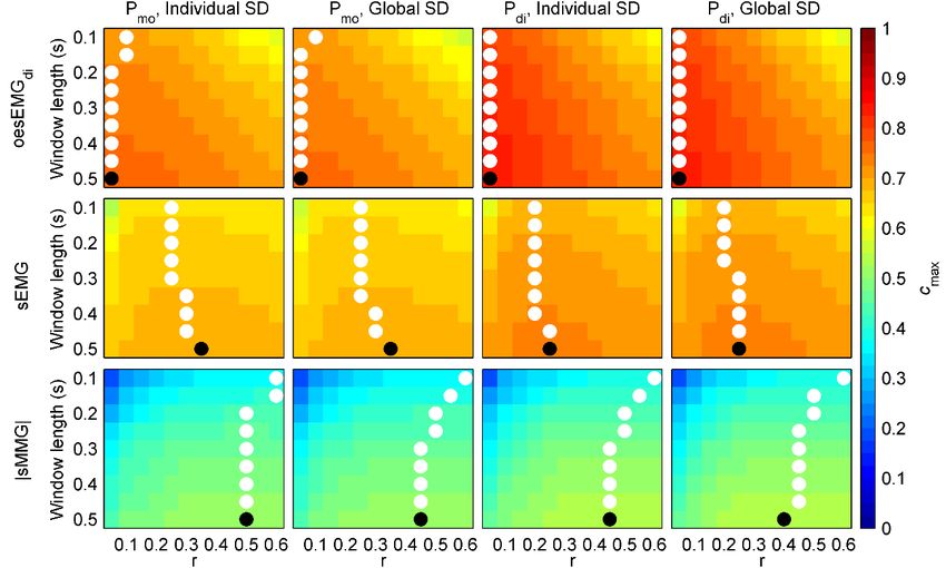
Figure 4 shows cmax values for healthy subjects. First, mean cmax of each group of signals during
resting breathing and threshold loading was calculated for each subject and combination of fSampEn
parameters (window length, r and SD). Then, mean cmax values of all healthy subjects were averaged.Entropy 2019, 21, 183 7 of 16
Entropy 2018,
Entropy 20,20,
2018, x FOR PEER
x FOR REVIEW
PEER REVIEW 7 of 1616
7 of
Figure
Figure Figure3. 3.
Boxplot
3. Boxplot Boxplotdistributions
distributions
distributions ofof
individual
of individual SDs
SDs
individual (H(H
SDs for
for
(H healthy
healthy
for subjects
subjects
healthy and
and
subjects PP for
and for patients)
patients)
P for are
are
patients) shown
areshown
shown
using
using black black
usingboxes. boxes.
black boxes. Red
Red lines lines represent
Red represent unique
unique
lines represent individual
individual
unique SDs
SDsSDs
individual (mean
(mean value).
value).
(mean Boxplot
Boxplot
value). Boxplotdistributions
distributions of
distributionsofof
global
global SDs SDs
areSDs
global are
shown shown using
using using
are shown blue boxes.
blue boxes. Red lines
Red lines
blue boxes. represent
represent
Red lines unique
unique
represent global
global
unique SDs
SDsSDs
global (mean
(mean
(mean value).
value).
value).
Figure 4. Similarity
Figure
Figure (cmax(c)max
4. 4.Similarity
Similarity between
(c ) between
max
fSampEn
) between fSampEn time-series
fSampEn time-series
time-seriesofofinspiratory
ofinspiratory
inspiratory muscle
muscle
muscle myographic
myographic signals
myographicsignals
signals
(oesEMG
(oesEMG ,
(oesEMG
di sEMG
di , sEMGand |sMMG|)
and |sMMG|) and pressure
and pressuresignals
di, sEMG and |sMMG|) and pressure signalsmo signals(P (P and P ) in healthy subjects. For
(Pmo anddiPdi) in healthy subjects. Foreach
mo and P di in healthy subjects. For each
each
comparison,
comparison, different values for window length (from 0.1 to 0.5 s), r (from 0.05 to 0.6) andSDSD
comparison, different different
values values
for for
window window
length length
(from (from
0.1 to 0.1
0.5 s), to
r 0.5
(froms), r
0.05(from
to 0.05
0.6) andto
SD0.6) and
(individual
or global) were tested. White dots indicate the location of the highest cmax of each row. Black dots
indicate the location of the highest cmax of the whole matrix.Entropy 2019, 21, 183 8 of 16
Very similar cmax values were obtained for oesEMGdi , sEMG and |sMMG| using individual and
global SDs, suggesting that global SDs could reasonably be used in order to make fSampEn time-series
of different subjects comparable.
Using global SDs, the highest cmax values in all cases were obtained for a global window length of
0.5 s. However, optimal global r varied among groups of signals: 0.05 for oesEMGdi , 0.35 and 0.25 for
sEMG, and 0.45 and 0.4 for |sMMG|.
The effect of using global instead of individual fSampEn parameters was measured for each
healthy subject as the absolute difference, ∆cmax , between the cmax value obtained using global
parameters and that obtained using individual parameters (Tables 1–3). ∆cmax values were expressed
as percentages of the cmax value obtained using individual parameters.
Table 1. Difference between individual and global fSampEn parameters in the cross-covariance of
oesEMGdi fSampEn time-series and pressure signals of healthy subjects.
oesEMGdi vs. Pmo oesEMGdi vs. Pdi
Subject Ind. SD Glob. SD ∆cmax Ind. SD Glob. SD ∆cmax
Ind. r cmax Glob. r cmax (%) Ind. r cmax Glob. r cmax (%)
H1 0.20 0.767 0.05 0.758 1.15 0.05 0.869 0.05 0.870 0.06
H2 0.05 0.690 0.05 0.691 0.22 0.05 0.750 0.05 0.751 0.08
H3 0.10 0.776 0.05 0.775 0.09 0.05 0.782 0.05 0.779 0.32
H4 0.20 0.751 0.05 0.742 1.18 0.10 0.879 0.05 0.871 0.84
H5 0.40 0.723 0.05 0.691 4.53 0.05 0.883 0.05 0.883 0.05
H6 0.10 0.823 0.05 0.818 0.60 0.05 0.784 0.05 0.784 0.03
H7 0.25 0.743 0.05 0.725 2.36 0.05 0.941 0.05 0.942 0.06
H8 0.05 0.814 0.05 0.814 0.05 0.05 0.907 0.05 0.909 0.18
H9 0.05 0.799 0.05 0.798 0.10 0.05 0.848 0.05 0.845 0.43
H10 0.15 0.822 0.05 0.811 1.27 0.05 0.860 0.05 0.860 0.00
H11 0.30 0.726 0.05 0.716 1.32 0.05 0.868 0.05 0.869 0.10
H12 0.05 0.774 0.05 0.772 0.28 0.05 0.728 0.05 0.725 0.42
Median 0.13 0.770 0.05 0.765 0.88 0.05 0.864 0.05 0.865 0.09
(IQR) (0.05–0.21) (0.739–0.803) (0.05–0.05) (0.723–0.801) (0.19–1.28) (0.05–0.05) (0.784–0.880) (0.05–0.05) (0.783–0.874) (0.06–0.35)
Ind.: Individual; Glob.: Global.
Table 2. Difference between individual and global fSampEn parameters in the cross-covariance of
sEMG fSampEn time-series and pressure signals of healthy subjects.
sEMG vs. Pmo sEMG vs. Pdi
Subject Ind. SD Glob. SD ∆cmax Ind. SD Glob. SD ∆cmax
Ind. r cmax Glob. r cmax (%) Ind. r cmax Glob. r cmax (%)
H1 0.60 0.765 0.35 0.747 2.32 0.40 0.850 0.25 0.829 2.54
H2 0.60 0.575 0.35 0.572 0.47 0.60 0.639 0.25 0.619 3.18
H3 0.60 0.663 0.35 0.664 0.12 0.60 0.658 0.25 0.653 0.80
H4 0.30 0.666 0.35 0.666 0.07 0.30 0.647 0.25 0.643 0.72
H5 0.60 0.707 0.35 0.703 0.57 0.35 0.801 0.25 0.800 0.04
H6 0.35 0.736 0.35 0.732 0.63 0.30 0.697 0.25 0.696 0.18
H7 0.50 0.714 0.35 0.706 1.12 0.20 0.842 0.25 0.842 0.00
H8 0.30 0.822 0.35 0.822 0.01 0.30 0.864 0.25 0.862 0.18
H9 0.10 0.709 0.35 0.689 2.69 0.15 0.720 0.25 0.718 0.35
H10 0.20 0.779 0.35 0.779 0.10 0.10 0.840 0.25 0.834 0.65
H11 0.45 0.585 0.35 0.568 2.87 0.25 0.701 0.25 0.699 0.27
H12 0.10 0.680 0.35 0.606 11.01 0.10 0.643 0.25 0.604 5.97
Median 0.40 0.708 0.35 0.696 0.60 0.30 0.711 0.25 0.708 0.50
(IQR) (0.28–0.60) (0.665–0.744) (0.35–0.35) (0.649–0.736) (0.11–2.41) (0.19–0.36) (0.655–0.840) (0.25–0.25) (0.650–0.830) (0.18–1.23)
Ind.: Individual; Glob.: Global.Table 3. Difference between individual and global fSampEn parameters in the cross-covariance of
|sMMG| fSampEn time-series and pressure signals of healthy subjects.
Entropy 2019, 21, 183 |sMMG| vs. Pmo |sMMG| vs. Pdi 9 of 16
Subject Ind. SD Glob. SD Ind. SD Glob. SD Δcmax
Δcmax (%)
Ind. r cmax Glob. r cmax Ind. r cmax Glob. r cmax (%)
H1 3. Difference
Table 0.55 between
0.642 individual
0.45 and
0.641global fSampEn
0.10 parameters
0.50 in the cross-covariance
0.649 0.40 0.648of 0.13
|sMMG|
H2 fSampEn
0.50 time-series
0.388 and
0.45pressure signals of0.62
0.385 healthy subjects.
0.50 0.431 0.40 0.427 0.91
H3 0.50 0.443 0.45 0.439 0.94 0.60 0.431 0.40 0.432 0.21
|sMMG| vs. Pmo |sMMG| vs. Pdi
H4 0.55 0.394 0.45 0.392 0.52 0.25 0.471 0.40 0.469 0.51
Subject Ind. SD Glob. SD ∆cmax Ind. SD Glob. SD ∆cmax
H5 Ind. r
0.55 cmax
0.529 0.45
Glob. r
0.529
cmax (%) 0.02 Ind. r
0.50 cmax
0.562 Glob. r
0.40 cmax
0.562 (%) 0.03
H1
H6 0.55
0.40 0.532
0.642 0.45
0.45 0.530
0.641 0.10
0.20 0.50
0.50 0.649
0.480 0.40
0.40 0.648
0.479 0.13
0.16
H2H7 0.50 0.15 0.530
0.388 0.450.45 0.524 0.62 1.29 0.50 0.20 0.431 0.653 0.40 0.40 0.427 0.6500.91
0.385 0.33
H3 0.50 0.443 0.45 0.439 0.94 0.60 0.431 0.40 0.432 0.21
H4
H8 0.55
0.60 0.666
0.394 0.45
0.45 0.652
0.392 0.52
2.02 0.25
0.60 0.471
0.717 0.40
0.40 0.469
0.700 0.51
2.39
H5H9 0.55 0.45 0.470
0.529 0.450.45 0.469 0.02 0.24 0.50 0.50 0.562 0.478 0.40 0.40 0.562 0.4780.03
0.529 0.06
H6 0.40 0.532 0.45 0.530 0.20 0.50 0.480 0.40 0.479 0.16
H10
H7 0.15
0.40 0.654
0.530 0.45
0.45 0.654
0.524 1.29
0.01 0.20
0.35 0.653
0.690 0.40
0.40 0.650
0.690 0.33
0.05
H11
H8 0.60 0.55 0.536
0.666 0.450.45 0.532 2.02 0.62 0.60 0.50 0.717 0.642 0.40 0.40 0.700 0.6392.39
0.652 0.47
H9 0.45 0.470 0.45 0.469 0.24 0.50 0.478 0.40 0.478 0.06
H12
H10 0.40
0.50 0.471
0.654 0.45
0.45 0.468
0.654 0.01
0.62 0.35
0.10 0.690
0.408 0.40
0.40 0.690
0.400 0.05
2.04
H11 0.55 0.50 0.530
0.536 0.450.45 0.526 0.62 0.57 0.50 0.50 0.642 0.521 0.40 0.40 0.639 0.5200.47
0.532 0.27
Median
H12 0.50 0.471 0.45 0.468 0.62 0.10 0.408 0.40 0.400 2.04
Median (0.44- (0.463- (0.45- (0.461- (0.18- (0.33- (0.461- (0.40- (0.460- (0.11-
(IQR) 0.50 0.530 0.45 0.526 0.57 0.50 0.521 0.40 0.520 0.27
(IQR) 0.55)(0.463–0.562)
(0.44–0.55) 0.562) (0.45–0.45)
0.45) (0.461–0.560)
0.560) (0.18–0.70)
0.70)(0.33–0.50)0.50) 0.650)
(0.461–0.650) (0.40–0.40)0.40) 0.649)
(0.460–0.649) (0.11–0.61) 0.61)
Ind.:Ind.:
Individual; Glob.:Glob.:
Individual; Global.Global.
Median ∆cmaxΔc
Median were belowbelow
max were 3% for 3%
all groups
for allofgroups
signals,ofindicating
signals, that the performance
indicating of fSampEn of
that the performance
in healthy
fSampEnsubjects is not subjects
in healthy affected is
using global instead
not affected of individual
using global insteadfSampEn parameters.
of individual fSampEn parameters.
3.3. Performance of fSampEn
3.3. Performance in COPD
of fSampEn Patients
in COPD Patients
Figure 5 shows
Figure cmax values
5 shows for COPD
cmax values patients.
for COPD First,First,
patients. mean cmax cof
mean maxeach group
of each of signals
group during
of signals during
resting breathing and threshold loading was calculated for each patient and combination of
resting breathing and threshold loading was calculated for each patient and combination of fSampEnfSampEn
parameters (window
parameters length,
(window r andr SD).
length, Then,
and SD). Then, cmax cvalues
meanmean of all
max values ofCOPD
all COPDpatients werewere
patients averaged.
averaged.
Figure 5. Similarity
Figure (cmax )(cbetween
5. Similarity fSampEn
max) between fSampEn time-series of inspiratory
time-series muscle
of inspiratory myographic
muscle signals
myographic signals
(sEMG and |sMMG|) and pressure signals (P ) in COPD patients. For each comparison,
(sEMG and |sMMG|) and pressure signals (Pmo) in COPD patients. For each comparison, different
mo different
values for window
values for windowlength (from(from
length 0.1 to0.1 r (from
0.5tos),0.5 0.05 to
s), r (from 0.6)
0.05 toand
0.6) SD
and(individual or global)
SD (individual werewere
or global)
tested. White dots indicate the location of the highest
tested. White dots indicate the location of the highest c of each row. Black dots indicate the location
max cmax of each row. Black dots indicate the location
of theofhighest cmax of
the highest cmaxthe
of whole matrix.
the whole matrix.
As in
Ashealthy subjects,
in healthy veryvery
subjects, similar cmax values
similar werewere
cmax values obtained for sEMG
obtained and |sMMG|
for sEMG and |sMMG| using
using
individual global SDs in COPD patients. Using global SDs, the highest c
individual and global SDs in COPD patients. Using global SDs, the highest cmax values incases
and max values in all all cases
were obtained, again, for a global window length of 0.5 s. Regarding optimal global r, it varied from
0.2 for sEMG to 0.55 and 0.5 for |sMMG|.
The effect of using global instead of individual fSampEn parameters was measured for each
COPD patient as the difference between the cmax value obtained using global parameters and that
obtained using individual parameters (Table 4).Entropy 2019, 21, 183 10 of 16
In COPD patients, median ∆cmax were below 1% for both sEMG and |sMMG| groups of signals,
indicating that the performance of fSampEn in these patients is not affected using global instead of
individual fSampEn parameters.
Table 4. Difference between individual and global fSampEn parameters in the cross-covariance of
sEMG and |sMMG| fSampEn time-series and Pmo of COPD patients.
sEMG vs. Pmo |sMMG| vs. Pmo
Subject
Ind. SD Glob. SD ∆cmax Ind. SD Glob. SD ∆cmax
Ind. r cmax Glob. r cmax (%) Ind. r cmax Glob. r cmax (%)
P1 0.20 0.649 0.20 0.648 0.10 0.60 0.376 0.50 0.377 0.22
P2 0.15 0.634 0.20 0.634 0.07 0.60 0.334 0.50 0.330 1.29
P3 0.15 0.555 0.20 0.545 1.72 0.60 0.425 0.50 0.425 0.11
P4 0.10 0.571 0.20 0.569 0.35 0.35 0.434 0.50 0.428 1.26
P5 0.15 0.822 0.20 0.822 0.00 0.60 0.551 0.50 0.550 0.29
P6 0.55 0.705 0.20 0.698 1.11 0.40 0.553 0.50 0.551 0.26
P7 0.40 0.756 0.20 0.753 0.32 0.60 0.630 0.50 0.632 0.24
P8 0.20 0.793 0.20 0.791 0.15 0.40 0.585 0.50 0.584 0.29
P9 0.50 0.665 0.20 0.659 0.82 0.20 0.247 0.50 0.233 5.75
P10 0.15 0.815 0.20 0.815 0.01 0.30 0.445 0.50 0.442 0.81
P11 0.30 0.794 0.20 0.793 0.11 0.60 0.523 0.50 0.524 0.21
P12 0.25 0.692 0.20 0.691 0.03 0.60 0.450 0.50 0.446 0.84
P13 0.15 0.628 0.20 0.627 0.19 0.50 0.554 0.50 0.553 0.14
P14 0.30 0.792 0.20 0.783 1.17 0.30 0.574 0.50 0.574 0.11
Median 0.20 0.698 0.20 0.694 0.17 0.55 0.487 0.50 0.485 0.27
(IQR) (0.15–0.30) (0.638–0.793) (0.20–0.20) (0.637–0.789) (0.08–0.7) (0.36–0.60) (0.427–0.553) (0.50–0.50) (0.426–0.552) (0.21–0.83)
Ind.: Individual; Glob.: Global.
3.4. General fSampEn Parameters
In light of the very similar global SDs in healthy subjects and COPD patients (Figure 3), and the
robustness of fSampEn to the use of global instead of individual fSampEn parameters, the following
general fSampEn parameters are proposed to be used in healthy subjects as well as in COPD patients:
0.5 s moving window with 90% overlap, m equal to 2 and tolerance equal to 0.05 × 0.0121 (oesEMGdi ),
0.3 × 0.0022 (sEMG) and 0.5 × 0.0060 (|sMMG|).
Figures 6 and 7 show representative recordings from a healthy subject and a COPD patient,
respectively, during resting breathing and the inspiratory threshold loading protocol. fSampEn
time-series of these representative subjects’ myographic signals were calculated using the proposed
general fSampEn parameters.
As observed in Figures 6 and 7, fSampEn performed very well in all myographic signals, tracking
amplitude changes evoked by inspiratory muscle EMG and MMG activity during the inspiratory
threshold loading protocol as well as being robust to cardiac noise.Entropy 21, 183
2019,2018,
Entropy 20, x FOR PEER REVIEW 11 of1116of 16
Figure 6. Measurements recorded during the inspiratory threshold loading protocol in a healthy
Figure 6. Measurements recorded during the inspiratory threshold loading protocol in a healthy
subject. Two respiratory cycles are shown for quiet breathing and threshold loading. The oesEMGdi
subject. Two respiratory cycles are shown for quiet breathing and threshold loading. The oesEMGdi
signal corresponds to the electrode pair 1. fSampEn time-series of the oesEMGdi , sEMG and |sMMG|
signal corresponds to the electrode pair 1. fSampEn time-series of the oesEMGdi, sEMG and |sMMG|
signals were calculated using the general fSampEn parameters proposed in this section.
signals were calculated using the general fSampEn parameters proposed in this section.Entropy 21, 183
2019,2018,
Entropy 20, x FOR PEER REVIEW 12 of1216of 16
Figure 7. Measurements recorded during the inspiratory threshold loading protocol in a COPD patient.
TwoFigure 7. Measurements
respiratory recorded
cycles are shown during
for quiet the inspiratory
breathing threshold
and threshold loading
loading. fSampEnprotocol in a COPD
time-series of
patient.
the sEMG andTwo respiratory
|sMMG| cycles
signals arecalculated
were shown forusing
quietthe
breathing
generaland threshold
fSampEn loading.proposed
parameters fSampEnintime-
this series
section.of the sEMG and |sMMG| signals were calculated using the general fSampEn parameters
proposed in this section.
4. Discussion and Conclusions
4.InDiscussion
this study, and Conclusions
we conducted an evaluation of the performance of fSampEn for inspiratory muscle
EMG andInMMG analysis,
this study, which led us
we conducted anto propose, for
evaluation theperformance
of the first time, a set of optimalfor
of fSampEn general fSampEn
inspiratory muscle
parameters adapted to each type of myographic signal.
EMG and MMG analysis, which led us to propose, for the first time, a set of optimal general fSampEn
Evaluatingadapted
parameters the amplitude
to each of
typemyographic
of myographic signals recorded from different respiratory muscles
signal.
using conventional
Evaluatingamplitude
the amplitudeestimators is difficult signals
of myographic due to the cardiacfrom
recorded muscle interference.
different In order
respiratory to
muscles
reduce crosstalk from the heart in myographic respiratory signals, the use of
using conventional amplitude estimators is difficult due to the cardiac muscle interference. In order fSampEn was adopted
in our previous
to reduce studies
crosstalk from[6,9,15,17,19].
the heart in This technique
myographic is basedsignals,
respiratory on SampEn,the usewhich has previously
of fSampEn was adopted
contributed to improving the understanding of the underlying mechanisms
in our previous studies [6,9,15,17,19]. This technique is based on SampEn, which has previously of physiological processes
in a contributed
wide numbertoofimprovingclinical applications [16,32–34]. of the underlying mechanisms of physiological
the understanding
Depending
processes in aon the number
wide application, fSampEn
of clinical an adjustment of its input parameters m, r
requires[16,32–34].
applications
and window length. However, even for the same application,
Depending on the application, fSampEn requires an adjustment such as of respiratory muscle activity
its input parameters m, r and
estimation,
windowseveral length.values
However,have been
even suggested
for the same for fSampEn parameters.
application, such as Arespiratory
previous study muscleby activity
our
group demonstrated
estimation, severalthat r and
values havewindow length arefor
been suggested thefSampEn
most critical parameters,
parameters. influencing
A previous study the by our
magnitude and shape of fSampEn time-series [26]. It was reported that
group demonstrated that r and window length are the most critical parameters, influencing the a window length of 1 s and
either m equal toand
magnitude 1 and r ranging
shape of fSampEn to 0.64 or m[26].
from 0.1time-series equal to 2 and
It was r ranging
reported that from 0.13 tolength
a window 0.45 could
of 1 be s and
suitable values for respiratory muscle activity estimation using fSampEn in sEMG
either m equal to 1 and r ranging from 0.1 to 0.64 or m equal to 2 and r ranginglicfrom 0.13 to 0.45 could signals. However,
some belimitations were acknowledged
suitable values for respiratory in this
muscleprevious study.
activity Firstly, only
estimation sEMG
using lic signals
fSampEn inweresEMGanalysed
lic signals.
for aHowever,
healthy subject. However, other
some limitations weremyographic
acknowledged signals, such
in this as sMMG
previous lic or Firstly,
study. oesEMGonly di , could
sEMG require
lic signals
different fSampEn parameters. Moreover, impaired respiratory mechanics
were analysed for a healthy subject. However, other myographic signals, such as sMMG (e.g., in COPD patients) maylic or
affect the amplitude
oesEMG di, couldand complexity
require differentof fSampEn
myographic respiratoryMoreover,
parameters. signals [1,5,35],
impaired and respiratory
therefore fSampEn
mechanics
(e.g. in COPD patients) may affect the amplitude and complexity of myographic respiratory signalsEntropy 2019, 21, 183 13 of 16
parameters different to those used in healthy subjects may be required. Furthermore, the performance
of different combinations of fSampEn parameters was evaluated by means of Pearson’s correlation
coefficient between fSampEn time-series and Pmo , which was used as an approximation of the overall
mechanical output of the inspiratory muscles during breathing. However, Pmo was conditioned by the
threshold loads imposed by the inspiratory muscle trainer, which was attached to the distal end of the
pneumotachograph in series with a mouthpiece.
In this current study, we have thoroughly investigated the performance of fSampEn, for the first
time, in different types of myographic respiratory signals, including oesEMGdi , sEMGlic , sEMGpara ,
sMMGlic and sMMGpara . Moreover, the participants in the present study cohort were twelve healthy
subjects with an age range from 21 to 44 years and fourteen COPD patients with an age range from
53 to 80 years. This heterogeneous population covers a wide age range and both normal and impaired
respiratory mechanics. Furthermore, the inspiratory threshold loading protocol provided the potential
to acquire inspiratory muscle EMG and MMG signals under a wide range of loads on the inspiratory
muscles, allowing the performance of fSampEn to be evaluated for a variety of inspiratory muscle
activation levels. The performance of fSampEn was assessed by means of the cross-covariance of
fSampEn time-series and both non-invasive measurements of Pmo and the gold standard invasive
measurements of Pdi . While Pmo assesses global inspiratory muscle strength, Pdi is more specific for
the diaphragm.
The kernel of the fSampEn algorithm is the search for similar sequences within a signal, so that
more similar sequences indicate lower complexity and yield lower fSampEn values. Two sequences
are similar if the maximum difference of their corresponding values is less than or equal to the
tolerance parameter, which is usually set as r times the SD of the signal analysed. When calculated
within a moving window using a fixed tolerance for all windows, fSampEn can quantify amplitude
variations on a signal. In this study, for a given subject and type of myographic respiratory signal,
choosing a unique individual tolerance, and therefore a unique individual SD, allows fSampEn to track
amplitude variations during the inspiratory threshold loading protocol and optimises the performance
of fSampEn, mainly in terms of cardiac noise attenuation, for that subject. On the other hand, a unique
global SD is required in order to compare fSampEn time-series of different subjects. Due to differences
in myographic respiratory signal amplitude among subjects, using a global SD, however, may not be
optimal in all subjects and may compromise the performance of fSampEn in some subjects.
In the present study we compared, for the first time, the performance of fSampEn in three groups
of myographic respiratory signals (oesEMGdi , sEMG and |sMMG|) using different r values (0.05–0.6)
and both individual and global SDs. Intra- and inter-subject variability of the SD of myographic
respiratory signals was firstly analysed (see Figure 3). Intra-subject variability was due to different
levels of inspiratory muscle activation during the inspiratory threshold loading protocol. Inter-subject
variability, however, was related to different patterns of inspiratory muscle activation during the
inspiratory threshold loading protocol, since subjects were not provided with any specific instructions
to adopt a certain breathing pattern. Moreover, inspiratory muscle EMG and MMG signals were
not normalised, and therefore myographic signal amplitudes varied among subjects. Despite the
inter-subject variability of SDs, individual and global SDs performed similarly in both healthy subjects
and COPD patients. Moreover, global SDs of healthy subjects were very similar to those of COPD
patients, for both sEMG and |sMMG| signals.
In light of these results, we have proposed general tolerance (r × SD) values of 0.05 × 0.0121
(oesEMGdi ), 0.3 × 0.0022 (sEMG) and 0.5 × 0.0060 (|sMMG|) to be used in healthy subjects as well as
in COPD patients. The use of these general SDs allows fSampEn time-series to be optimally calculated
and compared in different subjects.
Different combinations of window lengths between 0.1 s and 0.5 s were tested, and a window
length of 0.5 s maximised the performance of fSampEn in all study participants and myographic
respiratory signals. In general, the longer the window, the smoother the fSampEn time-series [26].
Long windows are therefore desirable to minimise the influence of cardiac noise or short-time variationsEntropy 2019, 21, 183 14 of 16
of myographic respiratory signals, e.g., in neural respiratory drive estimation [9] or in inspiratory
muscle activity estimation from the area under the curve of inspiratory EMG and MMG signals [6].
Conversely, short windows are required to evaluate short-time variations of myographic respiratory
signals, e.g., in onset estimation of the neural inspiratory time [18].
In this study, fSampEn time-series were compared with pressure signals, which are low-frequency
signals and vary slowly over time, and therefore longer windows performed better than shorter
windows. On the other hand, a lower threshold of 0.5 s was applied to respiratory phase durations
during segmentation of myographic signals. Therefore, in order to focus the analysis of myographic
respiratory signals on inspiratory activity, window lengths longer than 0.5 s were not tested. Yentes et al.
proposed a minimum window length of 200 samples for the correct performance of fSampEn [36].
The window length value of 0.5 s proposed in this study meets this criterion, even for |sMMG| signals
(500 Hz x 0.5 s = 250 samples).
The m parameter is typically set at 1 or 2 [16,37]. In recent studies by our group, no relevant
differences were found between m = 1 and m = 2 for fSampEn calculation in sEMGlic signals [26],
and m = 2 has been properly used for the analysis of different myographic respiratory signals [6].
Accordingly, m = 2 has also been used in the present study for the analysis of myographic
respiratory signals.
Sampling frequency is another parameter to be taken into account in fSampEn calculation. It has
been reported that higher sampling frequencies yield lower fSampEn values [26]. However, the effect
of sampling frequency in fSampEn time-series can be omitted if sampling frequency is correctly chosen
based on the effective bandwidth of each type of myographic respiratory signal, as in the present study.
In summary, we performed a thorough evaluation of the most relevant parameters of fSampEn,
that are r and window length, for a proper estimation of inspiratory muscle activity from EMG and
MMG signals. The evaluation of fSampEn performed in a heterogeneous study population, including
healthy subjects and COPD patients within a wide age range, allowed us to propose general values
for fSampEn parameters that permit optimal calculation and comparison of fSampEn time-series of
different subjects, thus contributing to improving the assessment of inspiratory muscle activity in
health and disease.
Author Contributions: Conceptualization, M.L.G., L.E. and R.J.; Methodology, M.L.G. and R.J.; Software, M.L.G.;
Validation, M.L.G., L.E. and R.J.; Formal analysis, M.L.G.; Investigation, M.L.G., L.E. and R.J.; Writing—original
draft preparation, M.L.G. and L.E.; Writing—review and editing, M.L.G., L.E. and R.J.; Visualization, M.L.G.;
Supervision, R.J.
Funding: This research was funded by the Government of Catalonia (CERCA Programme). This work was
supported in part by the Secretaria d’Universitats i Recerca del Departament d’Empresa i Coneixement de la
Generalitat de Catalunya (Consolidated research group GRC 2017 SGR 01770), the Spanish Ministry of Economy
and Competitiveness (project DPI2015-68820-R MINECO/FEDER), and the Biomedical Research Networking
Centre in Bioengineering, Biomaterials and Nanomedicine (CIBER-BBN, Instituto de Salud Carlos III/FEDER).
Manuel Lozano-García and Luis Estrada were the recipients of two European Respiratory Society Fellowships
(ERS LTRF 2015-5185 and ERS LTRF 2017 01-00086 respectively). Luis Estrada was also supported by the Instituto
para la Formación y Aprovechamiento de Recursos Humanos and Secretaría Nacional de Ciencia, Tecnología e
Innovación (IFARHU-SENACYT) of the Government of Panama (grant 270-2012-273).
Conflicts of Interest: The authors declare no conflict of interest. The funders had no role in the design of the
study; in the collection, analyses, or interpretation of data; in the writing of the manuscript, or in the decision to
publish the results.
References
1. Jolley, C.J.; Luo, Y.M.; Steier, J.; Reilly, C.; Seymour, J.; Lunt, A.; Ward, K.; Rafferty, G.F.; Polkey, M.I.;
Moxham, J. Neural respiratory drive in healthy subjects and in COPD. Eur. Respir. J. 2009, 33, 289–297.
[CrossRef] [PubMed]
2. American Thoracic Society/European Respiratory Society. ATS/ERS statement on respiratory muscle testing.
Am. J. Respir. Crit. Care Med. 2002, 166, 518–624. [CrossRef] [PubMed]Entropy 2019, 21, 183 15 of 16
3. Mead, J.; Loring, S.H. Analysis of volume displacement and length changes of the diaphragm during
breathing. J. Appl. Physiol. 1982, 53, 750–755. [CrossRef] [PubMed]
4. Luo, Y.M.; Moxham, J.; Polkey, M.I. Diaphragm electromyography using an oesophageal catheter: Current
concepts. Clin. Sci. (Lond.) 2008, 115, 233–244. [CrossRef] [PubMed]
5. Sarlabous, L.; Torres, A.; Fiz, J.A.; Martínez-Llorens, J.M.; Gea, J.; Jané, R. Inspiratory muscle activation
increases with COPD severity as confirmed by non-invasive mechanomyographic analysis. PLoS ONE 2017,
12, e0177730. [CrossRef] [PubMed]
6. Lozano-García, M.; Sarlabous, L.; Moxham, J.; Rafferty, G.F.; Torres, A.; Jané, R.; Jolley, C.J. Surface
mechanomyography and electromyography provide non-invasive indices of inspiratory muscle force and
activation in healthy subjects. Sci. Rep. 2018, 8, 16921. [CrossRef] [PubMed]
7. Reilly, C.C.; Ward, K.; Jolley, C.J.; Lunt, A.C.; Steier, J.; Elston, C.; Polkey, M.I.; Rafferty, G.F.; Moxham, J.
Neural respiratory drive, pulmonary mechanics and breathlessness in patients with cystic fibrosis. Thorax
2011, 66, 240–246. [CrossRef] [PubMed]
8. MacBean, V.; Hughes, C.; Nicol, G.; Reilly, C.C.; Rafferty, G.F. Measurement of neural respiratory drive
via parasternal intercostal electromyography in healthy adult subjects. Physiol. Meas. 2016, 37, 2050–2063.
[CrossRef]
9. Estrada, L.; Torres, A.; Sarlabous, L.; Jané, R. Improvement in neural respiratory drive estimation from
diaphragm electromyographic signals using fixed sample entropy. IEEE J. Biomed. Health Inform. 2016, 20,
476–485. [CrossRef]
10. Lin, L.; Guan, L.; Wu, W.; Chen, R. Correlation of surface respiratory electromyography with esophageal
diaphragm electromyography. Respir. Physiol. Neurobiol. 2018. [CrossRef]
11. González-Izal, M.; Malanda, A.; Gorostiaga, E.; Izquierdo, M. Electromyographic models to assess
muscle fatigue. J. Electromyogr. Kinesiol. 2012, 22, 501–512. [CrossRef] [PubMed]
12. Ibitoye, M.O.; Hamzaid, N.A.; Zuniga, J.M.; Hasnan, N.; Wahab, A.K.A. Mechanomyographic parameter
extraction methods: An appraisal for clinical applications. Sensors 2014, 14, 22940–22970. [CrossRef]
[PubMed]
13. Sinderby, C.; Lindström, L.; Grassino, A.E. Automatic assessment of electromyogram quality. J. Appl. Physiol.
1995, 79, 1803–1815. [CrossRef] [PubMed]
14. Sinderby, C.A.; Beck, J.C.; Lindström, L.H.; Grassino, A.E. Enhancement of signal quality in esophageal
recordings of diaphragm EMG. J. Appl. Physiol. 1997, 82, 1370–1377. [CrossRef] [PubMed]
15. Sarlabous, L.; Torres, A.; Fiz, J.A.; Jané, R. Evidence towards improved estimation of respiratory muscle effort
from diaphragm mechanomyographic signals with cardiac vibration interference using sample entropy with
fixed tolerance values. PLoS ONE 2014, 9, e88902. [CrossRef] [PubMed]
16. Richman, J.S.; Moorman, J.R. Physiological time-series analysis using approximate entropy and sample
entropy. Am. J. Physiol. Heart Circ. Physiol. 2000, 278, H2039–H2049. [CrossRef] [PubMed]
17. Ràfols-de-Urquía, M.; Estrada, L.; Estévez-Piorno, J.; Sarlabous, L.; Jané, R.; Torres, A. Evaluation of
a wearable device to determine cardiorespiratory parameters from surface diaphragm electromyography.
IEEE J. Biomed. Health Inform. 2018. [CrossRef] [PubMed]
18. Estrada, L.; Torres, A.; Sarlabous, L.; Jané, R. Onset and Offset Estimation of the Neural Inspiratory Time in
Surface Diaphragm Electromyography: A Pilot Study in Healthy Subjects. IEEE J. Biomed. Health Inform.
2018, 22, 67–76. [CrossRef] [PubMed]
19. Sarlabous, L.; Torres, A.; Fiz, J.A.; Gea, J.; Martinez-Llorens, J.M.; Jané, R. Efficiency of mechanical activation
of inspiratory muscles in COPD using sample entropy. Eur. Respir. J. 2015, 46, 1808–1811. [CrossRef]
[PubMed]
20. Zhang, X.; Zhou, P. Sample entropy analysis of surface EMG for improved muscle activity onset detection
against spurious background spikes. J. Electromyogr. Kinesiol. 2012, 22, 901–907. [CrossRef] [PubMed]
21. Zhou, P.; Zhang, X. A novel technique for muscle onset detection using surface EMG signals without removal
of ECG artifacts. Physiol. Meas. 2014, 35, 45–54. [CrossRef] [PubMed]
22. Liu, J.; Liu, Q. Use of the integrated profile for voluntary muscle activity detection using EMG signals with
spurious background spikes: A study with incomplete spinal cord injury. Biomed. Signal Process. Control
2016, 24, 19–24. [CrossRef]You can also read