Skeletal Muscle Lipid Droplets and the Athlete's Paradox - MDPI
←
→
Page content transcription
If your browser does not render page correctly, please read the page content below
cells
Review
Skeletal Muscle Lipid Droplets and the
Athlete’s Paradox
Xuehan Li 1,† , Zemin Li 1,† , Minghua Zhao 1 , Yingxi Nie 1 , Pingsheng Liu 2,3 , Yili Zhu 1, * and
Xuelin Zhang 1, *
 1    School of Kinesiology and Health, Capital University of Physical Education and Sports, Beijing 100191,
      China; lixuehan@cupes.edu.cn (X.L.); lizemin@cupes.edu.cn (Z.L.); zhaominghua@cupes.edu.cn (M.Z.);
      nieyingxi2018@cupes.edu.cn (Y.N.)
 2    National Laboratory of Biomacromolecules, CAS Center for Excellence in Biomacromolecules, Institute of
      Biophysics, Chinese Academy of Sciences, Beijing 100101, China; pliu@ibp.ac.cn
 3    University of Chinese Academy of Sciences, Beijing 100049, China
 *    Correspondence: zhuyili@cupes.edu.cn (Y.Z.); zhangxuelin@cupes.edu.cn (X.Z.)
 †    These authors contributed equally to this work.

 Received: 28 January 2019; Accepted: 12 March 2019; Published: 15 March 2019

 Abstract: The lipid droplet (LD) is an organelle enveloped by a monolayer phospholipid membrane
 with a core of neutral lipids, which is conserved from bacteria to humans. The available evidence
 suggests that the LD is essential to maintaining lipid homeostasis in almost all organisms. As a
 consequence, LDs also play an important role in pathological metabolic processes involving the
 ectopic storage of neutral lipids, including type 2 diabetes mellitus (T2DM), atherosclerosis, steatosis,
 and obesity. The degree of insulin resistance in T2DM patients is positively correlated with the size
 of skeletal muscle LDs. Aerobic exercise can reduce the occurrence and development of various
 metabolic diseases. However, trained athletes accumulate lipids in their skeletal muscle, and LD size
 in their muscle tissue is positively correlated with insulin sensitivity. This phenomenon is called the
 athlete’s paradox. This review will summarize previous studies on the relationship between LDs in
 skeletal muscle and metabolic diseases and will discuss the paradox at the level of LDs.

 Keywords: lipid droplet; skeletal muscle; metabolic diseases; athlete’s paradox

1. Introduction
     The lipid droplet (LD) is an organelle that stores neutral lipids in cells and plays an important role
in maintaining lipid homeostasis in almost all organisms [1]. There is abundant experimental evidence
that LDs interact with other organelles, and that this is mediated by regulatory proteins and enzymes
embedded in their surface. LD proteins are also responsible for regulating the size, shape, and stability
of LDs, parameters that are associated with various physiological states [2]. Some of the interactions
between LDs and other organelles and aspects of LD dynamics are diagramed in Figure 1.
     In the past several decades, there has been a worldwide increase in the incidence of lipid metabolic
diseases. As high caloric diets have become more affordable and lifestyles more sedentary, an increasing
fraction of the world’s population ingests lipids and other calories above their metabolic needs.
A chronic positive energy balance results in elevated blood triacylglycerol (TAG) and free fatty acid
(FFA) content. This, in turn, leads to the ectopic storage of neutral lipids in non-adipose tissues, such
as skeletal muscle, liver, and heart [3]. Two major human adipose tissues are described: white adipose
tissue (WAT) and brown adipose tissue (BAT). WAT is used mainly to store energy, while BAT is used
mainly to produce heat [4]. Ectopic lipid storage [5,6] refers to the lipids that cannot be consumed and
stored in adipose tissues but are stored in non-adipose tissues, which, in turn, causes lipid metabolism
disorder as well as lipid toxicity.

Cells 2019, 8, 249; doi:10.3390/cells8030249                                      www.mdpi.com/journal/cells
Cells 2019, 8, x FOR PEER REVIEW 2 of 9
lipids that cannot be consumed and stored in adipose tissues but are stored in non-adipose tissues,
Cells 2019,
which, 8, 249 causes lipid metabolism disorder as well as lipid toxicity.
in turn, 2 of 9
Figure
Figure1.1.Dynamics
Dynamicsof oflipid
lipid droplets (LDs). As
droplets (LDs). Asan
anorganelle,
organelle,a lipid
a lipid droplet
droplet presents
presents a very
a very active
active status
status
in cells, including movement, interaction with other cellular organelles, and size change. (A) LDs(A)
in cells, including movement, interaction with other cellular organelles, and size change. can
LDs can interact
interact with thewith the endoplasmic
endoplasmic reticulumreticulum (ER) through
(ER) through LD-ER sites.
LD-ER contact contact
Thesites. Theisaction
action is to
to exchange
exchange
materialmaterial and information
and information between between the two organelles.
the two organelles. (B) A large(B)LDA can
large
beLD can be
divided divided
into smaller into
LDs
smaller LDs by two means: fission and budding. (C) Two small LDs can form a large
by two means: fission and budding. (C) Two small LDs can form a large LD using fusion and neutralLD using fusion
and neutral
lipid lipid transportation.
transportation.
Lipidtoxicity
Lipid toxicityis isthought
thoughttotointerfere
interferewith
withnormal
normalcellular
cellularfunctions
functionsinina aprocess
processcalled
calledlipid
lipid
toxicity[7,8].
toxicity [7,8].This
Thisis isthe
thehypothesis
hypothesisthatthatexcessive,
excessive,atypical
atypicalstorage
storageofoflipids
lipidsininnon-adipose
non-adiposetissuestissues
influences the metabolic homeostasis of the impacted tissues and organs. Lipid toxicity leads toto
influences the metabolic homeostasis of the impacted tissues and organs. Lipid toxicity leads
disturbances
disturbances inin cell
cell signaling
signaling andand
thethe development
development of insulin
of insulin resistance,
resistance, which,
which, in turn,
in turn, can can
resultresult
in
a in a series
series of related
of related diseases
diseases including
including typetype 2 diabetes
2 diabetes (T2DM)
(T2DM) andand non-alcoholic
non-alcoholic fatty
fatty liverdisease
liver disease
(NAFLD)[9].
(NAFLD) [9].
Duetotoitsitsgreat
Due greattissue
tissuemass
massandandlarge
largecontribution
contributiontotometabolic
metabolicdemand,
demand,skeletal
skeletalmuscle
muscleis isa a
particularly
particularly consequential
consequential cell
cell type
type forfor pathologies
pathologies of lipid
of lipid homeostasis
homeostasis [10,11].
[10,11]. Lipids
Lipids are stored
are stored as
as TAG in LDs within skeletal muscle cells, called intramyocellular lipid (IMCL) [12,13]. To meet
energy demands, IMCL can be hydrolyzed into FFAs, which are processed through β-oxidation in
Cells 2019, 8, 249                                                                                   3 of 9

mitochondria to generate ATP and heat in skeletal muscle [12,14]. Physical training can induce
an increase in IMCL pools, which is reflected in an increased LD number and accompanying
morphological changes. Interestingly, while a diet-induced increase in IMCL is associated with insulin
resistance, similar IMCL accumulation in response to exercise is not [15]. This apparent contradiction
is referred to as is the athlete’s paradox [16]. A resolution of the paradox has yet to be fully achieved.
      There have been advances in methodological approaches permitting more accurate measurements
of IMCL and LDs in skeletal muscle, including the biochemical extraction of TAG, magnetic resonance
spectrometry, histochemical staining with immunofluorescence microscopy, and transmission electron
microscopy (TEM) [12,13]. Methods have also been recently established for the isolation of muscle
LDs permitting proteomic analysis [17]. These new techniques provide more detailed observation of
skeletal muscle LDs, which raises a new perspective for the study of the athlete’s paradox.

2. Diabetes Mellitus
     Diabetes mellitus (DM) is a complex, chronic metabolic disease with multiple causes. The most
obvious feature of the disease is a sustained elevation of blood glucose levels, accompanied by
long-term disorders in glucose, lipid, and protein metabolism caused by insufficient insulin secretion
or insulin non-responsiveness [18]. The definition of DM contains several criteria including fasting
blood glucose above 7.0 mmol/L [19]. If DM is not controlled through proper lifestyle and medical
intervention, the sequelae include organ failure and peripheral nerve necrosis [20].
     There are three common types of DM. Type 1 diabetes mellitus (T1DM) was previously
called insulin-dependent diabetes. This type of autoimmune disease is most common in children
and adolescents. Immunological destruction of the pancreatic beta cells results in insufficient
insulin secretion to stimulate glucose uptake [21]. T2DM is a type of metabolic disease that is
characterized mainly by a reduction in insulin sensitivity (insulin resistance), which in turn decreases
insulin-stimulated uptake of blood glucose [22]. The third type is gestational diabetes mellitus (GDM).
This type occurs in pregnant women and represents a serious complication of childbirth. The condition
is specific to pregnant women who have not previously been diagnosed with diabetes [23].
     T2DM is by far the most common form of the disease, accounting for over 90% of cases. The early
stages of the disease are marked by increasing insulin resistance in the peripheral tissues including
muscle, fat, and liver. At first, a compensatory increase in insulin secretion is able to maintain normal
glycemic levels [24,25]. However, over time, the heavy demand leads to beta-cell exhaustion and
apoptosis. The resulting decrease in insulin secretion along with increasing insulin resistance leads to
a loss in glycemic control and dangerous elevations in blood glucose levels [26–28].
     At the cellular level, lipid homeostasis includes the balance between the absorption of free fatty
acids from the blood and the synthesis and hydrolysis of lipids in cells under normal conditions [29].
The ectopic accumulation of lipids in peripheral tissues can interfere with these cellular functions,
disturbing lipid homeostasis at the cellular level. Ultimately, this can lead to diseases including,
commonly, T2DM. Due to its increasing prevalence and mounting societal costs, T2DM has become
the focus of global attention.
     When lipids accumulate beyond the capacity of adipose tissue to efficiently store it, the rate of
lipid hydrolysis exceeds that of esterification. This results in a sharp rise in blood FFA levels, which
has two downstream consequences. First, it drives insulin resistance through fatty acid receptors
on the cell surface. Second, FFAs are absorbed by the peripheral tissues, which further disturbs
insulin signaling. Since skeletal muscle accounts for more than 70% of the body’s blood glucose
intake [30], the high concentration of free fatty acids in the blood of obese patients has the greatest
impact on the insulin response of skeletal muscle [31]. The degree of IMCL is positively correlated
with insulin resistance [32,33]. Furthermore, the increase in IMCL in skeletal muscle is accompanied
by an accumulation of the metabolic intermediates diacylglycerol (DAG) [34] and neuroamide [35],
which have also been linked to insulin resistance. The levels of DAG accumulation in skeletal muscle
of endurance training and sedentary obese rats are similar [36]. In addition, the concentration of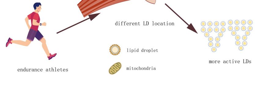
Cells 2019, 8, 249                                                                                    4 of 9

phosphatidylethanolamine molecular species containing palmitoleate is increased in endurance-trained
rats, but the opposite is true in sedentary obese rats. These findings indicate that endurance exercise
can affect the lipid composition of skeletal muscle. Unfortunately, the LD lipidome of skeletal muscle
with or without endurance exercise remains unknown.

3. Skeletal Muscle Lipid Droplets and the Athlete’s Paradox
     The adoption of an exercise program can alleviate many chronic diseases [37]. An exercise
regimen can accelerate metabolism, improve cardiovascular function, and enhance immunity [38].
Indeed, a short-term exercise intervention was found to alleviate induced insulin resistance but also
to increase the expression of genes associated with IMCL synthesis, resulting in the accumulation of
TAG in skeletal muscles [39]. Another observational study found that physically trained individuals
had increased skeletal muscle TAG relative to lean, sedentary people [16]. This is consistent with
the known role of IMCL in meeting the energy demand of skeletal muscle [12]. Other studies have
found a positive correlation between IMCL levels and insulin sensitivity in those engaged in aerobic
training [40,41]. However, this association is surprising since IMCL has also been positively correlated
with the development of metabolic diseases and insulin resistance in the general population [11,42,43].
This is the athlete’s paradox.
     It is likely that the assessment of IMCL levels is too crude a measure and that this simple metric
belies important biochemical differences between diet-induced and exercise-induced accumulation of
TAG in skeletal muscle. A more nuanced analysis is required to resolve the paradox. Improvements
in biopsy and imaging techniques for studying muscle are allowing for more detailed analysis of the
phenomenon linking LDs and insulin sensitivity in muscle tissue.

4. Subcellular Compartmentalization of Lipid Droplets
     LDs are found in two distinct locations in muscle, just beneath the cell membrane (subsarcolemmal
LDs) and between myofibrils (intermyofibrillar LDs) [44]. The total cellular volume of intermyofibrillar
LDs greatly exceeds that of the subsarcolemmal pool [44]. There is a substantial drop in total IMCL
following acute exercise suggesting the use of IMCL as an energy reservoir for the muscle [11]. Multiple
methodologies including stable isotope tracing, magnetic resonance spectroscopy, and fluorescence
and electron microscopy support that conclusion [12]. There is evidence that LDs in both subcellular
locations contribute to some degree to the energy needs of muscle tissue [11]. However, the pools
appear to be biochemically distinct.
     An analysis of LDs in muscle tissue before and after exhaustive exercise found a measurable
drop in the cellular fraction of the intermyofibrillar but not the subsarcolemmal LDs [44]. Similarly,
in another study, a reduction in the intermyofibrillar lipid pools was seen in endurance athletes after
moderate and high-intensity exercise [45]. Thus, it is primarily the intermyofibrillar LDs that contribute
energy to muscles during periods of high demand. Furthermore, LDs are more abundant in fast-twitch,
type I fibers than type II fibers and this distribution is more pronounced in trained athletes [44,46]. LDs
in the muscle of trained athletes are relatively small, are predominantly intermyofibrillar, and are more
prevalent in fast-twitch, type I fibers. In contrast, T2DM patients accumulate larger, subsarcolemmal
LDs that are more abund ant in type II fibers [45]. A measure of total IMCL does not distinguish
between these pools, which likely underlies the athlete’s paradox.

5. Lipid Droplets and Mitochondria
     LDs interact with mitochondria, and the distribution of LDs and mitochondria in skeletal
muscle type I muscle fibers have been observed by traditional light microscope and laser confocal
three-dimensional reconstruction technique. It was found that LDs are mainly distributed in the
aggregation of mitochondria [47]. Meanwhile, peridroplet mitochondria (PDM) in brown adipocytes
support LD expansion because Perilipin-5 induces mitochondrial recruitment to LDs and increases the
synthesis of TAG dependent on ATP synthase [48].
Cells 2019, 8, 249 5 of 9
It has also been reported that the protein content of each intermyofibrillar mitochondrion in
rat skeletal muscle is nearly twice as high as that of Subsarcolemmal mitochondria, which means
that intermyofibrillar mitochondria have higher activity [49]. At the protein level, SNAP23 is one
of the proteins found to regulate the interaction between LDs and mitochondria [50]. In skeletal
muscle, SNAP23 is partially localized on the cell membrane and is involved in the translocation of
insulin-sensitive glucose transporter 4 (GLUT4) to the cell membrane [51]. In fatty acid treated cells
with increased LDs, SNAP23 is more localized on the surface of LDs, which enhances the interaction
between LDs and mitochondria and reduces the plasma membrane GLUT4, which, in turn, decreases
glucose uptake [51].
Multiple lines of evidence have established that LDs can physically associate with mitochondria,
suggesting a functional link in the mobilization and use of energy reserves [40]. In addition to the
accumulation of intramyofibrillar LDs, long-term endurance training also increases the biogenesis of
mitochondria [52]. There is evidence that mitochondrial dysfunction can lead to insulin resistance [53].
The activity of the electron transfer chain in submembranous mitochondria of patients with type
2 diabetes and obesity is significantly lower than that of thin volunteers. It suggests that mitochondrial
dysfunction may also lead to type 2 diabetes. Therefore, a more detailed understanding of the role of
IMCL in health and disease will require the techniques of cell biology and biochemistry.
6. New Approaches to the Study of Muscle Physiology
The traditional techniques of total lipid extraction and microscopy of biopsies are complemented
by LD isolation and spectroscopy. It remains technically difficult for many laboratories to purify LDs
from muscle tissue. Our group established a method to isolate LDs as early as 2004 [54] and then
improved the method [55]. Using this technique, we have successfully isolated LDs from mouse
skeletal muscle and found a close association between LDs and mitochondria. The isolation method
provides a means to carry out morphological, biochemical, and functional analyses of muscle LDs [56].
This technique may help illuminate the role of LD contact with mitochondria or other organelles in
health and disease along with molecular details of muscle LD physiology.
The application of magnetic resonance spectroscopy (MRS) to muscle physiology is a new
approach permitting a non-invasive examination of muscle performance. The technique can be
used to measure multiple parameters in a single experimental protocol, including acetylcarnitine,
phosphocreatine, IMCL, and maximum oxidative capacity (Qmax). In one study, multiple parameters
were measured in the quadriceps of the left leg of thirteen Ironman volunteers and ten normal
volunteers using MRS. The athletes had a higher IMCL content than the normal volunteers, as well as
a higher Qmax and faster phosphocreatine resynthesis and recovery [57]. However, both LD isolation
and MRS are currently unable to distinguish between intermyofibrillar and subsarcolemmal LDs.
7. Conclusions and Prospect
There are two distinct LD populations in skeletal muscle cells. Intermyofibrillar LDs are highly
metabolically active, serving as an energy reservoir during acute exercise while subsarcolemmal LDs
are fewer in number and are less active. T2DM patients accumulate lipids in large subsarcolemmal LDs.
In contrast, athletes accumulate lipids in intermyofibrillar LDs, which are smaller and more numerous
(Figure 2). The high surface area to volume ratio allows for more efficient and rapid liberation of their
energy stores than those of the subsarcolemma.Cells 2019, 8, 249 6 of 9
Cells 2019, 8, x FOR PEER REVIEW 6 of 9
Figure 2. Size and location of LDs in skeletal muscle. Endurance athletes have the same amount
of intermyofibrillar lipid in their skeletal muscle LDs as type 2 diabetes mellitus (T2DM) patients.
Figure 2. their
However, Size LDs
and location of LDs
are smaller in skeletal
in volume than muscle.
the LDsEndurance athletes
in the skeletal have
muscle the same
of T2DM amount
and, of
therefore,
intermyofibrillar
the larger surface lipid in their skeletal
area provides muscle LDs
higher lipolysis as type
activity. The2 LDs
diabetes mellitus (T2DM)
of endurance athletes patients.
may also
However, their LDs are smaller in volume than the LDs in the skeletal muscle
be contacted with mitochondria more than LDs in the skeletal muscle of T2DM, which provides of T2DM and,
the
therefore, the larger surface area provides higher lipolysis
required energy for skeletal muscle with higher efficiency. activity. The LDs of endurance athletes
may also be contacted with mitochondria more than LDs in the skeletal muscle of T2DM, which
Itprovides
remainsthetorequired energy for
be discovered skeletal
how thesemuscle
two with higher efficiency.
LD populations differ at the molecular level. It is
possible that proteins specifically enriched in the intermyofibrillar LDs mediate a close association
It remains to be
with mitochondria, discoveredenergy
facilitating how these
use. two LDare
There populations
likely otherdiffer at the molecular
molecular differences level.
that Italso
is
possible that proteins specifically enriched in the intermyofibrillar LDs mediate a close
explain why lipids can safely accumulate in intermyofibrillar LDs while diet-induced lipid storage in association
with mitochondria,
subsarcolemmal LDs facilitating energy
leads to lipid use.and
toxicity There are likely
metabolic other molecular
dysfunction. differencesgoverning
The mechanisms that also
explain why lipids can safely accumulate in intermyofibrillar LDs while diet-induced
the differential storage of lipids in these two populations remain unknown. Further advances lipid storage in
insubsarcolemmal
spectroscopic, LDs leads to lipid
microscopic, and toxicity and metabolic
biochemical methodsdysfunction. The mechanisms
will be required to establishgoverning
a deeper
the differential storage of lipids in these two populations remain unknown. Further advances in
understanding of the molecular mechanisms underlying the athlete’s paradox.
spectroscopic, microscopic, and biochemical methods will be required to establish a deeper
understanding
Author of theX.L.
Contributions: molecular
and Z.L.mechanisms underlying
wrote the manuscript andthe athlete’s
revised paradox.
it, they are contributed equally to this
work. M.Z. and Y.N. contributed to review and edit the manuscript. The main review was performed by P.L., Y.Z.
and X.Z. All
Author authors readX.L.
Contributions: andand
approved the final
Z.L. wrote manuscript.and revised it, they are contributed equally to this
the manuscript
work. M.Z.
Funding: and
This workY.N. contributed
was supportedtobyreview and edit
the National the manuscript.
Natural The mainofreview
Science Foundation Chinawas performed
(31771314 by P.L.,
and 31100854)
and
Y.Z.the Scientific
and X.Z. AllResearch Common
authors read Program of
and approved theBeijing Municipal Commission of Education (KM201810029002).
final manuscript.
Acknowledgments:
Funding: This work The authors
was thank by
supported John Zehmer
the for his
National critical
Natural reading
Science and usefulof
Foundation suggestions.
China (31771314 and
31100854) and the Scientific Research Common Program of
Conflicts of Interest: The authors declare no conflict of interest. Beijing Municipal Commission of Education
(KM201810029002).
References
Acknowledgments: The authors thank Dr. John Zehmer for his critical reading and useful suggestions.
1.Conflicts
Martin,of S.; Parton,
Interest: R.G.
The Opinion:
authors Lipid
declare no Droplets:
conflict of A Unified
interest . View of a Dynamic Organelle. Nat. Rev. Mol.
Cell Biol. 2006, 7, 373. [CrossRef] [PubMed]
2.References
Barneda, D.; Frontini, A.; Cinti, S.; Christian, M. Dynamic Changes in Lipid Droplet-Associated Proteins in
the “Browning” of White Adipose Tissues. Biochim. Et Biophys. Acta (BBA)-Mol. Cell Biol. Lipids 2013, 1831,
1. Martin, S.; Parton, R.G. Opinion: Lipid Droplets: A Unified View of a Dynamic Organelle. Nat. Rev. Mol.
924–933. [CrossRef] [PubMed]
Cell Biol. 2006, 7, 373.Cells 2019, 8, 249 7 of 9
3. Van Hees, A.M.; Saris, W.H.; Hul, G.B.; Schaper, N.C.; Timmerman, B.E.; Lovegrove, J.A.; Roche, H.M.;
Blaak, E.E. Effects of Dietary Fat Modification on Skeletal Muscle Fatty Acid Handling in the Metabolic
Syndrome. Int. J. Obes. 2010, 34, 859. [CrossRef] [PubMed]
4. Van Dam, A.D.; Boon, M.R.; Berbée, J.F.P.; Rensen, P.C.N.; van Harmelen, V. Targeting White, Brown and
Perivascular Adipose Tissue in Atherosclerosis Development. Eur. J. Pharmacol. 2017, 816, 82–92. [CrossRef]
[PubMed]
5. Boren, J.; Taskinen, M.-R.; Olofsson, S.-O.; Levin, M. Ectopic Lipid Storage and Insulin Resistance: A Harmful
Relationship. J. Intern. Med. 2013, 274, 25–40. [CrossRef] [PubMed]
6. Loher, H.; Kreis, R.; Boesch, C.; Christ, E. The Flexibility of Ectopic Lipids. Int. J. Mol. Sci. 2016, 17, 1554.
[CrossRef] [PubMed]
7. Krahmer, N.; Guo, Y.; Farese, R.V.; Walther, T.C. Snapshot: Lipid Droplets. Cell 2009, 139, 1024. [CrossRef]
[PubMed]
8. Daemen, S.; van Polanen, N.; Hesselink, M.K.C. The Effect of Diet and Exercise on Lipid Droplet Dynamics
in Human Muscle Tissue. J. Exp. Biol. 2018, 221 (Suppl. 1), jeb167015. [CrossRef] [PubMed]
9. Unger, R.H. Lipid Overload and Overflow: Metabolic Trauma and the Metabolic Syndrome. Trends Endocrinol.
Metab. 2003, 14, 398–403. [CrossRef] [PubMed]
10. Greenberg, A.S.; Coleman, R.A.; Kraemer, F.B.; McManaman, J.L.; Obin, M.S.; Puri, V.; Yan, Qi.; Miyoshi, H.;
Mashek, D.G. The Role of Lipid Droplets in Metabolic Disease in Rodents and Humans. J. Clin. Investig.
2011, 121, 2102–2110. [CrossRef] [PubMed]
11. Badin, P.-M.; Langin, D.; Moro, C. Dynamics of Skeletal Muscle Lipid Pools. Trends Endocrinol. Metab. 2013,
24, 607–615. [CrossRef] [PubMed]
12. Van Loon, L.J.C. Use of Intramuscular Triacylglycerol as a Substrate Source During Exercise in Humans.
J. Appl. Physiol. 2004, 97, 1170–1187. [CrossRef] [PubMed]
13. Kiens, B. Skeletal Muscle Lipid Metabolism in Exercise and Insulin Resistance. Physiol. Rev. 2006, 86, 205–243.
[CrossRef] [PubMed]
14. Bosma, M. Lipid Homeostasis in Exercise. Drug Discov. Today 2014, 19, 1019–1023. [CrossRef] [PubMed]
15. Zhang, P.; Gao, J.; Pu, C.; Zhang, Y. Apolipoprotein Status in Type 2 Diabetes Mellitus and Its Complications.
Mol. Med. Rep. 2017, 16, 9279–9286. [CrossRef] [PubMed]
16. Goodpaster, B.H.; He, J.; Watkins, S.; Kelley, D.E. Skeletal Muscle Lipid Content and Insulin Resistance:
Evidence for a Paradox in Endurance-Trained Athletes. J. Clin. Endocrinol. Metab. 2001, 86, 5755–5761.
[CrossRef] [PubMed]
17. Li, L.; Zhang, H.; Wang, W.; Hong, Y.; Wang, J.; Zhang, S.; Xu, S.; Shu, Q.; Li, J.; Yang, F.
Comparative Proteomics Reveals Abnormal Binding of Atgl and Dysferlin on Lipid Droplets from Pressure
Overload-Induced Dysfunctional Rat Hearts. Sci. Rep. 2016, 6, 19782. [CrossRef] [PubMed]
18. American Diabetes Association. Diagnosis and Classification of Diabetes Mellitus. Diabetes Care 2014, 37
(Suppl. 1), S81–S90. [CrossRef] [PubMed]
19. Cheng, A.Y.Y.; Lau, D.C.W. The Canadian Diabetes Association 2013 Clinical Practice Guidelines—Raising
the Bar and Setting Higher Standards! Can. J. Diabetes 2013, 37, 137–138. [CrossRef] [PubMed]
20. Alam, U.; Asghar, O.; Azmi, S.; Malik, R.A. General Aspects of Diabetes Mellitus. In Handbook of Clinical
Neurology; Elsevier: Amsterdam, The Netherlands, 2014; pp. 211–222.
21. Sharif, K.; Watad, A.; Coplan, L.; Amital, H.; Shoenfeld, Y.; Afek, A. Psychological Stress and Type 1 Diabetes
Mellitus: What Is the Link? Expert Rev. Clin. Immunol. 2018, 14, 1081–1088. [CrossRef] [PubMed]
22. Saini, V. Molecular Mechanisms of Insulin Resistance in Type 2 Diabetes Mellitus. World J. Diabetes 2010, 1,
68. [CrossRef] [PubMed]
23. Kampmann, U.; Madsen, L.R.; Skajaa, G.O.; Iversen, D.S.; Moeller, N.; Ovesen, P. Gestational Diabetes: A
Clinical Update. World J. Diabetes 2015, 6, 1065. [CrossRef] [PubMed]
24. Reaven, G.M. The Insulin Resistance Syndrome: Definition and Dietary Approaches to Treatment.
Annu. Rev. Nutr. 2005, 25, 391–406. [CrossRef] [PubMed]
25. Kahn, S.E. The Relative Contributions of Insulin Resistance and Beta-Cell Dysfunction to the Pathophysiology
of Type 2 Diabetes. Diabetologia 2003, 46, 3–19. [CrossRef] [PubMed]
26. Stumvoll, M.; Goldstein, B.J.; van Haeften, T.W. Type 2 Diabetes: Principles of Pathogenesis and Therapy.
Lancet 2005, 365, 1333–1346. [CrossRef]Cells 2019, 8, 249 8 of 9
27. Facchini, F.S.; Hua, N.; Abbasi, F.; Reaven, G.M. Insulin Resistance as a Predictor of Age-Related Diseases.
J. Clin. Endocrinol. Metab. 2001, 86, 3574–3578. [CrossRef] [PubMed]
28. Kasuga, M. Insulin Resistance and Pancreatic B Cell Failure. J. Clin. Investig. 2006, 116, 1756–1760. [CrossRef]
[PubMed]
29. Agmon, E.; Stockwell, B.R. Lipid Homeostasis and Regulated Cell Death. Curr. Opin. Chem. Biol. 2017, 39,
83–89. [CrossRef] [PubMed]
30. Jensen, J.; Rustad, P.I.; Kolnes, A.J.; Lai, Y. The Role of Skeletal Muscle Glycogen Breakdown for Regulation
of Insulin Sensitivity by Exercise. Front. Physiol. 2011, 2, 112. [CrossRef] [PubMed]
31. Samuel, V.T.; Shulman, G.I. Mechanisms for Insulin Resistance: Common Threads and Missing Links. Cell
2012, 148, 852–871. [CrossRef] [PubMed]
32. Krssak, M.F.P.K.; Petersen, K.F.; Dresner, A.; DiPietro, L.; Vogel, S.M.; Rothman, D.L.; Shulman, G.I.;
Roden, M. Intramyocellular Lipid Concentrations Are Correlated with Insulin Sensitivity in Humans: A 1h
Nmr Spectroscopy Study. Diabetologia 1999, 42, 113–116. [CrossRef] [PubMed]
33. Perseghin, G. Intramyocellular Triglyceride Content Is a Determinant of in Vivo Insulin Resistance in
Humans: A 1h-13c Nuclear Magnetic Resonance Spectroscopy Assessment in Offspring of Type 2 Diabetic
Parents. Diabetes 1999, 48, 1600–1606. [CrossRef] [PubMed]
34. Amati, F. Revisiting the Diacylglycerol-Induced Insulin Resistance Hypothesis. Obes. Rev. 2012, 13, 40–50.
[CrossRef] [PubMed]
35. Holland, W.L.; Brozinick, J.T.; Wang, L.; Hawkins, E.D.; Sargent, K.M.; Liu, Y.; Narra, K.; Hoehn, K.L.;
Knotts, T.A.; Siesky, A. Inhibition of Ceramide Synthesis Ameliorates Glucocorticoid-, Saturated-Fat-, and
Obesity-Induced Insulin Resistance. Cell Metab. 2007, 5, 167–179. [CrossRef] [PubMed]
36. Kawanishi, N.; Takagi, K.; Lee, H.; Nakano, D.; Okuno, T.; Yokomizo, T.; Machida, S. Endurance Exercise
Training and High-Fat Diet Differentially Affect Composition of Diacylglycerol Molecular Species in Rat
Skeletal Muscle. Am. J. Physiol.-Regul. Integr. Comp. Physiol. 2018, 314, R892–R901. [CrossRef] [PubMed]
37. Halabchi, F.; Alizadeh, Z.; Sahraian, M.A.; Abolhasani, M. Exercise Prescription for Patients with Multiple
Sclerosis; Potential Benefits and Practical Recommendations. BMC Neurol. 2017, 17, 185. [CrossRef]
38. Oja, P.; Titze, S.; Kokko, S.; Kujala, U.M.; Heinonen, A.; Kelly, P.; Koski, P.; Foster, C. Health Benefits of
Different Sport Disciplines for Adults: Systematic Review of Observational and Intervention Studies with
Meta-Analysis. Br. J. Sports Med. 2015, 49, 434–440. [CrossRef] [PubMed]
39. Schenk, S.; Horowitz, J.F. Acute Exercise Increases Triglyceride Synthesis in Skeletal Muscle and Prevents
Fatty Acid–Induced Insulin Resistance. J. Clin. Investig. 2007, 117, 1690–1698. [CrossRef] [PubMed]
40. Tarnopolsky, M.A.; Rennie, C.D.; Robertshaw, H.A.; Fedak-Tarnopolsky, S.N.; Devries, M.C.; Hamadeh, M.J.
Influence of Endurance Exercise Training and Sex on Intramyocellular Lipid and Mitochondrial Ultrastructure,
Substrate Use, and Mitochondrial Enzyme Activity. Am. J. Physiol.-Regul. Integr. Comp. Physiol. 2007, 292,
R1271–R1278. [CrossRef] [PubMed]
41. Van Loon, L.J.C.; Goodpaster, B.H. Increased Intramuscular Lipid Storage in the Insulin-Resistant and
Endurance-Trained State. Pflügers Arch. 2006, 451, 606–616. [CrossRef] [PubMed]
42. Jacob, S.; Machann, J.; Rett, K.; Brechtel, K.; Volk, A.; Renn, W.; Maerker, E.; Matthaei, S.; Schick, F.;
Claussen, C. Association of Increased Intramyocellular Lipid Content with Insulin Resistance in Lean
Nondiabetic Offspring of Type 2 Diabetic Subjects. Diabetes 1999, 48, 1113–1119. [CrossRef] [PubMed]
43. Bonen, A.; Parolin, M.L.; Steinberg, G.R.; Calles-Escandon, J.; Tandon, N.N.; Glatz, J.C.; Luiken, J.J.F.P.;
Heigenhauser, G.J.F.; Dyck, D.J. Triacylglycerol Accumulation in Human Obesity and Type 2 Diabetes
Is Associated with Increased Rates of Skeletal Muscle Fatty Acid Transport and Increased Sarcolemmal
Fat/Cd36. FASEB J. 2004, 18, 1144–1146. [CrossRef] [PubMed]
44. Koh, H.-C.E.; Nielsen, J.; Saltin, B.; Holmberg, H.; Ørtenblad, N. Pronounced Limb and Fibre Type Differences
in Subcellular Lipid Droplet Content and Distribution in Elite Skiers before and after Exhaustive Exercise.
J. Physiol. 2017, 595, 5781–5795. [CrossRef] [PubMed]
45. Daemen, S.; Gemmink, A.; Brouwers, B.; Meex, R.C.R.; Huntjens, P.R.; Schaart, G.; Moonen-Kornips, E.;
Jörgensen, J.; Hoeks, J.; Schrauwen, P. Distinct Lipid Droplet Characteristics and Distribution Unmask the
Apparent Contradiction of the Athlete’s Paradox. Mol. Metab. 2018, 17, 71–81. [CrossRef] [PubMed]Cells 2019, 8, 249 9 of 9
46. Van Loon, L.J.C.; Koopman, R.; Manders, R.; van der Weegen, W.; van Kranenburg, G.P.; Keizer, H.A.
Intramyocellular Lipid Content in Type 2 Diabetes Patients Compared to Overweight Sedentary Men and
Highly Trained Endurance Athletes. Am. J. Physiol.-Endocrinol. Metab. 2004, 287, E558–E565. [CrossRef]
[PubMed]
47. Shaw, C.S.; Jones, D.A.; Wagenmakers, A.J.M. Network Distribution of Mitochondria and Lipid Droplets in
Human Muscle Fibres. Histochem. Cell Biol. 2008, 129, 65–72. [CrossRef] [PubMed]
48. Benador, I.Y.; Veliova, M.; Mahdaviani, K.; Petcherski, A.; Wikstrom, J.D.; Assali, E.A.; Acín-Pérez, R.;
Shum, M.; Oliveira, M.F.; Cinti, S. Mitochondria Bound to Lipid Droplets Have Unique Bioenergetics,
Composition, and Dynamics That Support Lipid Droplet Expansion. Cell Metab. 2018, 27, 869–885. [CrossRef]
[PubMed]
49. Ferreira, R.; Vitorino, R.; Alves, R.M.P.; Appell, H.J.; Powers, S.K.; Duarte, J.A.; Amado, F. Subsarcolemmal
and Intermyofibrillar Mitochondria Proteome Differences Disclose Functional Specializations in Skeletal
Muscle. Proteomics 2010, 10, 3142–3154. [CrossRef] [PubMed]
50. Jägerström, S.; Polesie, S.; Wickström, Y.; Johansson, B.R.; Schröder, H.D.; Højlund, K.; Boström, P. Lipid
Droplets Interact with Mitochondria Using Snap23. Cell Biol. Int. 2009, 33, 934–940. [CrossRef] [PubMed]
51. Boström, P.; Andersson, L.; Vind, B.; Håversen, L.; Rutberg, M.; Wickström, Y.; Larsson, E.; Jansson, P.;
Svensson, M.K.; Brånemark, R. The Snare Protein Snap23 and the Snare-Interacting Protein Munc18c in
Human Skeletal Muscle Are Implicated in Insulin Resistance/Type 2 Diabetes. Diabetes 2010, 59, 1870–1878.
[CrossRef] [PubMed]
52. Groennebaek, T.; Vissing, K. Impact of Resistance Training on Skeletal Muscle Mitochondrial Biogenesis,
Content, and Function. Front. Physiol. 2017, 8, 713. [CrossRef] [PubMed]
53. Ritov, V.B.; Menshikova, E.V.; He, J.; Ferrell, R.E.; Goodpaster, B.H.; Kelley, D.E. Deficiency of Subsarcolemmal
Mitochondria in Obesity and Type 2 Diabetes. Diabetes 2005, 54, 8–14. [CrossRef] [PubMed]
54. Liu, P.; Ying, Y.; Zhao, Y.; Mundy, D.I.; Zhu, M.; Anderson, R.G.W. Chinese Hamster Ovary K2 Cell Lipid
Droplets Appear to Be Metabolic Organelles Involved in Membrane Traffic. J. Biol. Chem. 2004, 279,
3787–3792. [CrossRef] [PubMed]
55. Ding, Y.; Zhang, S.; Yang, L.; Na, H.; Zhang, P.; Zhang, H.; Wang, Y.; Chen, Y.; Yu, J.; Huo, C. Isolating Lipid
Droplets from Multiple Species. Nat. Protoc. 2013, 8, 43. [CrossRef] [PubMed]
56. Zhang, H.; Wang, Y.; Li, J.; Yu, J.; Pu, J.; Li, L.; Zhang, H.; Zhang, S.; Peng, G.; Yang, F. Proteome of Skeletal
Muscle Lipid Droplet Reveals Association with Mitochondria and Apolipoprotein Ai. J. Proteome Res. 2011,
10, 4757–4768. [CrossRef] [PubMed]
57. Klepochová, R.; Valkovič, L.; Hochwartner, T.; Triska, C.; Bachl, N.; Tschan, H.; Trattnig, S.; Krebs, M.;
Krššák, M. Differences in Muscle Metabolism between Triathletes and Normally Active Volunteers
Investigated Using Multinuclear Magnetic Resonance Spectroscopy at 7 t. Front. Physiol. 2018, 9, 300.
[CrossRef] [PubMed]
© 2019 by the authors. Licensee MDPI, Basel, Switzerland. This article is an open access
article distributed under the terms and conditions of the Creative Commons Attribution
(CC BY) license (http://creativecommons.org/licenses/by/4.0/).You can also read