An interplay between two EGF-receptor ligands, Vein and Spitz, is required for the formation of a subset of muscle precursors in Drosophila
←
→
Page content transcription
If your browser does not render page correctly, please read the page content below
Mechanisms of Development 79 (1998) 73–82
An interplay between two EGF-receptor ligands, Vein and Spitz, is required
for the formation of a subset of muscle precursors in Drosophila
Talia Yarnitzky, Li Min, Talila Volk*
Department of Molecular Genetics, The Weizmann Institute of Science, 76100 Rehovot, Israel
Received 13 August 1998; accepted 21 September 1998
Abstract
Activation of the Drosophila EGF-receptor (DER) is spatially and temporally controlled by the release of its various ligands. DER and its
ligand Spitz mediate the formation of specific somatic muscle precursors. We show that a second DER ligand, Vein, complements the
activity of Spitz in the development of various somatic muscle precursors. In vn mutant embryos, the DER-dependent muscle precursors do
not form in some of the segments. This phenotype is significantly enhanced in embryos carrying only one copy of wild type spitz. Our
analysis suggests that Vein activation of DER differs qualitatively from that of Spitz in that it does not lead to the expression of the
inhibitory protein Argos, possibly leading to a continuous activation of the DER signaling pathway. 1998 Elsevier Science Ireland Ltd.
All rights reserved
Keywords: Muscle; Drosophila; Mesoderm; Receptor tyrosine kinase; Egfr/DER; Vein; Spitz; RTK
1. Introduction The founder myoblasts are the product of distinct
lineages that are initiated by the segregation of a special
Locomotion of the Drosophila larva is achieved through class of muscle progenitors. Each progenitor divides asym-
coordinated contractions of an array of somatic muscles metrically to give rise to two distinct founder myoblasts, or
attached to the larval epidermis (Bate, 1990, 1993). The a founder and the precursor of an adult muscle (Carmena et
identity of each of the ~30 types of somatic muscle fibers al., 1995; Baylies et al., 1998). The activity of inscutable
is determined during early embryonic stages from a set of and numb genes in the progenitor asymmetric cell division
muscle founder cells (Rushton et al., 1995). Each founder has been recently described (Ruiz Gomez and Bate, 1997;
cell defines a specific muscle fiber identity, characterized by Carmena et al., 1998). Muscle progenitors are selected in a
a unique size, position, and orientation for reviews see process reminiscent of neuroblast segregation which simi-
(Bate, 1993; Abmayr et al., 1995). Muscle differentiation larly requires the neurogenic genes (Hartenstein et al.,
is achieved when myoblasts expressing high Twist levels 1992). A single progenitor is selected from a cluster of
fuse to a given founder myoblast and follow its specific cells expressing lethal of scute (l’scute) to retain high levels
pattern of gene expression (Baylies and Bate, 1996). Differ- of L’scute (Ruiz Gomez et al., 1997) and in addition it
ent founder myoblasts express a specific repertoire of genes expresses a particular subset of identity genes (mentioned
characteristic of their distinct fate, e.g. S59 (Dohrmann et above). Specification of the progenitor cell is influenced by
al., 1990), even-skipped (Frasch et al., 1987), vestigial (Wil- an extrinsic mechanism involving intercellular signaling
liams et al., 1991), apterous (Bourgouin et al., 1992), nau- molecules such as those encoded by the wingless (wg) and
tilus (Michelson et al., 1990), connectin (Nose et al., 1992), decapentaplegic (dpp) genes (Staehling-Hampton et al.,
and krüppel (Gaul et al., 1987). 1994; Baylies et al., 1995; Frasch, 1995; Lawrence et al.,
1995; Park et al., 1996; Ranganayakulu et al., 1996).
Another mechanism essential for the specification of a
subset of muscle progenitors is the Drosophila EGF-recep-
* Corresponding author. Tel.: +972-8-934-2426; fax: +972-8-934-4108;
e-mail: lgvolk@wiccmail.weizmann.ac.il tor pathway (Buff et al., 1998). In the absence of the recep-
0925-4773/98/$ - see front matter 1998 Elsevier Science Ireland Ltd. All rights reserved
PII S0925-4773 (98 )0 0175-0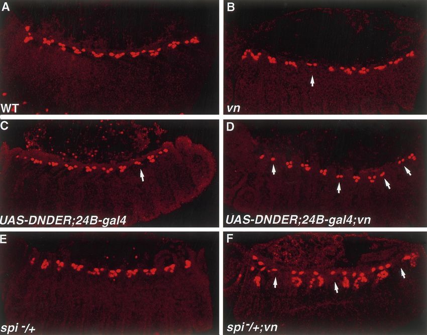
74                                   T. Yarnitzky et al. / Mechanisms of Development 79 (1998) 73–82

tor (DER/Egfr), or its ligand, Spitz, specific muscle founder           trant, but is significantly enhanced in embryos carrying only
cells, including the precursor of DA1 (dorsal acute muscle              one copy of wild type spitz, or in embryos in which the
no. 1), VA2 (ventral acute muscle no. 2) and LL1 (lateral               activity of DER is reduced by expressing a dominant-nega-
longitudinal muscle no. 1), are missing, leading to a loss of           tive form of the receptor. Ectopic expression of Vein in the
these specific muscle fibers. The loss of DA1 muscle foun-              mesoderm is sufficient to induce supernumerary DER-
der myoblast in DER or spitz mutant embryos results from a              dependent muscle founders in wild type, spitz, or rhomboid
defected determination of its specific muscle progenitor                mutant embryos. Unlike Spitz, Vein activation of DER does
P15. In addition, it was suggested that the development of              not lead to the expression of the inhibitory protein, Argos.
individual muscles is differentially sensitive to variations in         These results led us to suggest a model in which Vein is
the level of signaling by DER (Buff et al., 1998). DER is a             required to complement Spitz activity in the formation of
receptor tyrosine kinase, essential for the determination of a          specific muscle precursors by inducing a continuous activa-
wide array of cell fates during embryonic development (Ray              tion of the DER signaling pathway, following the activation
and Schupbach, 1996; Perrimon and Perkins, 1997;                        of this receptor by Spitz.
Schweitzer and Shilo, 1997). The regulation of the activity
of the DER-signaling cascade is thought to be accomplished
by localized release of its ligands, since DER is widely                2. Results
expressed in all germ layers throughout embryonic devel-
opment. Among DER ligands, Spitz, a TGFa homologue, is                  2.1. Vein contributes to the specification of the DER-
a major ligand active in a number of tissues (Rutledge et al.,          dependent muscle precursors
1992). Spitz is produced as a membrane-bound protein and
is processed to produce an extracellular, active form                      vein mRNA expression is detected in the ventral ecto-
(Schweitzer et al., 1995b). A function for Rhomboid in                  derm, somatic mesoderm, tracheal pits and chordotonal
the regulation of Spitz processing has been suggested                   organs (Schnepp et al., 1996). During, and following germ
(Golembo et al., 1996a).                                                band retraction, vein mRNA expression is observed in a
   Vein, an additional ligand for DER (Schnepp et al., 1996;            reiterated segmental pattern in a number of muscle precur-
Yarnitzky et al., 1997), is a secreted growth factor contain-           sor cells (Fig. 1A,B). Double labeling with anti-Vein and
ing Ig-like and EGF-like domains at its C-terminal. The                 anti-Krüppel antibodies reveals that the Krüppel-positive
embryonic expression of vein mRNA is dynamic and                        cells also express Vein (an overlap between Krüppel and
marks a number of tissues where the Spitz ligand was                    Vein staining in various muscle precursors is demonstrated
reported to be the active ligand. These tissues include the             in Fig. 1C–E).
ventral ectoderm (Schnepp et al., 1996), tracheal pits                     Buff et al. (1998), showed that development of the
(Schnepp et al., 1996), and somatic muscle precursors                   Krüppel-expressing muscle founders DA1, LL1 and VA2,
(Buff et al., 1998; and see also the present work). Despite             depends on DER activity. In the absence of Spitz, or in
its restricted embryonic expression pattern, no obvious phe-            embryos expressing dominant negative DER (DNDER),
notype in these tissues has been reported in vein mutant                these muscle founders are missing from all the segments.
embryos. During late stages of embryonic development,                   At stages 12–13 of embryonic development, the DA1 mus-
Vein is expressed in all the somatic muscles, and is required,          cle precursor is identified at the dorsal region of the meso-
independently of Spitz, for the muscle-dependent differen-              derm by the common expression of Eve and Krüppel in a
tiation of tendon cells (Yarnitzky et al., 1997).                       cluster of several nuclei, representing myoblasts fused to the
   Genetic evidence described in Schnepp et al. (1996) sug-             DA1 founder cell. The precursors of muscles LL1 and VA2
gests a functional link between both DER ligands. For                   express Krüppel, and can be identified by their relative loca-
example, reducing Vein levels in a spitz null genotype wor-             tions in an embryo at stages 12–13. In vein (vn) mutant
sened the phenotype to produce a collapsed embryo with an               embryos at stages 12–13 of embryonic development, the
extruded head skeleton. In addition, ventral denticles and              DA1 muscle precursor is missing in one (or occasionally
head skeleton defects in spitz;vein double mutants were                 two) segments of mutant embryos (figurehere>Fig. 2B,D).
more severe than the predicted additive phenotype of the                The penetrance of this phenotype is ~50% (out of 100
single mutants. This led to the hypothesis that both ligands            embryos scored). The Krüppel-positive muscle precursors
function in a synergistic manner in certain tissues. Recently,          LL1 and VA2 are also missing in vein mutant embryos in a
both Vein and Spitz were found to be required for specifica-            similar frequency as observed for DA1 (Fig. 2F). No altera-
tion of neuroblasts along the dorsal-ventral axis during early          tions in the severity of the phenotype were noticed in var-
embryonic development (Skeath, 1998).                                   ious vn heteroallelic combinations, including vnD25/
   In the present work, we show that Vein is required to                Def(3L)XAS96, or vnP1749/vndddL6. Thus, we concluded that
complement Spitz in the development of a subset of muscle               the loss of the various muscle precursor cells observed in
precursors whose differentiation depends on DER activity.               vein mutant embryos is in line with the expression of Vein
In vein mutant embryos these muscle precursors do not form              in these cells. In contrast to the phenotype observed in spitz
in some of the segments. This phenotype is not fully pene-              mutant embryos, in which all the DA1, LL1 and VA2 mus-
T. Yarnitzky et al. / Mechanisms of Development 79 (1998) 73–82                                             75

cle precursors are missing (Buff et al., 1998), the vn mutant                    precursor in embryos in which the activity of DER was
phenotype is mild, raising the question of the actual con-                       reduced by expressing a dominant-negative form of DER
tribution of Vein to that process.                                               (DNDER) in the mesoderm. This was achieved by inducing
   While the occasional loss of specific muscles is detected                     the expression of UAS-DNDER using the mesoderm-speci-
in vn mutant embryos at earlier developmental stages (e.g.                       fic 24B-gal4 line. In such embryos, grown at 22°C, the for-
stage 12), during late stages of embryonic development (e.g.                     mation of the DA1 muscle precursor (detected by Eve
stage 15–16), a significant number of the somatic muscle                         expression), is either normal or affected in not more than
fibers exhibit multiple, mis-guided membrane extensions at                       one segment (Fig. 3C). In embryos carrying both a vein
their leading edges (Fig. 2G). This phenotype is the conse-                      mutation (vnP1749) and a UAS-DNDER construct induced
quence of a defective differentiation of the tendon cells and                    by 24B-gal4 (at 22°C), the loss of DA1 muscle precursor
is not restricted to the specific muscles (e.g. DA1, VA2 and                     is enhanced and observed in three to six segments (Fig. 3D).
LL1) mentioned above (Yarnitzky et al., 1997).                                   In contrast to the 50% penetrance observed in the vein
                                                                                 mutants, this phenotype is fully penetrant.
2.2. The requirement for Vein is enhanced when activation                           To test more directly the model that Vein complements
of DER is reduced                                                                the activity of Spitz in the development of the DA1 muscle
                                                                                 precursor, we examined the formation of DA1 muscle pre-
   The relatively moderate contribution of Vein to the devel-                    cursor in vn mutant embryos carrying only one copy of wild
opment of the DER-dependent muscle precursor cells raises                        type spitz. While the DA1 muscle precursor is formed in all
the possibility that Vein complements or provides a backup                       the segments of embryos heterozygous for the spitz muta-
mechanism for the activity of Spitz in these cells. In that                      tion, the vn mutant embryos carrying only one copy of wild
case it is expected that reducing the activity of DER signal-                    type spitz, show significant enhancement in the penetrance
ing pathway in the muscle precursor cells during their for-                      (100% compare to 50%) of the phenotype (Fig. 3F). In
mation, may enhance the effect of loss of Vein. To test this                     addition, the number of affected segments was elevated
possibility we followed the formation of the DA1 muscle                          (three instead of one), as demonstrated in Fig. 3. Both

Fig. 1. vein is expressed in a subset of somatic muscle precursors. vein mRNA is detected in a subset of somatic muscle precursors during and following
embryonic germ band retraction (A,B). Double labeling with anti-Vein (C, red) and anti-Krüppel (D, green) antibodies, indicates that both proteins are co-
localized in an embryo at stage 12-13. Panel E represents the merged Vein/Krüppel image. The identity of the various muscles is indicated by arrowheads in
C–E.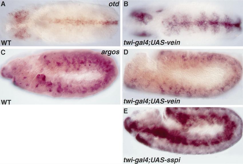
76                                          T. Yarnitzky et al. / Mechanisms of Development 79 (1998) 73–82

Fig. 2. A partial loss of the somatic muscle precursors is observed in vein mutant embryos. Wild type (A,C,E) and vein mutant (the embryos in B,D are
vndddL6/Df(3L)XAS96, the embryo in F is vnD25/Df(3L)XAS96, and the embryos in G,H are vnP1749) were stained with anti-Even-skipped (A,B), anti-Krüppel
(C–F), or anti-myosin heavy chain antibodies (G,H). At stage 12–13, a loss of DA1 muscle precursor, identified by its reactivity with anti Even-skipped (B),
or with anti Krüppel (D) antibodies, is observed in one segment of a vein mutant embryo (the defected segment is indicated by arrows in B and D). The
Krüppel positive muscle precursors LL1 and VA2 are also absent at a similar frequency in a vein mutant embryo (arrows in F). Note that in stage 15–16 (G,H)
various myotubes at the ventral aspects of the embryo exhibit mis-routed pattern (arrow in G). At the dorsal aspects of such embryo DA1 is missing in 2
segments (arrows in H), while DO1 is also missing in one segment, presumably due to abnormal guidance.

experiments support the model in which Vein activity com-                        sion of the active secreted Spitz ligand in the mesoderm
plements or provides a backup mechanism for the critical                         results in supernumerary muscle founder cells (Buff et al.,
contribution of Spitz to the formation of DA1 muscle pre-                        1998; and our unpublished results). To test the potential of
cursor.                                                                          Vein to induce the formation of supernumerary muscle
                                                                                 founder cells, we overexpressed Vein in the mesoderm.
2.3. Vein is sufficient to induce the formation of the DER-                      Such overexpression using the twist-gal4 inducer line,
dependent muscle founders                                                        results in the formation of supernumerary Even-skipped
                                                                                 positive clusters in the dorsal domain of the embryo (Fig.
  Vein appears to be a relatively weak EGF-receptor ligand                       4B), presumably representing overproduction of DA1 mus-
compared to Spitz (Schnepp et al., 1998). Ectopic expres-                        cle founder cells. Additional clusters of supernumerary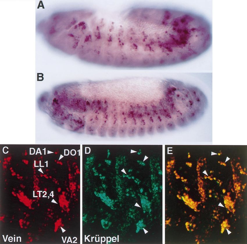
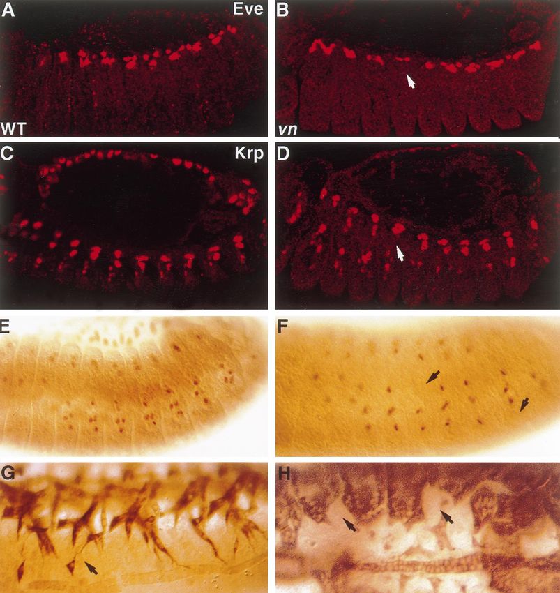
T. Yarnitzky et al. / Mechanisms of Development 79 (1998) 73–82                                            77

Fig. 3. vein mutant phenotype (B) is enhanced in embryos in which the activity of DER is reduced. The formation of DA1 muscle precursor (detected with
anti Even-skipped antibody) is slightly affected (about one segment is affected), in an embryo expressing a dominant-negative form of DER (UAS-DNDER),
in combination with the mesoderm-specific 24B-gal4 at 22°C (C). This phenotype is significantly enhanced (three to six segments are affected) in such
embryos (as in C), that are in addition, homozygous mutant for vein (vndddL6/Df(3L)XAS96 in D). A similar phenotypic enhancement is observed in vein
mutant embryos (vnP1749) heterozygous for a spitz (spiOE92) mutation (F), while in heterozygous spitz mutant embryos the DA1 muscle precursors are formed
in all segments (E). Arrows in B–D,F show the segments in which DA1 is missing. In each experiment at least 100 embryos were scored. Panels B,D,F show
a representative phenotype of a given experiment.

Krüppel positive cells (probably LL1 and VA2 muscle foun-                      2.4. Vein-mediated activation of the DER pathway does not
ders) were observed (Fig. 4D). This confirms that Vein is                       lead to the induction of Argos
capable of inducing the formation of DER-dependent mus-
cle founders. A similar result was obtained using the ecto-                        Schnepp et al. (1998) showed that the addition of Vein to
derm-specific gal4 line, krüppel-gal4, although the number                     S2 cultured cells leads to a relative lower levels of activated
of ectopic cells formed was lower (not shown). In addition,                     MAPK, compared to Spitz. We demonstrate that in vivo,
we observed that ectopic Eve-positive clusters are formed                       overexpression of Vein leads to lower levels of activated
not only in wild type embryos but also in spitz mutant                          MAPK as well. An equal number of embryos carrying either
embryos, or in rhomboid mutant embryos, when ectopic                            UAS-ssp (the secreted active form of Spitz; Schweitzer et
Vein is induced by the 69B-gal4 inducer line (Fig. 4F,G).                       al., 1995b) or UAS-vein, together with the heat-shock gal4
   Thus, although Vein appears to be a weak activator of the                    inducer (hs-gal4), were treated with heat shock and ana-
EGF-receptor pathway compared to Spitz, it is capable, like                     lyzed by Western for the levels of activated MAPK. The
Spitz, of inducing over production of DER-dependent mus-                        anti di-phospho MAPK antibody (anti dp-ERK; Gabay et
cle founder cells, even in the absence of functional Spitz.                     al., 1997) was utilized in the Western analysis. A represen-
These experiments also confirm the ability of Vein to act                       tative Western blot (of three independent repetitions)
non-autonomously on neighboring cells.                                          together with the calculated intensity ratio of the MAPK
78 T. Yarnitzky et al. / Mechanisms of Development 79 (1998) 73–82
Fig. 4. Ectopic expression of Vein induces supernumerary muscle founders. A higher number of Eve and/or Krüppel expressing cells is detected in embryos
expressing ectopic Vein, induced by twist-gal4 (B,D), or 69B-gal4 (E–G), compared to wild type embryos (A,C). The embryos were stained with anti Even-
skipped (A,B,E-G) or anti Krüppel antibodies (C,D). Supernumerary Eve- or Krüppel expressing cells are observed (arrows in B,D,E–F). Note that the effect
of ectopic Vein is detected not only in a wild type background but also in spitz (F) or rhomboid (G) mutant embryos. Although 69B-gal4 and twist-gal4
induce expression in distinct tissues (ectoderm or mesoderm respectively), no significant difference in the resulting phenotype of ectopic Vein was noted.
reactive band, normalized against the control band, is pre-
sented in Fig. 5. The level of dp-ERK induced in embryos
overexpressing Vein is lower relative to embryos expressing
sSpitz. Similar results were obtained when comparing addi-
tional independent transgenic lines carrying the UAS-vein or
UAS-sspitz constructs.
This observation, together with the results described by
Schnepp et al. (1998), supports the idea that Vein is a weak
activator of the DER-mediated signaling pathway. Lower
levels of activated MAPK may result in a distinct profile of
gene expression induced by Vein. Spitz activation of the
DER-pathway leads to positive transcriptional regulation
of argos, an inhibitor of the DER pathway (Schweitzer et Fig. 5. Spitz and Vein activate the EGF receptor pathway to different
levels. Representative Western blot analysis (of three independent repeti-
al., 1995a). Argos secretion from cells in which the DER tions), using anti di-phospho-ERK antibody, as detected in wild type (hs-
signaling pathway has been activated is essential to prevent gal4/ + ) embryos, embryos expressing ectopic Vein (hs-gal4;UAS-vein),
Spitz from affecting neighboring cells (Golembo et al., and embryos expressing ectopic Spitz (hs-gal4;UAS-sspitz) is shown. Each
1996b; Freeman, 1997). We wished to test the ability of lane represents protein extract made from 15 embryos, which were heat-
Vein to induce transcription of argos, when overexpressed shocked for 20 min, incubated for additional 3 h and subjected to Western
analysis. The intensity of the activated MAPK is presented in arbitrary
in the mesoderm. Ectopic expression of sSpitz by the twist- units (see materials and methods). Note that the level of activated MAPK
gal4 inducer leads to the ectopic expression of argos mRNA in embryos expressing sSpitz is significantly higher compared to embryos
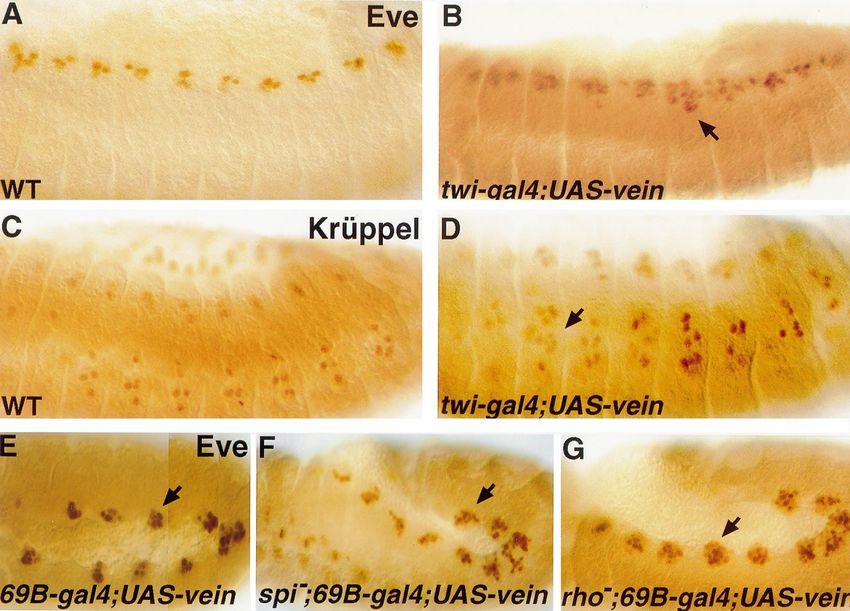
in the entire mesoderm (Fig. 6E). Unlike Spitz, the ectopic expressing Vein.T. Yarnitzky et al. / Mechanisms of Development 79 (1998) 73–82 79 Fig. 6. Ectopic Vein does not lead to ectopic expression of argos. Like Spitz, ectopic expression of Vein induced by twist-gal4 leads to the ectopic expression of otd in the ventral ectoderm (B). Compare otd expression in the embryo in panel B, to otd expression in a wild type embryo (A). In contrast to its effect on the expression of otd, no effect on argos mRNA expression is detected in embryos expressing ectopic Vein (D), utilizing the same gal4/UAS constructs, compare D to wild type argos expression in C. Unlike Vein, ectopic expression of sSpitz (using the same twist-gal4 inducer) leads to high levels of ectopic argos mRNA (E). expression of Vein, induced by the same twist-gal4 inducer, dependent activation, a prolonged activity of Vein is possi- does not lead to overexpression of argos mRNA in the ble only in case the latter is refractive to Argos. mesoderm (Fig. 6D). The transgenic line carrying the UAS-vein construct (used in this experiment) represents the strongest insertion line that we have obtained. Embryos 3. Discussion which are the progeny of this line, and in addition carry the twist-gal4 inducer (used above), show ectopic expression of The Drosophila EGF receptor, DER, mediates an array of orthodenticle (otd) in the ventral ectoderm (demonstrated in differentiation events during embryonic, larval and adult Fig. 6B). The mRNA expression of otd is observed in a row development (Ray and Schupbach, 1996; Perrimon and Per- of 2–4 cells wide along the midline at stage 10 of embryonic kins, 1997; Schweitzer and Shilo, 1997). An interplay development (Finkelstein et al., 1990; Kim and Crews, between the different DER ligands may result in alterations 1993). This expression is highly dependent on the activation at the level of receptor activation, or duration of the recep- of the DER-pathway in this region (Gabay et al., 1996). The tor-mediated signaling within the cell, contributing to dif- level of ectopic otd in the UAS-vein flies is indistinguishable ferent developmental consequences. In the present paper we of that observed when sSpitz is induced by the twist-gal4 show that coordinated activities of two DER ligands, Spitz line (Golembo et al., 1996a). We concluded that Vein acti- and Vein, are essential for the correct development of a vation of the DER pathway differs from that of Spitz with subset of embryonic somatic muscles. respect to the induction of Argos. While both Spitz and Vein activate the DER-signaling Assuming that the ectopic experiments reflect the in vivo pathway, their potential to activate this pathway differs sig- situation in the muscle founder cells, it appears that activa- nificantly. The levels of activated MAPK induced by the tion of DER by Vein in these cells may result in prolonged addition of secreted Spitz to DER-transfected S2 tissue cul- DER activation, since it is not inhibited by Argos. However, tured cells, are significantly higher compared to those since Argos protein may be present due to previous Spitz- induced by Vein (Schnepp et al., 1998). This is consistent
80                                    T. Yarnitzky et al. / Mechanisms of Development 79 (1998) 73–82

with our in vivo analysis of the levels of activated MAPK                mutant embryos and the role of Vein in these tissues is
induced by either secreted Spitz or Vein. The lower activa-              yet to be elucidated.
tion of DER by Vein may lead to qualitatively different
consequences in terms of gene expression. For example                    3.2. Vein functions sequentially to Spitz
we have shown that unlike Spitz, Vein overexpression
does not lead to Argos expression. A prolonged expression                   Vein appears to act sequentially to Spitz since vein
of Vein in the muscle founders, thus, may lead to a contin-              mRNA in the somatic mesodermal cells follows Rhomboid
uous activation of the EGF-receptor pathway in these cells.              expression in the progenitors (Buff et al., 1998). Rhomboid
   Since Vein is a secreted protein, while Spitz is produced             expression appears to represent the local activation of Spitz
as a membrane-bound, non-active DER ligand, distinct                     (Schweitzer et al., 1995b). The delayed expression of vein
mechanisms control their mode of DER activation. Regula-                 mRNA, is consistent with the suggestive function of Vein as
tion of Vein activity appears to be at the transcriptional               a ligand that complements Spitz activity. It is possible that
level, since its ectopic expression in various tissues (e.g.             Spitz activation of DER positively regulates the transcrip-
ectoderm or mesoderm) leads to measurable biological con-                tion of vein in the founder cells. In line with this possibility
sequences (Schnepp et al., 1998; and our results). Spitz                 is our observation that overexpression of Spitz leads to the
activity is controlled by the expression of Rhomboid or                  expression of Vein (Golembo et al., unpublished).
Star, presumably by its processing into an active secreted                  The nature of the inter-relationship between Vein and
form (Schweitzer et al., 1995b).                                         Argos is yet to be elucidated. Since activation of DER by
                                                                         Spitz leads to Argos expression (Golembo et al., 1996b), it
3.1. Different modes of Vein activity during embryonic                   may inhibit Vein activity. Vein activity in the muscle foun-
development                                                              ders suggests that these cells are either refractive to Argos or
                                                                         that Argos does not inhibit Vein activity.
   Although Vein is a less potent activator of DER, it func-                In summary, an interplay between the temporal expression
tions as a single ligand in certain tissues, such as in tendon           and differential activities of two EGF-receptor ligands des-
cell differentiation (Yarnitzky et al., 1997). In this tissue,           cribed in this work, may be essential for a robust receptor
the limited activation of DER by Vein may be compensated                 activation required to accomplish specific differentiation pro-
by concentrating it at the site of activity, i.e. at the muscle-         grams.
tendon junctional site. The accumulation of Vein at this site
may be also essential to restrict the number of affected cells.
   Spitz functions in a wide array of embryonic tissues                  4. Materials and methods
where Vein mRNA is also detected, e.g. the somatic meso-
derm, ventral mesectodermal cells, tracheal pits, and chor-              4.1. Fly stocks
dotonal organs. The biological contribution of Vein in
tissues where Spitz appears to be the major ligand (accord-                 The following gal4 inducers were used: 69B-gal4, twist-
ing to the resulting mutant phenotype), could be explained               gal4, 24B-gal4 (A. Brand, Cambridge, UK), krüppel-gal4
by two alternative mechanisms: Vein could act synergisti-                (M. Leptin, Köln, Germany), hs-gal4 (K25-2Xsev hsp70-
cally with Spitz, or, Vein may act sequentially to Spitz to              GAL4, obtained from E. Hafen, Zürich, Switzerland). In
provide a low, but continuous level of DER activation                    addition the following strains were used: y w (wild-type
which is required to backup, or complement Spitz activity.               strain); Df(3L)XAS96 (Dr. W.A. Johnson, Iowa, WI); spit-
Simultaneous expression of Vein together with sSpitz in                  zoe92 (N. Perrimon, MA, USA); veinD25 (created in our lab by
embryos, using heat-shock-gal4, did not lead to higher                   excision of the P-element in vnP1749), veindddL6 (A. Simcox,
levels of activated MAPK (our unpublished results). There-               OH, USA), vnP1749 (A.C. Spradling, MD, USA); UAS-
fore we do not favor the model of synergistic activation of              DNDER, UAS-Secreted-Spitz 17a; UAS-Secreted-Spitz 4b
DER by both ligands. To this end, our results support the                (B. Shilo, Rehovot, Israel). UAS-vein flies were produced
idea that the role of Vein in tissues where Spitz is the major           in our lab (Yarnitzky et al., 1997). The UAS-vein 110 inser-
ligand is to complement Spitz activity. This explains the                tion induces a high expression level and UAS-vein 98 repre-
extremely weak vein phenotype observed in these tissues,                 sents a weaker line. Mutant embryos were identified using
in comparison to a significant and measurable phenotype                  ‘blue balancers’ and anti-b-galactosidase staining.
obtained in tissues where Vein functions as a single ligand.
It is also consistent with the genetic interaction between               4.2. Immunochemical reagents
vein and spitz, described by Schnepp et al. (1996).
   vein mRNA is also observed in domains in which Spitz                    Vein was visualized either by in situ hybridization with a
or Rhomboid contribution has not yet been described, e.g.                digoxigenin-labeled 3.4 kb vein cDNA fragment, or by anti-
the amnioserosa, the dorsal most row of ectodermal cells                 body against GST-Vein fusion protein, raised in rats. Argos
during germ band retraction and head structures. No                      expression was monitored by in situ hybridization using
obvious phenotype is observed in these tissues in vein                   argos RNA as probe (M. Freeman, MRC, Cambridge). An
T. Yarnitzky et al. / Mechanisms of Development 79 (1998) 73–82 81
otd clone was obtained from R. Finklestein (University of BioRad MRC 1024 confocal microscope coupled to a
Pennsylvania, PA). Anti Krüppel antibody was obtained Zeiss Axiovert 135M microscope. Bright field and fluores-
from C. Rushlow (NYU, New York, NY) and anti Even- cent digital images were processed using Photoshop version
skipped antibody was obtained from M. Frasch (Mount 3.0 (Adobe Systems Inc., CA, USA).
Sinai Hospital, New York, NY). Anti-myosin heavy chain
(MHC) antibody was obtained from P. Fisher (Stony Brook,
NY). The level of activated MAPK was monitored by anti Acknowledgements
diphospho-ERK (Sigma). Anti-b-galactosidase antibodies
were purchased from Cappel (USA). We thank A. Brand, M. Leptin, N. Perrimon, E. Hafen, B.
Secondary antibodies included HRP, Fluorescein, Rhoda- Shilo, A. Simcox and W.A. Johnson for various fly strains;
min or Cy3-conjugated Goat or Donkey anti Rabbit or anti C. Rushlow, M. Frasch, M. Freeman, P. Fisher, and B. Shilo
Rat IgG (Jackson, USA), and anti-dig-AP antibody (Boeh- for antibodies and cDNA probes; B. Shilo, and E. Schejter
ringer Mannheim, Germany). for critical reading of the manuscript; B. Shilo, M. Golembo
and A. Michelson for fruitful discussions and suggestions.
4.3. Whole mount embryonic staining This work was supported by a grant from the Israel Science
Foundation (T.V.).
Staining was performed essentially as described (Ashbur-
ner, 1989). In brief, embryos were collected and incubated
as indicated, dechorionated and fixed in a mixture of 3% References
paraformaldehyde and heptane. Following two washes,
embryos were stained with X-gal staining solution, permea- Abmayr, S.M., Erickson, M.S., Bour, B.A., 1995. Embryonic development
bilized, and stained with primary antibody, and second anti- of the larval body wall musculature of Drosophila melanogaster. Trends
body. Embryos labeled with fluorescent antibody were Genet. 11, 153–159.
double-labeled with anti-b-galactosidase antibody to iden- Ashburner, M., 1989. Drosophila a Laboratory Manual. Cold Spring Har-
bor Laboratory Press, Cold Spring Harbor, NY, pp. 44–49.
tify mutant embryos in situ hybridization was performed by Bate, M., 1990. The embryonic development of larval muscles in
the method of Tautz and Pfeiffle (1989). Drosophila. Development 110, 791–804.
Stained embryos were examined under a Zeiss Axioscope Bate, M., 1993. The mesoderm and its derivatives. In: Bate, M., Martines
microscope. Arias, A. (Eds.), The Development of Drosophila Melanogaster. Cold
Spring Harbor Laboratory Press, Cold Spring Harbor, NY, pp. 1013–
1090.
4.4. Flat preparation of embryos
Baylies, M.K., Bate, M., 1996. twist: a myogenic switch in Drosophila.
Science 272, 1481–1484.
Flat preparations (used for fluorescent staining in Fig. 1), Baylies, M.K., Bate, M., Ruiz Gomez, M., 1998. Myogenesis: a view from
were prepared essentially according to Bate (1990); live Drosophila. Cell 93, 921–927.
embryos were dechorionated and the vitteline membrane Baylies, M.K., Martinez Arias, A., Bate, M., 1995. wingless is required for
was removed by hand. The embryos were opened and flat- the formation of a subset of muscle founder cells during Drosophila
tened on poly-l-Lysine covered coverslips, fixed with 3% embryogenesis. Development 121, 3829–3837.
Bourgouin, C., Lundgren, S.E., Thomas, J.B., 1992. apterous is a Droso-
paraformaldehyde in PBS, and stained. phila LIM domain gene required for the development of a subset of
embryonic muscles. Neuron 9, 549–561.
4.5. Western analysis Buff, E., Carmena, A., Gisselbrecht, S., Jimenez, F., Michelson, A.M.,
1998. Signalling by the Drosophila epidermal growth factor receptor
Embryos containing either UAS-vein or UAS-sspitz, in is required for the specification and diversification of embryonic muscle
addition to hs-gal4 inducer and also hs-gal4/ + embryos, progenitors. Development 125, 2075–2086.
were collected for 2 h, incubated for 3 h at 25°C, heat- Carmena, A., Bate, M., Jimenez, F., 1995. Lethal of scute, a proneural
gene, participates in the specification of muscle progenitors during Dro-
shocked at 37°C for 20 min, and returned to 29°C for 3 h.
sophila embryogenesis. Genes Dev. 9, 2373–2383.
Embryos were boiled in sample buffer and subjected to SDS- Carmena, A., Murugasu-Oei, B., Menon, D., Jimenez, F., Chia, W., 1998.
PAGE and Western analysis. HRP-conjugated-antibody was Inscuteable and numb mediate asymmetric muscle progenitor cell divi-
used and detected by Super signal substrate (Pierce). Each sions during Drosophila myogenesis. Genes Dev. 12, 304–315.
lane represents 15 embryos. The intensity of the bands was Dohrmann, C., Azpiazu, N., Frasch, M., 1990. A new Drosophila homeo-
box gene is expressed in mesodermal precursor cells of distinct muscles
analyzed using NIH Image version 1.55 (NIH, MD, USA).
during embryogenesis. Genes Dev. 4, 2098–2111.
The intensities of the MAPK reactive band and of a control Finkelstein, R., Smouse, D., Capaci, T.M., Spradling, A.C., Perrimon, N.,
band, were calculated for each cross, and the ratio between 1990. The orthodenticle gene encodes a novel homeo domain protein
these numbers was compared to the corresponding ratio of involved in the development of the Drosophila nervous system and
the control experiment (hs-gal4/ + embryos). ocellar visual structures. Genes Dev. 4, 1516–1527.
Frasch, M., 1995. Induction of visceral and cardiac mesoderm by ectoder-
mal Dpp in the early Drosophila embryo. Nature 374, 464–467.
4.6. Confocal microscopy Frasch, M., Hoey, T., Rushlow, C., Doyle, H., Levine, M., 1987. Char-
acterization and localization of the even-skipped protein of Drosophila.
Fluorescent labeled preparations were imaged using a EMBO J. 6, 749–759.82 T. Yarnitzky et al. / Mechanisms of Development 79 (1998) 73–82 Freeman, M., 1997. Cell determination strategies in the Drosophila eye. Ruiz Gomez, M., Bate, M., 1997. Segregation of myogenic lineages in Development 124, 261–279. Drosophila requires numb. Development 124, 4857–4866. Gabay, L., Scholz, H., Golembo, M., Klaes, A., Shilo, B.Z., Klambt, C., Ruiz Gomez, M., Romani, S., Hartmann, C., Jäckle, H., Bate, M., 1997. 1996. EGF receptor signaling induces pointed P1 transcription and inac- Specific muscle identities are regulated by Kruppel during Drosophila tivates Yan protein in the Drosophila embryonic ventral ectoderm. embryogenesis. Development 124, 3407–3414. Development 122, 3355–3362. Rushton, E., Drysdale, R., Abmayr, S.M., Michelson, A.M., Bate, M., Gabay, L., Seger, R., Shilo, B.Z., 1997. In situ activation pattern of Dro- 1995. Mutations in a novel gene, myoblast city, provide evidence in sophila EGF receptor pathway during development. Science 277, 1103– support of the founder cell hypothesis for Drosophila muscle 1106. development. Development 121, 1979–1988. Gaul, U., Seifert, E., Schuh, R., Jäckle, H., 1987. Analysis of Kruppel Rutledge, B.J., Zhang, K., Bier, E., Jan, Y.N., Perrimon, N., 1992. The protein distribution during early Drosophila development reveals post- Drosophila spitz gene encodes a putative EGF-like growth factor transcriptional regulation. Cell 50, 639–647. involved in dorsal-ventral axis formation and neurogenesis. Genes Golembo, M., Raz, E., Shilo, B.Z., 1996. The Drosophila embryonic mid- Dev. 6, 1503–1517. line is the site of Spitz processing, and induces activation of the EGF Schnepp, B., Donaldson, T., Grumbling, G., Ostrowski, S., Schweitzer, R., receptor in the ventral ectoderm. Development 122, 3363–3370. Shilo, B.Z., Simcox, A., 1998. EGF domain swap converts a Drosophila Golembo, M., Schweitzer, R., Freeman, M., Shilo, B.Z., 1996. Argos EGF receptor activator into an inhibitor. Genes Dev. 12, 908–913. transcription is induced by the Drosophila EGF receptor pathway to Schnepp, B., Grumbling, G., Donaldson, T., Simcox, A., 1996. Vein is a form an inhibitory feedback loop. Development 122, 223–230. novel component in the Drosophila epidermal growth factor receptor Hartenstein, A.Y., Rugendorff, A., Tepass, U., Hartenstein, V., 1992. The pathway with similarity to the neuregulins. Genes Dev. 10, 2302–2313. function of the neurogenic genes during epithelial development in the Schweitzer, R., Howes, R., Smith, R., Shilo, B.Z., Freeman, M., 1995. Drosophila embryo. Development 116, 1203–1220. Inhibition of Drosophila EGF receptor activation by the secreted protein Kim, S.H., Crews, S.T., 1993. Influence of Drosophila ventral epidermal Argos. Nature 376, 699–702. development by the CNS midline cells and spitz class genes. Schweitzer, R., Shaharabany, M., Seger, R., Shilo, B.Z., 1995. Secreted Development 118, 893–901. Spitz triggers the DER signaling pathway and is a limiting component in Lawrence, P.A., Bodmer, R., Vincent, J.P., 1995. Segmental patterning of embryonic ventral ectoderm determination. Genes Dev. 9, 1518–1529. heart precursors in Drosophila. Development 121, 4303–4308. Schweitzer, R., Shilo, B.Z., 1997. A thousand and one roles for Drosophila Michelson, A., Abmayr, S., Bate, M., Martinez Arias, A., Maniatis, T., EGF receptor. Trends Genet. 13, 191–196. 1990. Expression of a MyoD family member prefigures muscle pattern Skeath, J.B., 1998. The Drosophila EGF receptor controls the formation in Drosophila embryos. Genes Dev. 4, 2086–2097. and specification of neuroblasts along the dorsal-ventral axis of the Nose, A., Mahajan, V.B., Goodman, C.S., 1992. Connectin: a homophilic Drosophila embryo. Development 125, 3301–3312. cell adhesion molecule expressed on a subset of muscles and the moto- Staehling-Hampton, K., Hoffmann, F.M., Baylies, M.K., Rushton, E., neurons that innervate them in Drosophila. Cell 70, 553–567. Bate, M., 1994. dpp induces mesodermal gene expression in Park, M., Wu, X., Golden, K., Axelrod, J.D., Bodmer, R., 1996. The Drosophila. Nature 372, 783–786. wingless signaling pathway is directly involved in Drosophila heart Tautz, D., Pfeiffle, C., 1989. A nonradioactive in situ hybridization method development. Dev. Biol. 177, 104–116. for the localization of specific RNAs in Drosophila embryos reveals a Perrimon, N., Perkins, L.A., 1997. There must be 50 ways to rule the translational control of the segmentation gene hunchback. Chromosoma signal: the case of the Drosophila EGF receptor. Cell 89, 13–16. 98, 81–85. Ranganayakulu, G., Schulz, R.A., Olson, E.N., 1996. Wingless signaling Williams, J.A., Bell, J.B., Carroll, S.B., 1991. Control of Drosophila wing induces nautilus expression in the ventral mesoderm of the Drosophila and haltere development by the nuclear vestigial gene product. Genes embryo. Dev. Biol. 176, 143–148. Dev. 5, 2481–2495. Ray, R.P., Schupbach, T., 1996. Intercellular signaling and the polariza- Yarnitzky, T., Min, L., Volk, T., 1997. The Drosophila neuregulin homo- tion of body axes during Drosophila oogenesis. Genes Dev. 10, 1711– log Vein mediates inductive interactions between myotubes and their 1723. epidermal attachment cells. Genes Dev. 11, 2691–2700.
You can also read