Water Soluble Triarylborane Chromophores for One and Two Photon Excited Fluorescence Imaging of Mitochondria in Cells
←
→
Page content transcription
If your browser does not render page correctly, please read the page content below
Water‐Soluble Triarylborane Chromophores for One‐and Two‐Photon
Excited Fluorescence Imaging of Mitochondria in Cells
Griesbeck, S., Zhang, Z., Gutmann, M., Luehmann, T., Edkins, R. M., Clermont, G., Lazar, A. N., Haehnel, M.,
Edkins, K., Eichhorn, A., Blanchard-Desce, M., Meinel, L., & Marder, T. B. (2016). Water‐Soluble Triarylborane
Chromophores for One‐and Two‐Photon Excited Fluorescence Imaging of Mitochondria in Cells. Chemistry - A
European Journal, 22(41), 14701-14706. https://doi.org/10.1002/chem.201602639
Published in:
Chemistry - A European Journal

Document Version:
Peer reviewed version

Queen's University Belfast - Research Portal:
Link to publication record in Queen's University Belfast Research Portal

Publisher rights
Copyright 2016 Wiley-VCH Verlag GmbH &Co. KGaA. This work is made available online in accordance with the publisher’s policies. Please
refer to any applicable terms of use of the publisher.

General rights
Copyright for the publications made accessible via the Queen's University Belfast Research Portal is retained by the author(s) and / or other
copyright owners and it is a condition of accessing these publications that users recognise and abide by the legal requirements associated
with these rights.

Take down policy
The Research Portal is Queen's institutional repository that provides access to Queen's research output. Every effort has been made to
ensure that content in the Research Portal does not infringe any person's rights, or applicable UK laws. If you discover content in the
Research Portal that you believe breaches copyright or violates any law, please contact openaccess@qub.ac.uk.

Download date:09. Sep. 2021
FULL PAPER
Water-Soluble Triarylborane Chromophores for One- and Two-
Photon Excited Fluorescence Imaging of Mitochondria in Cells
Stefanie Griesbeck,[a] Zuolun Zhang,[a,b] Marcus Gutmann,[c] Tessa Lühmann,[c] Robert M. Edkins,[a,d]
Guillaume Clermont,[e] Adina N. Lazar,[e] Martin Haehnel,[a] Katharina Edkins,[a,f] Antonius Eichhorn,[a]
Mireille Blanchard-Desce,*[e] Lorenz Meinel,*[c] Todd B. Marder*[a]
Abstract: Three water-soluble tetracationic quadrupolar attack by nucleophilies, resulting in bond cleavage or the
chromophores comprising two three-coordinate boron -acceptor formation of a four-coordinate boron species, which inhibits the
groups bridged by thiophene-containing moieties were synthesised boron atom from being part of the delocalised -system. To
for biological imaging applications. The derivative 3 containing the prevent the attack of nucleophiles such as water, kinetic
bulkier 5-(3,5-Me2C6H2)-2,2′-(C4H2S)2-5′-(3,5-Me2C6H2) bridge is stabilisation can be achieved by introducing sterically demanding
stable over a long period of time, exhibits a high fluorescence substituents, such as mesityl (Mes) or 2,4,6-(CF3)3C6H2 (FMes),[2]
quantum yield and strong one- (OPA) and two-photon absorption to the boron atom, or by incorporation of the boron atom in a rigid,
(TPA), with a TPA cross-section of 268 GM at 800 nm in water. planar structure.[3] Only small anions, such as fluoride or cyanide,
Confocal laser scanning fluorescence microscopy studies in live cells are able to overcome the steric bulk and attack the boron centre,
indicate localisation of the chromophore at the mitochondria; which is exploited for anion-selective sensing.[4] Triarylboranes
moreover, cytotoxicity measurements prove biocompatibilty. Thus, are also used in organic light-emitting diodes (OLEDs) as
chromophore 3 has excellent potential for one- and two-photon electron-transporting, emitting and hole-blocking materials.[5]
excited fluorescence imaging of mitochondrial function in cells. The large change in dipole moment upon excitation of
compounds including a triarylborane moiety as an electron
acceptor results in large first and second-order molecular
hyperpolarisabilities and .[6] These interesting 2nd and 3rd order
Introduction non-linear optical properties indicate that triarylboranes should be
excellent components of chromophores that undergo two-photon
Three-coordinate organoboron compounds have aroused much absorption (TPA). Several octupolar and quadrupolar compounds
interest for various optical and electronic applications. [1] Due to its using this boron acceptor were reported and their TPA cross-
vacant pz-orbital, three-coordinate boron is a strong -electron sections (2) and fluorescence quantum yields (Φf) were
acceptor when connected to a conjugated -system. The trigonal measured to develop structure-TPA relationships.[7] In previous
planar geometry and Lewis acidity of the boron atom facilitate work, we reported that the insertion of thiophene rings into the -
bridge of A--A chromophores (A = boryl acceptor; here,
B(Mes)2) results in a remarkable increase of the TPA cross-
[a] S. Griesbeck, Dr. Z. Zhang, Dr. R. M. Edkins, Dr. M. Haehnel, Dr. K.
Edkins, A. Eichhorn, Prof. Dr. T. B. Marder, section, and synthesised a quadrupolar compound with a TPA
Institut für Anorganische Chemie cross-section of 1930 GM at 770 nm that is, as far as we know,
Julius-Maximilians-Universität Würzburg the highest 2/m.w. of all compounds containing B(Mes)2 and
Am Hubland, 97074 Würzburg (Germany)
thiophene groups reported to date.[8] Because the TPA maximum
E-mail: todd.marder@uni-wuerzburg.de
[b] Dr. Z. Zhang of each of these chromophores is located in the near-infrared
State Key Laboratory of Supramolecular Structure and Materials (NIR) biological transparent window, the reported chromophores
College of Chemistry, Jilin University are potentially good candidates for two-photon excited
Changchun 130012 (China)
fluorescence (TPEF) microscopy of living cells and tissues.
[c] M. Gutmann, Dr. T. Lühmann, Prof. Dr. L. Meinel
Institut für Pharmazie und Lebensmittelchemie However, these prototype compounds were not designed to be
Julius-Maximilians-Universität Würzburg water-soluble, posing formidable challenges for in vitro or in vivo
Am Hubland, 97074 Würzburg (Germany) application, and it is this important aspect that we develop in this
E-mail: lorenz.meinel@uni-wuerzburg.de
study while maintaining their aqueous stability and favourable
[d] Dr. R. M. Edkins
Chemistry Research Laboratory optical properties.
University of Oxford Only a few examples of water-soluble triarylboranes have
12 Mansfield Road, Oxford OX1 3TA (United Kingdom) been reported to date.[9] Gabbaï and co-workers achieved water-
[e] G. Clermont, A. N. Lazar, Dr. M. Blanchard-Desce
solubility by successively replacing the para-methyl groups of
Institut des Sciences Moléculaires
Univ. Bordeaux, ISM (CNRS UMR5255) trimesitylborane with cationic ammonium substituents, and used
Bâtiment A12, 351 cours de la libération, 33405 TALENCE cedex two such compounds as efficient cyanide sensors in water.[10]
(France) They and two other groups made use of a similar method for the
E-mail: mireille.blanchard-desce@u-bordeaux.fr
preparation of water-soluble triarylboranes with phosphonium
[f] Dr. K. Edkins
School of Medicine, Pharmacy and Health substituents for anion sensing.[11] Recently, a water-soluble,
Durham University, University Boulevard nonionic triarylborane, containing polyethylene glycol chains as
Stockton-on-Tees, TS17 6BH, (United Kingdom) the hydrophilic groups, was reported by Yang and co-workers as
Supporting information for this article is given via a link at the end of an efficient fluorescence indicator for ATP in the cytoplasm and
the document. cell membrane.[12] Furthermore, while our work was in progress,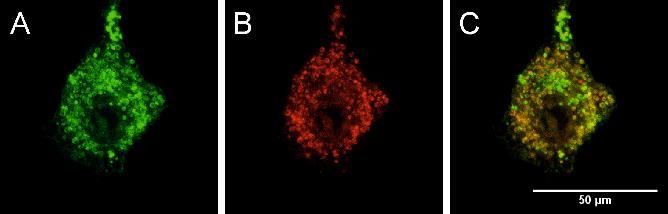
FULL PAPER
Scheme 1. Synthesis of the compounds 1-3. a) n-BuLi, THF, –78 °C to r.t.; b) CH2Cl2, r.t.; c) t-BuLi, THF, –78 °C to r.t.; d) B2pin2, [Ir(µ-OMe)(COD)]2, dtbpy, THF,
80 °C; e) Pd2(dba)3, SPhos, KOH, toluene, H2O, 85 °C; f) MeOTf, CH2Cl2/Et2O 1:3, r.t.
the same authors reported a water-soluble triarylborane cross section in water. Co-localisation and cytotoxicity studies
containing Cu(II)-cyclen. While non-fluorescent itself, it can serve show that dye 3 is an excellent candidate for the use as a new
as a one- and two-photon excited fluorescence turn-on probe for mitochondrial imaging agent.
H2S at mitochondria.[13]
For TPEF imaging in biological systems, we prepared
analogues of our previous quadrupolar compounds with Results and Discussion
trimethylammonium substituents for enhanced water-solubility.
These substituents are not just promising due to their hydrophilic Thiophene-boron directly connected chromophores 1 and 2
character, but are also expected to enhance the accumulation in
the mitochondria.[14] These features profile the molecules as Synthesis. The synthesis of the compounds 1 and 2 is
potential sensors for the mitochondrial membrane potential, summarised in Scheme 1. Compounds 5 and 6 were prepared via
providing direct information about the status of a cell’s power reaction of bis[4-(N,N-dimethylamino)-2,6-xylyl]fluoroborane 4[18]
plants.[15] Importantly, the use of such dyes, if amenable for TPA, with dilithiated bithiophene or quaterthiophene. Neutral
is potentially beyond in vitro use on single cells, populations of compounds 5 and 6 were methylated with methyl triflate in CH2Cl2,
cells, or united cell structures, but may very well expand into in and the products 1 and 2 precipitated in quantitative yield. Both
vivo applications by virtue of the aforementioned accessibility of compounds 1 and 2 were found to be water-soluble at
deeper cell layers and tissues for NIR light. Measurements of concentrations suitable for fluorescence microscopy (Table 1),
time-lapse acute mitochondrial responses to, e.g., drug exposure, especially noting that commercially available chromophores for
inducible gene knock-in/-out or exposure to other challenges mitochondrial imaging are generally dissolved in dimethyl
could provide immediate information on secondary respiratory sulfoxide (DMSO).
challenges to mitochondria, thereby providing on-the-fly read-out
of cell damage. Other potential applications include live cell Linear optical properties. The absorption and emission spectra
imaging of diseased vs. healthy tissue, e.g., to understand the of the methylated dyes 1 and 2 were measured in water (Fig. 1A)
underlying mechanisms of dynamic transport in and MeCN (Figs. S30 and S31, see ESI). The absorption spectra
neurodegenerative diseases such as glaucoma[16] or Alzheimer’s recorded in water exhibit a broad band at 426 nm for compound
disease.[17] 1, whereas an elongated quaterthiophene -system shifts the
In this paper, we present three quadrupolar chromophores, 1- absorption by ca. 30 nm to the red for chromophore 2. The
3, containing cationic triarylborane acceptors (Scheme 1). The - extinction coefficients, measured in MeCN, due to enhanced
bridge has been modified by the number of thiophene spacers stability (vide infra) and solubility, range from 28 000 to
and the nature of the aryl substituent adjacent to the boron atom. 48 000 M-1 cm-1 (Table 1). The emission spectra are broad, with
Their linear photophysical properties were examined maxima spread over a ca. 150 nm range for the different
experimentally and theoretically. With the water-stable derivative compounds. The smaller quadrupolar compound, 1, has an
3 we demonstrate herein one- and two-photon excited emission maximum in water at 451 nm, with a small Stokes shift
fluorescence imaging of the mitochondria in cells, due to its of 1 300 cm-1. By insertion of two more thiophene rings into the
remarkable fluorescence quantum yield and high two-photon bridging unit, the emission of 2 is bathochromicallyFULL PAPER
/ x 103 cm-1 / x 103 cm-1
Methylation of 9 was achieved with methyl triflate in a CH2Cl2/Et2O
A 35 30 25 20 15
B 34 32 30 28 26 24 22 20 18
1.0 1 1.0 start mixture giving a 76% isolated yield of 3. Compound 3 is air and
2 10 min
20 min moisture stable and can be stored on the bench as a solid at room
Normalized Intensity
0.8 0.8 30 min
Relative Intensity
1h temperature, in contrast to the commercially available
1 h 30 min
0.6 0.6
2h MitoTrackers, for which storage at less than –20 °C and exclusion
3h
0.4 0.4 4h of light are recommended in their instructions.[20] Chromophore 3
5h
0.2 0.2
6h
24 h
was also found to be water-soluble in the required concentration
96 h range.
0.0 0.0
300 350 400 450 500 550 600 650 700 750 800 300 350 400 450 500 550 600
/ nm / nm
Linear optical properties. The absorption maximum of 3 shifts
/ x 103 cm-1 / x 103 cm-1
34 32 30 28 26
C 1. (A) Absorption (solid) and emission
D 24 22 20 18 34 32 30 28 26 24 22 20 18 hypsochromically to 425 nm in water, compared to 2. This blue
Figure (dashed) spectra of 1 and 2 in
1.0
water.
HO
(B) Absorption spectrum of MeCN 1.0
1 in water as a function of time.
HO 2 2 shift can be explained by somewhat diminished electronic
MeCN
delocalisation of the planar -system to the boron pz-orbital, as
Normalized Absorption
Normalized Absorption
DMSO DMSO
0.8 MeOH 0.8 MeOH
EtOH EtOH shown in TD-DFT calculations (Figs. S53-S55), as a result of
0.6 0.6
shifted by more than 100 nm, resulting in a much larger Stokes increased twisting in the ground state due to the increased steric
0.4 0.4
shift. The fluorescence quantum yields for 1 and 2 in water are hindrance at the boron centre. The emission maximum is not
remarkably
0.2
large at 0.19 and 0.20, 0.2respectively, whereas they are affected as much as the absorption maximum; hence, the Stokes
even larger
0.0 in MeCN, both being 0.41.
0.0 The fluorescence lifetimes shift is further increased to 6 000 cm-1. The fluorescence quantum
300 350 400 450 500 550 600 300 350 400 450 500 550 600
are relatively short /being
nm 1.2 and 0.7 ns in water, respectively,
/ nm and yield of 3 in MeCN of 0.41 is the same as those of 1 and 2,
are similar in MeCN. whereas in water it is 0.10 which, while lower than the other two
UV-vis absorption and emission spectra monitored over chromophores, is still remarkable. This decrease is a result of the
extended time periods demonstrate slow degradation of 1 and 2 ca. ten-times higher non-radiative decay rate in H2O compared to
in water as shown in Figs. 1B, S33, S38 and S39. Mass MeCN. While there is almost no difference between these two
spectrometry of the degradation product confirmed hydrolysis at solvents for compounds 1 and 2, the lifetime is shortened
the boron centre. We note that light and oxygen speeds up the drastically from 1.9 ns to 300 ps for 3 with increasing solvent
decomposition of 1 and 2 (Figs. S34-S37, S40-41 and S48). polarity. Chromophore 3 shows almost no solvatochromism in its
absorption spectra
Water-stable chromophore 3 / x 103 cm-1 / x 103 cm-1
32 28 24 20 16 34 32 30 28 26 24 22 20 18
A B
H2O start
1.0 1.0
Synthesis. To improve the water-stability of the chromophore for MeCN 1h
2h
DMSO
Normalized Intensity
bioimaging applications, we synthesised compound 3 in which the 0.8 MeOH 0.8 3h
Relative Intensity
EtOH 8h
additional two ortho-methyl groups provide significantly enhanced 0.6 0.6
24 h
48 h
steric protection at the boron centre. Therefore, 4 was reacted 0.4 0.4
72 h
with 2-lithio-m-xylene giving bis[4-(N,N-dimethylamino)-2,6-xylyl]-
0.2 0.2
2,6-xylylborane, 7 in 63% isolated yield (Scheme 1). For use in
Suzuki-Miyaura reactions, the para-position of this borane was 0.0
300 350 400 450 500 550 600 650 700 750 800
0.0
300 350 400 450 500 550 600
/ nm / nm
borylated in an iridium-catalysed CH-activation reaction in 91%
isolated yield.[19] Borylated species 8 was coupled with 5,5′-
dibromo-2,2′-bithiophene to prepare the neutral precursor 9 in Figure 2. (A) Absorption (solid) and emission (dashed) spectra of 3 in different
solvents. (B) Absorption spectra of 3 in water as a function of time.
82% isolated yield, using Pd2(dba)3 as the catalyst precursor,
SPhos as the ligand and potassium hydroxide as the base.
Table 1. Photophysical data for chromophores 1, 2, 3 and MitoTracker Red CMXRos (MTRC).
Stokes
Solubility abs ε em kr knr 2OPA TPAmax σ2maxΦ σ2max
Solvent shift f
/ mM / nm / M-1 cm-1 / nm / ns / 108 s / 108 s / ns / nm / nm / GM / GM
/ cm-1
H2O 5.0 426 - 451 1 300 0.19 1.2 1.6 6.8 6.3 - - - -
1
MeCN - 426 28 000 448 1 200 0.41 1.7 2.3 3.4 4.2 - - - -
H2O 0.5 458 - 583 4 700 0.20 0.7 2.8 11.4 3.5 - - - -
2
MeCN - 467 48 000 555 3 400 0.41 0.7 5.9 8.5 1.7 - - - -
800 27 268
H2O 1.0 425 - 570 6 000 0.10 0.3 3.3 30 3.0 850
870 6 58
3
800 285 693
MeCN - 428 51 000 554 5 300 0.41 1.9 2.1 3.1 4.6 856
870 58 140
MitoTracker
Red H2O 1.0 577 - 602 720 - - - - - - - - -
CMXRosFULL PAPER
(Fig. 2A and Table S2). A progressive blue-shift of the emission molecules.[22] The lowest-excited state is, however, slightly OPA
is observed on going from water to (polar aprotic or protic) organic allowed (as indicated by the shoulder between 850 and 900 nm),
solvents of decreasing polarity. UV-vis measurements over 72 h most probably due to conformational freedom responsible for
clearly demonstrate that the increased steric protection provided slight deviations from ideal centrosymmetry.
by the additional methyl groups in 3 dramatically enhances It was not possible to determine the actual maximum of the
stability in water (Fig. 2B). Furthermore, 3 is more stable in water TPA (Fig. 3), which is calculated to be at 792 nm (Table S8), as
than the commercially available imaging chromophore we have not measured beyond 800 nm, but at 800 nm, we
MitoTracker Red CMXRos (Figs. S46-S47). observe a very large TPA cross section of 268 GM in H2O, which
is increased to 693 GM in MeCN (Fig. S49). This value is more
DFT calculations than five times higher than that reported recently for the only other
example of a triarylborane mitochondrial imaging chromophore
Density Functional Theory (DFT) calculations were carried out in (120 GM at 765 nm in DMSO).[13] Due to the sizeable fluorescence
order to examine the effects of the linker groups on the dihedral quantum yields, relatively large values of the two-photon
angles around the boron centre and its influence on conjugation. brightness (27 and 285 GM in water and MeCN, respectively)
The geometry of each of the compounds 1-3 was optimised using have been measured, making dye 3 of much promise for two-
DFT (B3LYP/6-31G*, gas phase). The structures all show the photon imaging in tissues.
expected propeller arrangement of the aryl groups about the
rigorously trigonal planar boron centre. The exchange of the / nm
400 420 440 460 480 500 520
thiendiyl linkers at the boron atom in 1 and 2 for xylylene groups 160
250
in 3, leads to an increased twist of the aryl group with respect to 140
120
the BC3 plane (1: 16.2°; 2: 13.7°; and 3: 43.2°). This reduced 200
/ x 103 M-1 cm-1
100
conjugation with the boron atom leads to the LUMO of 3 being
2 / GM
150
80
0.37 eV higher in energy than that of 2. The -bridge backbone 60
100
remains relatively planar in all cases (inter-ring dihedral angles 40
50
1:14.3°; 2: 1.5 and 10.3°; and 3: 18.9 and 12.4°). 20
Time Dependent-DFT (TD-DFT) calculations were carried 0 0
800 850 900 950 1000
out in order to obtain information on the nature of the optical TPA / nm
transitions, and to compute the expected UV-vis absorption
spectra of the compounds and compare this with the experimental
spectra. TD-DFT calculations excellently reproduced the Figure 3. One-photon absorption (OPA) (black) and two-photon absorption
(TPA) spectra (red) of 3 in H2O.
experimental absorption maxima of the lowest energy bands, the
deviation in energy being within 0.02-0.15 eV in simulated MeCN
solution. Full details of these results and those in the gas phase
Cell cytotoxicity and live cell imaging
are presented in the ESI (Figs. S53-S55). Introduction of the
xylylene groups in 3 leads to a hypsochromically shifted In light of the above photophysical properties and water-stability
absorption spectrum relative to 2, in line with the experimental of 3, and thus its potential as a chromophore for live cell imaging,
spectra. As seen in the natural transition orbitals (NTOs) (Figs. we next examined its possible cytotoxicity in cells. Therefore, we
S53-S55) the S1←S0 transitions of all three compounds contain a exposed three different cell lines - murine-fibroblasts (NIH 3T3),
significant contribution from population of the empty boron pz- human embryonal kidney (HEK 293T), and human-hepatic origin
orbital, albeit that the transitions are predominantly -*. The (HepG2-16) - to serial dilutions of 3 and also LiOTf and studied
boron acceptor thus increases the conjugation length of the - the cell metabolic activity with a colorimetric (WST-1) assay (Figs.
system. For TPA and TPEF applications, we also considered the 4 and S50-S52). These experiments confirmed that compound 3
S2←S0 transition, as this is the lowest energy allowed TPA did not influence the cell viability at concentrations as high as
transition in a quadrupolar chromophore; thus, the NTOs for these 10 µM. We have also checked the triflate counterion, as its lithium
transitions are plotted in Figs. S53-S55. salt, for cytotoxicity and found that it showed no toxicity up to
100 µM (for cytotoxicity results of 1 and 2, see ESI Figs. S50-S52).
Two-photon absorption We therefore suggest that compound 3 can be safely used in live
cell imaging applications and that this class of compounds shows
Table 1 summarises the results of TPA measurements of
potential for the development of in vivo diagnostics to probe
chromophore 3 by using a two-photon excited fluorescence
mitochondrial function.
method.[21] Due to the quantum selection rules for
centrosymmetric molecules, the TPA maximum does not occur at
the doubled wavelength of the one-photon absorption (OPA)
maximum, but is located at a shorter wavelength. Indeed, as
observed in Fig. 3, in which the TPA and rescaled OPA are
compared, the maximum TPA is observed at higher energy,
corresponding to an excited state which is not one-photon allowed.
This is in agreement with the typical behavior of quadrupolarFULL PAPER
24 h 24 h
A B
140 120
living cells vs. control [%]
120
living cells vs. control [%]
100
100
80
80
60
60
40
40
20 20 As clearly shown in Fig. 6, fluorescence images under standard
0 0 one-photon and two-photon excitation show the same localisation
control 100nM 1µM 10µM 100µM control 100nM 1µM 10µM 100µM
of the dye. Furthermore, emission spectra of the uptaken dyes
were acquired demonstrating that the dye structure is retained
Figure 4. The metabolic activity of NIH 3T3 cells was measured after 24 h with upon internalisation in the cells. Hence, the steric protection
serial dilution of 3 (A) and LiOTf (B) using a colorimetric assay. The results show
the relative cell viability as a percentage of the untreated control (white bars) +
around the empty pz-orbital at the boron atom not only confers
the standard deviation. stability of dye 3 in pure water, but is also sufficient to make it
stable in a cellular environment. The blue-shifted emission
compared to that observed in pure water can be related to
Thus, living mouse embryo fibroblast cells (NIH 3T3) were environmental effects, suggesting that the dye is located in a less
treated with a 10 µM concentration of chromophore 3. polar environment.
Visualisation by confocal laser scanning fluorescence microscopy
showed that 3 penetrated the cell membrane of living cells and
localised at the mitochondria as confirmed by co-localisation Conclusions
experiments with the commercial mitochondrial imaging agent
MitoTracker Red CMXRos (Fig. 5). In conclusion, three water-soluble quadrupolar chromophores
with triarylborane acceptors were synthesised. The two
Figure 5. Confocal microscope live cell image of murine NIH 3T3 fibroblast cells compounds 1 and 2 are bright emitters in water, but were shown
with (A) 10 µM of dye 3 and (B) 125 nM of MitoTracker Red CMXRos. (C) Merged
to decompose, due to hydrolysis at the boron centre. In contrast,
image indicating the co-localisation of compound 3 with mitochondria.
3 has enough steric protection around the empty pz-orbital at the
boron atom that it is sufficiently stable in water. We have proved
Figure 6. Confocal microscope image of POS 1 cells after 8 h of incubation with as well that 3 localises in mitochondria by co-localisation
dye 3 (300 nm in the media): merge of transmission image (in grey) and experiments, and that our new chromophore is not toxic at
fluorescence image (green) showing the internalisation of the dye within the concentrations suitable for imaging purposes. We have shown
cells: (A) one-photon excitation (ex = 405nm;em = 500-600 nm) and (B) two-
that 3 is more stable than the commercial available MitoTracker
photon excitation (ex = 800 nm ; em = 500-600 nm); (C) emission spectrum
upon one-photon excitation (ex = 405nm; 20 nm step) of the dye within the Red CMXRos, while the solubility in water remains the same. We
cell; (D) emission spectrum upon two-photon excitation (ex = 800 nm; 20 nm report here the first TPA cross-section measurement of a
step) of the dye within the cell. triarylborane in water, being 268 GM at 800 nm, which is very
large; hence, 3 is suitable for two-photon live cell microscopy. We
have reported herein a three-coordinate boron-containing
chromophore for mitochondrial TPEF imaging, profiling this
compound as a water-soluble, biocompatible mitochondrial
tracker for in vitro live cell imaging applications. Future application
as a diagnostic tool for clinical use should, in spite of the
promising data set obtained with respect to (cellular)
biocompatibility, be re-assessed in light of the outcome from
robust (pre-)clinical safety studies. Optimisation of such
chromophores to enhance quantum yields and TPA cross-
sections and to tune emission wavelengths is currently in
progress.
Acknowledgements
T. B. M. is grateful for generous financial support from the
Based on the sizeable two-photon brightness of dye 3 in water, Bavarian State Ministry of Science, Research, and the Arts for the
we also tested 3 as a two-photon dye to stain POS 1 cells - a cell Collaborative Research Network "Solar Technologies go Hybrid".
line derived from an osteosarcoma tumour. The two-photon Z.Z. and R.M.E. thank the Alexander von Humboldt Foundation
imaging experiments (and parallel confocal imaging under one- for Postdoctoral Research Fellowships. R.M.E also thanks The
photon excitation) were performed using a 300 n M concentration. Royal Commission for the Exhibition of 1851 for a researchFULL PAPER
fellowship. M. B. D. is grateful for generous funding from the S. Yuan, Z.-Q. Liu, Y.-X. Shen, H.-F. Chen, Org. Lett. 2010, 12, 5192-
Région Aquitaine (chair of excellence). This study has also been 5195; f) Y. Chen, G.-Q. Liu, Y.-Y. Wang, P. Yu, Z. Liu, Q. Fang, Synth.
Met. 2012, 162, 291-295; g) N. S. Makarov, S. Mukhopadhyay, K.
carried out with financial support from the French State, managed
Yesudas, J.-L. Brédas, J. W. Perry, A. Pron, M. Kivala, K. Müllen, J. Phys.
by the French National Research Agency (ANR) in the frame of Chem A 2012, 116, 3781-3793; h) M. Charlot, L. Porrès, C. D. Entwistle,
“the Investments for the future” Programme IdEx Bordeaux – A. Beeby, T. B. Marder, M. Blanchard-Desce, Phys. Chem. Chem. Phys.
LAPHIA (ANR-10-IDEX-03-02). The two-photon microscopy was 2005, 7, 600-606; i) C. D. Entwistle, J. C. Collings, A. Steffen, L.-O.
carried out at the Bordeaux Imaging Center, a service unit of the Pålsson, A. Beeby, D. Albesa-Jové, J. M. Burke, A. S. Batsanov, J. A. K.
CNRS-INSERM and Bordeaux University, a member of the Howard, J. A. Mosely, S.-Y. Poon, W.-Y. Wong, F. Ibersiene, S. Fathallah,
A. Boucekkine, J.-F. Halet, T. B. Marder, J. Mater. Chem. 2009, 19,
national infrastructure France BioImaging. Financial support by
7532-7544; j) J. C. Collings, S.-Y. Poon, C. Le Droumaguet, M. Charlot,
Deutsche Forschungs-gemeinschaft (DFG; ME - 3820/3-1) is
C. Katan, L.-O. Pålsson, A. Beeby, J. A. Mosely, H. M. Kaiser, D.
gratefully acknowledged by L. M. Kaufmann, W.-Y. Wong, M. Blanchard-Desce, T. B. Marder, Chem. Eur.
J. 2009, 15, 198-208.
Keywords: borane • luminescence • two-photon excited [8] L. Ji, R. M. Edkins, L. J. Sewell, A. Beeby, A. S. Batsanov, K. Fucke, M.
fluorescence • mitochondrion • cell imaging Drafz, J. A. K. Howard, O. Moutounet, F. Ibersiene, A. Boucekkine, E.
Furet, Z. Liu, J.-F. Halet, C. Katan, T. B. Marder, Chem. Eur. J. 2014, 20,
13618-13635.
[1] a) C. D. Entwistle, T. B. Marder, Angew. Chem. Int. Ed. 2002, 41, 2927-
[9] a) H. Zhao, L. A. Leamer, F. P. Gabbaï, Dalton Trans. 2013, 42, 8164-
2931; b) C. D. Entwistle, T. B. Marder, Chem. Mater. 2004, 16, 4574-
8178; b) T. W. Hudnall, F. P. Gabbaï, J. Am. Chem. Soc. 2007, 129,
4585; c) S. Yamaguchi, A. Wakamiya, Pure Appl. Chem. 2006, 78, 1413-
11978-11986.
1424; d) F. Jäkle, Coord. Chem. Rev. 2006, 250, 1107-1121; e) F. Jäkle,
[10] C.-W. Chiu, Y. Kim, F. P. Gabbaï, J. Am. Chem. Soc. 2009, 131, 60-61.
Chem. Rev. 2010, 110, 3985-4022; f) Z. M. Hudson, S. Wang, Dalton
[11] a) Y. Kim, F. P. Gabbaï, J. Am. Chem. Soc. 2009, 131, 3363-3369; b) T.
Trans. 2011, 40, 7805-7816.
Agou, M. Sekine, J. Kobayashi, T. Kawashima, Chem. Eur. J. 2009, 15,
[2] Z. Zhang, R. M. Edkins, J. Nitsch, K. Fucke, A. Steffen, L. E. Longobardi,
5056-5062; c) K. C. Song, K. M. Lee, N. V. Nghia, W. Y. Sung, Y. Do, M.
D. W. Stephan, C. Lambert, T. B. Marder, Chem. Sci. 2015, 6, 308-321.
H. Lee, Organometallics 2013, 32, 817-823.
[3] Z. Zhou, A. Wakamiya, T. Kushida, S. Yamaguchi, J. Am. Chem. Soc.
[12] X. Li, X. Guo, L. Cao, Z. Xun, S. Wang, S. Li, Y. Li, G. Yang, Angew.
2012, 134, 4529-4532.
Chem. Int. Ed. 2014, 53, 7809-7813.
[4] a) T. W. Hudnall, C.-W. Chiu, F. P. Gabbaï, Acc. Chem. Res. 2009, 42,
[13] J. Liu, X. Guo, R. Hu, X. Liu, S. Wang, S. Li, Y. Li, G. Yang, Anal. Chem.
388-397; b) Z. M. Hudson, S. Wang, Acc. Chem. Res. 2009, 42, 1584-
2016, 88, 1052-1057.
1596; c) C. R. Wade, A. E. J. Broomsgrove, S. Aldridge, F. P. Gabbaï,
[14] a) X. Zhao, Y. Li, D. Jin, Y. Xing, X. Yan, L. Chen, Chem. Commun. 2015,
Chem. Rev. 2010, 110, 3958-3984; d) E. Galbraith, T. D. James, Chem.
51, 11721-11724; b) M. Grzybowski, E. Glodkowska-Mrowka, V. Hugues,
Soc. Rev. 2010, 39, 3831-3842.
W. Brutkowski, M. Blanchard-Desce, D. T. Gryko, Chem. Eur. J. 2015,
[5] a) T. Noda, Y. Shirota, J. Am. Chem. Soc. 1998, 120, 9714-9715; b) Y.
21, 9101-9110; c) N. Jiang, J. Fan, F. Xu, X. Peng, H. Mu, J. Wang, X.
Shirota, H. Kageyama, Chem. Rev. 2007, 107, 953-1010; c) F. Li, W. Jia,
Xiong, Angew. Chem. Int. Ed. 2015, 54, 2510-2514; d) M. Homma, Y.
S. Wang, Y. Zhao, Z.-H. Lu, J. Appl. Phys. 2008, 103, 034509-1-034509-
Takei, A. Murata, T. Inoue, S. Takeoka, Chem. Commun. 2015, 51,
6; d) C. Sun, Z. M. Hudson, M. G. Helander, Z.-H. Lu, S. Wang,
6194-6197.
Organometallics 2011, 30, 5552-5555; e) A. Shuto, T. Kushida, T.
[15] S. W. Perry, J. P. Norman, J. Barbieri, E. B. Brown, H. A. Gelbard,
Fukushima, H. Kaji, S. Yamaguchi, Org. Lett. 2013, 15, 6234-6237.
BioTechniques 2011, 50, 98-115.
[6] a) Z. Yuan, N. J. Taylor, T. B. Marder, I. D. Williams, S. K. Kurtz, L.-T.
[16] Y. Takihara, M. Inatani, K. Eto, T. Inoue, A. Kreymerman, S. Miyake, S.
Cheng, J. Chem. Soc., Chem. Commun. 1990, 21, 1489-1492; b) Z.
Ueno, M. Nagaya, A. Nakanishi, K. Iwao, Y. Takamura, H. Sakamoto, K.
Yuan, N. J. Taylor, R. Ramachandran, T. B. Marder, Appl. Organomet.
Satoh, M. Kondo, T. Sakamoto, J. L. Goldberg, J. Nabekura, H. Tanihara,
Chem. 1996, 10, 305-316; c) C. Branger, M. Lequan, R. M. Lequan, M.
Proc. Natl. Acad. Sci. USA 2015, 112, 10515-10520.
Barzoukas, A. Fort, J. Mater. Chem. 1996, 6, 555-558; d) Z. Yuan, C. D.
[17] Z.-H. Sheng, Q. Cai, Nat. Rev. Neurosci. 2012, 13, 77-93.
Entwistle, J. C. Collings, D. Albesa-Jové, A. S. Batsanov, J. A. K. Howard,
[18] C.-W. Chiu, F. P. Gabbaï, Organometallics 2008, 27, 1657-1659.
N. J. Taylor, H. M. Kaiser, D. E. Kaufmann, S.-Y. Poon, W.-Y. Wong, C.
[19] I. A. I. Mkhalid, J. H. Barnard, T. B. Marder, J. M. Murphy, J. F. Hartwig,
Jardin, S. Fathallah, A. Boucekkine, J.-F. Halet, T. B. Marder, Chem. Eur.
Chem. Rev. 2010, 110, 890-931.
J. 2006, 12, 2758-2771.
[20] Molecular Probes, Inc., WP 07510, Eugene, 2008.
[7] a) Z.-Q. Liu, Q. Fang, D. Wang, G. Xue, W.-T. Yu, Z.-S. Shao, M.-H.
[21] a) C. Xu, W. W. Webb, J. Opt. Soc. Am. B 1996, 13, 481-491; b) M. A.
Jiang, Chem. Commun. 2002, 23, 2900-2901; b) Z.-Q. Liu, Q. Fang, D.
Albota, C. Xu, W. W. Webb, Appl. Opt. 1998, 37, 7352-7356.
Wang, D.-X. Cao, G. Xue, W.-T. Yu, H. Lei, Chem. Eur. J. 2003, 9, 5074-
[22] M. Barzoukas, M. Blanchard-Desce, J. Chem. Phys. 2000, 113, 3951-
5084; c) Z.-Q. Liu, Q. Fang, D.-X. Cao, D. Wang, G.-B. Xu, Org. Lett.
3959.
2004, 6, 2933-2936; d) Z.-Q. Liu, M. Shi, F.-Y. Li, Q. Fang, Z.-H. Chen,
T. Yi, C.-H. Huang, Org. Lett. 2005, 7, 5481-5484; e) L. Ji, Q. Fang, M.-FULL PAPER
Entry for the Table of Contents (Please choose one layout)
Layout 1:
FULL PAPER
Text for Table of Contents Author(s), Corresponding Author(s)*
Page No. – Page No.
Title
((Insert TOC Graphic here: max.
width: 5.5 cm; max. height: 5.0 cm))
Layout 2:
FULL PAPER
Stefanie Griesbeck, Zuolun Zhang,
Marcus Gutmann, Tessa Lühmann,
Robert M. Edkins, Guillaume Clermont,
Adina N. Lazar, Martin Haehnel,
Katharina Edkins, Antonius Eichhorn,
Mireille Blanchard-Desce,* Lorenz
Meinel,* Todd B. Marder*
Very positive image of boron: A new tetracationic quadrupolar 3-coordinate boron
compound is water-soluble and stable, and has a large two-photon absorption cross Page No. – Page No.
section and fluorescence quantum yield, even in water. It localises at mitochondria
Water-Soluble Triarylborane
and is non-toxic to cells at concentrations required for one- and two-photon excited
Chromophores for One- and Two-
fluorescence imaging of mitochondrial function.
Photon Excited Fluorescence
Imaging of Mitochondria in CellsYou can also read