Pathogenicity Potential of the Bacteria in Selected Locations of Rapid Creek
←
→
Page content transcription
If your browser does not render page correctly, please read the page content below
Pathogenicity Potential of the Bacteria in Selected Locations of Rapid Creek
         Murray, Kelsey E.1, Erickson, Molly2, Schaefer, Morgan2, Kunza, Lisa. A.1,2,3,
                                    DeVeaux, Linda. C.1,2

                            South Dakota School of Mines and Technology
                                    1
                                      Biomedical Engineering Program
                        2
                         Department of Chemistry and Applied Biological Sciences
                          3
                            Program of Atmospheric and Environmental Sciences

1.1 Introduction

Bacterial levels, particularly fecal coliforms such as E. coli, are standard water quality indicators
of fecal contamination. The EPA estimates 25% of national groundwater systems exceed the Total
Coliform Rule, suggesting widespread dissemination of enteric organisms into the environment
[1, 2]. South Dakota surface water is not immune to fecal contamination, and according to the
2016 South Dakota Integrated Report for Surface Water Quality Assessment, sections of Rapid
Creek are affected by unacceptably high levels of indicator bacteria, particularly E. coli [3].

Certain E. coli strains are a normal inhabitant of the human intestinal tract, and when ingested do
not cause disease, while other strains are highly pathogenic to humans, causing severe and
potentially fatal illnesses. Pathogenic strains are considered emerging zoonotic infectious agents,
and have been linked to outbreaks of severe diarrhea and hemolytic uremic syndrome, an often
lethal condition in children. Infection by pathogenic variants, predominantly Shiga-toxigenic E.
coli serotype O157:H7, accounts for approximately 73,000 annual cases of reported illnesses in
the U.S. alone [4]. Although disease-causing in humans, many pathogenic E. coli strains are
common inhabitants of the guts of both domestic and wild animals, particularly ruminants, where
they appear to cause no symptoms. Both municipal drinking water and water used recreationally
are known to harbor Shiga toxin-producing organisms, and transmission of Shiga-toxigenic E. coli
(STEC) to humans often occurs through fecal loading of local water sources by domestic and wild
animals, storm water drainage, septic tank leakage, and wastewater and municipal sewage
discharge [5].

The difference between harmless and dangerous bacteria lies in the genetic make-up of the
bacteria. Pathogenic organisms have acquired genes that allow for production of Shiga toxins and
other factors which allow successful invasion of human hosts. Bacteria have many mechanisms,
collectively known as horizontal gene transfer, by which they can share their genes to other
members of the microbial community, regardless of species. The more potentially harmful genes
a single bacterium acquires, the more likely the bacterium will be able to cause disease in humans.
Routine water quality testing provides a snapshot of fecal coliform abundance; however, such
sampling does not account for the potential human health risk due to bacterial presence.
In order to better inform the public about water safety, West Dakota Water Development District funded the current project to assess the pathogenicity potential of bacteria in Rapid Creek. Using a novel water quality monitoring metric developed by our research group at South Dakota School of Mines and Technology, we tested for the presence of approximately 20 known virulence genes in surface water samples and bacteria isolates, allowing for an extensive characterization of the pathogenicity potential of the resident bacteria. 2.1 Materials and Methods 2.1.1 Sampling Scheme. The sampling effort for this project was a collaborative effort with H2E and USGS, with Emily Martinez, Galen Hoogestraat, and Jaime Haueter performing sample collection. Samples were collected from four sites along Rapid Creek: Dark Canyon (WQM 69), Canyon Lake (USGS 6413220), East St. Patrick Street (WQM 92), and the below the wastewater treatment plant (USGS 06421500) [Refer to figure 4.1]. Samples were collected monthly on the following dates: November 21 and December 13, 2016, and January 10, February 15, March 13, April 10, May 8, June 12, July 10, August 7, September 20, and October 11, 2017. 2.1.2 Sample Collecting and Processing. Surface water was collected in sterile polypropylene bottles and were transported on ice to the laboratory, where they were processed within 24 hours of collection. Samples were agitated prior to filtration to resuspend settled solids and homogenize the sample. A subsample of 100 mL of each sample was filtered through 0.45 um mixed cellulose ester membranes using a vacuum manifold. Total DNA from filter paper was extracted from surface water using PowerWater DNA Extraction Kit (MoBio Laboratories), and plasmid DNA was extracted using GeneJet Plasmid Kit (Thermo Scientific) per the respective manufacturer protocols. Bacterial isolate DNA was prepared with the GNOME DNA Isolation Kit (MP Biomedicals). All DNA samples were stored at -20 ˚C. Environmental bacteria were collected by membrane filtration and were cultured on MacConkey agar to select for gram-negative, enteric bacilli and differentiate colonies based on lactose fermentation. Isolated colonies were replica plated onto Sorbitol MacConkey Agar, and Sorbitol MacConkey Agar supplemented with potassium tellurite. DNA was extracted as described above. 2.1.3 Pathogenicity Profiling (Path-STREAM). DNA extracts were used as template in polymerase chain reactions (PCR) using oligonucleotide primer pairs (Fisher Scientific) specific for E. coli pathogenicity genes associated with the virulence profiling tool designed by our research group. Presence of a particular gene was confirmed if the appropriately sized DNA fragment was visible when amplified DNA samples were subjected to agarose gel electrophoresis and UV transillumination upon ethidium bromide staining. 3.1 Results
3.1.1 Path-STREAM Analysis of Surface Water. In order to assess the temporal and spatial
virulence profiles of the bacterial communities within Rapid Creek, Path-STREAM was
implemented in surface water samples from various sites over the course of 12 months. Genes
detected in the highest percentage of Rapid Creek surface water samples were stx2 (55%), eae
(22%), exhA (20%) and espP (17%), and genes infrequently detected were eaeA (3%) and einV
(2.5%) [Figure 1]. The stx1 gene was not detected in the total bacterial population of Rapid Creek.
The stx2 gene was detected at least once during every month of sampling, whereas eaeA was only
detected in June 2017, einV was only detected in February 2017, and espP was frequently detected
between February and April 2017. The highest number of gene detections (28 total) was in
February 2017, and the lowest number of gene detections was in December 2016 (4 total). Genes
detected from Dark Canyon, Canyon Lake, and East St. Patrick Street were relatively uniform,
with 43, 44, and 44 respective gene detections. Below the wastewater treatment plant, however,
there were 69 total gene detections over the course of the project.

There may be an unanticipated inverse relationship between water temperature and gene detection.
The total number of genes detected from November 2016 to April 2017, when the average water
temperature was 3.5°C, was 108, compared to 79 gene detections between May and October 2017,
when the average water temperature was 14.9°C. As expected, an inverse relationship between
water temperature and dissolved oxygen (DO) was observed. However, there was no relationship
between DO and gene detection.

3.1.2 Path-STREAM Analysis of Bacterial Isolates from Surface Water. Path-STREAM
analysis of surface water demonstrated the presence of multiple virulence genes within Rapid
Creek. In order to determine whether a single organism carried multiple pathogenicity genes, or
if the various virulence genes were carried individually, surface water bacteria were isolated and
characterized for their respective pathogenicity gene profiles. Bacterial isolates were obtained
from the July 2107 samples by culturing surface water filtrates on MacConkey agar, which selects
for gram-negative and enteric bacilli, and differentiates based on lactose fermentation. A freeze-
thaw lysis method was used to obtain template for subsequent pathogenicity gene and 16S PCR
reactions. In total, 255 isolates were characterized: 22 from Dark Canyon, 61 from Canyon Lake,
86 from East St. Patrick Street, and 86 from below the wastewater treatment plant. The varying
numbers of isolates obtained from each site were due to differential bacterial loads from each
respective site.

Of the 255 bacterial isolates, 31% did not carry any Path-STREAM virulence genes, 32% carried
only one virulence gene, 30.5% carried two virulence genes, 4% carried 3 virulence genes and
1.5% carried 4 virulence genes [Figure 2]. Interestingly, stx1 was detected in isolates, but was
noticeably absent from the community analysis. In this case, stx1 was presumably below the limit
of detection of the assay, an obstacle that can be overcome by further development of the assay,
possibly in the form of a microarray. In isolates carrying a single virulence gene, stx1, stx2, eaeA,
einV, or espP were detected, with the highest percentage of isolates carrying the eaeA (13%) or
stx2 gene (7%). In isolates carrying two virulence genes, the stx2/eaeA combination was the most
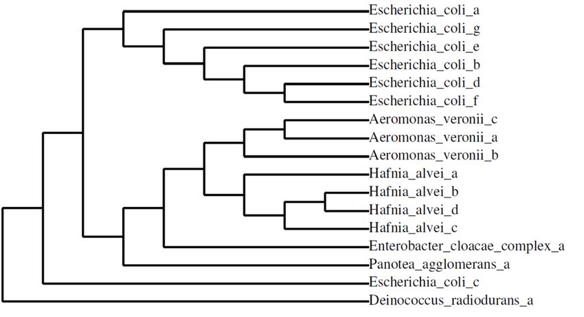
prevalent (13% of isolates). Combinations of three or four virulence genes were detected within
a single organism in a small fraction of the total isolates. While isolates carrying 3 or 4 virulence
genes in tandem were relatively rare, they were more likely to be associated with Canyon Lake or
the wastewater treatment plant [Figure 3].

When analyzing bacterial isolates by site, Dark Canyon had 22 total gene detections, Canyon Lake
had 106, East St. Patrick St. had 83, and below the wastewater treatment plant had 120 gene
detections. Stx2 was the most detected gene with 109 total detections, followed by eaeA with 107,
espP with 51, einV with 39, and stx1 with 25 total gene detections. While stx1 was not detected in
in the total bacterial community of Rapid Creek, the gene was present in a small fraction of isolates.
The eae and exhA genes were not detected in any bacterial isolates, but they were present in the
total bacterial population of Rapid Creek.

3.1.3 16S rRNA Sequencing of Bacterial Isolates from Surface Water. From the 255 bacterial
isolates that were characterized for their respective pathogenicity profiles, 16 isolates were chosen
for 16S rRNA sequencing for species identification: 2 isolates were obtained from Dark Canon, 5
from Canyon Lake, 4 from E. St. Patrick St., and 5 from the wastewater treatment plant.
Escherichia coli, Aeromonas veronii, Hafnia alvei, organisms from the Enterobacter cloacae
complex, and Panotea agglomerans were identified as species that carry the virulence genes
detected within Rapid Creek [Table 1]. All species of bacteria identified as carrying E. coli
virulence genes are highly related, and all have been previously reported as stx carriers [Figure 3]
[6-11]. Interestingly, certain gene combinations were carried by multiple species of bacteria,
which may be indicative of high levels of horizontal gene transfer among the resident bacteria.
For example, stx2/eaeA/einV was present in 6 total organisms from 4 different species: Hafnia
alvei, Enterobacter cloacae complex, Escherichia coli, and Aeromonas veronii. Overall, most of
the virulence gene-carrying organisms were identified as E. coli, as 7 of the 16 isolates were from
this species. Certain organisms were associated with specific sites; Hafnia alvei, for instance, was
identified only in Canyon Lake, and Aeromonas veronii was solely associated with the wastewater
treatment plant. E. coli, however, was identified at all sites along Rapid Creek.

4.1 Discussion

E. coli is a genetically, phenotypically, and metabolically diverse and abundant species of bacteria.
The primary niche for E. coli is the mammalian gut, thus when found in the environment, E. coli
are often indicators of fecal contamination. Surface water is particularly susceptible to fecal inputs,
and can therefore harbor potentially pathogenic organisms, largely attributed to loading from
domestic and wild animals, stormwater runoff, septic tank leakage, and wastewater and municipal
discharge [12]. Current water quality testing relies on measuring fecal coliforms, or, more
specifically, E. coli. However, since bacteria have varying levels of harmful attributes,
identification of a water sample as carrying E. coli provides limited information about the potential
danger of consumption or recreational use of the water.
When bacteria adapt to new environments, they are able to acquire and exchange genes that confer
a selective advantage. But, often the genes that allow survival in a secondary habitat are also genes
that contribute to pathogenicity in humans [13]. Horizontal gene transfer is a source of bacterial
evolution that allows for genes to be passed easily between bacteria, even among distantly related
or unrelated species, and the pathogenic capacity of a bacterium is directly related to the acquisition
and expression of virulence genes. Shiga toxins are critical pathogenicity factors that contribute
to the disease-causing potential of E. coli. In addition to Shiga toxin, other E. coli pathogenicity
factors including adhesins, toxins, invasins, cytotoxic necrotizing factors, protein secretion
systems, and iron uptake systems may be shared between individuals in a given bacterial
population, especially in environments where bacterial levels are high [14]. Therefore, existing
harmless E. coli may acquire genes carried by other organisms, transforming them into dangerous
human pathogens. In surface water, where there are diverse populations of bacterial communities
originating from various sources, the likelihood of acquisition of novel combinations of virulence
attributes within a single organism is high.

Because of the availability of DNA from neighboring cells within an environment, the entire
genetic complement must be considered. Historically, environments are fairly isolated, with
minimal mixing; however, human activity has affected this in a number of ways. Both urbanization
and agricultural practices have introduced new sources of fecal contamination to waterways that
were not previously impacted, and animal-human interactions result in spreading of their biota to
the environment. Under certain stresses, including pollution of waterways, wastewater treatment
processes, and changes in nutrients within a waterbody, the rate of lateral gene transfer increases,
often resulting in exchange of traits that allow for pathogenicity in humans.

4.1.1 Indicator Bacteria and Pathogenicity Profiling. E. coli and/or coliform levels are
commonly used as indicators of fecal contamination. Portions of Rapid Creek are currently listed
as impaired by SD DENR. This is of particular concern during high recreational use. In the current
work, pathogenicity gene presence was not related to the abundance of E. coli or total fecal
coliforms. It has been established that neither presence nor abundance of virulence factors is
directly linked to fecal indicator bacteria levels [19-21]. For example, it is possible for surface
water to have an E. coli load that is below the EPA’s threshold of impairment, while prevalence of
virulence factors is high [16, 19, 22]. Conversely, it is possible for impaired sites to completely
lack E. coli virulence genes [16]. Thus, the tool developed by our lab group improves upon the
current water quality metric.

4.1.2 Pathogenicity Genes. A wide and diverse array of microorganisms are capable of producing
Shiga toxin, including STEC, a human pathogen. Such bacteria possess at least one gene for Shiga
toxin, an essential virulence factor that contributes to the disease-causing potential. The Shiga
toxin genes (stx), which comprise two antigenically distinct variants (stx1 and stx2), have not only
been found in the coliform bacteria E. coli, Enterobacter, and Citrobacter, and the aquatic
bacterium Aeromonas, but in the soil bacterium Acinetobacter, the food pathogens Camplyobacter
and Shigella, and even an insect endosymbiont, Hamiltonella [23, 24]. Bacteria, particularly E.
coli, are capable of encoding and expressing more than one Shiga toxin variant. In addition to
Shiga toxin, other virulence factors including adhesins, toxins, invasins, cytotoxic necrotizing
factors, protein secretion systems, and iron uptake systems may be shared between individuals in
a given bacterial population [25]. The eae virulence factors (eae, eaeA, eaeγ2), encode proteins
responsible for the attaching and effacing lesions in the gut mucosa of infected patients, and ehxA,
a protein that damages red blood cells, genes are often transmitted in E. coli along with Shiga
toxins. Presence of einV is indicative of enteroinvasive E. coli (EIEC), which can cause a
Shigellosis-like disease with watery diarrhea and dysentery. EIEC’s mechanism of pathogenesis
involves epithelial invasion of the colon leading to inflammation and ulceration of the mucosal
lining. The espP gene encodes a serine protease which has homologs in Neisseria gonorrhoeae,
Haemophilus influenzae, and E. coli O157:H7. Serine proteases contributes to mucosal
hemorrhage in hemorrhagic colitis and hemolytic uremic syndrome. These virulence factors in
pathogenic E. coli originated from horizontal gene transfer events, and such gene sharing is
observed in environments where bacterial levels are high.

4.1.3 Potential Health Risk. Virulence gene profiling of the bacterial community within surface
water provides a foundation for risk assessment associated with contacting fecally contaminated
waterways [27]. The presence of E. coli pathogenicity genes that are carried by various organisms
within Rapid Creek indicates significant horizontal gene transfer among the resident bacteria, and
their presence may indicate a potential risk to human health. Almost all water samples containing
a Shiga toxin gene also had eaeA, which is required for full expression of virulence within EHEC
strains [16]. Even though there were fewer gene detections during months where recreational-use
is presumed to be higher, it should be noted there is a potential risk of infection as long as such
pathogenicity factors are present within the system, even when the total E. coli numbers are not
high [28].

With the elevated levels of total E. coli in portions of Rapid Creek, there is a potential to create
new combinations of these genes through horizontal gene transfer, and thus, new human
pathogens. Such evolution has been documented recently in Germany, where the acquisition of
the stx2 gene by an E. coli subtype containing the eaeA gene caused an outbreak in which more
than 50 people died from ingestion of the pathogen [29]. The stx1, stx2, and eaeA genes occur
concurrently in many strains of pathogenic E. coli, and are all found in E. coli O157:H7. Since
this gene combination was present within several different species within Rapid Creek, there is
potential for this gene combination to be transferred to E. coli or other opportunistic pathogens.
Presence of pathogenicity factors in Rapid Creek indicates a potential risk to human health. Future
studies should focus on the magnitude of pathogenicity gene presence within the waterways.

Acknowledgements | We are grateful for West Dakota Water Development District for funding
this project and the joint work done with H2E and USGS. This work was completed in part with
support from South Dakota School of Mines and Technology.
Figure 1 | Path-STREAM analysis of Rapid Creek. Genes detected using Path-STREAM included stx2, eae, eaeA, einV, espP and ehxA. The stx1 gene was not
detected in any samples. The magnitude of the bar represents the number of replicate samples positive for each respective gene. Sites are abbreviated as follows:
DC: Dark Canyon, CL: Canyon Lake, SP: East St. Patrick Street, TP: below the wastewater treatment plant.
45

                                     40
   % Of Isolates according to site

                                     35

                                     30

                                     25

                                     20

                                     15

                                     10

                                     5

                                     0
                                          0    1                2               3             4
                                              Number of Pathogenicty Genes Detected

                                                     DC   CL   SP   TP

Figure 2 | Number of pathogenicity genes detected in Rapid Creek isolates, expressed as a percentage of isolates from
each individual site. Sites are abbreviated as follows: DC: Dark Canyon, CL: Canyon Lake, SP: East St. Patrick Street,
TP: below the wastewater treatment plant.
14
12
10
% Total Isolates
8
6
4
2
0
DC CL SP TP
Figure 3 | Percent of bacterial isolates carrying specific virulence gene combinations. Sites are abbreviated as follows:
DC: Dark Canyon, CL: Canyon Lake, SP: East St. Patrick Street, TP: below the wastewater treatment plant.Table 1 | Species identification of bacterial isolates carrying various combinations of virulence genes.
Gene Combination Site Species %
Identity
stx1/einV DC Escherichia coli 96
stx1/stx2/eaeA SP Escherichia coli 88
stx1/stx2/eaeA/einV TP Escherichia coli 98
stx1/stx2/eaeA/espP CL Hafnia alvei 97
TP Aeromonas veronii 95
stx2/eaeA DC Pantoea agglomerans 97
stx2/eaeA/einV CL Hafnia alvei 97
SP Enterobacter cloacae 95
complex
SP Escherichia coli 97
SP Escherichia coli 97
TP Aeromonas veronii 98
TP Escherichia coli 97
stx2/eaeA/espP CL Hafnia alvei 96
CL Hafnia alvei 96
TP Aeromonas veronii 99
CL Escherichia coli 96
Figure 3 | Phylogenetic tree of isolates sequenced from Rapid Creek [30-36].5.1 Literature Cited
1. Daniel SL, Hung KF, Janezic KJ, Ferry B, Hendricks EW, Janiga BA, Johnson T, Murphy S, Roberts ME, Scott
SM: Phenotypic and genotypic characterization of Escherichia coli isolated from untreated surface
waters. The open microbiology journal 2013, 7:9-19.
2. Macler BA, Merkle JC: Current knowledge on groundwater microbial pathogens and their control.
Hydrogeology Journal 2000, 8(1):29-40.
3. Pirner SM: The 2016 South Dakota Integrated Report for Surface Water Quality Assessment. In.: South
Dakota Department of Environment and Natural Resources; 2016.
4. CDC: National Enteric Disease Surveillance: Shiga toxin-producing Escherichia coli (STEC) Annual
Report, 2012. National Center for Emerging and Zoonotic Infectious Diseases; Division of Foodborne,
Waterborne, and Environmental Diseases Centers for Disease Control 2012.
5. Ishii S, Sadowsky MJ: Escherichia coli in the environment: implications for water quality and human
health. Microbes and Environments 2008, 23(2):101-108.
6. Probert WS, McQuaid C, Schrader K: Isolation and identification of an Enterobacter cloacae strain
producing a novel subtype of Shiga toxin type 1. Journal of clinical microbiology 2014, 52(7):2346-
2351.
7. Mezzatesta ML, Gona F, Stefani S: Enterobacter cloacae complex: clinical impact and emerging
antibiotic resistance. Future microbiology 2012, 7(7):887-902.
8. Parker JL, Shaw JG: Aeromonas spp. clinical microbiology and disease. Journal of Infection 2011,
62(2):109-118.
9. Albert MJ, Alam K, Islam M, Montanaro J, Rahaman A, Haider K, Hossain MA, Kibriya A, Tzipori S: Hafnia
alvei, a probable cause of diarrhea in humans. Infection and immunity 1991, 59(4):1507-1513.
10. Figueras MJ, Aldea MJ, Fernández N, Aspíroz C, Alperi A, Guarro J: Aeromonas hemolytic uremic
syndrome. A case and a review of the literature. Diagnostic microbiology and infectious disease 2007,
58(2):231-234.
11. Alperi A, Figueras M: Human isolates of Aeromonas possess Shiga toxin genes (stx1 and stx2) highly
similar to the most virulent gene variants of Escherichia coli. Clinical Microbiology and Infection 2010,
16(10):1563-1567.
12. Agarwal M, Tomar RS, Jyoti A: Detection of water-borne pathogenic bacteria: where molecular methods
rule. Int J of Multi Curr Researc 2014, 2:351-358.
13. Brinkmeyer R, Amon RMW, Schwarz JR, Saxton T, Roberts D, Harrison S, Ellis N, Fox J, DiGuardi K,
Hochman M et al: Distribution and persistence of Escherichia coli and Enterococci in stream bed and
bank sediments from two urban streams in Houston, TX. Science of The Total Environment 2015,
502:650-658.
14. Gerrish RS, Lee JE, Reed J, Williams J, Farrell LD, Spiegel KM, Sheridan PP, Shields MS: PCR versus
hybridization for detecting virulence genes of enterohemorrhagic Escherichia coli. Emerg Infect Dis
2007, 13(8):1253-1255.
15. Ahmed W, Sawant S, Huygens F, Goonetilleke A, Gardner T: Prevalence and occurrence of zoonotic
bacterial pathogens in surface waters determined by quantitative PCR. water research 2009,
43(19):4918-4928.
16. Masters N, Wiegand A, Ahmed W, Katouli M: Escherichia coli virulence genes profile of surface waters
as an indicator of water quality. water research 2011, 45(19):6321-6333.
17. Ram S, Vajpayee P, Shanker R: Prevalence of multi-antimicrobial-agent resistant, shiga toxin and
enterotoxin producing Escherichia coli in surface waters of river Ganga. Environmental science &
technology 2007, 41(21):7383-7388.
18. Poma V, Mamani N, Iñiguez V: Impact of urban contamination of the La Paz River basin on
thermotolerant coliform density and occurrence of multiple antibiotic resistant enteric pathogens in
river water, irrigated soil and fresh vegetables. SpringerPlus 2016, 5(1):499.
19. Smith CJ, Olszewski AM, Mauro SA: Correlation of Shiga toxin gene frequency with commonly used
microbial indicators of recreational water quality. Applied and environmental microbiology 2009,
75(2):316-321.
20. Bonetta S, Pignata C, Lorenzi E, De Ceglia M, Meucci L, Bonetta S, Gilli G, Carraro E: Detection of
pathogenic Campylobacter, E. coli O157: H7 and Salmonella spp. in wastewater by PCR assay.
Environmental Science and Pollution Research 2016:1-8.Pathogenicity Potential of the Bacteria in Selected Locations of Rapid Creek
21. Haack SK, Duris JW, Fogarty LR, Kolpin DW, Focazio MJ, Furlong ET, Meyer MT: Comparing wastewater
chemicals, indicator bacteria concentrations, and bacterial pathogen genes as fecal pollution
indicators. Journal of environmental quality 2009, 38(1):248-258.
22. Shelton DR, Karns JS, Coppock C, Patel J, Sharma M, Pachepsky YA: Relationship between eae and stx
virulence genes and Escherichia coli in an agricultural watershed: implications for irrigation water
standards and leafy green commodities. Journal of food protection 2011, 74(1):18-23.
23. Mauro SA, Koudelka GB: Shiga toxin: expression, distribution, and its role in the environment. Toxins
2011, 3(6):608-625.
24. Degnan PH, Yu Y, Sisneros N, Wing RA, Moran NA: Hamiltonella defensa, genome evolution of
protective bacterial endosymbiont from pathogenic ancestors. Proceedings of the National Academy of
Sciences 2009, 106(22):9063-9068.
25. Pass M, Odedra R, Batt R: Multiplex PCRs for identification of Escherichia coli virulence genes. Journal of
clinical microbiology 2000, 38(5):2001-2004.
26. Tozzoli R, Caprioli A, Morabito S: Detection of toxB, a plasmid virulence gene of Escherichia coli O157,
in enterohemorrhagic and enteropathogenic E. coli. Journal of clinical microbiology 2005, 43(8):4052-
4056.
27. Bugarel M, Martin A, Fach P, Beutin L: Virulence gene profiling of enterohemorrhagic (EHEC) and
enteropathogenic (EPEC) Escherichia coli strains: a basis for molecular risk assessment of typical and
atypical EPEC strains. BMC microbiology 2011, 11(1):1.
28. Sidhu JP, Ahmed W, Hodgers L, Toze S: Occurrence of virulence genes associated with diarrheagenic
pathotypes in Escherichia coli isolates from surface water. Applied and environmental microbiology
2013, 79(1):328-335.
29. Brzuszkiewicz E, Thürmer A, Schuldes J, Leimbach A, Liesegang H, Meyer F-D, Boelter J, Petersen H,
Gottschalk G, Daniel R: Genome sequence analyses of two isolates from the recent Escherichia coli
outbreak in Germany reveal the emergence of a new pathotype: Entero-Aggregative-Haemorrhagic
Escherichia coli (EAHEC). Archives of microbiology 2011, 193(12):883-891.
30. Dereeper A, Audic S, Claverie J-M, Blanc G: BLAST-EXPLORER helps you building datasets for
phylogenetic analysis. BMC evolutionary biology 2010, 10(1):8.
31. Dereeper A, Guignon V, Blanc G, Audic S, Buffet S, Chevenet F, Dufayard J-F, Guindon S, Lefort V, Lescot
M: Phylogeny. fr: robust phylogenetic analysis for the non-specialist. Nucleic acids research 2008,
36(suppl_2):W465-W469.
32. Edgar RC: MUSCLE: multiple sequence alignment with high accuracy and high throughput. Nucleic acids
research 2004, 32(5):1792-1797.
33. Castresana J: Selection of conserved blocks from multiple alignments for their use in phylogenetic
analysis. Molecular biology and evolution 2000, 17(4):540-552.
34. Guindon S, Gascuel O: A simple, fast, and accurate algorithm to estimate large phylogenies by
maximum likelihood. Systematic biology 2003, 52(5):696-704.
35. Anisimova M, Gascuel O: Approximate likelihood-ratio test for branches: a fast, accurate, and powerful
alternative. Systematic biology 2006, 55(4):539-552.
36. Chevenet F, Brun C, Bañuls A-L, Jacq B, Christen R: TreeDyn: towards dynamic graphics and annotations
for analyses of trees. BMC bioinformatics 2006, 7(1):439.
Page 12You can also read