Eucalypt MADS-Box Genes Expressed in Developing Flowers
←
→
Page content transcription
If your browser does not render page correctly, please read the page content below
Plant Physiol. (1998) 118: 365–372

 Eucalypt MADS-Box Genes Expressed in Developing Flowers

                  Simon G. Southerton1*, Heidi Marshall2, Aidyn Mouradov, and Robert D. Teasdale
                ForBio Research Pty. Ltd., 50 Meiers Road, Indooroopilly, Queensland 4068, Australia

                                                                             et al., 1995), asparagus (Miller et al., 1995), and tomato
   Three MADS-box genes were identified from a cDNA library                  (Pnueli et al., 1991). They have also been found in gymno-
derived from young flowers of Eucalyptus grandis W. Hill ex                  sperms (Tandre et al., 1995; Mouradov et al., 1996, 1997).
Maiden. The three egm genes are single-copy genes and are ex-                The function of many of these MADS-box genes is not
pressed almost exclusively in flowers. The egm1 and egm3 genes               known; however, most are almost exclusively expressed in
shared strongest homology with other plant MADS-box genes,
                                                                             flowers, suggesting that they are likely to play a role in
which mediate between the floral meristem and the organ-identity
                                                                             flower development.
genes. The egm3 gene was also expressed strongly in the receptacle
or floral tube, which surrounds the carpels in the eucalypt flower              The MADS-box genes are likely to have played an im-
and bears the sepals, petals, and numerous stamens. There appeared           portant role in floral diversification because they regulate
to be a group of genes in eucalypts with strong homology with the            the major steps in floral morphogenesis. Phylogenetic anal-
3* region of the egm1 gene. The egm2 gene was expressed in                   ysis of the plant MADS-box genes has revealed that they
eucalypt petals and stamens and was most homologous to MADS-                 are organized into distinct groups, the members of which
box genes, which belong to the globosa group of genes, which                 share similar functional roles in flower development (Pu-
regulate organogenesis of the second and third floral whorls. The
                                                                             rugganan et al., 1995; Theissen and Saedler, 1995). MADS-
possible role of these three genes in eucalypt floral development is
                                                                             box genes control floral meristem formation (Huijser et al.,
discussed.
                                                                             1992; Mandel et al., 1992), floral organ formation (Sommer
                                                                             et al., 1990; Yanofsky et al., 1990; Angenent et al., 1992,
                                                                             1993; Jack et al., 1992; Mandel et al., 1992; Tröbner et al.,
   The basic organization of the angiosperm flower is re-                    1992; Bradley et al., 1993; Goto and Meyerowitz, 1994), and
markably conserved among all species. The central gener-                     mediate between these meristem and organ-identity func-
ative organs (stamens and carpels) are always surrounded                     tions (Angenent et al., 1994; Pnueli et al., 1994). Molecular
by perianth structures (sepals and petals). Alterations in                   evolutionary analysis has revealed that the ancestral gene
the number, shape, size, color, and arrangement of these                     in each of these groups of genes arose before the appear-
floral organs, usually adaptations to particular pollination                 ance of the flowering plants (Purugganan et al., 1995).
strategies (Richards, 1986), have led to the evolution of an
                                                                                In eucalypts, flowers are generally arranged in a dicha-
enormous range of floral structures. The diversification of
                                                                             sium, which terminates in a single flower, the subtending
patterns and structures in plants is attributable in part to
                                                                             bracts (deciduous) of which enclose all other flowers in the
evolution of the structure and regulation of genes that
                                                                             developing inflorescence. During flower development the
control morphogenesis (Doebley, 1993). In recent years
                                                                             sepal primordia of the first whorl (outermost) rapidly fuse
there has been rapid expansion of our understanding of the
                                                                             to each other, as do the petal primordia of the second
genes controlling floral morphogenesis.
                                                                             whorl. These two fused structures, separately or fused to
   Genetic and molecular studies in Arabidopsis and snap-
                                                                             each other, form a structure called the operculum, which
dragon, among other plant species, have led to the identi-
fication of genes that direct floral development. Many of                    covers and protects the developing reproductive structures
these genes contain a MADS-box domain, which is in-                          until anthesis. In another modification, the calyx, corolla,
volved in DNA binding, and a K-box domain, which is                          and numerous stamens are borne on the summit of a floral
involved in protein dimerization (Schwarz-Sommer et al.,                     tube, which surrounds and is fused to the wall of the
1992; Davies et al., 1996). The MADS-box is highly con-                      inferior ovary (Beadle et al., 1972). Figure 1 shows the
served, about 56 aa long, and usually at the N terminus of                   arrangement of floral organs in a typical eucalypt flower.
the protein, whereas less homology is observed in the                           In this paper we report the isolation of three MADS-box
K-box (Ma et al., 1991; Pnueli et al., 1991). More than 12                   genes from Eucalyptus grandis W. Hill ex Maiden, which are
MADS-box genes have been observed in angiosperm spe-                         predominantly expressed in flowers. We compare the se-
cies, including Arabidopsis (Ma et al., 1991), maize (Mena                   quences of these genes with those of other plant MADS-box
                                                                             genes, and examine the expression of these genes in devel-
  1
    Present address: Department of Biochemistry, University of               oping eucalypt flowers. Analysis of gene sequence and
Queensland, Queensland 4072, Australia.                                      expression revealed similarities to other plant MADS-box
  2
    Present address: The Queensland Institute of Medical Re-                 genes with functional roles in floral development.
search, Brisbane, Queensland 4029, Australia.
  * Corresponding author; e-mail southert@biosci.uq.edu.au; fax
617–3365– 4699.                                                                Abbreviation: aa, amino acid(s).
                                                                       365
                                Downloaded on January 29, 2021. - Published by https://plantphysiol.org
                               Copyright (c) 2020 American Society of Plant Biologists. All rights reserved.
366 Southerton et al. Plant Physiol. Vol. 118, 1998
tion of 2 volumes of ethanol and placed at 270°C for 1 h,
pelleted by centrifugation at 10,000g in a microcentrifuge at
4°C, and the pellet was dried before dissolving in diethyl
pyrocarbonate-treated water.
Library Construction and Screening
Poly(A1) RNA was purified using oligo(dT)25 Dyna-
beads, according to the manufacturer’s methods (Dynal,
Oslo, Norway), from RNA isolated from young E. grandis
flowers, which were initiating formation of floral organ
primordia. This poly(A1) RNA was used to construct a
cDNA library using the Pharmacia Time Saver cDNA Syn-
thesis kit and the cloning vector Lambda ExCell. Approx-
imately 100,000 clones were screened at low stringency (53
Figure 1. Diagrammatic median vertical section through a typical
eucalypt flower before anthesis. c, Carpel; p, petal; pd, peduncle; r,
receptacle; s, style; se, sepal; st, stamen. The fused sepals and fused
petals, separately or fused together, form the operculum.
MATERIALS AND METHODS
Plant Material
Eucalyptus grandis W. Hill ex Maiden floral and foliar
material was obtained on a number of occasions between
May 1994 and December 1995, from a mature specimen
growing at Lone Pine Koala Sanctuary, Fig Tree Pocket,
Queensland, Australia. Roots were obtained from seed-
lings germinated aseptically and grown in a growth room
for 1 month. Eucalyptus globulus Labill. flowers were col-
lected in July 1996, by Peter Buxton from Australian Paper
Plantations in Gippsland, Victoria. The large E. globulus
flowers allowed easy dissection of floral organs, including
the petals (inner operculum), stamens, carpels, receptacle,
and style for northern-blot analysis. The sepals (outer oper-
culum) had already abscised from these flowers.
Nucleic Acid Extraction
DNA was extracted from E. grandis leaves by the method
described by Keil and Griffin (1994). RNA was extracted
from various plant tissues using the method of Chang et al.
(1993) with modifications. Three grams of tissue was
ground in liquid nitrogen to a fine powder using a mortar
and pestle, then added to 10 mL of extraction buffer (2%
cetyl-trimethyl-ammonium bromide, 2% PVP 40, 100 mm
Tris-HCl, pH 8.0, 25 mm EDTA, 2 m NaCl, 0.5 g/L spermi-
dine, and 2% b-mercaptoethanol) at 65°C, and the tube was
shaken thoroughly and incubated for 5 min at 65°C. The
solutions were then extracted twice with an equal volume
of chloroform, with centrifugation at 7500g for 15 min after
each extraction. The supernatant from the second extrac-
tion was mixed with 0.25 volume of 10 m LiCl, and the
Figure 2. A comparison of the deduced aa sequences of the cDNAs
RNA precipitated overnight at 4°C. RNA was pelleted by egm1 (a), egm2 (b), and egm3 (c), with similar sequences from other
centrifugation at 7500g for 20 min, dissolved in 500 mL of plant species. The MADS-box is underlined and the K-box is in
SSTE (0.5% SDS, 1 m NaCl, 10 mm Tris-HCl, pH 8.0, and 1 italics. Dots indicate aa identical to the particular egm reference
mm EDTA), and then extracted with an equal volume of sequence, and dashes indicate gaps in the sequence alignment.
chloroform. The purified RNA was precipitated by addi- Numbers indicate aa position in the egm sequences.
Downloaded on January 29, 2021. - Published by https://plantphysiol.org
Copyright (c) 2020 American Society of Plant Biologists. All rights reserved.Eucalypt MADS-Box Genes 367
Figure 3. A comparison of the deduced aa se-
quence of the MADS-box domain of the egm
genes with the MADS-box domain of other plant
genes. Dots indicate aa identical to the consen-
sus sequence.
SSC, 48°C) using, as a probe, a mixture of 32P-labeled period of 2.5 h with gentle agitation. Sections were baked
MADS-box domain sequences that had been amplified onto slides coated in 3-aminopropyltriethoxysilane at 65°C
from the tomato tdr1, tdr4, tdr5, and tdr8 genes using de- for 18 h. The RNA probe was subjected to mild alkali
generate primers matching each end of the MADS-box. hydrolysis by heating at 60°C for 1 h in 100 mm carbonate
buffer (pH 10.2) before adding to 120 mL of hybridization
buffer (Jack et al., 1992), and coated on each slide. Slides
Sequence Analysis
were incubated overnight at 42°C, then washed using the
DNA sequences were determined using dye-terminator
chemistry on an automated sequencer (model 377, ABI,
Columbia, MD). All sequence analysis was performed us-
ing programs in the University of Wisconsin Genetics
Computer Group package (Madison). Genetic distance
analysis of nucleotide sequences from the MIK region (Pu-
rugganan et al., 1995) of several plant MADS-box genes
were performed using the ClustalX program.
DNA and RNA Gel-Blot Analysis
Southern-blot analysis using standard methods was per-
formed on 5-mg samples of DNA digested with EcoRI,
HindIII, or BamHI. Northern-blot analysis was performed
using standard methods on 10-mg samples of total RNA.
In Situ Hybridization
33
P-labeled riboprobes were synthesized by T7 or SP6
RNA polymerase, using template plasmids linearized with
restriction enzymes to enable synthesis of sense or anti-
sense probes (minus the MADS-box) of each of the three E.
grandis MADS-box genes.
Young flowers, roots, and leaves, germinated seedlings,
and vegetative apices were fixed in 3.7% formaldehyde,
dehydrated, cleared, and embedded in wax, and sections
were prepared for in situ hybridization according to the
method of Jack et al. (1992), with minor modifications.
Histoclear (National Diagnostics, Atlanta, GA) was used Figure 4. DNA gel-blot analysis of the egm genes. DNA isolated
instead of xylene throughout the method. Thick plant tis- from E. grandis was digested with EcoRI, HindIII, or Xbal and hy-
sues were presectioned to a thickness of about 1 mm to aid bridized with the egm gene minus the MADS-box under high strin-
infiltration of the fixative. Tissues were typically fixed for a gency. Each lane contains 5 mg of digested genomic DNA.
Downloaded on January 29, 2021. - Published by https://plantphysiol.org
Copyright (c) 2020 American Society of Plant Biologists. All rights reserved.368 Southerton et al. Plant Physiol. Vol. 118, 1998
Figure 5. Phylogenetic tree of several dicot
MADS-box genes and the three eucalypt egm
genes based on genetic distance analysis of the
MIK region. The lengths of the horizontal
branches are proportional to the number of base
substitutions. Numbers next to the nodes indi-
cate bootstrap values from 100 replicates. Nodes
with ,50% bootstrap support are collapsed.
method of Jack et al. (1992). Slides were then coated in protein and the tomato TDR5 protein (83% identity over
liquid emulsion and exposed for approximately 7 d. Slides 173 aa). The cDNA egm2 encodes a predicted protein of 208
were viewed under dark-field illumination, in which sig- aa and shares 62% homology over 213 aa with the petunia
nals appeared as white grains against a dark background. FBP1 protein, and 60% homology over 215 aa with the
snapdragon GLOBOSA protein. The cDNA egm3 encodes a
predicted protein of 245 aa, which is most homologous to
RESULTS
the Arabidopsis AGL4 protein (63% identity over 254 aa).
Sequence Analysis An alignment of the MADS-box region of the three euca-
lypt genes with the MADS-box region of several closely
Three different cDNAs (egm1, accession no. AF029975;
related plant MADS-box genes is shown in Figure 3.
egm2, accession no. AF029976; and egm3, accession no.
AF029977) were isolated from an E. grandis cDNA library
derived from young floral tissue as described in “Materials
Genomic Organization of the egm Genes
and Methods.” These cDNAs were sequenced and found to
contain the MADS-box. The sequences for egm2 and egm3 To determine the number of copies of the respective egm
each contained a full-length coding region and 59 and 39 genes in the eucalypt genome, DNA gel-blot analysis was
untranslated sequences. The egm1 cDNA lacked 17 bases of performed on E. grandis genomic DNA digested with var-
the 59 end of the coding region. The sequence of this ious restriction enzymes. Figure 4 shows genomic DNA
portion of the egm1 gene was obtained by sequencing a hybridizing to 39 probes specific for each of the three egm
genomic clone obtained from an E. grandis genomic library. genes. In all three blots, single, strongly hybridizing bands
Figure 2 shows the alignment of the predicted protein were observed under high-stringency conditions, indicat-
sequences of the three egm genes with those of similar ing that each of the three egm genes occurs as a single copy
MADS-box genes from other plants. A predicted protein of in the eucalypt genome. In the egm1 blots, the weakly
183 aa is encoded by egm1, which shares the strongest aa hybridizing bands indicated the presence of a number of
homology (85% identity over 170 aa) with the petunia FBP2 genes with significant homology to the 39 region of the
Downloaded on January 29, 2021. - Published by https://plantphysiol.org
Copyright (c) 2020 American Society of Plant Biologists. All rights reserved.Eucalypt MADS-Box Genes 369
egm1 sequence outside the MADS-box. Hybridizations
with the 59 region of all of the egm genes revealed a large
number of bands, indicating that there are a large number
of MADS-box genes in the eucalypt genome (results not
shown).
Figure 5 shows a phylogenetic tree of plant MADS-box
genes based on analysis of the MIK region. Both egm1 and
egm3 were included in the Agl2 group. Some of the genes in
this group are involved in interactions between the meris-
tem and the organ-identity genes. The egm2 gene was
included in the Pistillata/globosa group, which, together
with the Ap3/deficiens group of genes, is involved in regu-
lating the development of petals and stamens.
Expression of the egm Genes in Eucalypts
To determine the pattern of expression of the egm genes,
northern blots of total RNA from a range of tissues were
probed with each of the three genes. As shown in Figure 6a,
all three genes were strongly expressed in floral tissue.
Expression of egm3 was virtually exclusive to floral tissue,
since in overexposed northern blots only a very weak
signal was detected in nonfloral tissue. Weak expression of
the egm1 gene, and to a lesser extent the egm2 gene, was
detected in vegetative tissue, including vegetative shoots,
leaves, stems, and seedlings. The expression pattern of the
egm1 gene may also include expression of some of the other
genes sharing homology to the egm1 gene as indicated by
Southern-blot analysis.
Further analysis of egm2 and egm3 expression within
floral organs was performed on total RNA isolated from
flowers of E. globulus. These flowers are large, allowing Figure 6. Analysis of the expression of the egm genes in different
easier dissection of floral organs and collection of sufficient eucalypt tissues. a, RNA gel-blot analysis of egm1, egm 2, and egm3
material for RNA extraction. Unfortunately, RNA from the expression in roots, seedlings, stems, shoots, leaves, and flowers. b,
RNA gel-blot analysis of egm2 and egm3 expression in floral recep-
sepals (outer operculum) could not be collected because
tacles, petals, stamens, carpels, and styles. The blots contained 10 mg
they are shed early in floral development, when the floral of total RNA and were hybridized with 39 regions of egm1, egm2,
organs are difficult to dissect. As shown in Figure 6b, a and egm3 genes. The lengths of each of the three egm messages are
strong egm2 signal was detected in stamens, and a weaker estimated at 1.1 kb.
signal was observed in petals. No expression was detected
in the receptacle, carpels, and the style. The expression of
DISCUSSION
egm3 was strongest in the receptacle, moderate in petals,
carpels, and stamens, and absent in the style. In this study we report the isolation and characterization
In situ hybridization was used to localize the expression of three MADS-box genes from E. grandis. The eucalypt
of the egm genes in eucalypt floral tissue. In sections probed genome, in common with the genomes of other plants (Ma
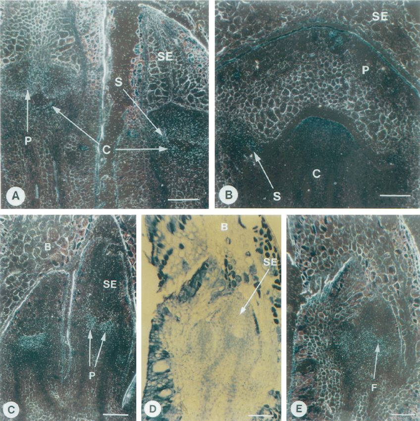
with the egm1 probe, a signal was detected in the primordia et al., 1991; Pnueli et al., 1991; Mena et al., 1995; Miller et al.,
of developing petals, stamens, and carpels (Fig. 7A). 1995; Tandre et al., 1995), contains a family of MADS-box
Within the carpels at later stages of development, expres- genes. All three eucalypt genes are expressed predomi-
sion was strongest in the developing ovules. The egm2 gene nantly in flowers, like most other identified MADS-box
was expressed in the petals and stamens at the primordial genes. Similarities between the eucalypt genes and other
stages (Fig. 7C), and weakly in these organs during later plant MADS-box genes were identified by sequence and
stages as the flower enlarged (Fig. 7B). Expression of egm3 expression analysis to provide clues to the part these genes
was detected in very young flowers in the floral meristem play in eucalypt flower development.
and the base of the sepals (Fig. 7, D and E), but was not The predicted protein of the egm1 gene shares strong
detected later in development. Failure to detect expression homology with the petunia f bp2 (Angenent et al., 1994) and
of the three egm genes later in floral development is not tomato tdr5 (Pnueli et al., 1994) genes in the Agl2 group.
consistent with the RNA-blot analysis. We have been un- Both the f bp2 and tdr5 genes are strongly expressed in the
able to detect in situ expression of several other eucalypt inner three whorls (petals, stamens, and carpels) of the
genes in mature flowers, which we have shown by flower. Functional analysis of these genes revealed that
northern-blot analysis to be strongly expressed in these they are required for determination of the three inner
tissues. whorls of the flower in each species (Angenent et al., 1994;
Downloaded on January 29, 2021. - Published by https://plantphysiol.org
Copyright (c) 2020 American Society of Plant Biologists. All rights reserved.370 Southerton et al. Plant Physiol. Vol. 118, 1998
Figure 7. RNA in situ-hybridization analysis of egm2 and egm3 expression in eucalypt flowers. B, Bract; C, carpel; F, floral
meristem; P, petal; SE, sepal; S, stamen. Bar 5 100 mm. A, Dark-field view of a longitudinal section through two E. grandis
flowers, with developing petal, stamen, and carpel primordia hybridized to the egm1 gene probe. B, Dark-field view of a
longitudinal section through an E. grandis flower initiating stamen primordia, hybridized to the egm2 gene probe. C,
Dark-field view of a longitudinal section through two E. grandis flowers initiating petal primordia, hybridized to the egm2
gene probe. D, Bright-field view of a longitudinal section through two young E. grandis flowers with developing sepal
primordia. E, Dark-field view of the section in D hybridized to the egm3 gene probe.
Pnueli et al., 1994). Incomplete homeotic transformation of The predicted protein of the egm2 gene shares strong
all three whorls is observed in plants lacking the function homology with the petunia f bp1 and snapdragon globosa
of these genes. Analysis of the egm1 gene in floral devel- genes. The genes f bp1 (Angenent et al., 1993) and globosa
opment is complicated by the occurrence of a small family (Tröbner et al., 1992) have been shown to function as
of genes in eucalypts that share significant homology to organ-identity genes specifying second- and third-whorl
egm1 outside of the MADS-box region. RNA gel-blot anal- petal and stamen organogenesis. They function as class B
ysis revealed that expression of egm1 is likely to be almost genes according to the A, B, and C model, which describes
exclusive to flowers. In situ analysis with an egm1 probe the way in which the combinatorial expression of homeotic
revealed expression in primordia of petals, stamens, and genes specifies floral organ identity. Both the f bp1 and
carpels, and in the ovules within the developing carpels; globosa genes are expressed in petals and stamen primor-
however, these results may also depict expression of egm1 dia, and more strongly during their development. Expres-
homologs. sion of egm2 was detected only in petals and stamens of
Downloaded on January 29, 2021. - Published by https://plantphysiol.org
Copyright (c) 2020 American Society of Plant Biologists. All rights reserved.Eucalypt MADS-Box Genes 371
eucalypt flowers. RNA-blot analysis revealed much stron- LITERATURE CITED
ger expression of egm2 in stamens than in petals. Expres-
Angenent GC, Busscher M, Franken J, Mol JNM, van Tunen AJ
sion in the petals may have been low because the flowers (1992) Differential expression of two MADS-box genes in wild-
sampled were close to anthesis, when the operculum type and mutant petunia flowers. Plant Cell 4: 983–993
(fused petals) is shed from the flower. It is also possible Angenent GC, Franken J, Busscher M, Colombo L, van Tunen AJ
that high expression of the egm2 gene is associated with the (1993) Petal and stamen formation in petunia is regulated by the
formation of the large number of stamens typically found homeotic gene f bp1. Plant J 4: 101–112
in eucalypt flowers. Angenent GC, Franken J, Busscher M, Weiss D, van Tunen AJ
(1994) Co-suppression of the petunia homeotic gene fbp2 affects
Like egm1, the egm3 gene shares closest homology to the the identity of the generative meristem. Plant J 5: 33–44
Agl2 group of genes. Its expression in all four floral whorls Beadle NCW, Evans OD, Carolin RC (1972) Flora of the Sydney
and in the receptacle suggests functional divergence from Region. Reed, Sydney, Australia
egm1. In situ analysis revealed expression in the floral Bradley D, Carpenter R, Sommer H, Hartley N, Coen E (1993)
meristem and in the sepal primordia. As the sepals devel- Complementary floral homeotic phenotypes result from oppo-
oped, egm3 expression was restricted to the base of the site orientations of a transposon at the plena locus of Antirrhi-
num. Cell 72: 85–95
sepals. In eucalypts the receptacle is modified into a floral Chang S, Puryear J, Cairney J (1993) A simple and efficient
tube that surrounds and sometimes protrudes above the method for isolating RNA from pine trees. Plant Mol Biol Rep
carpels. Sepals, petals, and stamens are borne on the apex 11: 113–116
of the receptacle. These results suggest that the floral tube Davies B, Egea-Cortines M, de Andrade Silva E, Saedler H,
is initiated from the floral meristem, and may be derived Sommer H (1996) Multiple interactions amongst floral homeotic
from the base of the developing sepals. It would be inter- MADS-box proteins. EMBO J 15: 4330–4343
Doebley J (1993) Genetics, development and plant evolution. Curr
esting to determine the function of the egm3 gene in the Opin Genet Dev 3: 865–872
development of the floral tube in eucalypt flowers. Goto K, Meyerowitz EM (1994) Function and regulation of the
Attempting to determine gene function on the basis of Arabidopsis floral homeotic gene Pistillata. Genes Dev 8: 1548–
similarities in gene sequence or expression pattern, partic- 1560
ularly with regulatory proteins (Gruenberg et al., 1992), can Gruenberg DA, Natesan S, Alexandre C, Gilman MZ (1992)
be misleading. Despite this, recent phylogenetic analysis of Human and Drosophila homeodomain proteins that enhance the
DNA binding activity of serum response factor. Science 257:
plant MADS-box genes has revealed that the gene family 1089–1095
sorts into groups of genes that share a similar functional Huijser P, Klein J, Lonnig W-E, Meijer H, Saedler H, Sommer H
role in flower development (Purugganan et al., 1995; (1992) Bracteomania, an inflorescence anomaly, is caused by the
Theissen and Saedler, 1995). The members of these groups loss of function of the MADS-box gene squamosa in Antirrhinum
also share similar expression patterns and sequence homol- majus. EMBO J 11: 1239–1249
ogy. Our analysis of the MADS-box gene family is in Jack T, Brockman LL, Meyerowitz EM (1992) The homeotic gene
APETALA3 of Arabidopsis thaliana encodes a MADS-box and is
agreement with this work. expressed in petals and stamens. Cell 68: 683–697
The three egm genes clustered with genes with which Keil M, Griffin AR (1994) Use of random amplified polymorphic
they shared strongest overall sequence and expression pat- DNA (RAPD) markers in the discrimination and verification of
tern homology. This provides a clue to the part these genes genotypes in Eucalyptus. Theor Appl Genet 89: 442–456
may play in eucalypt flower development. The results sug- Ma H, Yanofsky ME, Meyerowitz EM (1991) AGL1-AGL6, an
gest that the egm1 gene may function in mediating between Arabidopsis gene family with similarity to floral homeotic and
transcription factor genes. Genes Dev 5: 484–495
the floral meristem and the organ-identity genes in a man-
Mandel MA, Gustafson-Brown C, Savidge B, Yanofsky ME
ner similar to the f bp2 and tdr5 genes in petunia and (1992) Molecular characterisation of the Arabidopsis floral ho-
tomato, respectively. The egm2 gene is likely to be a ho- meotic gene APETALA1. Nature 360: 273–277
meotic B function gene regulating organogenesis of the Mena M, Mandel MA, Lerner DR, Yanofsky MF, Schmidt RJ
petals and stamens in eucalypt flowers in a manner similar (1995) A characterisation of the MADS-box gene family in maize.
to the globosa gene in snapdragon (Tröbner et al., 1992). The Plant J 8: 845–854
Miller HG, Kocher TD, Loy B (1995) New MADS-box domains in
strong expression of egm3 in the floral receptacle provides
Asparagus officinalis L. Sex Plant Reprod 8: 318–320
an interesting lead for further investigations to elucidate its Mouradov A, Glassick T, Teasdale R (1997) Isolation and charac-
function. Further analysis, either by heterologous comple- terization of a new MADS-box cDNA from Pinus radiata (acces-
mentation of floral mutants or by gene suppression sion no. U76726) (PGR 97–027). Plant Physiol 113: 664
in eucalypts, may assist in the determination of the func- Mouradov A, Glassick T, Vivian-Smith A, Teasdale R (1996)
tion of these three MADS-box genes in eucalypt floral Isolation of a MADS-box gene family from Pinus radiata (acces-
sion nos. U42399 and U42400) (PGR 96–002). Plant Physiol 110:
morphogenesis.
1047
Pnueli L, Abu-Abeid M, Zamir D, Nacken W, Schwarz-Sommer
ACKNOWLEDGMENTS Z, Lifschitz E (1991) The MADS-box gene family in tomato:
temporal expression during floral development, conserved sec-
The authors thank Peter Buxton for assistance with collection of
ondary structures and homology with homeotic genes from
plant material, Albert Ng and Adrian Steele for assistance with
Antirrhinum and Arabidopsis. Plant J 1: 255–266
computer analysis, and Corinna Lange for critical reading and
Pnueli L, Hareven D, Broday L, Lifschitz E (1994) The TM5 [tdr5]
preparation of the manuscript. MADS-box gene mediates organ differentiation in the three
inner whorls of tomato flowers. Plant Cell 6: 175–186
Received May 26, 1998; accepted June 29, 1998. Purugganan MD, Rounsley SD, Schmidt RJ, Yanofsky ME (1995)
Copyright Clearance Center: 0032–0889/98/118/0365/08. Molecular evolution of flower development: diversification of
Downloaded on January 29, 2021. - Published by https://plantphysiol.org
Copyright (c) 2020 American Society of Plant Biologists. All rights reserved.372 Southerton et al. Plant Physiol. Vol. 118, 1998
the plant MADS-box regulatory gene family. Genetics 140: Tandre K, Albert VA, Sundas A, Engström P (1995) Conifer
345–356 homologues to genes that control floral development in angio-
Richards AJ (1986) Plant Breeding Systems. Allen and Unwin, sperms. Plant Mol Biol 27: 69–78
Winchester, MA Theissen G, Saedler H (1995) MADS-box genes in plant ontogeny
Schwarz-Sommer Z, Hue I, Huijser R, Flor RJ, Hansen R, Tetens and phylogeny: Haeckel’s ‘biogenetic law’ revisited. Curr Opin
E, Lönnig W, Saedler H, Sommer H (1992) Characterisation of Genet Dev 5: 628–639
the Antirrhinum floral homeotic MADS-box gene deficiens: evi- Tröbner W, Ramirez L, Motte P, Hue I, Huijser P, Lönnig W-E,
dence for DNA binding and autoregulation of its persistent Saedler H, Sommer H, Schwarz-Sommer Z (1992) Globosa: a
expression throughout flower development. EMBO J 11: 251–263 homeotic gene which interacts with deficiens in the control of
Sommer H, Beltran J-R, Huijser R, Pape H, Lönnig W-E, Saedler Antirrhinum floral organogenesis. EMBO J 11: 4693–4704
H, Schwarz-Sommer Z (1990) Deficiens, a homeotic gene in- Yanofsky ME, Ma H, Bowman JL, Drews GN, Feldmann KA,
volved in the control of flower morphogenesis in Antirrhinum Meyerowitz EM (1990) The protein encoded by the Arabidopsis
majus: the protein shows homology to transcription factors. homeotic gene agamous resembles transcription factors. Nature
EMBO J 9: 605–613 346: 35–39
Downloaded on January 29, 2021. - Published by https://plantphysiol.org
Copyright (c) 2020 American Society of Plant Biologists. All rights reserved.You can also read