HOXA5 counteracts the function of pathological scar-derived fibroblasts by partially activating p53 signaling - Nature
←
→
Page content transcription
If your browser does not render page correctly, please read the page content below
Liang et al. Cell Death and Disease (2021)12:40
                                    https://doi.org/10.1038/s41419-020-03323-x                                                                                                             Cell Death & Disease

                                     ARTICLE                                                                                                                                                    Open Access

                                    HOXA5 counteracts the function of pathological
                                    scar-derived fibroblasts by partially activating
                                    p53 signaling
                                    Yimin Liang1, Renpeng Zhou1, Xiujun Fu1, Chen Wang1 and Danru Wang1

                                      Abstract
                                      The inactivation of p53 can lead to the formation of pathological scars, including hypertrophic scars and keloids.
                                      HOXA5 has been reported to be a critical transcription factor in the p53 pathway in cancers. However, whether HOXA5
                                      also plays a role in pathological scar progression through activating p53 signaling remains unknown. In this study, we
                                      first demonstrated that HOXA5 overexpression in hypertrophic scar-or keloids-derived fibroblasts decreased cell
                                      proliferation, migration and collagen synthesis, whereas increased cell apoptosis. Furthermore, the results of luciferase
                                      activity assays and ChIP PCR assays indicated that HOXA5 transactivated p53 by binding to the ATTA-rich core motif in
                                      the p53 promoter. HOXA5 also increased the levels of p21 and Mdm2, which are downstream targets of p53.
                                      Interestingly, silencing p53 in these pathological scar-derived fibroblasts partially attenuated HOXA5-mediated growth
                                      inhibition effect and HOXA5-induced apoptosis. In addition, 9-cis-retinoic acid augmented the expression of HOXA5
                                      and promoted the effects of HOXA5 on pathological scar-derived fibroblasts, and these effects could be suppressed by
                                      HOXA5 knockdown. Thus, our study reveals a role of HOXA5 in mediating the cellular processes of pathological scar-
                  1234567890():,;
1234567890():,;
                  1234567890():,;
1234567890():,;

                                      derived fibroblasts by transcriptionally activating the p53 signaling pathway, and 9-cis-retinoic acid may be a potential
                                      therapy for pathological scars.

                                    Introduction                                                                                        and then quit after the tasks have been completed.
                                      Pathological scars, including keloids and hypertrophic                                            Apoptosis is confirmed to be the primary mechanism for
                                    scars that are characteristic of excessive dermal fibrosis,                                          cells quit2. Previous studies have demonstrated that
                                    are caused by excessive cell proliferation and collagen                                             pathological scars usually form due to defective apoptotic
                                    synthesis during the wound healing process. Pathological                                            signals that lead to fibroblast dysfunction3–5. The p53
                                    scars can result in severe organ malformation and dys-                                              tumor suppressor plays a crucial role in regulating
                                    function and are consistently difficult to cure. Therefore,                                          apoptosis6,7. However, the molecular mechanisms by
                                    the underlying molecular mechanism of scar formation                                                which p53 participates in pathological scarring remain
                                    and the anti-scarring strategy are the research hotspots.                                           unclear and controversial.
                                      The wound healing process includes three overlapping                                                In human breast cancer, the loss of p53 expression may
                                    but distinct stages: inflammation, granulation tissue for-                                           be caused by a lack of homeobox A5 (HOXA5) expres-
                                    mation, and remodeling1. Each group of stage-specific                                                sion8. Homeobox genes are a group of genes that play key
                                    cells sequentially occur at the wounded site, proliferate,                                          roles in both development and postnatal regeneration9.
                                                                                                                                        Our previous study revealed that HOXA5 was able to
                                                                                                                                        suppress keratinocyte growth and epidermal formation10,
                                    Correspondence: Danru Wang (wangdanru1776@163.com)                                                  we also showed that HOXA5 was substantially activated
                                    1
                                     Department of Plastic and Reconstructive Surgery, Shanghai 9th People’s                            by mechanical stretch in renewed skin, suggesting a role
                                    Hospital, Shanghai Jiao Tong University School of Medicine, 200011 Shanghai,
                                                                                                                                        for HOXA5 in regulating skin regeneration, including
                                    China
                                    These authors contributed equally: Yimin Liang, Renpeng Zhou                                        scarless wound healing11. While HOXA5 can promote
                                    Edited by D. Aberdam

                                    © The Author(s) 2021
                                                        Open Access This article is licensed under a Creative Commons Attribution 4.0 International License, which permits use, sharing, adaptation, distribution and reproduction
                                                        in any medium or format, as long as you give appropriate credit to the original author(s) and the source, provide a link to the Creative Commons license, and indicate if
                                    changes were made. The images or other third party material in this article are included in the article’s Creative Commons license, unless indicated otherwise in a credit line to the material. If
                                    material is not included in the article’s Creative Commons license and your intended use is not permitted by statutory regulation or exceeds the permitted use, you will need to obtain
                                    permission directly from the copyright holder. To view a copy of this license, visit http://creativecommons.org/licenses/by/4.0/.

                                    Official journal of the Cell Death Differentiation Association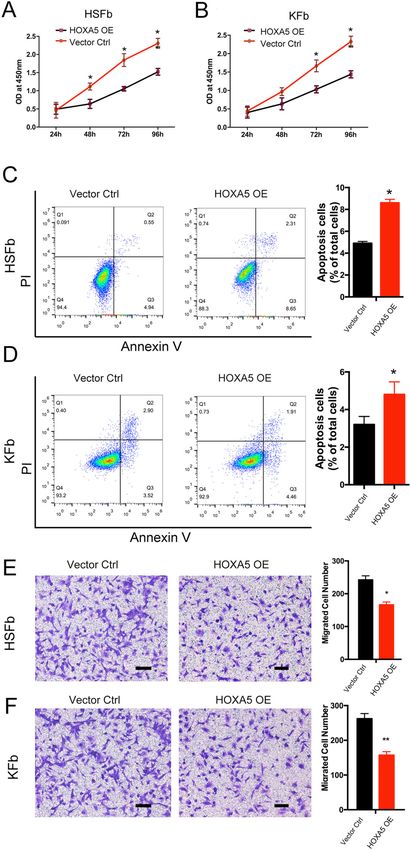
Liang et al. Cell Death and Disease (2021)12:40                                                                  Page 2 of 12

breast cancer cell death through a p53-dependent apop-          modified Eagle’s medium (DMEM) for 24 h and then
totic pathway8, the mechanism by which HOXA5 tran-              transfected with supernatants containing the lentivirus.
scriptionally regulates p53 in pathological scars remains
unknown.                                                        CCK-8 assay
  Here, we hypothesized that HOXA5 could inhibit the              The cell proliferation of fibroblasts was measured by the
pathological scarring by activating the p53 pathway.            Cell Counting Kit-8 assay (CCK-8 assay, Sigma) according
Therefore, we investigated the expression and role of           to the manufacturer’s instructions. At 24 h posttransfec-
HOXA5 in the cellular processes of pathological scar-           tion with HOXA5, the fibroblasts were seeded in 96-well
derived fibroblasts, both from keloids (KFb) and hyper-          plates at 3000 cells/well. The absorbance was measured at
trophic scars (HSFb), as well as the underlying molecular       450 nm at the indicated time points.
mechanism by which HOXA5 regulates the p53 pathway.
In addition, the effects of 9-cis-retinoic acid on HOXA5        Cell viability and apoptosis analysis
expression and KFb and HSFb cellular processes were               Cell viability and apoptosis were detected using the
examined to identify a potential therapy to prevent             Annexin V-FITC Apoptosis Detection Kit (Sigma,
scarring.                                                       APOAF). HSFb and KFb were harvested, centrifuged at
                                                                300 × g and resuspended in binding buffer. Then 5 μl
Materials and methods                                           Annexin V-FITC and 10 μl propidium iodide solution
Tissue collection and fibroblasts culture                        were added to each cell suspension and incubated in the
  Normal primary fibroblasts (Fb) were isolated and cul-         dark. The samples were kept on ice and analyzed imme-
tured from the normal skin of patients who underwent            diately analyzed by flow cytometry with a FACSCalibur
abdominoplasty. Written consent was obtained. Primary           instrument (BD Biosciences, USA). The data were further
fibroblasts (HSFb and KFb) were isolated and cultured            analyzed with FlowJo software (FlowJo, OR) to calculate
from the hypertrophic scars and keloids, respectively, of       the proportion of apoptotic cells.
15 patients after obtaining informed consent of the
patients and approval of the Shanghai ninth hospital’s          Transwell assay
Ethical Committee. The mean patient age was 32.7 years            HSFb and KFb were digested with 0.25% trypsin and
(range 22–45 years) and there were nine women and six           suspended in serum-free medium, 1 × 10⁴ cells were see-
men. Briefly, the harvested tissue samples were washed           ded in a Matrigel-coated Transwell chamber (8-µm pore,
with phosphate-buffered saline, cut into pieces and             size,Corning). Complete medium (500 µl) was added to
digested with 0.3% collagenase type I (Serva,Germany).          the lower chamber. After incubation for 24 h, the non-
The digested tissue samples were filtered through a 200-         migrated fibroblasts were removed from the upper surface
µm mesh filter, and centrifuged at 500 × g for 10 min. The       of the membrane. The migrated fibroblasts were fixed in
supernatant was discarded, and the pellet was resus-            4% paraformaldehyde and stained with 0.4% crystal violet
pended in DMEM (HyClone, USA) and cultured in                   solution (Beyotime, China). The migrated fibroblasts were
DMEM supplemented with 10% fetal bovine serum                   visualized under an inverted microscope (Olympus,
(Hyclone, USA) in 5% CO2 at 37 °C. Primary cells at             Japan), and the average numbers of fibroblasts were cal-
passages 2–3 were used for the experiments.                     culated by quantifying the cells in ten randomly selected
                                                                fields.
Expression vector construction and transfection
procedures                                                      Fibroblast-populated collagen lattice (FPCL)
  The sequence of HOXA5 was amplified from wild-type               Rat tail type I collagen (Gibco,USA) solutions were
complementary DNA (cDNA) by PCR. The following                  mixed with HSFb or KFb at a density of 3 × 10⁵/ml. The
primer sequences were used: forward: 5ʹ-GAGGATCC                mixture was seeded into the 24-well plates and then
CCGGGTACCGGTCGCCACCATGAGCTCTTATTTT                              incubated at 37 °C. The collagen gels were imaged after
GTAAAC-3ʹ; reverse: 5ʹ-TCACCATGGTGGCGACCGG                      48 h and the degree of gel contraction was determined by
GGGACGGAAGGCCCCTCCTG-3ʹ. The HOXA5 PCR                          calculating the ratio of the final contracted gel areas to the
fragments and the pGC-FU vector (Shanghai GeneChem,             initial gel areas.
China) were digested with Age I and then ligated with T4
DNA ligase to produce the pGC-FU-HOXA5. The empty               Western blot
pGC-FU vector was used as the control vector. 293T cells          HSFb and KFb were lysed with RIPA buffer (Beyotime,
were transfected with pGC-FU-HOXA5, pHelper 1.0 and             China) and the supernatants were harvested. The 25 μg
pHelper 2.0 (Shanghai GeneChem, China) to generate the          protein samples were separated by 10% sodium dodecyl
viral particles. Then fibroblasts were plated into six-well      sulfate polyacrylamide gel electrophoresis and transferred
plates, cultured with 10% fetal bovine serum/Dulbecco’s         to polyvinylidene fluoride membranes. The membrane

Official journal of the Cell Death Differentiation Association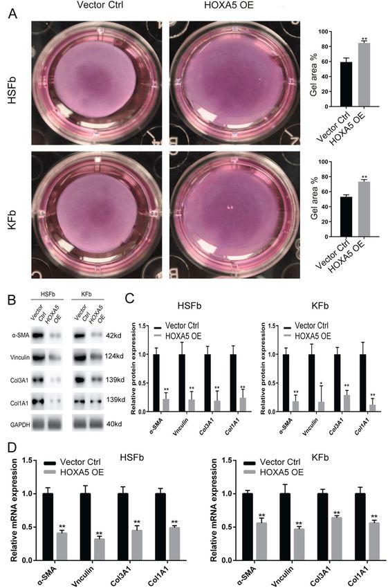
Liang et al. Cell Death and Disease (2021)12:40                                                                Page 3 of 12

was blocked for 1 h at room temperature and incubated           Chromatin immunoprecipitation (ChIP) PCR
with primary antibodies overnight at 4 °C. The antibodies         HSFb and KFb in the HOXA5-overexpressing group or
used were as follows: mouse anti-GAPDH(1:2000,                  control group were crosslinked in 1% formaldehyde for
Abcam) mouse anti-α-SMA (1:500, Abcam), mouse anti-             10 min at room temperature. The fibroblasts were lysed and
vinculin (1:500, Abcam), rabbit anti-collagen I (1:500,         sonicated to obtain 200–500 bp genomic DNA fragments.
Abcam), rabbit anti-collagen III (1:500, Abcam), mouse          The sheared fragments were incubated overnight at 4 °C
anti-p21 (1:500, Abcam), mouse anti-Mdm2 (1:500,                with rabbit anti-HOXA5 or with control rabbit IgG. After
Abcam), Rabbit anti-HOXA5(1:1000, Acam), mouse anti-            blocking in 0.5% BSA, the Dynabeads (Invitrogen) were
p53(1:1000, Abcam). The membranes were washed three             added to couple the antibodies. The precipitated DNA
times with PBST and incubated with the corresponding            fragments were purified and used for PCR analysis. Primers
HRP-conjugated secondary antibodies for 1 h at room             designed to analyze the binding of the p53 promoter were as
temperature. The protein levels were detected using an          follows: forward TGCTCAAGACTGGCGCTAAA; reverse
ECL reagent kit (Thermo Fisher, USA). The ChemiDoc              GGAGCTTACCCAATCCAGGG.
MP imaging system (Bio-Rad, USA) was used to detect
the protein expression. The data were analyzed using            Small-interfering RNA (siRNA) transfection
ImageJ software (NIH, USA). GAPDH was used as the                 Synthetic siRNA targeting human HOXA5 or p53 and
endogenous reference for normalization.                         nonspecific control siRNA were purchased from Gene-
                                                                Chem (Shanghai, China). Fibroblasts were seeded into 6-
Quantitative real-time PCR                                      well plates and cultured to 60% confluence and then
  Total RNA was extracted using TRIzol and the RNA              transfected with HOXA5 siRNA or p53 siRNA or control
concentration was determined using a NanoDrop2000               siRNAs. At 48 h after transfection, the cells were har-
spectrophotometer (Thermo Scientific). Then 500 ng RNA           vested for qPCR detection and 72 h after transfection, the
was used to synthesize cDNA with reverse transcriptase          cells were harvested for western blotting analysis.
(Takara) and the messenger RNAs (mRNAs) levels were
quantified using the GoTaq qPCR Master Mix(Promega,              9-cis-retinoic acid(9-cis-RA) treatment
USA). The sequences of primers were as follows: GA                For the treatment of HBFb and KFb, 9-cis-retinoic acid
PDH forward: GCACCGTCAAGGCTGAGAAC, GAPDH                        (9-cis-RA, Sigma) was dissolved in dimethyl sulfoxide.
reverse: TGGTGAAGACGCCAGTGGA, α-SMA forward:                    Cultured HSFb and KFb were treated with 0.1 µmol/l 9-
AGGTAACGAGTCAGAGCTTTGGC, α-SMA reverse: CT                      cis-RA, and the cells in the control group were treated
CTCTGTCCACCTTCCAGCAG, vinculin forward: AGAG                    with standard medium.
ACTGTTCAGACCACTGAG, vinculin reverse: CATTGA
GTTCACCAACATCAC, Col1A1 forward: AAGAGCTCG                      Statistical analyses
TGGGAAAGCCTGGATGG, Col1A1 reverse: AAAGAT                         All the experiments were repeated three times. The data
CTTTTGGGACTTACTGTCTTCGT, Col3A1 forward: CC                     were analyzed by SPSS12.0. One-way analysis of variance
CAGAACATCACATATCAC, Col3A1 reverse: CAAGAGG                     (ANOVA) test was used to identify significant differences
AACACATATGGAG. Mdm2 forward: GAATCATCGGAC                       among experimental groups. The t-test was performed to
TCAGGTACATC, Mdm2 reverse: TCTGTCTCACTAAT                       identify statistically significant differences between two
TGCTCTCCT; p53 forward: CCTCAGCATCTTATCCG                       groups. The results are expressed as the mean ± standard
AGTGG, reverse:TGGATGGTGGTACAGTCAGAGC; p21                      deviation.
forward: AGGTGGACCTGGAGACTCTCAG, reverse: TC
CTCTTGGAGAAGATCAGCCG. The reactions were per-                   Results
formed in triplicate. The mRNA levels were normalized to        Effects of HOXA5 overexpression on pathological scar-
that of GAPDH by using the delta-delta Ct method.               derived fibroblasts
                                                                  To investigate the role of HOXA5 in the cell biology
Luciferase reporter assay                                       of fibroblasts derived from keloids (KFb) or from
  A luciferase reporter construct containing the p53            hypertrophic scars (HSFb), we established HOXA5
promoter was purchased from Shanghai Gema. Before               overexpression in both types of scar-derived fibroblasts
transfection, HSFb or KFb were seeded into 24-well              (Fig. S1). The CCK-8 assay showed that compared with
plates. The fibroblasts were transfected with the luciferase     the control (Vector Ctrl), HOXA5 overexpression
promoter constructs and either a HOXA5 overexpression           (HOXA5 OE) significantly inhibited the cell prolifera-
construct or an empty vector. The cells were lysed at 24 h      tion of HSFb at 48 (1.12 ± 0.09 vs. 0.63 ± 0.12), 72
after transfection and the luciferase activity was measured     (1.85 ± 0.17 vs. 1.05 ± 0.06) and 96 (2.31 ± 0.13 vs.
using the luciferase reporter assay system (Promega)            1.52 ± 0.09) hours (Fig. 1A) and decreased the KFb cell
according to the manufacturer’s instructions.                   proliferation at 72 (1.66 ± 0.16 vs. 1.03 ± 0.09) and 96

Official journal of the Cell Death Differentiation Association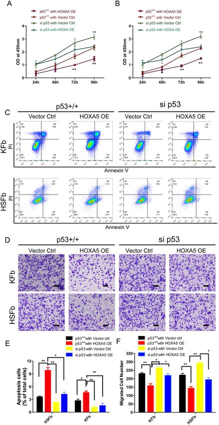
Liang et al. Cell Death and Disease (2021)12:40                                                                                       Page 4 of 12

  Fig. 1 Effects of HOXA5 overexpression on pathological scar-derived fibroblasts. A, B A CCK-8 assay was performed to determine the cell
  proliferation rates of fibroblasts derived from keloids (KFb) or from hypertrophic scars (HSFb) with/without HOXA5 overexpression. C, D Flow
  cytometric analysis was performed to determine the percentages of apoptotic KFb and HSFb in each group. E, F Transwell analysis was performed to
  determine the cell migration ability of KFb and HSFb in each group. *p < 0.05, **p < 0.01. scale bar = 100 μm.

Official journal of the Cell Death Differentiation Association
Liang et al. Cell Death and Disease (2021)12:40 Page 5 of 12
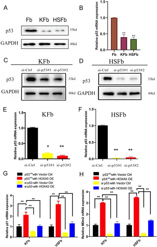
(2.53 ± 0.15 vs. 1.43 ± 0.09) hours (Fig. 1B). We next Knockdown of p53 partially weakened HOXA5-mediated
investigated the effects of the HOXA5 overexpression regulation in both HSFb and KFb
on the apoptosis of KFb and HSFb by flow cytometry. To investigate the potential mechanism underlying
As shown in Fig. 1C, D, compared with the percentage HOXA5-mediated p53 upregulation in HSFb and KFb, we
of apoptotic HSFb and KFb in the control group (HSFb: further analyzed p53. The qPCR and western blot analyses
4.9 ± 0.2%; KFb:3.2 ± 0.4%), the percentage of apoptotic showed that the expression of p53 was significantly lower
cells in the HOXA5 overexpression group was sig- in KFb and HSFb than in normal fibroblasts (Fig. 4A, B).
nificantly higher (HSFb:8.6 ± 0.3%; KFb:4.8 ± 0.6%). We To determine whether p53 facilitated the HOXA5-
also performed the transwell assay to investigate the mediated regulation on both the KFb and the HSFb, we
potential function of HOXA5 in the migration of scar- knocked down (KD) p53 using two siRNAs targeting p53.
derived fibroblasts. Compared with the number of The kockdown efficiency was confirmed by qPCR and
migrated KFb and HSFb in the control group western blot (Fig. 4C–F).
(HSFb:262 ± 15; KFb:242 ± 12), the number of migrated To further elucidate whether p53 contributes to
KFb and HSFb in the HOXA5 overexpression group HOXA5-mediated phenotypes in pathological scar-
(HSFb:166 ± 8; KFb:157 ± 10) was significantly reduced derived fibroblasts, we conducted reconstitution experi-
(Fig.1E, F). Then, we further measured the effects of ments by transfecting HSFb and KFb with si-p53 or the
HOXA5 on cell contractive ability using a fibroblast- HOXA5 overexpression plasmid alone or with si-p53 and
populated collagen lattice (FPCL). The data showed the HOXA5 overexpression plasmid in combination.
that the KFb and HSFb in the HOXA5 overexpression Compared with control (p53+/+ with vector ctrl group),
group exhibited impaired cell contractive ability (Fig. HOXA5 overexpression alone (p53+/+ with HOXA5 OE
2A). We also performed qPCR and western blot assays group) promoted the expression of p21 and Mdm2 in
to investigate the effects of HOXA5 on extracellular both HSFb and KFb. However, this effect was attenuated
matrix secretion, and the data showed that the HOXA5 by the knockdown of p53 (si p53 with HOXA5 OE group)
overexpression decreased the expression of α-SMA, (Fig. 4G, H).
Vinculin, ColI and Col III (Fig. 2B–D). In addition, Then, we performed proliferation, migration and
when compared with the control group, the HOXA5 apoptosis assays. Compared with control (p53+/+ with
knockdow group exhibited significantly increased vector ctrl group), HOXA5 overexpression alone (p53+/+
protein level of α-SMA, Vinculin, ColI and Col III with HOXA5 OE group) inhibited cell proliferation in
(Fig. S2). both HSFb and KFb. However, this effect was attenuated
by p53 knockdown (si p53 with HOXA5 OE group),
HOXA5 binds to the p53 promoter and activates indicating a critical role of p53 in the HOXA5-mediated
p53 signaling in both HSFb and KFb inhibition of proliferation (Fig. 5A, B). Similarly, in the
To investigate whether HOXA5 affects pathological cellular apoptosis analyses with Annexin V-FITC/PI
phenotypes by regulating p53 expression, we co- double staining, HOXA5 (HSFb:9.8 ± 0.3%; KFb:4.6 ± 0.2%
transfected both the HOXA5-overexpressing plasmid in p53 + /+ with HOXA5 OE group) induced cell apop-
and the p53 promoter luciferase reporter construct (the tosis in both HSFb and KFb compared with the control
initiation of transcription is at the 2 kb upstream) into group(HSFb:3.6 ± 0.2%; KFb:2.7 ± 0.3% in p53 + /+ with
both HSFb and KFb, and then detected the luciferase vector ctrl group), and si-p53 (HSFb:4.2 ± 0.4%; KFb:1.5 ±
activity. The data showed that HOXA5 was able to acti- 0.2% in si p53 with HOXA5 OE group)partially abolished
vate the expression of the p53 promoter reporter gene in the HOXA5-mediated cell apoptosis (Fig. 5C, E). More-
both kinds of cells (Fig. 3A, B). To confirm a direct over, HOXA5 overexpression alone (HSFb:143 ± 9;
interaction between HOXA5 and p53, we further detected KFb:158 ± 12) inhibited the cell migration of both HSFb
the binding of HOXA5 and the ATTA-rich core motif in and KFb compared with control (HSFb:221 ± 11;
the p53 promoter using chromatin immunoprecipitation KFb:230 ± 6), and this effect was attenuated when p53 was
(ChIP) coupled with quantitative PCR. We found that knocked down (HSFb:194 ± 13; KFb:219 ± 11) (Fig. 5D, F).
after the overexpression of HOXA5, the level of binding These results indicated that HOXA5 regulated the cell
was enhanced (Fig. 3C, D). Furthermore, both the mRNA function of HSFb and KFb, at least partially by regulating
expression and the protein levels of p21 and Mdm2, p53.
which are the downstream targets of p53, were sig-
nificantly upregulated in both kinds of fibroblasts in the 9-cis-retinoic acid increases HOXA5 expression and
HOXA5 overexpression group (Fig. 3E, F). Taken toge- promotes HOXA5-mediated effects on HSFb and KFb
ther, these data suggested that HOXA5 may affect Retinoic acid is known to stimulate the expression of
pathological phenotypes by transcriptionally upregulating HOXA5 in some cancer cells and has been evaluated as a
the expression of p53. preventive and therapeutic agent12–14. To test whether the
Official journal of the Cell Death Differentiation Association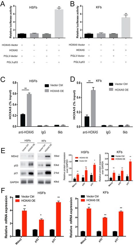
Liang et al. Cell Death and Disease (2021)12:40                                                                                          Page 6 of 12

  Fig. 2 Role of HOXA5 in pathological scar-derived fibroblasts. A A fibroblast-populated collagen lattice was used to measure the contractive
  abilities of KFb and HSFb in each group. B–D The expression of fibroproliferative biomarkers, including α-SMA, Vinculin, ColI, and Col III in KFb and
  HSFb in each group was detected by western blot and qPCR analysis. *p < 0.05, **p < 0.01.

Official journal of the Cell Death Differentiation Association
Liang et al. Cell Death and Disease (2021)12:40                                                                                         Page 7 of 12

  Fig. 3 HOXA5 binds to the p53 promoter and activates p53 signaling in both HSFb and KFb. A, B Luciferase assay data showed that HOXA5
  activated the expression of p53 promoter reporter gene in KFb and HSFb. C, D ChIP data showed a direct interaction between HOXA5 and p53. 6 kb:
  the 6 kb upstream of the transcription start site, was used as a negative control for the transcription start site. E, F qPCR and western blot data
  showed that the overexpression of HOXA5 enhanced the expression of p21 and Mdm2, which are downstream targets of p53. *p < 0.05, **p < 0.01.

Official journal of the Cell Death Differentiation Association
Liang et al. Cell Death and Disease (2021)12:40                                                                                     Page 8 of 12

  Fig. 4 Knockdown of p53 in both the HSFb and the KFb. A, B The expression of p53 in KFb, HSFb and normal fibroblasts was detected by qPCR
  and western blot. C, D p53 protein levels in KFb and HSFb were detected by western blot. E, F p53 mRNA levels were detected by QPCR in KFb and
  HSFb. G, H Knockdown of p53 partially weakened the HOXA5-mediated regulation of p21 and Mdm2 in KFb and HSFb. *p < 0.05, **p < 0.01.

Official journal of the Cell Death Differentiation Association
Liang et al. Cell Death and Disease (2021)12:40                                                                                     Page 9 of 12

  Fig. 5 Knockdown of p53 partially weakens HOXA5-mediated regulation in both HSFb and KFb. A, B Knockdown of p53 partially weakened
  the HOXA5-mediated regulation of cell proliferation in KFb and HSFb. C, E Knockdown of p53 partially weakened the HOXA5-mediated regulation of
  cell apoptosis in KFb and HSFb. D, F Knockdown of p53 partially weakened -mediated regulation of cell migration in KFb and HSFb. *p < 0.05,
  **p < 0.01. scale bar = 100 μm.

Official journal of the Cell Death Differentiation Association
Liang et al. Cell Death and Disease (2021)12:40 Page 10 of 12
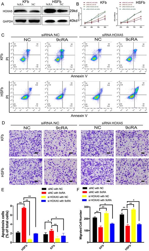
9-cis-retinoic acid (9cRA) is an agent that could poten- P53 is widely known as a transcription factor that
tially be used for the treatment of pathological scars, controls cell proliferation and apoptosis. However, the
HSFb and KFb were treated with 9cRA alone, knockdown expression and role of p53 in hypertrophic scars and
HOXA5 alone or 9cRA and HOXA5 in combination. The keloids remain controversial. Ladin et al.17 showed that
data showed that the expression of HOXA5 in patholo- the p53 is significantly upregulated in keloids and keloid-
gical scar-derived fibroblasts significantly increased when derived fibroblasts compared with normal tissue. Tanaka
the cells were treated with 9cRA (Fig. 6A). Then we et al.18 also reported that the expression of p53 is sig-
established and confirmed the HOXA5 knockdown in nificantly higher in keloids and hypertrophic scars than in
both scar-derived fibroblasts (Fig. S3). 9cRA treatment normal skin. On the other hand, low levels, and even no
alone (siRNA NC with 9cRA group) inhibited cell pro- expression, of p53 were detected in hypertrophic scars
liferation compared with the control (siRNA NC with NC and keloids in several previous studies6,19. Previous stu-
group) in both HSFb and KFb. However, this effect was dies also showed that the overexpression of p53 in HSFb
attenuated upon knockdown of HOXA5 (si-HOXA5 with promoted autophagy and inhibited collagen expression7,
9cRA group), indicating a critical role of HOXA5 in the while p53 knockout promoted hypertrophic scar forma-
9cRA-mediated inhibition of proliferation (Fig. 6B). tion20. Here, we showed that p53 is significantly down-
Similarity, in cellular apoptosis assay, 9cRA treatment regulated in both HSFb and KFb, and we further
(HSFb:7.5 ± 0.2%; KFb:2.5 ± 0.1% in the siRNA NC with demonstrated that knockdown of p53 promoted cell
9cRA group) induced cell apoptosis in both HSFb and proliferation, migration and inhibited apoptosis in HSFb
KFb compared with the control group (HSFb:2.1 ± 0.1%; and KFb. It was reported that 5-fluorouracil (5-Fu) can
KFb:1.76 ± 0.1% in siRNA NC with NC group), and induce the accumulation of p53 in keloids21, and the
HOXA5 knockdown (HSFb:1.9 ± 0.3%; KFb:1.4 ± 0.3% in combination of topical 5-Fu with steroids is a treatment
si-HOXA5 with 9cRA group) attenuated the 9cRA- strategy for hypertrophic scars and keloids in clinic22,23.
induced cell apoptosis (Fig. 6C, E). Moreover, 9cRA Although further investigation is needed, it is interesting
treatment alone(HSFb:138 ± 13; KFb:126 ± 10) inhibited to hypothesize that pathological scar tissues that pre-
the cell migration of both HSFb and KFb compared with viously received 5-Fu treatment may have a higher
the control (HSFb:234 ± 15; KFb:208 ± 7), and this effect expression of p53. In addition, although keloids and
was attenuated when HOXA5 was knocked down hypertrophic scars have common characteristics includ-
(HSFb:151 ± 7; KFb:156 ± 6) (Fig. 6D, F). The results indi- ing excessive collagen accumulation, keloids tend to
cated that HOXA5 is a key effector of 9-cis-retinoic acid. extend beyond the site of injury while hypertrophic scars
tend to regress. The different roles of the 9-cis-RA/
Discussion HOXA5/P53 pathway in each fibrotic condition require
Our data revealed that the overexpression of HOXA5 in further investigation.
both kinds of fibroblasts decreased proliferation, migration HOXA5 plays a role in activating the p53-dependent
and collagen synthesis and increased cell apoptosis. The pathway in several cancers8,9, however, the expression of
gain/loss-of-function studies showed that HOXA5 was the HOXA5 and its molecular mechanism in hypertrophic
upstream regulator responsible for the aberrant expression scars or keloids have not been investigated. Our data
of p53 during scar formation and progression. Moreover, demonstrated that HOXA5 is a positive regulator of p53
HOXA5 transcriptionally activated p53 and its downstream transcription and function in cultured fibroblasts derived
targets (p21 and Mdm2) by regulating binding to the p53 from pathological scars, although its function is not
promoter. In addition, 9-cis-retinoic acid induced the completely dependent on p53. In breast cancer cells, it has
expression of HOXA5, while knockdown of HOXA5 abol- been shown that HOXA5 can induce cell apoptosis
ished the function of 9-cis- retinoic acid in both KFb and through a p53-dependent8 and p53-independent apopto-
HSFb. Therefore, these findings confirmed the importance tic pathway24. Thus, whether HOXA5 induces apoptosis
of the HOXA5-p53 signaling pathway in pathological scar through alternative pathways in keloids and hypertrophic
progression and indicated a potential role of 9-cis-retinoic scars requires further investigation.
acid in therapeutic strategies to prevent scarring. Retinoic acid has been used as a therapeutic agent for a
Hypertrophic scars and keloids are fibrotic diseases variety of cancers and HOXA5 is a critical mediator of
characterized by aberrant fibroblast proliferation and this effect14,25. In contrast to a previous study claimed that
collagen deposition. The formation of these two patho- HOXA5 knockdown only partially inhibited RA-induced
logical scars involves many factors, and the etiology apoptosis in breast cancer cells14, our data showed that 9-
remains unclear. Treatment aimed at molecular targets is cis-retinoic acid transfection could strongly induce
considered as a promising option15,16. Here, we tried to HOXA5 expression and inhibit cell proliferation and
investigate the effect of p53 on pathological scars, as well migration. Moreover, the knockdown of HOXA5 com-
as the underlying mechanism. pletely inhibited the function of the 9- cis-retinoic acid
Official journal of the Cell Death Differentiation AssociationLiang et al. Cell Death and Disease (2021)12:40 Page 11 of 12 Fig. 6 9-cis-retinoic acid increases HOXA5 expression and promotes HOXA5-mediated effects on HSFb and KFb. A 9-cis-retinoic acid (9cRA) enhanced the expression of HOXA5 in KFb and HSFb. B Knockdown of HOXA5 inhibited the 9cRA-mediated regulation of cell proliferation. C, E Knockdown of HOXA5 inhibited the 9cRA-mediated regulation of cell migration in KFb and HSFb. D, F Knockdown of HOXA5 inhibited the 9cRA- mediated regulation of cell apoptosis in KFb and HSFb. *p < 0.05, **p < 0.01. scale bar = 100 μm. Official journal of the Cell Death Differentiation Association
Liang et al. Cell Death and Disease (2021)12:40                                                                                                             Page 12 of 12

  In addition, an important finding of our present work                                  5. Deitch, E. A., Wheelahan, T. M., Rose, M. P., Clothier, J. & Cotter, J. Hypertrophic
was that HOXA5 regulated the expression of the fibrosis-                                    burn scars: analysis of variables. J. Trauma 23, 895–898 (1983).
                                                                                        6. Teofoli, P. et al. Expression of Bcl-2, p53, c-jun and c-fos protooncogenes in
related genes, Col1A1,Col3A1 and vinculin in fibroblasts                                    keloids and hypertrophic scars. J. Dermatol. Sci. 22, 31–37 (1999).
via the p53 pathway.                                                                    7. Shi, J. et al. Wild-type p53-modulated autophagy and autophagic fibroblast
  In conclusion, our results revealed a key role of HOXA5                                  apoptosis inhibit hypertrophic scar formation. Lab Invest. 98, 1423–1437
                                                                                           (2018).
in the inhibition of pathological scarring through the p53                              8. Raman, V. et al. Compromised HOXA5 function can limit p53 expression in
pathway and proved that 9cRA is a promising candidate                                      human breast tumours. Nature 405, 974–978 (2000).
for clinical treatment strategies.                                                      9. Nunes, F. D., de Almeida, F. C., Tucci, R. & de Sousa, S. C. Homeobox genes: a
                                                                                           molecular link between development and cancer. Pesqui. Odontol. Bras. 17,
                                                                                           94–98 (2003).
Acknowledgements
                                                                                       10. Liang, Y. et al. HOXA5 inhibits keratinocytes growth and epidermal for-
This work was supported by grants from National Natural Science Foundation
                                                                                           mation in organotypic cultures in vitro and in vivo. J. Dermatol. Sci. 66,
of China (81671923,81971839,82002037) and sponsored Shanghai Sailing
                                                                                           197–206 (2012).
Program (20YF1422800).
                                                                                       11. Yang, M. et al. A preliminary study of differentially expressed genes in
                                                                                           expanded skin and normal skin: implications for adult skin regeneration. Arch.
Author contributions                                                                       Dermatol. Res. 303, 125–133 (2011).
Y.M.L. designed and performed experiments, collected and analyzed the data,            12. Teo, W. W. et al. HOXA5 determines cell fate transition and impedes tumor
and wrote the paper. R.P.Z. provided the methodology and provided technical                initiation and progression in breast cancer through regulation of E-cadherin
and material support. X.J.F. and W.C. conducted the data collection and analysis.          and CD24. Oncogene 35, 5539–5551 (2016).
D.R.W. designed and organized the study and checked the manuscript.                    13. Yang, F., Miao, L., Mei, Y. & Wu, M. Retinoic acid-induced HOXA5 expression is
                                                                                           co-regulated by HuR and miR-130a. Cell Signal 25, 1476–1485 (2013).
Conflict of interest                                                                    14. Chen, H. et al. HOXA5 acts directly downstream of retinoic acid receptor beta
The authors declare that they have no conflict of interest.                                 and contributes to retinoic acid-induced apoptosis and growth inhibition.
                                                                                           Cancer Res. 67, 8007–8013 (2007).
Ethics statement                                                                       15. Shirakami, E., Yamakawa, S. & Hayashida, K. Strategies to prevent hypertrophic
The experiments were approved by the Shanghai ninth hospital’s Ethical                     scar formation: a review of therapeutic interventions based on molecular
Committee.                                                                                 evidence. Burns Trauma 27, tkz003 (2020).
                                                                                       16. Huang C. & Ogawa R. Systemic factors that shape cutaneous pathological
Publisher’s note                                                                           scarring. FASEB J, 34, 13171–13184 (2020).
Springer Nature remains neutral with regard to jurisdictional claims in                17. Ladin, D. A. et al. p53 and apoptosis alterations in keloids and keloid fibro-
published maps and institutional affiliations.                                              blasts. Wound Repair Regen. 6, 28–37 (1998).
                                                                                       18. Tanaka, A. et al. Expression of p53 family in scars. J. Dermatol. Sci. 34, 17–24
Supplementary Information accompanies this paper at (https://doi.org/                      (2004).
10.1038/s41419-020-03323-x).                                                           19. Chang, M. et al. Potential molecular mechanisms involved in 5-aminolevulinic
                                                                                           acid-based photodynamic therapy against human hypertrophic scars. Plast.
                                                                                           Reconstr. Surg. 136, 715–727 (2015).
Received: 19 July 2020 Revised: 1 December 2020 Accepted: 3 December                   20. Aarabi, S. et al. Mechanical load initiates hypertrophic scar formation through
2020                                                                                       decreased cellular apoptosis. FASEB J. 21, 3250–3261 (2007).
                                                                                       21. Huang, L. et al. Low-dose 5-fluorouracil induces cell cycle G2 arrest and
                                                                                           apoptosis in keloid fibroblasts. Br. J. Dermatol. 163, 1181–1185 (2010).
                                                                                       22. Lee, H. J. & Jang, Y. J. Recent understandings of biology, prophylaxis and
                                                                                           treatment strategies for hypertrophic scars and keloids. Int. J. Mol. Sci. 19, 711
References                                                                                 (2018).
 1. Gurtner, G. C., Werner, S., Barrandon, Y. & Longaker, M. T. Wound repair and       23. Khalid, F. A. et al. Comparison of efficacy and safety of intralesional triamci-
     regeneration. Nature 453, 314–321 (2008).                                             nolone and combination of triamcinolone with 5-fluorouracil in the treatment
 2. Greenhalgh, D. G. The role of apoptosis in wound healing. Int J. Biochem. Cell         of keloids and hypertrophic scars: Randomised control trial. Burns 45, 69–75
     Biol. 30, 1019–1030 (1998).                                                           (2019).
 3. Shih, B., Garside, E., McGrouther, D. A. & Bayat, A. Molecular dissection of       24. Chen, H., Chung, S. & Sukumar, S. HOXA5-induced apoptosis in breast cancer
     abnormal wound healing processes resulting in keloid disease. Wound Repair            cells is mediated by caspases 2 and 8. Mol. Cell Biol. 24, 924–935 (2004).
     Regen. 18, 139–153 (2010).                                                        25. Marshall, H., Morrison, A., Studer, M., Pöpperl, H. & Krumlauf, R. Retinoids and
 4. Butler, P. D., Longaker, M. T. & Yang, G. P. Current progress in keloid research       Hox genes. FASEB J. 10, 969–978 (1996).
     and treatment. J. Am. Coll. Surg. 206, 731–741 (2008).

Official journal of the Cell Death Differentiation Association
You can also read