Invasion of Skeletal and Smooth Muscle by L1210 Leukemia1
←
→
Page content transcription
If your browser does not render page correctly, please read the page content below
[CANCER   RESEARCH   27 Part 1, 2159-2178, November   1967]

Invasion of Skeletal and Smooth Muscle
by L1210 Leukemia1
DAVID     BRANDES,2 ELSA ANTON,                  AND BRIAN SCHOFIELD

Departments of Pathology, The Johns Hopkins University School of Medicine, and Baltimore City Hospitals, Baltimore, Maryland

SUMMARY                                                               hensive study on the LI 210 leukemia in our laboratory, were
                                                                      undertaken to determine whether electron microscopic and histo-
   Invasion of skeletal and smooth muscle fibers by L1210 leu-
kemie cells was studied by electron microscopy and histochemis-       chemical examination of invaded muscle would provide further
                                                                      information on the events at the host-tumor interzone that would
try. In the host-tumor interzone, degenerative changes were first
                                                                      shed some light on specific factors related to invasiveness.
detected in the tumor cells, and their cytoplasmic components,
especially ribosomes, appeared free in the interstitial spaces.
Many of the images strongly suggested that such tumor cell            MATERIALS       AND METHODS
components may have been taken up by the muscle cells, es                The solid and ascitcs forms of the LI 210 leukemia were carried
pecially in areas of fusion between normal and malignant ele          in DHA/2 mice weighing approximately 20 gm. In the case of the
ments. In some areas, the presence of tumor cell material seemed      solid form, 0.1 ml of a saline suspension (1:10) of the spleen from
to exert a stimulatory effect on the normal structures, reflected     a leukemic donor was injected intramuscularly. For the ascites
by hyperplasia of cell organelles rather than cellular multiplica     form, a 1:10 saline suspension of ascites fluid from a leukemic
tion. Degenerative changes and lysis of muscle fibers occurred        donor was prepared, and 0.1 ml was injected intraperitoneally.
predominantly in areas of tumor cell degradation, rather than in      Twenty-four animals were injected in each group and were sacri
the presence of healthy tumor cells, indicating that products         ficed on the fifth and sixth day after inoculation.
derived from altered tumor cells may play an important role in           The local tumor developed in the skeletal muscle at the site of
neoplastic invasiveness. Our results also indicate that in the case   inoculation of the solid form, and areas of infiltration of the
of this particular tumor, lysosomes probably do not play a role       intestinal wall and the diaphragm in the mice injected with the
in the destruction of normal structures.                              ascites cells were rapidly removed. All tissues were fixed in 3%
                                                                      glutaraldehyde in cacodylate buffer (24) for 2 hours at approxi
INTRODUCTION                                                          mately 4°Cand washed and stored overnight in the same buffer.
   The actual mechanisms of invasion and replacement of normal        For electron microscopy, the tissues were dehydrated in alcohols,
structures by malignant cells has given rise to much speculation      embedded in Epon 812, and ultrathin sections were cut with the
in the past, based greatly on inferences derived from histologie      LKB Ultrotome or with the Porter-Blum microtome. The pro
observations (6). The invasive capacity of neoplastic cells has       cedures for the histochemical preparations, both at the light and
been attributed to their power of progressive and rapid multipli      electron microscopy levels, have been described in detail in a
cation, to their motility and phagocytic capacity, to the elabora     previous paper (9). The grids were counterstained with uranyl
tion of lytic or toxic products, or to the loss of growth restraint   acetate and were observed and photographed with the RCA
normally exercised by cells on each other. These classic ideas have   EMU-3F electron microscope.
been reviewed by Willis (33) and later by Berenblum (6).
   More recently, the mechanisms of tumor invasion have been          RESULTS
reexamined in animal experimental studies and with the aid of
elaborate biologic models. Many of the results have pointed to        The Fine     Structure    and   Histochemislry     of Leukemic
the importance of proteolytic enzymes and toxic substances            Cells
from tumor cells as factors which may facilitate the destruction
of normal elements and the progression of the neoplasia [see             The L1210 leukemic cells were characterized by the presence of
Vasiliev (31, 32) and Sylvan (27, 29) for reviews].                   large nuclei occupying a great portion of the cell (Fig. 1). Abun
   The investigations reported here, which form part of a compre-     dant free ribosomes were found in the cytoplasm, but very few
                                                                      cisternae of the rough endoplasmic reticulum could be seen in
                                                                      these cells. Prominent Golgi complexes and virus i¡articleswere
   1This investigation was supported by (jrants CA 08518 from         seen in most cells. The ]¡articleswere located exclusively in the
the National Cancer Institute, and HD 00042 from the National         cytoplasm, either in the matrix or within cisternae of the endo
Institute of Child Health and Human Development, NIH, USPIIS.
   2Recipient of a Career Development Award, K3-CA-21,756-04.         plasmic reticulum. On the basis of their morphologic appearance
National Cancer Institute, USPHS.                                     and outer diameters, the viruses have been designated as A
  Received April 11, 1967; accepted July 11, 1U67.                    particles in accordance with the proposed classification of Dalton

NOVEMBER 1967                                                                                                                       2159

          Downloaded from cancerres.aacrjournals.org on January 17, 2021. © 1967 American Association for Cancer
                                                        Research.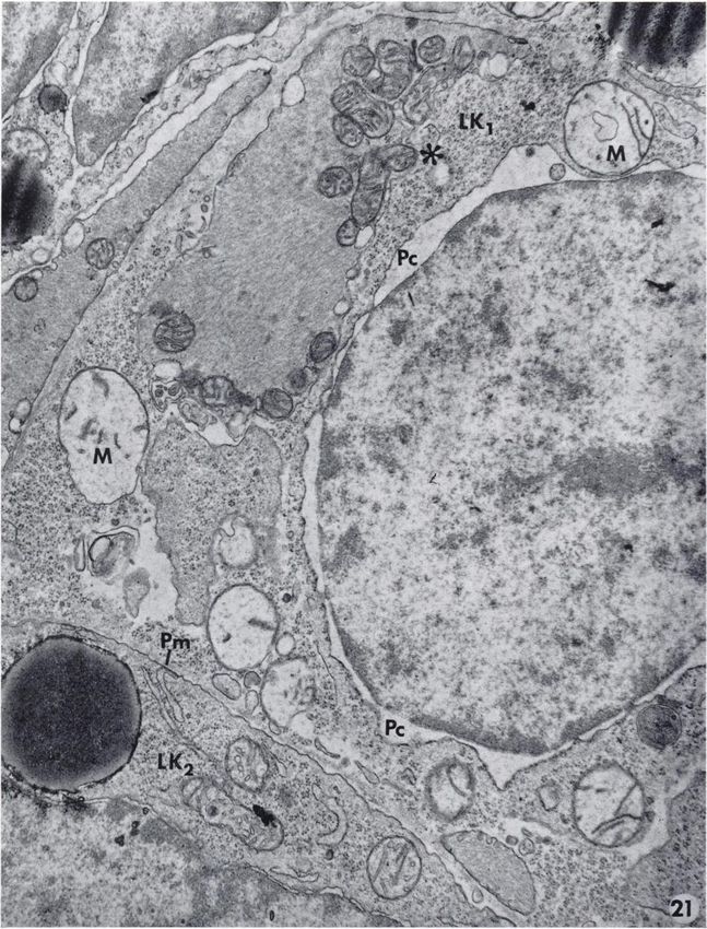
David Brandes, Elsa Anton, and Brian Schofield

(12).3 A more detailed description of the L1210 leukemie cells          of the invaded muscle. In some instances they contained myelin
and of the virus particles characteristic of this neoplasia are to be   bodies which seemed to be derived from degenerating cell or-
found in two recent publications (7, 8).                                ganelles such as mitochondria and ribosomes (Figs. 10, 11). The
   In light microscope preparations stained by the acid phos-           hyperplastic areas frequently appeared as protrusions which
phatase technic for the demonstration of lysosomes (Fig. 2), the        contained predominantly abundant ribosomes (Fig. 11) or hyper-
leukemie cells appeared predominantly negative, but a strong            trophic mitochondria and hyperplastic Golgi elements (Figs. 12,
positive reaction was observed in macrophages. The electron             13). Degenerative changes were detected in many of the marginal
histochemical preparations (Pig. 3) confirmed the paucity of            hypertrophie mitochondria (Fig. 13). The membranes of the
lysosomes in leukemie cells, which usually contained but a few          cristae lost their sharpness, and in many instances the mito
of those particles. In the preparations thus far examined, neither      chondria were occupied by interlacing bands of amorphous ma
lysosomes nor acid phosphatase activity could be seen in the            terial. Structures which seemed to represent residues of de
host-tumor interzone.                                                   generating mitochondria also appeared in these areas.
                                                                           Progressive alterations in skeletal muscle during leukemie cell
The    Host-Tu   mor   Interzone                                        invasion, which appeared directly related to their breakage and
                                                                        replacement of muscle fibers by tumor cells, were observed.
   Light Microscopy. The degree of infiltration and destruction            The sarcolemma disappeared, and the muscle showed a frayed
of skeletal muscle and smooth muscle on the fifth day after trans       irregular surface (Fig. 14). In more advanced stages the surface
plantation of the leukemia are illustrated in light micrographs         showed a frank moth-eaten appearance and the myofibrils had
(Figs. 4, 5). In both cases, the neoplastic cells have infiltrated      begun to disintegrate (Fig. 15). The loss of muscle substance gave
and widened the spaces between the muscle fibers, many of which         rise to the formation of lacunar spaces, and as this process ad
appear as isolated fragments.                                           vanced deeper into the muscle, the fibers became thinner and
   Electron Microscopy. Skektal Muscle Invasion. Before the             irregular (Fig. 16) and showed areas of constriction that appeared
onset of recognizable changes, the plasma membrane of the               to lead to actual fragmentation and disintegration (Figs. 17, 18).
leukemie cells, as well as the sarcolemma of the muscle fibers,            Intestinal Smooth Muscle Invasion. As in the case of skeletal
appeared as continuous dense structures (Fig. 6). The sarcolemma        muscle invasion, some of the smooth muscle fibers showed signs
was externally coated by a basement membrane, and a moderate            of stimulation reflected in enhanced pinocytosis, mitochondrial
amount of connective tissue fibers were seen between the leukemie       aggregates, and hyperplastic Golgi complexes (Fig. 19). Abundant
cells and the muscle. Aggregates of ribosomes, apparently de            clusters of free ribosomes in the interstitial spaces were observed
rived from leukemie cells, were frequently detected in the inter        in the host-tumor interzone, and very frequently the hyperplasia
stitial spaces t>etween leukemie cells and muscle, before any           of cell organelles occurred in the vicinity of the areas with extra
morphologic alterations occurred in either cell type (Fig. 7).          cellular ribosomes (Fig. 20).
  Progressive alterations in leukemie cells appeared to precede            Degenerative changes in the leukemie cells were also seen to
the changes in muscle fibers. Partial or almost complete disap          precede alteration in the smooth muscle fibers. In the neoplastic
pearance of the plasma membrane occurred in many of the leu             cells (Fig. 21), the perinuclear cisternae appeared very dilated,
kemie cells (Fig. 8), and their cytoplasmic content, especially         the mitochondria were swollen, and most of the cristae had dis
ribosomes, appeared free in the interstitial spaces in close ap         appeared. The plasma membrane was no longer visible, and the
position to the surface of the muscle fibers. Several degenerative      various cytoplasmic components of the leukemie cells, especially
changes including pycnosis and frank necrobiosis (Fig. 9), were         the ribosomes appeared free in the interstitial spaces. In some
observed in leukemie cells in the vicinity of morphologically in        areas, the plasma membrane of the smooth muscle cells appeared
tact muscle fibers, but virus particles could still be detected         interrupted, and the fusion of host-tumor cell cytoplasm became
among the debris of the neoplastic cells (Fig. 9, inset).               established (Fig. 21).
   Several changes were observed at the surface of the muscle              The areas of tumor cell-muscle fusion seemed to constitute a
fibers in areas where the plasma membrane of the leukemie cells         "no man's land" with cell organelles "intruding" into both sides
became discontinuous or disappeared, or in the presence of debris       of the line, for mitochondria and myofibrils were seen to lodge
from necrobiotic leukemie cells. Increased micropinocytotic             simultaneously in muscle and leukemie cells (Fig. 22). Pinocytotic
activity and the development of areas of hyperplastic marginal          vacuoles, externally coated or containing ribosomes apparently
sarcoplasm were among the earlier detectable changes in muscle          derived from those present in the interstitial spaces, were ob
fibers which otherwise appeared normal.                                 served in muscle cells, and in areas of fusion, tumor cell ribo
   The micropinocytotic vesicles appeared arranged in rows along        somes appeared to gain access into muscle fibers (Fig. 23). Initial
the muscle surface and contained structureless material of low-         lytic changes were observed in such areas (Fig. 23), and patterns
electron density (Figs. 8, 10, 11). The contents of the areas of        of disintegration of the normal structures in contact with altered
hyperplastic marginal sarcoplasm varied in the different portions       leukemie cells were similar to those described in relation to the
                                                                        invasion and replacement of the skeletal muscle by malignant
   3 It has been proposed, more recently, that the particles in the     cells.
cytoplasmic matrix be designated as ¡ntracytoplasmic A particles,
                                                                           The apparent stimulative effect of tumor cell material on nor
ami those in the eiuloplasmic reticulum as intracisternal   A parti     mal structures appeared to be exerted also on small blood vessels
cles. Suggestions for the Classification of Oncogenic RNA Viruses.      present in the areas of tumor cell invasion. In areas containing
Informal Meeting, New York, February 9, 19fi(i (J. Nati. Cancer         tumor cell debris, the cytoplasm of vascular endothelial cells
Inst,, 37: 395-397, 1966).                                              appeared broadened and contained abundant cell organelles,

2160                                                                                                     CANCER     RESEARCH      VOL. 27

          Downloaded from cancerres.aacrjournals.org on January 17, 2021. © 1967 American Association for Cancer
                                                        Research.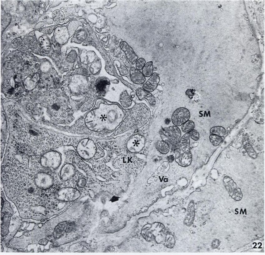
Invasion    of Muscle by LI210         Leukemia

particularly ribosomes (Fig. 24). The luminal surface was thrown          At the level of resolution of the electron microscope, however,
into complex projections and folding's, indicative of intense          the gap between tumor cells and skeletal muscle was considerably
pinocytosis, and hyperplasia of the Golgi apparatus and of the         larger, and the alterations of the normal structures appeared to
rough endoplasmic reticulum was also prevalent (Fig. 25).              occur in areas where tumor cells were breaking down, rather
                                                                       than in relation with vigorously multiplying leukemic cells.
DISCUSSION                                                                Noi (21) failed to detect morphologic or histochemical changes
                                                                       in kidney cells situated in front of or even in direct contact with
   The present results have indicated that during invasion of          growing tumor cells, and he questioned the idea that extracellular
skeletal and smooth muscle, alteration of the leukemic cells,          proteolysis or other destructive processes in normal tissues might
including necrobiosis, occurs before the onset of degenerative         precede tumor invasion.
changes in the normal tissues.                                            Those studies, however, were based on light microscopy ob
   Partial or complete disappearance of the plasma membranes           servations, and it is possible that the author failed to detect the
of the neoplastic cells exposed the surface of the muscle fibers       presence of tumor cell debris in areas of invasion and the initial
directly to the cytoplasmic components of the leukemic element         subcellular changes described in this study.
and particularly to the ribosome aggregates.                              Alteration and lysis of skeletal and smooth muscle fibers were
   In such areas, increased pinocytotic activity was detected at       also detected in areas not immediately in contact with growing
the surface of the muscle fibers, which may indicate the uptake        tumor cells, suggesting that substances extruded from altered
of material derived from the altered leukemic cells. More direct       neoplastic cells may have acted via the interstitial fluid. Lytic
evidence pointing to interchange of material between normal and        changes have been observed at distances greater than 1(X)ß
neoplastia elements was derived from the presence of numerous          from tumor cells, and on the basis of histochemical and biochem
areas of fusion between both cell types, in which cell organelles      ical evidence, this process has been referred to as "extracellular
appeared to protrude from one cell type into the other.                proteolysis" (25, 26), indicative of local abnormal characteristics
   The presence, in the interstitial spaces, of material derived       of the interstitial milieu prevailing in and around tumors (27).
from altered leukemic cells appeared at first to exert a stimu            Increased proteina.se activity at the host-tumor interzone, and
lative effect on the muscle fibers and also on the small blood         possible activation of these enzymes through imbalance of regu
vessels present in the areas of tumor cell invasion. In the skeletal   latory mechanisms, have been related to the destructive capacity
muscle fibers, the apparent stimulative effect was reflected in the    of neoplastic cells (27, 29). The possibility that cathepsin and
development of areas of marginal sarcoplasmic hyperplasia,             other lysosomal enzymes may be involved in tissue breakdown
increase in the number and size of the mitochondria, and hyper         in areas of tumor invasion has been discussed by Pearse and
trophy of the Golgi elements. Aggregates of mitochondria and           Hess (22), but these authors failed to detect high acid phospha-
hypertrophy of the Golgi components were also observed in the          tase activity in tumor cells, except in stromal macrophages.
smooth muscle fibers, and in some instances, both types of hyper-      Similar findings are reported here, where our preparations at the
plastic organelles appeared in close association. The stimulative      light and electron microscopic levels showed very few lysosomes
effect on blood vessels occurred in areas where the interstitial       in the leukemic cells and no acid phosphatase activity in areas
spaces were occupied by cytoplasmic products derived from              of muscle tissue breakdown.
neoplastic cells. The cytoplasm of the capillary endothelial cells        Structural defects in the organization of tumor-cell plasma
appeared broadened and studded with ribosomes, the endoplas-           membrane and increased permeability of this structure (1-3, 5,
mic reticulum and Golgi appeared unusually prominent, and the          27, 28), have been held res]x>nsible for the release of cytoplasmic
luminal surface was thrown into elaborate infoldings and finger-       material including enzymes, but these studies tend to indicate a
like projections which seemed to be actively engaged in the            more severe process, that is, the actual disappearance of the cell
formation of pinocytotic vacuoles.                                     membrane in the more peripheral tumor cells (type A cells of
   A stimulatory effect of tumor cells or their products on normal     Sylvénand Malmgren) (29).
tissues, including blood vessels and stroma, has been reported in         It is possible that some of the cytoplasmic constituents of the
numerous instances, both in vivo (4, 14, 17, 23, 30, 32), and in       leukemic cells, penetrating the normal structures in areas of
vitro (17-19, 23). In such instances, however, tumor cells or          fusion or by pinocytosis may have been responsible for the en
their extracts stimulated the proliferation of normal cells, but in    suing degenerative changes and final lysis of the muscle fibers.
our case only hyperplasia of cell organelles was encountered,          The marginal sarcoplasm of striated fibers as well as in some of
which may have been due to the nondividing nature of the               the smooth muscle fibers contained more ribosomes than are
tissues examined in this study.                                        usually found in normal conditions. Many of our electron micro
   What the biologic changes are that occur at the host-tumor          graphs seemed to indicate that some of the cell ribosomes were
interzone that lead from a condition of stimulation to one of          present in the interstitial spaces, and that incorporation into the
degradation and lysis of normal structures are not known with          muscle fibers may have occurred by pinocytosis or at the areas
certainty, but have attracted much interest and have been the          of fusion of malignant and normal cells.
subject of extensive experimental studies (see Ref. 11 for reviews).      The material extruded from tumor cells which had lost their
   In our light microscopic preparations, the areas of muscle cell     plasma membrane may have served other purposes than to act
invasion were packed with tumor cells, and it appeared that            as a toxic agent involved in the lysis of normal elements in the
compression alone, as suggested in classic textbooks of pa             course of invasion. As suggested by other authors (16, 31), the
thology, may have been a cause in the atrophy and breaking down        material released from the tumor cells may have contained a
of the normal structures.                                              conditioning or growth-stimulating factor required for the suc-

NOVEMBKR 1067

         Downloaded from cancerres.aacrjournals.org on January 17, 2021. © 1967 American Association for Cancer
                                                       Research.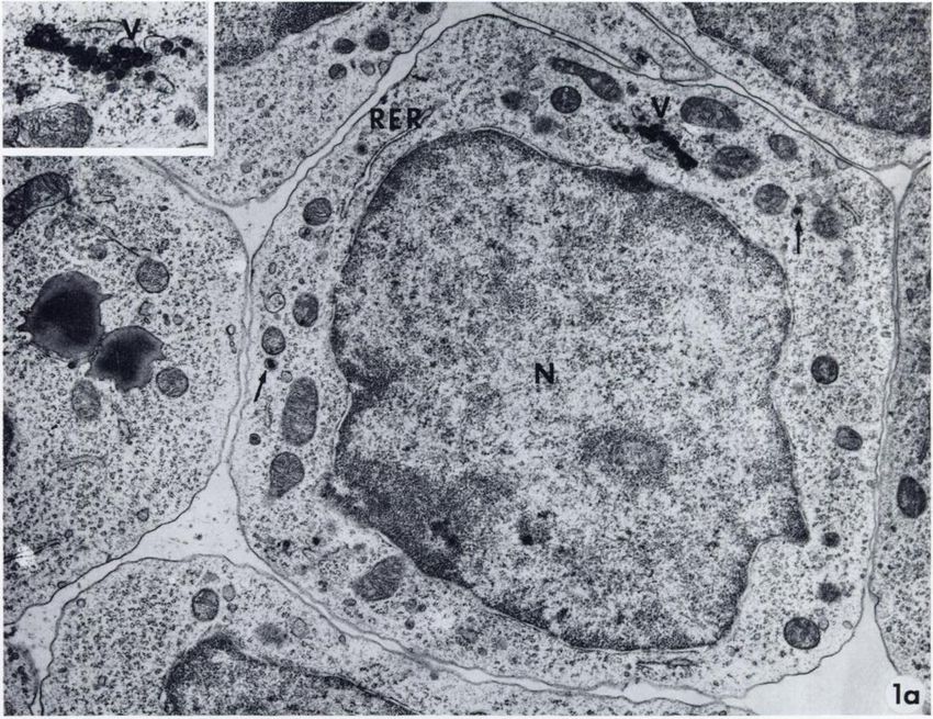
David Brandes, Elsa Anton, and Brian Schofield

cessful establishment of the isologous tumor graft, or may have    8. Brandes, D., Schofield, B. H., Slusser, R., and Anton, E. Stud
served as a source of nutritional material for other tumor cells.     ies of L 1210Leukemia. I. Ultrastrueture of Solid and Ascites
                                                                      Cells. J. Nati. Cancer Inst., 37: 467-485, 1966.
It may therefore be postulated that cell products, including en
zymes and cytoplasmic organelles, liberated by the tumor cells     9. Brandes, D., Sloan, K. W., Anton, E., and Bloedorn, F. Effect
                                                                      of X-Irrudiation on the Lysosomes of Mice Mammary Gland
may have fulfilled several functions such as stimulation of normal    Carcinoma. Cancer Res., 27: 731-746, 1967.
elements, conditioning the establishment of the tumor graft and   10. Braun, W. Effects of Cell Components on Cell Multiplication
served as a source of nutrition for other neoplastic cells. Such a    and Changes in Cell Populations. In: M. J. Brennan and
feedback-like mechanism as stimulation of one Å“il type by            W. L. Simpson (eds.), Biological Interactions in Normal and
products derived from the breaking up of other cell types has         Neoplastic Growth; Henry Ford Hospital International Sym
been considered as an extremely economic intercellular control        posium, pp. 187-197. Boston: Little Brown and Co., 19(i2.
mechanism (10). A lytic action on the invaded structures by       11. Brennan, M. J., and Simpson, W. L. (eds.). Biological Interac
activated enzymes or through the toxic effect of tumor cell           tions in Normal and Neoplastic Growth; Henry Ford Hospital
components would then constitute one facet of the many prop           International Symposium. Boston: Little Brown, and Co.,
erties attributed to the breakdown products derived from tumor        19IÌ2.
                                                                  12. Dalton, A. J. Micromorphology of Murine Tumor Viruses and
cells.                                                                of Affected Cells. Federation Proc., HI: 936-941, 1962.
   Some of the alterations observed in skeletal and smooth muscle 13. Engel, W. K. Mitochondrial Aggregates in Muscle Disease. J.
fibers in the areas of tumor cell infiltration greatly resembled      Histochem. Cytochem., 12: 40-48, 1904.
changes described in various muscle diseases.                     14. Foulds, L. The Histológica! Analysis of Tumors: A Critical
   Mitochondrial aggregates, such as those seen in both types of      Review. Am. J. Cancer, 39: 1-24, 1940.
fibers, have been described in patients with myotonia congenita,  15. Gustafsson, R., Tata, J. R., Lindberg, O., and Ernster, L.
paramyotonia congenita, hypokalemic periodic paralysis, and           The Relationship Between the Structure and Activity of Rat
adynamia episodica hereditaria (13). Hypertrophy and increase         Skeletal Muscle Mitochondria After Thyroidectomy and
                                                                      Thyroid Hormone Treatment. J. Cell Biol., 26: 555-578, 1905.
in mitochondrial population in perinuclear and subsareolemmic
regions such as those seen in this study have also been observed  10. Klein, G., and Klein, E. The Evolution of Independence From
                                                                       Specific Growth Stimulation and Inhibition in Mammalian
in rat skeletal muscle after thyroidectomy and thyroid hormone        Tumor Cell Populations. Symp. Soc. Exptl. Biol., //: 305-328,
stimulation (15). Areas of marginal sarcoplasmic hyperplasia           1957.
containing abundant ribosomes such as seen in this study have     17. Levi-Montalcini, R., and Angeletti. P. U. Considerations on a
also been described in striated muscle in the case of trichinosis      Nerve Growth Promoting Activity of some Tumors. In: M. J.
(20).                                                                  Brennan and W. L. Simpson (eds.), Biological Interactions in
   In our studies, these changes appeared to be localized ex           Normal and Neoplastic Growth; Henry Ford Hospital Inter
clusively in the areas of tumor cell infiltration, thus indicating a   national Symposium, pp. 225-237. Boston: Little Brown and
local reaction to the invading neoplastie celLs rather than a          Co., 1902.
systemic disease as in the case of most of the above conditions.  18. Ludford, R. J., and Barlow, H. The Influence of Malignant
                                                                       Cells Upon the Growth of Fibroblasts in vitro. Cancer Res.,
                                                                       4: 094-703, 1944.
ACKN OWLEDGMKNTS
                                                                  19. Ludford, R. J., and Barlow, H. Sarcomatous Transformation
  We wish to thank M. D. Tuduhl, Sr., for assistance in the            of the Stroma of Mammary Carcinomas that Stimulated
photography.                                                           Fibroblastic Growth in vitro. Cancer Res., 5: 257-264, 1945.
                                                                  20. Maeir, D. M., and Zaiman, II. The Development of Lysosomes
REFERENCES                                                             in Rat Skeletal Muscle in Trichinous Myositis. J. Histochem.
 1. Abercrombie, M., and Ambrose, K. I. The Surface Properties         Cytochem., 1J,:390-400, 1900.
    of Cancer Cells: A Review. Cancer Res., 32: 525-548, 1902.    21. Noi, V. J. Quoted by J. M. Vasiliev and V. I. Guelstein. In:
 2. Abercrombie, M., Heaysman, J. E. M., and Karthauser, H. M.         Local Cell Interactions in Neoplasms and in the Foci of Car-
    Social Behavior of Cells in Tissue Culture. III. Mutual Influ      cinogenesis. Progr. Exptl. Tumor Res., 8: 26-65, 1906.
    ence of Sarcoma Cells and Fibroblasta, Exptl. Cell Res., 13: 22. Pearse, A. G. E., Hess, R., and Snyder, R. Stromal and Tumor
    276-291, 1957.                                                     Cathepsins and Their Relationships to Metabolic Activity
 3. Ambrose, E. J., Easty, D. M., and Jones, P. C. T. Specific         and Invasiveness. In: M. J. Brennan and W. L. Simpson (eds.),
    Reactions of Poly-electrolytes with the Surfaces of Normal         Biological Interactions in Normal and Neoplastic Growth;
    and Tumor Cells. Brit. J. Cancer, 12: 439-447, 1958.                Henry Ford Hospital International Symposium, pp. 057-670.
 4. Argyris, B. F., and Argyris, T. S. Mechanism of Mammary            Boston- Little Brown and Co., 1962.
    Duct Stimulation by Tumor Transplants. Cancer Res., 21: 23. Ranadive, K. J., and Bhide, S. V. Tissue Interactions Between
    75-81, 1961.                                                       Normal and Malignant Cells In: M. J. Brennan and W. L.
 5. Bennet, L. R., and Cannon, F. E. Effects of Lytic Agents on        Simpson (eds.), Biological Interactions in Normal and Neo
    Plasma Membrane of Ehrlich Ascites Tumor Cells and Mouse           plastic Growth; Henry Ford Hospital International Sympo-
    Erythrocytes. J. Nati. Cancer Inst., 19: 999-1012, 1957.           posium, pp. 337-354. Boston: Little Brown and Co., 1902.
 6. Berenblum, I. The Nature of Tumor Growth. In: II. Florey 24. Sabatini, D. D., Bensch, K., and Barrnet, R. J. Cytochemistry
    (ed.), General Pathology, Ed. 3, pp. 528-550. Philadelphia:        and Electron Microscopy. The Preservation of Cellular Ultra-
    W. B. Saunders Co., 1962.                                          structure and Enzymatic Activity by Aldehyde Fixation. J.
 7. Brandes, D., Anton, E., and Lam, K. W. Studies of L 1210           Cell Biol., 17: 19-58, 1903.
    Leukemia. II. Ultrastructural and Cytochemical Changes after   25. Sylvcn, B. Ester Sulphuric Acids of High Molecular Weight
    Treatment with Cyclophosphamide and Vitamin A. J. Nati.            and Mast Cells in Mesenchymal Tumors. Acta Radio)., Suppl.
    Cancer Inst., in press.                                            59, 1945.

2162                                                                                               CANCER RESEARCH VOL. 27

         Downloaded from cancerres.aacrjournals.org on January 17, 2021. © 1967 American Association for Cancer
                                                       Research.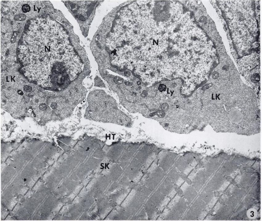
Invasion    of Muscle by L1210 Leukemia

20. Sylvén,B. The Biochemical Mechanism Underlying the De-           30 Vasiliev, J. M. The Role of Connective Tissue Proliferation in
    structive Growth of Tumors. Acta UnióIntern. Contra Can-              Invasive Growth of Normal and Malignant Tissues : A Review.
    crum, 14: 61-02, 1958.                                                 Brit. J. Cancer, 18: 524-536, 1958.
27. Sylvén,B. The Host-Tumor Interzone and Tumor Invasion.           'Al. Vasiliev, J. M. The Local Stimulatory Effect of Normal Tissues
    In: M. J. Brennan and W. L. Simpson (eds.), Biological Inter           Upon the Growth of Tumor Cells. In: M. J. Brennan and W. L.
    actions in Normal and Neoplastic Growth; Henry Ford Hos                Simpson (eds.), Biological Interactions in Normal and Neo
    pital International Symposium, pp. 635-655. Boston: Little             plastic Growth; Henry Ford Hospital International Sym
    Brown and Co., 1962.                                                   posium, pp. 299-309. Boston: Little Brown and Co., 19(i2.
28. Sylvén,B., and Malmgren, H. Topical Distribution of Pro-         32, Vasiliev, J. M., and Guelstein, V. I. Local Cell Interactions in
    teolytic Activities in Some Transplanted Mice Tumors. Exptl.           Neoplasms and in the Foci of Carcinogenesis. Progr. Exptl.
    Cell Res., 8: 575-577, 1955.                                           Tumor Res., 8: 26-65, 1966.
29. Sylvén, B., and Malmgren, II. The Histológica!Distribution of   33 Willis, R. A. The Direct Spread of Tumors. In: Pathology of
    Proteinase and Peptidase Activity in Solid Tumor Trans                 Tumors, Ed. 2, pp. 147-165. London: Butter-worth and Co.
    plants. Acta Radiol., Suppl. 154, 1957.                                (Publishers Ltd.), 1953.

NOVEMBER 19G7                                                                                                                       2103

         Downloaded from cancerres.aacrjournals.org on January 17, 2021. © 1967 American Association for Cancer
                                                       Research.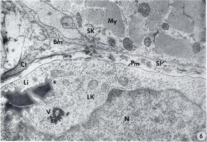
David Brandes, Elsa Anton, and Brian Schoficld
FIG. 1. Normal appearance of L1210 leukemic cells. The nuclei (.V) occupy a large portion of the cell. The cytoplasm is studded
with ribosomes and shows virus particles (V), some of which are of the intracisternal type (arrows). The Golgi complex ((?) is well
developed, hut the rough-surfaced endoplasmic reticulum (REIt) is scarce, a, X 10,500; 6, X 20,500; inset, X 30,000.
FIG. 2. Light micrograph. A positive reaction is seen in a few macrophages (arrows). The muscle fibers (asterisks) and the invading
leukemic cells show no apparent activity. Acid phosphatase stain, X 350.
FIG. 3. Electron micrograph. The leukemic cells (LK) contain few small lysosomes (Ly) which were not resolved in the light micro
graphs. Neither the muscle fiber (SK) nor the host-tumor (HT) interzone show any apparent activity. A', nuclei. Acid phosphatase
stain, X 11,500.
FIGS. 4 AND 5. One-micron sections from blocks embedded in Kpon from which ultrathin sections were then cut forelectron micros
copy. Toluidine blue stain.
FIG. 4. Invasion of smooth muscle of intestinal wall. Fragmented muscle fibers (arrows) are surrounded by leukemic cells. X 350.
FIG. 5. Invasion of skeletal muscle. Some fragmented fibers (arrows) are surrounded by leukemic cells, but others (asterisks) are seen
in areas showing no leukemic infiltration. X 350.
FIG. 6. Portion of a leukemic cell (LA') infiltrating a space between skeletal muscle fibers (SK). The plasma membrane (Pni) is still
intact and so is the sarcolemma (SI). The latter is externally coated by a thin layer of amorphous material with the appearance of a
basement membrane (Urn). The myofibrils (My) appear normal. N, nucleus; V, virus particles; Li, lipid droplets; Ct, connective tissue
fibers. X 20,000.
FIG. 7. Host-tumor interzone showing clusters of ribosomes (arrows) in the interstitial space. The plasma membrane of the leukemic
cell (LK) and the sarcolemma (SI) are still intact. JV, nucleus of leukemic cell; .V, nucleus of skeletal muscle fiber. X 10,500.
FIG. 8. Degenerating leukemic cell (/>K) in the midst of skeletal muscle fibers (SK). The nucleus (X) is undergoing pycnosis. The
plasma membrane is no longer visible, and the cytoplasm is in direct continuity with the interstitial space (arrows). The sarcolemma.
(Si) is intact and shows numerous pinocytotic vesicles. Virus particles are still present (V) in the tumor cell cytoplasm. X 21,500.
FIG. 9. Two necrobiotic leukemic cells (LK\, AA'2). Cytoplasmic debris have spread through the interstitial space (asterisk). An
adjacent muscle fiber (SK) appears normal and the sarcolemma (SI) is intact. Virus particles (V) are still seen in the necrobiotic leu
kemic cell. They are shown at higher magnification in the iipper right inset. Part of a macrophage (MP) is also visible. X 9,500.
Inset, X 18,500.
FIG. 10. Cytoplasmic materials from disintegrating leukemic cells are in continuity with the interstitial space. The nuclei (N) are
undergoing pycnosis. The skeletal muscle fiber (SK) contains numerous myelin bodies (My). X 18,500.
FIG. 11. A skeletal muscle fiber (SK) in contact with leukemic cell debris (asterisk) shows an area of hyperplastic marginal sarco-
plasm (star) containing abundant ribosomes and myelin bodies. X 20,000.
FIG. 12. Skeletal muscle fiber (SK) in an area of invasion. Hyperplastic marginal sarcoplasm containing an accumulation of hyper-
trophic mitochondria (M), abundant ribosomes, and a prominent Golgi complex (G). X 23,500.
FIG. 13. Area similar to that shown in Fig. 12 shows degenerative changes in mitochondria (arrows). A prominent Golgi complex (G)
is visible. SK, skeletal muscle fiber. X 21,500.
FIG. 14. Initial steps in the process of skeletal muscle disintegration. The sarcolemma is no longer visible and the surface of the
muscle fibers show a frayed appearance (arrow). A degenerating leukemic cell (LK) devoid of plasma membranes is present in this area.
X 11,500.
FIG. 15. The process of skeletal muscle degeneration is more advanced. The sarcolemma has disappeared, and the frayed appearance
of the surface is accentuated (arrows). The myofibrils start to disintegrate (asterisk). Abundant debris of tumor cell is seen in the inter
stitial space. X 23,500.
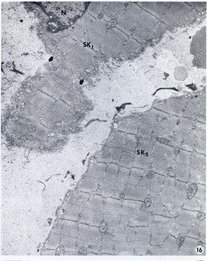
FIG. 16. Skeletal muscle fibers (SKi) in the process of fragmentation. The myofibrils are interrupted in an area of constriction (ar
rows). A second fiber (SKt) shows incipient fraying of the surface. No neoplastic cells are seen in this area, but the interstitial space
contains cell debris. N, nucleus of muscle cell. X 14,500.
FIG. 17. Isolated skeletal muscle fragments (SK) in an area which contains no tumor cells. Cellular debris are present in the inter
stitial spaces. N, nucleus of muscle cell. X 10,000.
FIG. 18. Diaphragmatic muscle in an area of ascites tumor cell invasion. The larger fragment (.4) contains mitochondria] aggregates
and shows striai ed fibrils (SF). Pinocytotic and finger-like projections at the cell surface are abundant. Small portions of muscle remain
attached to larger fragments by narrow Cytoplasmic bridges (arrows). No tumor cells are present in this area. X 10,000.
FIG. 19. Smooth muscle (SM) from intestinal wall, in the neighborhood of an area of leukemic infiltration. Aggregates of mitochondria
(M), hypertrophie Golgi elements (G), and increased pinocytotic activity are observed in many muscle cells. X 20,000.
FIG. 20. Smooth muscle fibers (SM) surrounded by leukemic cells (LK). Abundant rosettes of ribosomes are free in the interstitial
spaces (asterisk) between both cell types. Mitochondria, some with altered cristae, Golgi elements (G), myelin bodies (arrows), and ribo
somes are concentrated near such areas. X 21,500.
FIG. 21. Altered leukemic cell (AA'i) with marked dilation of the perinuclear cisterna (Pc) and swollen mitochondria (M). The plasma
membrane has almost completely disappeared, and areas of fusion of tumor cell and muscle cells are frequent (asterisk). Mitochondrial
aggregates are seen in such areas. The plasma membrane (Pm) of another leukemic cell (LA%) is intact. X 30,500.
FIG. 22. Extensive fusion of altered leukemic cell (LK) and smooth muscle (SM). Morphologic alterations in leukemic cell mitochon
dria (asterisk) are more obvious. Tumor cell ribosomes are in contact or within muscle cytoplasm which also shows vacuoles (Va) con
taining ribosomes. Initial lytic changes are seen in the muscle fiber at the area of the fusion with the leukemic cells (arrow). X 21,500.
FIG. 23. a, a mitochondrion (M) is partially within a leukemic cell (LK) and partially within a smooth muscle filier (SM). b, Arrow
shows where sarcolemma (SI) becomes interrupted. Muscle fibrils (star) have also penetrated the leukemic cell cytoplasm, a, X 10,500;
b, X 18,500.
FIG. 24. Capillary in area containing tumor cell debris. The endothelium is hypertrophie and shows enhanced pinocytotic activity
(arrous). X 11,500.
FIG. 25. Hyperplasia of Golgi elements (G) and of rough endoplasmic reticulum in capillary endolhelium in the area of tumor cell
invasion. X 18,500.
2164 CANCER RESEARCH VOL. 27
Downloaded from cancerres.aacrjournals.org on January 17, 2021. © 1967 American Association for Cancer
Research.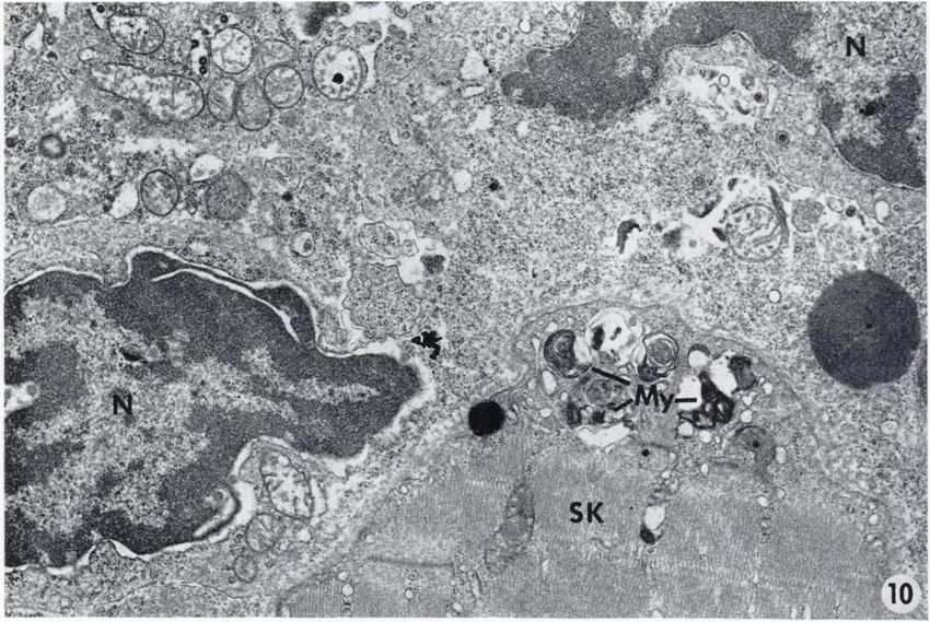
» \•ï'*:
                                 fC 'j' •
                                Jvfa*Sf     Ii

¡•"•A'-..
          •••••
                -         •••;-.
"'

     ^vÃ-$C!^¿>V-:::-.                                         -:^-; #•«$%••
Ì?
;^
^    »I
     _^nH       ^^L   *•
                         -^ ^   i*^5f i. * » v,   i    >'-^   4 .^BK   ff

Ã-^ü                                                                                N
                                                                                       ,

                      ••
                         *\ wt v »\-Ã-f- „y ,
                                                   •.-.1

                            _•'* ** v

                                                                                                                 -\vrÃ-. j ••'.
                                                                                                                 •fc   —

                 •
                 i* •

                                          RE R                                   G

                                                                                                              •
                                                                                                               •             >' "**{•"'*'
                                                                                                                                       ¿gy-v

&¿t                                                                                                    S^Äif>
"**
                                                                                                                                      wr xW

     NOVEMBER 1967                                                                                                                   2160

                      Downloaded from cancerres.aacrjournals.org on January 17, 2021. © 1967 American Association for Cancer
                                                                    Research.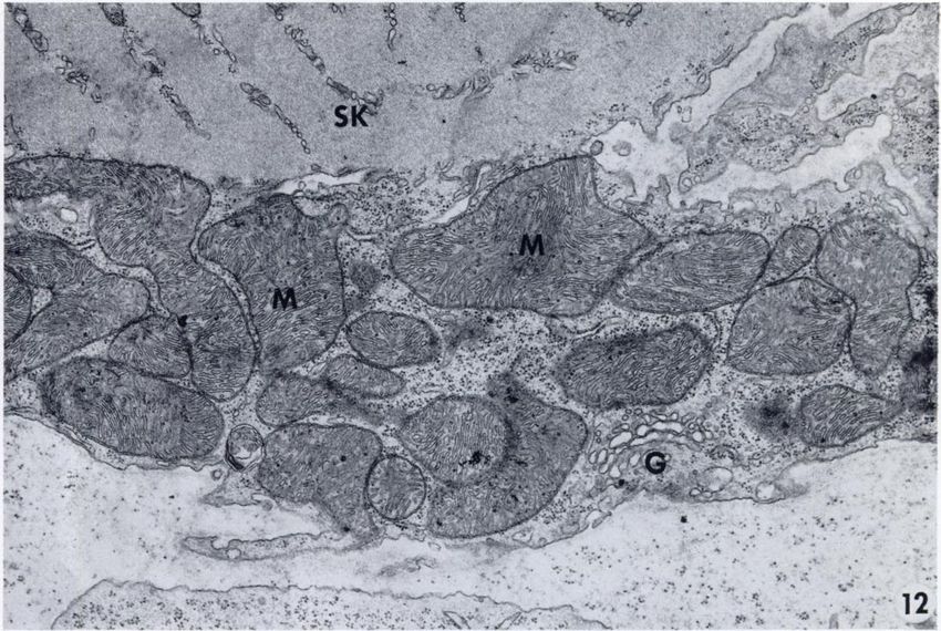
David Brandes, Elsa Anton, and Brian Schofield
                          •••'

                                *_**   *..

2166                                                                                      CANCER    RESEARCH    VOL. 27

       Downloaded from cancerres.aacrjournals.org on January 17, 2021. © 1967 American Association for Cancer
                                                     Research.
Invasion of Muscle by L1210 Leukemia

                                          «Si

NOVEMBER 1907                                                                                                  2167

      Downloaded from cancerres.aacrjournals.org on January 17, 2021. © 1967 American Association for Cancer
                                                    Research.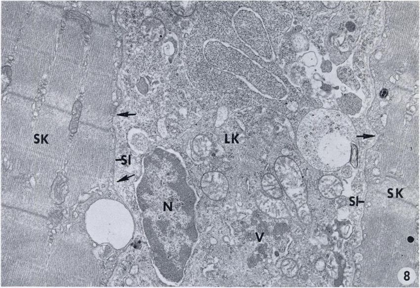
:
                                                       P;                My
                                                        SK

                                   —r—sr"Ã-""^
                                                                                             •v
                                                                              •-'•
                                                                                    •' ;
                                                                                                            v-
                                                                                                   .:   "

                                    v*7^
SK
sr Ã- 4»-•
-•*
» . •
. 0- '•••'•-
-m
«L. '. -
î^- ?Ã-'Ã- té^
" LK,r
•ii-^is^fe - <
. •-..•
:--
- •-4cS*
^>
•>
...
E^S^Atfei •' ; ' ï J
'"•"-^-^¿^'^•^ ;'.' ' .
SK
tu? i "•••^
Éa^v'v- ?iii
NOVEMBIÕH 19IÕ7
Downloaded from cancerres.aacrjournals.org on January 17, 2021. © 1967 American Association for Cancer
Research.N
'•»>
">
SK
10
SK
I
2170 CANCER RESEARCH VOL. 27
Downloaded from cancerres.aacrjournals.org on January 17, 2021. © 1967 American Association for Cancer
Research.?
'••••- ,
                     •
                      "/#--
               "    •
                     •••-"-            J   ''
                        •i- *'   .-   V.

                       1'

                                                                                                                 '•l-là •"
                                                                                                                            -

   •

                                                                                                                   14

                                                                                                                •*>>J.

                                                                                                            f

                                                                          .
                                                                                    '

                                                                                        C

                                                                              ,¡

2172                                                                                        CANCER   RESEARCH   VOL. 27

       Downloaded from cancerres.aacrjournals.org on January 17, 2021. © 1967 American Association for Cancer
                                                     Research.
Invasion of Muscle by LI210 Leukemia
SK
..,.**«.
; x;*<
SK2
M
>
1
16
NUVEMliKI! 1!)()7 2173
Downloaded from cancerres.aacrjournals.org on January 17, 2021. © 1967 American Association for Cancer
Research."•'- -*,rSj
• • •
"-
•
.
•.
•:,.?jtÃ-. ;-
CANCER RESEARCH VOL. 27
Downloaded from cancerres.aacrjournals.org on January 17, 2021. © 1967 American Association for Cancer
Research.Invasion of Muscle by Li210 Leukemia
19
IjP^f:
.' •* ' "^ - ¿' ,
Õ5£
fÇ^:^,^^~-r^^v *•&-•::'"
NOVEMBER 1907 2175
Downloaded from cancerres.aacrjournals.org on January 17, 2021. © 1967 American Association for Cancer
Research.Pm
PC
'LIC
2176 CÕNCER RESEARCH VOL. 27
Downloaded from cancerres.aacrjournals.org on January 17, 2021. © 1967 American Association for Cancer
Research...-
I
SM
:
* M
'*!?> -
; ^ : ^..
:''' VÖ ''
^
22
SM
'Si
LK 23
a »
" . *"'•-'
NOVEMBER 19G7 2177
Downloaded from cancerres.aacrjournals.org on January 17, 2021. © 1967 American Association for Cancer
Research.David Brandes, Elsa Anton, and Brian Schofield
*
o j - '
G
•-
\ . •>
¿f.
'
2178 CANCER RESEARCH VOL. 27
Downloaded from cancerres.aacrjournals.org on January 17, 2021. © 1967 American Association for Cancer
Research.Invasion of Skeletal and Smooth Muscle by L1210 Leukemia
David Brandes, Elsa Anton and Brian Schofield
Cancer Res 1967;27:2159-2178.
Updated version Access the most recent version of this article at:
http://cancerres.aacrjournals.org/content/27/11_Part_1/2159
E-mail alerts Sign up to receive free email-alerts related to this article or journal.
Reprints and To order reprints of this article or to subscribe to the journal, contact the AACR Publications
Subscriptions Department at pubs@aacr.org.
Permissions To request permission to re-use all or part of this article, use this link
http://cancerres.aacrjournals.org/content/27/11_Part_1/2159.
Click on "Request Permissions" which will take you to the Copyright Clearance Center's (CCC)
Rightslink site.
Downloaded from cancerres.aacrjournals.org on January 17, 2021. © 1967 American Association for Cancer
Research.You can also read


