Subtype-Directed Differentiation of Human iPSCs into Atrial and Ventricular Cardiomyocytes
←
→
Page content transcription
If your browser does not render page correctly, please read the page content below
Protocol
Subtype-Directed Differentiation of Human
iPSCs into Atrial and Ventricular Cardiomyocytes
                                                                                      Mandy Kleinsorge, Lukas
                                                                                      Cyganek
                                                                                      lukas.cyganek@gwdg.de

                                                                                      HIGHLIGHTS
                                                                                      Subtype-directed differentiation
                                                                                      of hiPSCs into atrial and
                                                                                      ventricular cardiomyocytes

                                                                                      Simple, robust, and efficient
                                                                                      monolayer-based differentiation
                                                                                      protocol

                                                                                      Approx. 90%–95% of the
                                                                                      intended cardiac subtype within
                                                                                      cTNT+ cells can be obtained

The generation of homogeneous populations of subtype-specific cardiomyocytes
derived from human induced pluripotent stem cells (hiPSCs) is crucial in
cardiovascular disease modeling as well as in drug discovery and cardiotoxicity
screenings. This protocol describes a simple, robust, and efficient monolayer-
based differentiation of hiPSCs into defined atrial and ventricular cardiomyocytes.

                                                                                      Kleinsorge & Cyganek, STAR Protocols --,
                                                                                      100026
                                                                                      --, 2020 ª 2020 The Authors.
                                                                                      https://doi.org/10.1016/j.xpro.2020.100026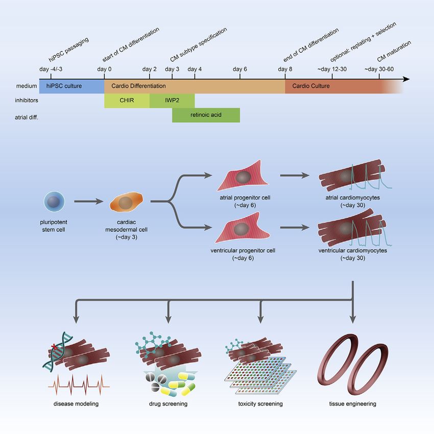
Please cite this article in press as: Kleinsorge and Cyganek, Subtype-Directed Differentiation of Human iPSCs into Atrial and Ventricular
 Cardiomyocytes, STAR Protocols (2020), https://doi.org/10.1016/j.xpro.2020.100026

Protocol
Subtype-Directed Differentiation of Human
iPSCs into Atrial and Ventricular Cardiomyocytes
Mandy Kleinsorge1,2 and Lukas Cyganek1,2,3,4,*
1Clinic for Cardiology and Pneumology, University Medical Center Göttingen, 37075 Göttingen, Germany

2DZHK   (German Center for Cardiovascular Research), partner site Göttingen, Göttingen, Germany
3Technical   Contact
4Lead   Contact
*Correspondence: lukas.cyganek@gwdg.de
https://doi.org/10.1016/j.xpro.2020.100026

SUMMARY
The generation of homogeneous populations of subtype-specific cardiomyo-
cytes derived from human induced pluripotent stem cells (hiPSCs) is crucial in
cardiovascular disease modeling as well as in drug discovery and cardiotoxicity
screenings. This protocol describes a simple, robust, and efficient monolayer-
based differentiation of hiPSCs into defined atrial and ventricular cardiomyo-
cytes.
For complete details on the use and execution of this protocol, please refer to
Cyganek et al., 2018.

BEFORE YOU BEGIN
Human induced pluripotent stem cells (hiPSCs) have to be obtained and used ac-
cording to the respective legal and ethical guidelines. hiPSCs and derivatives
thereof are maintained in a humidified incubator at 37 C and 5% CO2. All cell
culture procedures are performed in a Class II biosafety cabinet using sterile
conditions.

Coating of Culture Plates
For enhanced and homogeneous attachment of cells, culture plates must be coated
with extracellular matrix proteins or protein mixtures.

1. Dilute Matrigel:
   a. Thaw Matrigel (7-10 mg/ml) on ice at 4 C for 16-24 h.
   b. Distribute Matrigel in 250 ml aliquots into 50 ml centrifuge tubes using ice-cold
      pipette tips and tubes, and directly freeze.
   c. Store aliquots at -20 C for up to one year.
2. Coat cell culture-treated multi-well plates (e.g. 6-well plates):
   a. Resuspend a frozen Matrigel aliquot in 29.75 ml cold PBS to obtain the work-
      ing dilution of 1:120.
   b. Add 1 ml of diluted Matrigel per well of a 6-well plate (approx. 6-9 mg/cm2) and
      ensure that the entire well surface is covered.
   c. Incubate at 37 C for 60 min or at 4 C for 16-24 h.

   Note: Coated plates can be stored at 4 C for up to 2 weeks, but should not be
   used if the Matrigel has dried up.

                                                                                     STAR Protocols --, 100026, --, 2020 ª 2020 The Authors.          1
                                  This is an open access article under the CC BY-NC-ND license (http://creativecommons.org/licenses/by-nc-nd/4.0/).
Please cite this article in press as: Kleinsorge and Cyganek, Subtype-Directed Differentiation of Human iPSCs into Atrial and Ventricular
Cardiomyocytes, STAR Protocols (2020), https://doi.org/10.1016/j.xpro.2020.100026
Alternatives: Instead of Matrigel, plates can also be coated with Geltrex
(approx. 17 mg/cm2) or defined matrices such as recombinant Laminin-521
(approx. 0.9 mg/cm2).
KEY RESOURCES TABLE
REAGENT or RESOURCE SOURCE IDENTIFIER
Antibodies
a-actinin monoclonal mouse IgG1 Sigma-Aldrich Cat# A7811, RRID:AB_476766
CTNT monoclonal mouse IgG1 Thermo Fisher Cat# MA5-12960,
Scientific RRID:AB_11000742
RYR2 polyclonal rabbit IgG Sigma-Aldrich Cat# HPA020028,
RRID:AB_1856528
MLC2A monoclonal mouse IgG2a Synaptic Systems Cat# 311 011, RRID:AB_887737
MLC2V polyclonal rabbit IgG Proteintech Cat# 10906-1-AP,
RID:AB_2147453
Alexa Fluor 488 donkey anti-rabbit Thermo Fisher Cat#A21206,
Scientific RRID:AB_2535792
Alexa Fluor 555 donkey anti-mouse Thermo Fisher Cat#A31570,
Scientific RRID:AB_2536180
Chemicals, Peptides, and Recombinant Proteins
StemPro Accutase Thermo Fisher Cat#A1110501
Scientific
Albumin, Human Recombinant Sigma-Aldrich Cat#A9731
B-27 Supplement Thermo Fisher Cat#17504044
Scientific
B-27 Supplement minus Insulin Thermo Fisher Cat#A18956-01
Scientific
Bovine Serum Albumin (BSA) Sigma-Aldrich Cat#A2153
CHIR99021 Merck Millipore Cat#361559
Cryostor CS10 STEMCELL Cat#07930
Technologies
DAPI (4’,6-Diamidino-2-Phenylindole, Thermo Fisher Cat#D3571
Dilactate) Scientific
DMSO Sigma-Aldrich Cat#D2650
DPBS 13 Thermo Fisher Cat#14190094
Scientific
E8 Medium Thermo Fisher Cat#A1516901
Scientific
EDTA Carl Roth Cat#8040
Fetal Bovine Serum (FBS) Thermo Fisher Cat#10270-106
Scientific
Fluoromount-G Thermo Fisher Cat#00-4958-02
Scientific
Geltrex (Growth Factor Reduced) Thermo Fisher Cat#A1413302
Scientific
HEPES Buffer (pH 7.0-7.6) Sigma-Aldrich Cat#H3662
Hoechst 33342, Trihydrochloride, Thermo Fisher Cat#H3570
Trihydrate Scientific
ROTI Histofix 4% Carl Roth Cat#P087.5
(Continued on next page)
2 STAR Protocols --, 100026, --, 2020Please cite this article in press as: Kleinsorge and Cyganek, Subtype-Directed Differentiation of Human iPSCs into Atrial and Ventricular
Cardiomyocytes, STAR Protocols (2020), https://doi.org/10.1016/j.xpro.2020.100026
Continued
REAGENT or RESOURCE SOURCE IDENTIFIER
IWP2 Merck Millipore Cat#681671
IWP4 Reprocell Europe Cat#04-0036C
L-Ascorbic Acid 2-Phosphate Sigma-Aldrich Cat#A8960
Laminin 521 Biolamina Cat#LN521-05
Matrigel (Growth Factor Reduced) BD Cat#354230
Penicillin/Streptomycin Thermo Fisher Cat#15140-122
Scientific
Pro Survival Factor DDD00033325 Merck Millipore Cat#529659
Retinoic Acid Sigma-Aldrich Cat#R2625
ROCK Inhibitor Y27632 Merck Millipore Cat#688000
RPMI 1640 with GlutaMax and HEPES Thermo Fisher Cat#72400021
Scientific
RPMI 1640 (without glucose and Thermo Fisher Cat#11879020
HEPES) Scientific
Sodium Chloride Carl Roth Cat#9265.1
Sodium DL-Lactate Solution Sigma-Aldrich Cat#L4263
Stem MACS iPS Brew XF Medium Miltenyi Biotec Cat#130-104-368
StemPro Accutase Cell Dissociation Thermo Fisher Cat#A1110501
Reagent Scientific
StemFlex Medium Thermo Fisher Cat#A3349401
Scientific
Thiazovivin Merck Millipore Cat#420220
Triton X-100 Sigma-Aldrich Cat#T8787
103 TrypLE Select Thermo Fisher Cat#A1217701
Scientific
0.05% Trypsin-EDTA Thermo Fisher Cat#25300-054
Scientific
0.25% Trypsin-EDTA Thermo Fisher Cat#25200056
Scientific
Versene Solution (0.48 mM EDTA) Thermo Fisher Cat#15040033
Scientific
Wnt-C59 Peprotech Cat#1248913
MATERIALS AND EQUIPMENT
hiPSC Culture Medium
Complement Stem MACS iPS-Brew XF Basal medium with Stem MACS iPS-Brew XF
503 supplement. Store at 4 C for up to two weeks.
Alternatives: Instead of StemMACS iPS-Brew XF, E8 medium or StemFlex me-
dium can be used for the maintenance culture of hiPSCs.
EDTA Dissociation Buffer 0.5 mM
Add 1 ml/ml EDTA (from a stock solution of 0.5 M in ddH2O and pH 8.0) to PBS (Ca2+/
Mg2+-free). Add 1.8 mg/ml sodium chloride to adjust the osmolarity to ~340 mOsm.
Sterilize by filtration. Store at 4 C for up to 6 months.
Alternatives: Instead of preparing EDTA Dissociation Buffer, commercially
available EDTA solutions, such as Versene, can be used.
STAR Protocols --, 100026, --, 2020 3Please cite this article in press as: Kleinsorge and Cyganek, Subtype-Directed Differentiation of Human iPSCs into Atrial and Ventricular
Cardiomyocytes, STAR Protocols (2020), https://doi.org/10.1016/j.xpro.2020.100026
Thiazovivin 2 mM
Dissolve 0.62 mg/ml Thiazovivin in DMSO to obtain a 1,0003 stock solution of 2 mM.
Aliquot and store at -20 C for up to one year.
Alternatives: Instead of Thiazovivin, ROCK Inhibitor Y27632 or Pro Survival
Factor DDD00033325 can be used to minimize cell death during splitting/
digestion. Prepare a 5 mM stock solution (1,0003), by dissolving 1.60 mg/
ml Y27632 in DMSO or 1.47 mg/ml of DDD00033325 in DMSO. Apply either
of these at a final concentration of 5 mM.
hiPSC Cryopreservation Medium 23
Supplement hiPSC Culture Medium with 20% DMSO and a final concentration of
4 mM Thiazovivin (1:500 from 2 mM stock). Store at 4 C for up to one week.
Alternatives: If a hiPSC Culture Medium other than Stem MACS iPS-Brew XF is
used, the respective medium should also be used as basis for the Cryopreser-
vation Medium. Alternatively, a commercial cryopreservation medium such as
Cryostor CS10 may be used.
Cardio Differentiation Medium
Supplement RPMI 1640 (with GlutaMAX and HEPES) with 0.2 mg/ml L-ascorbic acid
2-phosphate and 0.5 mg/ml human recombinant albumin. Mix until dissolved and
filter to sterilize. Store at 4 C for up to one month.
Optional: Add antibiotics, such as 13 penicillin/streptomycin (from 1003).
Alternatives: If no satisfactory differentiation can be achieved with a cell line
using the above medium, a different medium can be applied instead: for
this, supplement RPMI 1640 (with GlutaMAX and HEPES) with a final concen-
tration of 13 B-27 Supplement minus insulin (from 503). Store at 4 C for up to
1 month.
Cardio Culture Medium
Supplement RPMI 1640 (with GlutaMAX and HEPES) with the final concentration of
13 B-27 Supplement (from 503). Store at 4 C for up to one month.
Optional: Add antibiotics, such as 13 penicillin/streptomycin (from 1003).
Cardio Recovery Medium
Supplement Cardio Culture Medium with 20% Fetal Bovine Serum and a final con-
centration of 2 mM Thiazovivin. Store at 4 C for up to two weeks.
Optional: Add antibiotics, such as 13 penicillin/streptomycin (from 1003).
Alternatives: Cardio Culture Medium with 2 mM Thiazovivin only can be used.
However, the addition of serum might be beneficial for cell survival.
Cardio Selection Medium
Supplement RPMI 1640 (without glucose and HEPES) with 0.2 mg/ml L-ascorbic acid
2-phosphate and 0.5 mg/ml human recombinant albumin, as well as a final concen-
tration of 4 mM lactate/HEPES (1:250 from 1 M stock). Store at 4 C for up to one
month.
4 STAR Protocols --, 100026, --, 2020Please cite this article in press as: Kleinsorge and Cyganek, Subtype-Directed Differentiation of Human iPSCs into Atrial and Ventricular
Cardiomyocytes, STAR Protocols (2020), https://doi.org/10.1016/j.xpro.2020.100026
Optional: Add antibiotics, such as 13 penicillin/streptomycin (from 1003).
Cardio Cryopreservation Medium 53
Mix 50% DMSO and 50% Fetal Bovine Serum. Add a final concentration of 10 mM
Thiazovivin (1:200 from 2 mM stock). Store at 4 C for up to one week.
Alternatives: Commercially available defined cryopreservation media such as
Cryostor CS10 can be used.
CHIR99021 10 mM
Dissolve 4.66 mg/ml CHIR99021 in DMSO to obtain a stock solution of 10 mM. In
case of precipitations, warm the solution to 37 C for 2–5 min. Aliquot and store at
-20 C for up to six months.
IWP2 5 mM
Dissolve 2.34 mg/ml IWP2 in DMSO to obtain a stock solution of 5 mM. Incubate at
37 C for 10-20 min, then aliquot and store at -20 C for up to one year.
Alternatives: Other Wnt signaling inhibitors such as IWP4 or Wnt-C59 can be
applied: Dissolve 2.48 mg/ml IWP4 in DMSO or 0.76 mg/ml Wnt-C59 in
DMSO to obtain a stock solution of 5 mM (1,0003) or 2 mM (1,0003), respec-
tively. Incubate at 37 C for 10-20 min, then aliquot and store at -20 C for up to
one year.
Retinoic Acid 1 mM
For a 100 mM master stock, dissolve 29.94 mg/ml retinoic acid in DMSO, vortex,
aliquot and store at -80 C. For the 1 mM working stock, dilute the 100 mM master
stock 1:100 in pure ethanol (95-100%) and store at -20 C for up to two weeks.
Note: Protect retinoic acid from UV light, air and oxidizing agents.
Lactate/HEPES Solution 1 M
Prepare a 1 M lactate/HEPES solution by mixing 14.3% of sodium DL-lactate solution
(60% [w/w] in H2O equals 7 M) with 85.7% of 1 M HEPES solution. Filter to sterilize.
Aliquot and store at -20 C for up to one year.
PBS with 1% BSA
Dissolve 10 mg/ml of Bovine serum albumin (BSA) in PBS. Aliquot and store at -20 C
for up to six months or at 4 C for up to two weeks.
PBS with 1% BSA and 0.1% Triton X-100
Add 1 ml/ml Triton X-100 to PBS with 1% BSA. Use immediately.
STEP-BY-STEP METHOD DETAILS
hiPSC Passaging and Maintenance
TIMING: 10–20 min
hiPSCs are maintained under feeder-free conditions with daily medium changes and
kept at 37 C and 5% CO2. When the cells reach 70-95% confluency, the cells can be
passaged (approx. every 3-5 days).
STAR Protocols --, 100026, --, 2020 5Please cite this article in press as: Kleinsorge and Cyganek, Subtype-Directed Differentiation of Human iPSCs into Atrial and Ventricular
Cardiomyocytes, STAR Protocols (2020), https://doi.org/10.1016/j.xpro.2020.100026
1. Prewarm Matrigel-coated plates, hiPSC Culture Medium and EDTA Dissociation
Buffer to 20-25 C.
2. Aspirate Matrigel from the precoated culture plates and add hiPSC Culture Me-
dium (2 ml per well of a 6-well plate).
3. Aspirate medium of hiPSCs. Cover the cells carefully with EDTA Dissociation
Buffer (e.g. 1 ml per well of a 6-well plate) and directly aspirate again.
4. Cover the cells carefully with EDTA Dissociation Buffer (e.g. 1 ml per well of a 6-
well plate) and incubate with minimal movement for 3-5 min.
Note: The longer the incubation, the smaller the cell clumps will be. The
optimal incubation time should be determined for each cell line; however,
3 min are sufficient for most cell lines.
5. Carefully aspirate solution and rapidly wash the cells off the well with 1 ml
hiPSC Culture Medium (for 6-well), but avoid excessive pipetting to keep
small clumps intact. The colonies will reattach quickly when not taken off the
plate.
Note: If overtreated with EDTA and already detached from the plate, the cells
could be collected, diluted in medium (e.g. 1:1), centrifuged at 200 3 g for
5 min, and resuspended in fresh medium.
6. Plate the desired amount onto the prepared Matrigel-coated plates with medium
(e.g. 1:6-1:10 or count and plate defined cell number). Add 1 ml/ml of 2 mM Thia-
zovivin (final concentration 2 mM) for the first 24 h.
7. Shake the plate carefully to distribute the cells evenly and incubate at 37 C and
5% CO2.
8. Change the medium every day to 2 ml hiPSC Culture Medium per well of a 6-well
plate.
Note: On one day per week (when the cells are less than 50% confluent), the
medium change can be skipped. For this, add 3 ml of hiPSC Culture Medium
per well of a 6-well plate the day before and then return to daily medium
changes after 48 h.
PAUSE POINT: For differentiation of hiPSCs into cardiomyocytes, continue
with step 22 (see ‘Subtype-Directed Differentiation of hiPSCs into Atrial and
Ventricular Cardiomyocytes’). If hiPSCs are not intended to be differentiated
shortly, they can be cryopreserved when they have reached a confluency of
70-95% (see ‘hiPSC Cryopreservation’, steps 9-14) and recovered for culture
when needed (see ‘hiPSC Recovery’, steps 15-21).
hiPSC Cryopreservation
TIMING: 10–20 min
For cryopreservation, hiPSCs can be released from the culture plates when they
reach 70-95% confluency.
9. Prewarm hiPSC Culture Medium, hiPSC Cryopreservation Medium and EDTA
Dissociation Buffer to 20-25 C.
10. Aspirate medium of hiPSCs. Cover the cells carefully with EDTA Dissociation
Buffer (e.g. 1 ml per well of a 6-well plate) and directly aspirate again.
6 STAR Protocols --, 100026, --, 2020Please cite this article in press as: Kleinsorge and Cyganek, Subtype-Directed Differentiation of Human iPSCs into Atrial and Ventricular
Cardiomyocytes, STAR Protocols (2020), https://doi.org/10.1016/j.xpro.2020.100026
11. Cover the cells carefully with EDTA Dissociation Buffer (e.g. 1 ml per well of a
6-well plate) and incubate with minimal movement for 3-5 min.
Note: The longer the incubation, the smaller the cell clumps will be. The
optimal incubation time should be determined for each cell line; however,
3 min are sufficient for most cell lines.
12. Carefully aspirate solution and rapidly wash the cells off the well with
0.75 ml hiPSC Culture Medium (for 6-well), but avoid excessive pipetting to
keep small clumps intact. The colonies will reattach quickly when not taken off
the plate.
Note: If overtreated with EDTA and already detached from the plate, the cells
could be collected, diluted in medium (e.g. 1:1), centrifuged at 200 3 g for
5 min, and resuspended in fresh medium.
13. In a dropwise manner, add 0.75 ml of Cryopreservation Medium to the well (for
6-well) and carefully mix.
14. Transfer the cells into cryogenic vials, freeze at -80 C in an isopropanol freezing
container for at least 2 h, then store in the vapor phase of a liquid nitrogen
freezer.
Note: Long-term storage in the vapor phase of liquid nitrogen (at a tempera-
ture between -140 C and -180 C) for more than 5 years was tested without
loss of pluripotency or loss of differentiation capacity.
hiPSC Recovery
TIMING: 10–20 min
Cryopreserved hiPSCs can be thawed as described here. Prior to starting the cardiac
differentiation, thawed hiPSCs should be allowed to recover for 2-3 passages.
Note: Cryogenic vials containing hiPSCs harvested from one well of a 6-well
plate can be plated to 2-3 new wells and usually reach confluency after 2-
3 days.
15. Prewarm Matrigel-coated plates and hiPSC Culture Medium to 20-25 C.
16. Aspirate Matrigel from the precoated culture plates and add hiPSC Culture Me-
dium (2 ml per well of a 6-well plate).
17. Add 9 ml of hiPSC Culture Medium to a 15 ml centrifuge tube.
18. Directly transfer the cryogenic vial to a 37 C water bath and observe the thawing
process. When most the content of the tube is thawed and only a small chunk of
ice remains, quickly take it out and clean thoroughly with 70% ethanol.
19. Carefully transfer the cells in a dropwise manner to the prepared 15 ml centri-
fuge tube with medium. Centrifuge at 200 3 g for 5 min.
20. Carefully aspirate the supernatant. Resuspend the pellet in hiPSC Culture
Medium (e.g. 1 ml) and plate on the prepared Matrigel-coated plates with
medium. Add 1 ml/ml of 2 mM Thiazovivin (final concentration 2 mM) for the first
24 h.
21. If the cells are well attached after 24 h, perform medium change with hiPSC Cul-
ture Medium. In case of low attachment, add 1 ml/ml of 2 mM Thiazovivin (final
concentration 2 mM) for another 24 h. From the next day onwards, change the
STAR Protocols --, 100026, --, 2020 7Please cite this article in press as: Kleinsorge and Cyganek, Subtype-Directed Differentiation of Human iPSCs into Atrial and Ventricular
Cardiomyocytes, STAR Protocols (2020), https://doi.org/10.1016/j.xpro.2020.100026
medium daily to 2 ml of hiPSC Culture Medium per well (6-well). Continue with
‘hiPSC passaging and maintenance’, steps 1-8.
Subtype-Directed Differentiation of hiPSCs into Atrial and Ventricular
Cardiomyocytes
TIMING: 10–12 days
The cardiac differentiation of hiPSCs can be conducted along the atrial or the ven-
tricular lineage. Both protocols comprise a series of medium changes as illustrated
in the graphical abstract, in between which the cells are kept at 37 C and 5% CO2.
Although atrial and ventricular hiPSC-cardiomyocytes exhibit similar changes in
morphology during differentiation (Figure 1), cultures of atrial cardiomyocytes typi-
cally contract significantly faster than ventricular cardiomyocyte cultures (see Videos
S1 and S2).
Note: It is recommended to use hiPSCs between passages 15 and 50 for effec-
tive differentiation. The hiPSC line should express pluripotency markers and
exhibit a normal karyotype (see Troubleshooting Problem 1).
22. Days -4 to -1: for both atrial and ventricular differentiation:
a. Passage hiPSCs as described in ‘hiPSC passaging and maintenance’ (steps
1-5). After releasing the cells, count them using an automated cell counter
or a hemocytometer, and plate defined cell numbers into a final volume of
2 ml per well (6-well).
Note: The optimal seeding density for plating is cell line-dependent; it can
range between 15,000-26,000 cells/cm2, i.e. 150,000-260,000 cells per well
(6-well).
Note: Each confluent well of a 6-well maintenance plate will yield 1-33106
cells for plating.
b. Change the hiPSC Culture Medium daily with 2 ml per well (6-well).
Note: Time for expansion to 80%-95% confluence can range between 2-
4 days.
23. Day 0: for both atrial and ventricular differentiation:
a. When the hiPSCs reach a confluency of 80-95%, start the differentiation
(= d0).
CRITICAL: The optimal cell density at the start of differentiation is crucial
and should be determined empirically for each cell line.
b. Freshly prepare Cardio Differentiation Medium with 4-5 mM CHIR99021 (0.4-
0.5 ml/ml of 10 mM stock solution).
CRITICAL: The optimal concentration of CHIR99021 can differ between
cell lines and should be determined empirically. We recommend 4 mM
as a starting point (see Troubleshooting Problem 2).
8 STAR Protocols --, 100026, --, 2020Please cite this article in press as: Kleinsorge and Cyganek, Subtype-Directed Differentiation of Human iPSCs into Atrial and Ventricular
Cardiomyocytes, STAR Protocols (2020), https://doi.org/10.1016/j.xpro.2020.100026
c. Aspirate the spent hiPSC Culture Medium and add 3 ml per well (6-well) of
Cardio Differentiation Medium with CHIR99021.
24. Day 2: for both atrial and ventricular differentiation:
a. Freshly prepare Cardio Differentiation Medium with 5 mM IWP2 (1 ml/ml of
5 mM stock solution). Aspirate the spent medium and carefully add 3 ml
per well (6-well) of Cardio Differentiation Medium with IWP2.
25. Day 3: for atrial differentiation:
a. Add 1 ml/ml of 1 mM retinoic acid (final concentration 1 mM) to the cells
without aspiration of the spent medium.
Note: For ventricular differentiation, no action is needed on day 3 of
differentiation.
26. Day 4: for atrial differentiation:
a. Freshly prepare Cardio Differentiation Medium with 1 mM retinoic acid
(1 ml/ml of 1 mM stock solution). Aspirate the spent medium and carefully
add 3 ml per well (6-well) of Cardio Differentiation Medium with retinoic acid.
27. Day 4: for ventricular differentiation:
a. Aspirate the spent medium and carefully add 3 ml per well (6-well) of fresh
Cardio Differentiation medium.
28. Day 6: for both atrial and ventricular differentiation:
a. Aspirate the spent medium and carefully add 3 ml per well (6-well) of fresh
Cardio Differentiation medium.
29. From day 8: for both atrial and ventricular differentiation:
a. From day 8 onwards, change the medium to 2 or 3 ml of Cardio Culture Me-
dium per well (6-well) every 2 or 3 days, respectively (e.g. 2 ml on Monday
and Wednesday, 3 ml on Friday).
30. Optionally, replate the cells in a lower density (see ‘Digestion of hiPSC-Derived
Cardiomyocytes’ steps 31-38) and perform cardiomyocyte selection (see ‘Meta-
bolic Selection of hiPSC-Derived Cardiomyocytes’ steps 39-43), ideally between
days 12-25 of differentiation.
Note: First spontaneous contractions of cultures can be observed after 8 days
of differentiation, robust spontaneous contractions after 10-12 days. If, after
day 15, no beating cells have been present on several consecutive days
of observation, the differentiation has likely failed. Please note that the spon-
taneous contraction is affected by several factors including medium contents
and environment temperature, so that, if necessary, additional characteristics
should be examined such as expression of cardiac markers via flow cytometry
or immunocytochemistry (see ‘Immunostaining of hiPSC-Derived Cardiomyo-
cytes’, steps 60-71).
Note: hiPSC-derived atrial and ventricular cardiomyocytes can be maintained
in culture for 2 months and longer (see Troubleshooting Problem 3).
PAUSE POINT: If differentiated atrial/ventricular cardiomyocytes are
not required for immediate use, they can be cryopreserved from day
15 after the start of differentiation onwards (see ‘Cryopreservation of
hiPSC-Derived Cardiomyocytes’ steps 44-51) and recovered for culture
when needed (see ‘Recovery of hiPSC-Derived Cardiomyocytes’, steps
52-59).
STAR Protocols --, 100026, --, 2020 9Please cite this article in press as: Kleinsorge and Cyganek, Subtype-Directed Differentiation of Human iPSCs into Atrial and Ventricular
Cardiomyocytes, STAR Protocols (2020), https://doi.org/10.1016/j.xpro.2020.100026
Figure 1. Subtype-Directed Differentiation of hiPSCs into Atrial and Ventricular Cardiomyocytes
hiPSCs undergoing ventricular cardiac differentiation. By day 8-10, the cells typically start to beat
and develop a characteristic ‘net-like’ morphology. Atrial cells exhibit similar morphological
changes, but can be distinguished from ventricular cells by their higher beating frequency (see
Videos S1 and S2). Consistent field of view over time is shown. Scale bar represents 100 mm.
Digestion of hiPSC-Derived Cardiomyocytes
TIMING: 30–45 min
Digestion of hiPSC-derived atrial and ventricular cardiomyocytes can be performed
in order to plate cells into desired culture vessel formats at defined cell numbers if
required (e.g. in lower cell density prior to metabolic selection).
31. Prewarm Cardio Recovery Medium, PBS or EDTA Dissociation Buffer, Matrigel-
coated plates, 0.25% trypsin-EDTA to 20-25 C.
32. Aspirate Matrigel from the precoated culture plates and add Cardio Recovery
Medium per well (2 ml per well of a 6-well plate).
33. Aspirate medium of the hiPSC-derived cardiomyocytes. Wash the cells once
with PBS or EDTA Dissociation Buffer (e.g. 1 ml per well of a 6-well plate) and
directly aspirate again.
34. Add 1 ml of 0.25% trypsin-EDTA per well (6-well) to the cells and incubate at
37 C for 8-12 min. Continue when the cells become round and start to detach
from the plate.
35. Carefully wash all cells from the plate and transfer them into a 15 ml centrifuge
tube. Add the same volume of Cardio Recovery Medium and centrifuge at 100 3
g for 10 min.
CRITICAL: Because the cells are very sensitive, handle them carefully and
avoid extensive pipetting.
36. Carefully aspirate the supernatant and resuspend the cells in a small volume (1-
2 ml) of Cardio Recovery Medium.
37. Plate the desired cell number (after counting or at a particular ratio) on prepared
coated plates.
Note: The cell viability should be greater than 55%.
10 STAR Protocols --, 100026, --, 2020Please cite this article in press as: Kleinsorge and Cyganek, Subtype-Directed Differentiation of Human iPSCs into Atrial and Ventricular
Cardiomyocytes, STAR Protocols (2020), https://doi.org/10.1016/j.xpro.2020.100026
38. After 2 days, change the medium to 2 ml of Cardio Culture Medium and change
this medium every 2 or 3 days thereafter (e.g. 2 ml on Monday and Wednesday,
3 ml on Friday).
Note: Before performing analysis of or further experiments with digested cardi-
omyocytes, let them recover on the cell culture plate for 4-7 days (see Figure 2).
Note: If digestion with 0.25% trypsin-EDTA seems too harsh for any particular
cell line, attempt to use a 0.05% dilution instead.
Alternatives: Instead of 0.25% trypsin-EDTA, you can use 103 TrypLE Select
or Accutase.
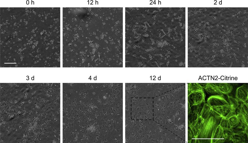
Figure 2. Recovery of hiPSC-Derived Cardiomyocytes after Replating/Thawing
A typical hiPSC-cardiomyocyte morphology can be observed from day 4 after replating or thawing
of the cells displayed for the ventricular subtype (atrial cells show similar morphological changes). A
CRISPR-engineered hiPSC line with endogenous tagging of ACTN2 with a fluorescence marker
illustrates the formation of sarcomeric structures at 12 days after replating. Consistent field of view
over time is shown. Scale bars represent 100 mm.
Metabolic Selection of hiPSC-Derived Cardiomyocytes
TIMING: 3–7 days
Metabolic selection can be performed in order to purify hiPSC-derived atrial and
ventricular cardiomyocytes after the differentiation. Provision of lactate as the only
energy source is permissive of the survival of cells with cardiac-like metabolism
only. To enhance selection efficiency and prevent detachment of cardiomyocytes,
replate the cells at a lower density, ideally around day 15. The optimal time point
for selection is between days 15-30, due to the cells’ high sensitivity to lactate asso-
ciated with their proliferative state at this age; however, selection is possible until
day 60.
39. Digest the differentiated cells as described in ‘Digestion of hiPSC-derived cardi-
omyocytes’ (steps 31-36). After resuspending the cells, count them using an
automated cell counter or a hemocytometer, and plate in a lower density, e.g.
at a ratio of 1:3 or at a defined cell number of 1-23106 cells per well (6-well).
STAR Protocols --, 100026, --, 2020 11Please cite this article in press as: Kleinsorge and Cyganek, Subtype-Directed Differentiation of Human iPSCs into Atrial and Ventricular
Cardiomyocytes, STAR Protocols (2020), https://doi.org/10.1016/j.xpro.2020.100026
40. Allow cells to regenerate in Cardio Culture Medium for 3-5 days before starting
the selection. Change the medium every 2-3 days.
41. Start the selection process by aspirating the spent medium and adding 2 ml per
well (6-well) of Cardio Selection Medium.
42. Change the medium to 2 or 3 ml Cardio Selection Medium every 2 or 3 days,
respectively, for the duration of the selection process (e.g. 2 ml on Monday
and Wednesday, 3 ml on Friday).
43. To stop the selection, switch to Cardio Culture Medium. Change the medium to
2 ml of Cardio Culture Medium and change this medium every 2 or 3 days there-
after (e.g. 2 ml on Monday and Wednesday, 3 ml on Friday).
Note: The incubation time in Cardio Selection Medium is dependent on the
age of the cardiomyocytes: 3-5 days before day 30, and 5-7 days for cells be-
tween days 30 and 60 (see Troubleshooting Problem 4).
Cryopreservation of hiPSC-Derived Cardiomyocytes
TIMING: 3 h
hiPSC-derived atrial and ventricular cardiomyocytes can be cryopreserved for long-
term storage. To ensure pure populations, the cells should ideally be frozen after
having passed through a digestion and selection process. Cryopreservation can
be performed from day 15 after the start of differentiation onwards.
44. Prewarm FBS, PBS or EDTA Dissociation Buffer, 0.25% trypsin-EDTA to 20-25 C.
45. Aspirate medium of hiPSC-derived cardiomyocytes. Wash the cells once with
PBS or EDTA Dissociation Buffer (e.g. 1 ml per well of a 6-well plate) and directly
aspirate again.
46. Add 1 ml of 0.25% trypsin-EDTA per well (6-well) to the cells and incubate at
37 C for 8-12 min. Continue when the cells become round and start to detach
from the plate.
47. Carefully wash all cells from the plate and transfer them into a 15 ml centrifuge
tube. Add the same volume of FBS and centrifuge at 100 3 g for 10 min.
CRITICAL: Because the cells are very sensitive, handle them carefully and
avoid extensive pipetting.
48. Carefully aspirate the supernatant and resuspend the cells in a small volume
(1-2 ml, depending on the size of the cell pellet) of FBS, and count the cells using
an automated cell counter or a hemocytometer.
49. According to the desired volume and cell number per cryogenic vial and the
consequent total number of cryogenic vials needed, fill up the cell suspension
with FBS to 80% of the total required volume. Add a final concentration of 13
Cardio Cryopreservation Medium (from 53), mix carefully and dispense the
cell suspension into the cryogenic vials.
Note: We suggest a cell density of 2-53106 cells in 1.5 ml of cryopreservation
medium per vial.
50. Transfer the tubes to a controlled rate freezing device (e.g. Cryotherm Planer)
and freeze the cells according to the protocol in Table 1.
Alternatives: If no automated controlled rate freezer is available, freeze the
cells in an isopropanol freezing container at -80 C for at least 2 h.
12 STAR Protocols --, 100026, --, 2020Please cite this article in press as: Kleinsorge and Cyganek, Subtype-Directed Differentiation of Human iPSCs into Atrial and Ventricular
Cardiomyocytes, STAR Protocols (2020), https://doi.org/10.1016/j.xpro.2020.100026
Table 1. Protocol for Controlled Rate Freezing of hiPSC-Derived Cardiomyocytes
Step Rate ( C/min) End Temperature ( C)
0 Starting temperature +4
1 -1 -4
2 -25 -40
3 +10 -12
4 -1 -40
5 -10 -90
6 Hold -90
51. After a minimum of 10 min at -90 C, store the tubes in the vapor phase of a liquid
nitrogen freezer.
Note: Long-term storage in the vapor phase of liquid nitrogen (at a tempera-
ture between -140 C and -180 C) for more than 2 years was tested without
obvious effects on cell quality.
Note: If digestion with 0.25% trypsin-EDTA seems too harsh for any particular
cell line, attempt to use a 0.05% dilution instead.
Alternatives: Instead of 0.25% trypsin-EDTA, you can use 103 TrypLE Select
or Accutase.
Recovery of hiPSC-Derived Cardiomyocytes
TIMING: 10–20 min
Cryopreserved hiPSC-derived atrial and ventricular cardiomyocytes can be recov-
ered into culture as described here.
Note: Recovery rates of cryopreserved hiPSC-cardiomyocytes might vary
between cell lines. Typically, plating efficiencies of 50%–-90% can be
expected.
52. Prewarm Matrigel-coated plates and Cardio Recovery Medium to 20-25 C.
53. Aspirate Matrigel from the precoated culture plates and add Cardio Recovery
Medium (2 ml per well of a 6-well plate).
54. Prepare a 15 ml centrifuge tube with 9 ml of Cardio Recovery Medium.
55. Transfer the cryogenic vial containing the cells from the liquid nitrogen
storage directly to a 37 C water bath and observe the thawing process.
When most of the content of the tube is thawed and only a small
chunk of ice remains, quickly take it out and clean thoroughly with 70%
ethanol.
56. Carefully add the cells dropwise into the prepared 15 ml centrifuge tube. Gently
mix and centrifuge at 100 3 g for 10 min.
57. Aspirate the supernatant and carefully resuspend the cells in 1 ml of Cardio Re-
covery Medium.
CRITICAL: Because the cells are very sensitive, handle them carefully and
avoid extensive pipetting.
STAR Protocols --, 100026, --, 2020 13Please cite this article in press as: Kleinsorge and Cyganek, Subtype-Directed Differentiation of Human iPSCs into Atrial and Ventricular
Cardiomyocytes, STAR Protocols (2020), https://doi.org/10.1016/j.xpro.2020.100026
58. Plate the desired cell number on the coated plates and incubate at 37 C and
5% CO2.
Note: Upon thawing, we recommend an initial plating of 1-33106 cells per
well of a 6-well plate to ensure optimal recovery of cells.
Optional: Recount the cells before plating (note that the recovery rate might
not be accurate at this point, so that the number of plated and attached cells
might be lower than the total number of living cells counted).
59. Change the medium to Cardio Culture Medium (2 ml per well of a 6-well plate)
after a recovery period of 2-3 days. Continue changing the medium regularly
every 2 or 3 days thereafter (e.g. 2 ml on Monday and Wednesday, 3 ml on
Friday).
Note: Before performing analysis of or further experiments with thawed cardi-
omyocytes, let them recover on the cell culture plate for 4-7 days (see
Figure 2).
Immunostaining of hiPSC-Derived Cardiomyocytes
TIMING: 2 days
For the characterization and quality control of hiPSC-derived atrial and ventricular
cardiomyocytes, they can be fixed and immunocytochemically stained for the
expression of cardiac markers, such as a-actinin, cTNT, RYR2, MLC2a, and MLC2v.
60. Carefully place sterile glass cover slips into a multi-well plate (e.g. cover
slips 20 mm in diameter into 12-well plates). Coat the cover slips with
Matrigel as described in ‘Before you begin’ (e.g. with 500 ml per 20 mm cover
slip).
Alternatives: Chamber slides with detachable rims can be coated with Matri-
gel, or – if the microscopic setup allows and the stained cells do not need to be
kept on mounted microscope slides – the cells can also be stained directly in
the cell culture plates.
61. Digest the differentiated cells as described in ‘Digestion of hiPSC-derived car-
diomyocytes’ (steps 31-36). After resuspending the cells, count them using an
automated cell counter or a hemocytometer, and plate at the desired cell den-
sity, e.g. 70,000-150,000 cells per 20 mm cover slip.
62. Allow the cells to regenerate in Cardio Culture Medium for 7-10 days before fix-
ation. Change the medium every 2-3 days.
63. Aspirate the medium of the cardiomyocytes on glass cover slips. Wash the cells
three times with PBS.
64. Incubate the cells in 4% Histofix (or other fixative solution) at 20-25 C for 20 min.
Wash them three times with PBS.
PAUSE POINT: Fixed cardiomyocytes can be stored in PBS with 1% BSA at
4 C for up to 1 month.
65. Dilute the primary antibody in PBS with 1% BSA with or without 0.1% Triton
X-100 depending on the antibody (see Table 2).
14 STAR Protocols --, 100026, --, 2020Please cite this article in press as: Kleinsorge and Cyganek, Subtype-Directed Differentiation of Human iPSCs into Atrial and Ventricular
Cardiomyocytes, STAR Protocols (2020), https://doi.org/10.1016/j.xpro.2020.100026
Table 2. Antibodies Used for the Immunocytochemical Staining of hiPSC-derived
Cardiomyocytes
Antibody Dilution Supplier
Primary antibody
a-actinin monoclonal mouse IgG1 1:1,000 Sigma-Aldrich
CTNT monoclonal mouse IgG1 1:200 Thermo Fisher Scientific
RYR2 polyclonal rabbit IgG 1:500 Sigma-Aldrich
MLC2A monoclonal mouse IgG2a 1:200 Synaptic Systems
MLC2V polyclonal rabbit IgG 1:200 Protein tech
Secondary antibody
Alexa Fluor 488 donkey anti-rabbit 1:1,000 Thermo Fisher Scientific
Alexa Fluor 555 donkey anti-mouse 1:1,000 Thermo Fisher Scientific
66. Aspirate the supernatant of the fixed cells and add 200 ml primary antibody dilu-
tion. Incubate at 4 C for 16-24 h.
67. Dilute the secondary antibody in PBS with 1% BSA with or without 0.1 % Triton
X-100 depending on the antibody (see Table 2).
68. Aspirate the primary antibody dilution and wash three times with PBS. Add the
secondary antibody dilution and incubate at 20-25 C in the dark for 1 h.
69. Aspirate the secondary antibody dilution and wash once with PBS. Add diluted
DAPI/Hoechst nuclear stain (1:2,000-1:3,000 in ddH2O) and incubate at 20-25 C
in the dark for 10 min.
70. Aspirate the solution and wash three times with PBS, followed by one time with
ddH2O. Mount the cover slips upside down onto clean glass slides with a few
drops of an aqueous mounting medium (e.g. Fluoromount/Vectashield). Be
careful not to create bubbles when lowering the cover slips onto the slides.
71. Let the mounting medium dry for 1 h and seal the edges of the coverslip with nail
varnish. Store the slides at 4 C in the dark.
EXPECTED OUTCOMES
The protocol for the differentiation of hiPSCs towards atrial or ventricular cardiomyo-
cytes described here has been adapted from and modified based on previously pub-
lished protocols (Zhang et al., 2011; Lian et al., 2013; Tohyama et al., 2013; Burridge
et al., 2014). It has been applied successfully to more than 50 hiPSC lines in our
hands, and robustly yields 80-90% of cTNT+ cardiomyocytes without significant dif-
ferences between atrial and ventricular specification. Upon metabolic selection, the
purity can be further increased. Within the cardiomyocyte population, this protocol
generates approximately 90-95% of the desired atrial or ventricular subtype. At day
30 after the start of differentiation, approximately 1-33106 atrial or ventricular car-
diomyocytes can be obtained per well of a 6-well plate, into which 150,000-
260,000 iPS cells have initially been plated 2-4 days before the start of differentia-
tion, i.e. for every iPS cell plated, approximately 10 cardiomyocytes can be
generated.
At ages of 20-90 days after start of differentiation (or even older), the cells can be
applied in experiments or analyzed for cardiac characteristics, such as the expression
of general cardiac markers (e.g. a-actinin, cTNT, RYR2) or subtype-specific markers
(e.g. MLC2v and MLC2a for ventricular and atrial cardiomyocytes, respectively).
However, regarding the choice of experimentation time point it should be noted
that the maturity of the cells will likely improve with prolonged culture time. The as-
sembly of hiPSC-derived atrial or ventricular cardiomyocytes and fibroblasts into
STAR Protocols --, 100026, --, 2020 15Please cite this article in press as: Kleinsorge and Cyganek, Subtype-Directed Differentiation of Human iPSCs into Atrial and Ventricular Cardiomyocytes, STAR Protocols (2020), https://doi.org/10.1016/j.xpro.2020.100026 engineered heart muscle will further mature the cardiac cells and enables their inves- tigation in a three-dimensional context (Cyganek et al., 2018). LIMITATIONS Our subtype-directed differentiation protocol provides relatively homogeneous cell populations for most hiPSC lines tested in our hands. However, differentiation effi- ciencies can vary significantly between cell lines. Although the protocol yields atrial/ventricular cardiomyocytes with purities of approximately 80-90%, a minimum of 10% cardiomyocytes of different lineages as well as non-cardiomyocytes can be found in the samples. The hiPSC-derived cardiomyocytes generated using this protocol display embry- onic- or fetal-like properties when compared to adult cardiac tissue, as evaluated by functional, transcriptomic and proteomic phenotyping (Cyganek et al., 2018). However, the generation of three-dimensional tissue has been shown to increase the maturity of the two-dimensional cell populations and already more closely re- flects the in vivo situation (Tiburcy et al., 2017). TROUBLESHOOTING Problem 1 The hiPSCs differentiate on their own already in maintenance culture OR the hiPSCs do not differentiate well into cardiac cells upon application of the directed differen- tiation protocol. Potential Solution Check for pluripotency of the starting hiPSC population, e.g. by assessing marker expression and trilineage differentiation potential. Verify that the hiPSC line displays a normal karyotype. Problem 2 Excessive cell death and detachment happens during/after CHIR99021 treatment. Potential Solution A substantial loss of cells is expected at this point. However, if most of the cells are lost, the differentiation will fail. Try to optimize the CHIR99021 concentration applied on day 0 of differentiation between 3-8 mM – this can vary between cell lines. Alternatively, try to optimize the starting cell density for the differentiation. Problem 3 During long-term culture of hiPSC-derived cardiomyocytes, clusters of beating cells start to detach. Potential Solution The differentiated cells can be digested and replated in a lower cell density (if they were too dense) onto new coated plates. Problem 4 Cardiac cells detach in whole clusters during metabolic selection. Potential Solution In order to eliminate dead cells from the cell cultures during the selection process and therefore save the cardiac cells, carefully collect the medium (including the clus- ters in suspension) and flush it through a 40 mm mesh. Then turn the filter and 16 STAR Protocols --, 100026, --, 2020
Please cite this article in press as: Kleinsorge and Cyganek, Subtype-Directed Differentiation of Human iPSCs into Atrial and Ventricular
  Cardiomyocytes, STAR Protocols (2020), https://doi.org/10.1016/j.xpro.2020.100026

carefully wash off the collected clusters into fresh Cardio Selection Medium. Transfer
the cell suspension into a new coated well.

SUPPLEMENTAL INFORMATION
Supplemental Information can be found online at https://doi.org/10.1016/j.xpro.
2020.100026.

ACKNOWLEDGMENTS
We thank Laura Cyganek, Nadine Gotzmann, Martina Grohe, Yvonne Hintz, Lisa
Krebs, and Yvonne Wedekind (all Stem Cell Unit, UMG) for excellent technical assis-
tance. This work was supported by the German Research Foundation (DFG) (CY 90/
1-1, CRC 1002 S01) and by the German Center for Cardiovascular Research (DZHK).

AUTHOR CONTRIBUTIONS
L.C. conceived the protocol and edited the manuscript. M.K. drafted the manuscript.

DECLARATION OF INTERESTS
The authors declare no competing interests.

Received: January 6, 2020
Revised: February 9, 2020
Accepted: February 25, 2020

REFERENCES
Burridge, P.W., Matsa, E., Shukla, P., Lin, Z.C.,     Lian, X., Zhang, J., Azarin, S.M., Zhu, K., Hazeltine,   Tohyama, S., Hattori, F., Sano, M., Hishiki, T.,
Churko, J.M., Ebert, A.D., Lan, F., Diecke, S.,       L.B., Bao, X., Hsiao, C., Kamp, T.J., and Palecek,       Nagahata, Y., Matsuura, T., Hashimoto, H., Suzuki,
Huber, B., Mordwinkin, N.M., et al. (2014).           S.P. (2013). Directed cardiomyocyte differentiation      T., Yamashita, H., Satoh, Y., et al. (2013). Distinct
Chemically defined generation of                      from human pluripotent stem cells by modulating          metabolic flow enables large-scale purification of
human cardiomyocytes. Nat. Methods 11,                Wnt/b-catenin signaling under fully defined              mouse and human pluripotent stem cell-derived
855–860.                                              conditions. Nat. Protoc. 8, 162–175.                     cardiomyocytes. Cell Stem Cell 12, 127–137.

Cyganek, L., Tiburcy, M., Sekeres, K., Gerstenberg,   Tiburcy, M., Hudson, J.E., Balfanz, P., Schlick, S.,     Zhang, Q., Jiang, J., Han, P., Yuan, Q., Zhang, J.,
K., Bohnenberger, H., Lenz, C., Henze, S., Stauske,   Meyer, T., Chang Liao, M.-L., Levent, E., Raad, F.,      Zhang, X., Xu, Y., Cao, H., Meng, Q., Chen, L., et al.
M., Salinas, G., Zimmermann, W.-H., et al. (2018).    Zeidler, S., Wingender, E., et al. (2017). Defined       (2011). Direct differentiation of atrial and ventricular
Deep phenotyping of human induced pluripotent         engineered human myocardium with advanced                myocytes from human embryonic stem cells
stem cell-derived atrial and ventricular              maturation for applications in heart failure             by alternating retinoid signals. Cell Res. 21,
cardiomyocytes. JCI Insight 3.                        modeling and repair. Circulation 135, 1832–1847.         579–587.

                                                                                                                STAR Protocols --, 100026, --, 2020                 17
You can also read