Deletion of Cbl- b inhibits CD8+ T- cell exhaustion and promotes CAR T- cell function
←
→
Page content transcription
If your browser does not render page correctly, please read the page content below
Open access Short report
J Immunother Cancer: first published as 10.1136/jitc-2020-001688 on 18 January 2021. Downloaded from http://jitc.bmj.com/ on February 18, 2021 by guest. Protected by copyright.
Deletion of Cbl-b inhibits CD8+ T-cell
exhaustion and promotes CAR T-
cell function
Jitendra Kumar,1,2 Ritesh Kumar,1,2 Amir Kumar Singh,1,2 Elviche L Tsakem,1,2
Mahesh Kathania,1,2 Matthew J Riese,3 Arianne L Theiss,4 Marco L Davila ,5
K Venuprasad 1,2
To cite: Kumar J, Kumar R, ABSTRACT the effector CD8+ T-cells that promote anti-
Kumar Singh A, et al. Deletion Background Chimeric antigen receptor (CAR) T-cell tumor immunity become dysfunctional and
of Cbl-b inhibits CD8+ T-cell therapy is an emerging option for cancer treatment, but
exhaustion and promotes CAR
exhausted in the tumor microenvironment
its efficacy is limited, especially in solid tumors. This is (TME). Exhausted T cells exhibit progressive
T-cell function. Journal for
partly because the CAR T cells become dysfunctional
ImmunoTherapy of Cancer loss of effector function (expression of inter-
2021;9:e001688. doi:10.1136/ and exhausted in the tumor microenvironment. However,
feron gamma (IFN-γ), tumor necrosis factor
jitc-2020-001688 the key pathways responsible for impaired function of
exhausted cells remain unclear, which is essential to alpha (TNF-α) and tumor cell killing) and
overcome CAR T-cell exhaustion. express inhibitory receptors (PD1, Tim3, and
►► Additional material is
published online only. To view, Methods Analysis of RNA-sequencing data from CD8+ LAG3).2 However, the key pathways respon-
please visit the journal online tumor-infiltrating lymphocytes (TILs) led to identification sible for impaired function of exhausted cells
(http://dx.doi.org/10.1136/jitc- of Cbl-b as a potential target. The sequencing data were remain unclear.
2020-001688). validated using a syngeneic MC38 colon cancer model. To Post-translational modification mediated
analyze the in vivo role of Cbl-b in T-cell exhaustion, tumor by ubiquitin conjugation plays an indispens-
Accepted 14 December 2020 growth, % PD1+Tim3+ cells, and expression of effector
able role in immune cells. Ubiquitination
cytokines were analyzed in cbl-b+/+ and cbl-b–/– mice.
To evaluate the therapeutic potential of Cbl-b depletion,
involves a cascade of biochemical reactions
we generated a new CAR construct, hCEAscFv-CD28- through ubiquitin activating (E1) enzymes,
CD3ζ.GFP, that recognizes human carcinoembryonic ubiquitin- conjugating (E2) enzymes, and
© Author(s) (or their antigen (CEA). cbl-b+/+ and cbl-b–/– CEA-CAR T cells were ubiquitin ligase (E3) enzymes. The E3 ubiq-
employer(s)) 2021. Re-use generated by retroviral transduction. Rag–/– mice bearing uitin ligases are critical components of this
permitted under CC BY-NC. No MC38-CEA cells were injected with cbl-b+/+ and cbl-b–/–; system because they recognize and target
commercial re-use. See rights CEA-CAR T cells, tumor growth, % PD1+Tim3+ cells and specific target proteins for ubiquitination.3
and permissions. Published by expression of effector cytokines were analyzed.
BMJ. Cbl-b is an E3 ubiquitin ligase that belongs
Results Our results show that the E3 ubiquitin ligase
1
Internal Medicine, UT to the Really Interesting New Gene (RING)
Cbl-b is upregulated in exhausted (PD1+Tim3+) CD8+
Southwestern Medical Center, TILs. CRISPR-Cas9-mediated inhibition of Cbl-b restores
family. Cbl- b contains an N- terminal tyro-
Dallas, Texas, USA
the effector function of exhausted CD8+ TILs. Importantly, sine kinase-binding domain, a RING finger,
2
Immunology, UT Southwestern and a C-terminal proline-rich sequence, and
the reduced growth of syngeneic MC38 tumors in
Medical Center, Dallas, Texas,
USA cbl-b–/– mice was associated with a marked reduction can thus function as both an E3 ligase and a
3
Division of Hematology and of PD1+Tim3+ CD8+ TILs. Depletion of Cbl-b inhibited molecular adaptor.3 Here, we demonstrate
Oncology, Department of CAR T-cell exhaustion, resulting in reduced MC38-CEA that Cbl-b is upregulated in exhausted CD8+
Medicine, Medical College of tumor growth, reduced PD1+Tim3+ cells and increased tumor- infiltrating lymphocytes (TILs) and
Wisconsin, Milwaukee, USA expression of interferon gamma, tumor necrosis factor plays a crucial role in dysfunction of tumor-
4
Division of Gastroenterology alpha, and increased tumor cell killing.
and Hepatology, Department of reactive TILs.
Conclusion Our studies demonstrate that deficiency of
Medicine, University of Colorado Cbl-b overcomes endogenous CD8+ T-cell exhaustion, and
School of Medicine, Aurora,
deletion of Cbl-b in CAR T cells renders them resistant to
Colorado, USA
5 exhaustion. Our results could facilitate the development of
Blood & Marrow Transplantation MATERIALS AND METHODS
and Cellular Immunotherapy, efficient CAR T-cell therapy for solid tumors by targeting
H Lee Moffitt Cancer Center Cbl-b. Mice
and Research Institute, Tampa, C57BL/6, Rag–/– and cbl-b–/– mice were
Florida, USA housed in microisolator cages, and the exper-
INTRODUCTION iments were performed in accordance with
Correspondence to
Dr K Venuprasad; Adoptive transfer T-cell therapy is an emerging the guidelines of the Institutional Animal
venuprasad.poojary@ option for cancer treatment,1 but its efficacy Care and Use Committee of UT Southwestern
utsouthwestern.edu is limited, especially in solid tumors, because Medical Center.
Kumar J, et al. J Immunother Cancer 2021;9:e001688. doi:10.1136/jitc-2020-001688 1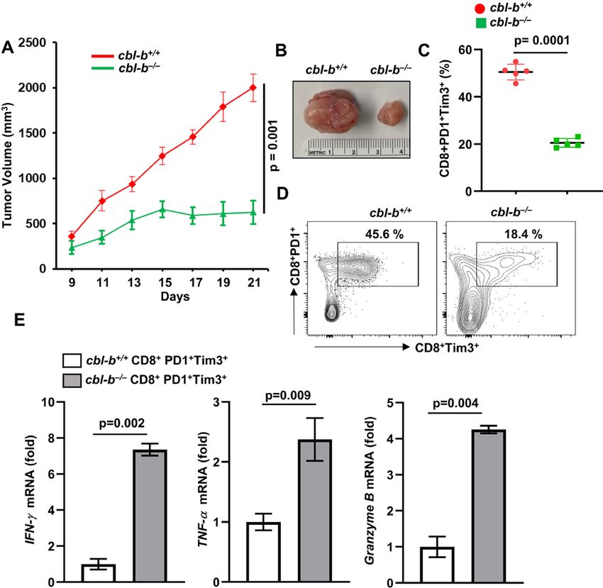
Open access
J Immunother Cancer: first published as 10.1136/jitc-2020-001688 on 18 January 2021. Downloaded from http://jitc.bmj.com/ on February 18, 2021 by guest. Protected by copyright.
with the transmembrane domain, and mouse stimulatory
domain, which included mouse CD28 and mouse CD3ζ.
CRISPR-Cas9 targeting
Three top-ranking guide sequences against Cbl-b exons
were designed and cloned into lenti-CRISPR v2 plasmid
(Addgene 52961). HEK293FT cells (Thermo Fisher)
were transfected with lenti- CRISPR v2 plasmid, along
with packaging plasmid pMD2.G (Addgene 12259) and
psPAX2 (Addgene 12260). Lentivirus was harvested
after 48–72 hours of incubation, passed through 0.45 µm
filter and stored at −80°C. The details of the guide RNAs
targeting Cbl-b gene are as follows:
Cbl-b guide RNA1: GATGGGATGTCGCATCCTCG.
Cbl-b guide RNA2: GCACTTCGTGCCTTACCGCG.
Cbl-b guide RNA3: GGCGAGTGTTCGAAAGTGCA.
Retroviral transduction, CAR T-cell production and adoptive
transfer
CD8+ T-cells were stimulated for 24 hours with anti-CD3
clone (1 µg/mL) and anti-CD28 (1 µg/mL) antibodies
and were cultured with mIL-2 (100 U/mL) and trans-
duced with the retrovirus by spinoculation method.7 On
the day of adoptive transfer, GFP+ cells were sorted by
flow cytometry and (GFP+CD8+ CAR T cells) adoptively
Figure 1 Cbl-b is upregulated in exhausted CD8+PD1+Tim3+ transferred via tail–vein injections.
TILs. (A) Geo dataset (GSE85947) was analyzed and
normalized read counts (TPM) for Cbl-b are shown. (B) Statistical analysis
Volcano plot of differentially expressed genes (−log10 p value
Statistical analysis was performed with the Prism V.8.0
vs log2 fold change) in PD1+Tim3+ versus PD1–Tim3– TILs. (C)
MC38 tumors were implanted subcutaneously into C57BL6
software, and all results were summarized as mean±SD.
mice; PD1–Tim3– and PD1+Tim3+ TILs were sorted; and Cbl-b Differences between groups were evaluated by two-tailed
messenger RNA levels were analyzed by real-time PCR. (D) Student’s t-test. Log-rank test was used for survival anal-
Immunoblot of Cbl-b protein using Fluorescence-activated ysis. A p value of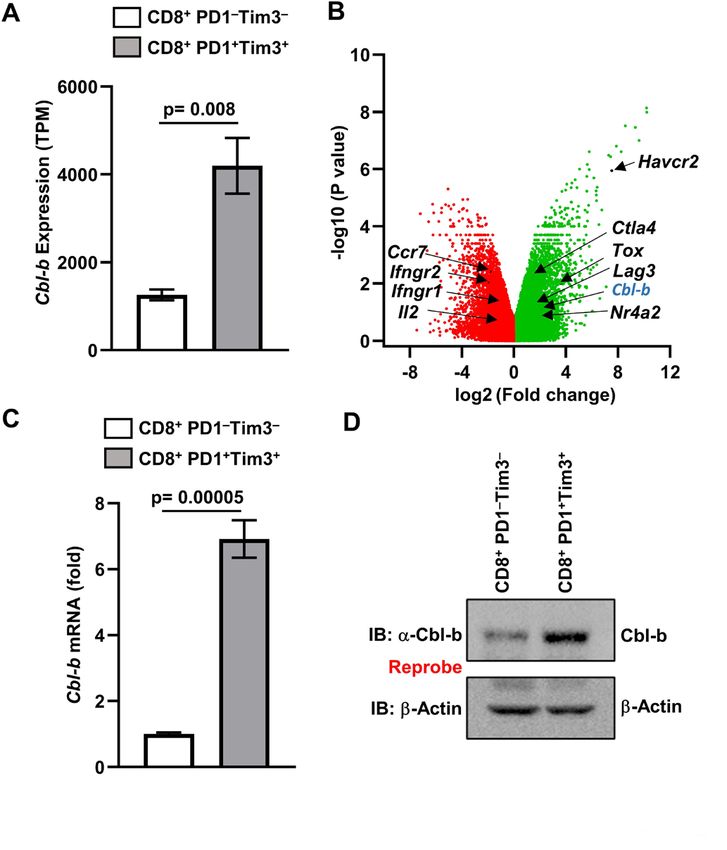
Open access
J Immunother Cancer: first published as 10.1136/jitc-2020-001688 on 18 January 2021. Downloaded from http://jitc.bmj.com/ on February 18, 2021 by guest. Protected by copyright.
Figure 3 Reduced MC38 tumor growth in cbl-b–/– mice.
cbl-b+/+ and cbl-b–/– mice (n=5) were subcutaneously
injected with MC38-CEA cells. (A) Tumor volume. (B)
Figure 2 Inhibition of Cbl-b restores effector function of Representative images of tumors. (C) Percentage of
CD8+PD1+Tim3+ TILs. Cbl-b was knocked out using Cbl-b- CD8+PD1+Tim3+ TILs. (D) Representative image showing
guide RNA-pLenti-CRISPR-v2 in sorted CD8+PD1+Tim3+cells percentage of CD8+PD1+Tim3+ TILs. (E) CD8+PD1+Tim3+
from MC38 tumor-bearing C57BL6 mice. (A) Expression of TILs were sorted and expression of IFN-γ, TNF-α and
IFN-γ, (B) TNF-α, (C) granzyme B and (D) IL-2 was measured granzyme B was analyzed by real-time PCR. The data are
by real-time PCR. The data are representative of three representative of three independent experiments. Statistics
independent experiments. Statistics are mean±SD, calculated are mean±SD, calculated by Student’s t-test (two-tailed).
by Student’s t-test (two tailed). IFN-γ, intergeron gamma; IL, CEA, carcinoembryonic antigen; IFN-γ, intergeron gamma;
interleukin; TIL, tumor-infiltrating lymphocyte; TNF-α, tumor TIL, tumor-infiltrating lymphocyte; TNF-α, tumor necrosis
necrosis factor alpha, gRNA. guide RNAs. factor alpha.
Inhibition of Cbl-b restores the effector function of exhausted number of CD8+ T-cell infiltration into MC38 tumors in cbl-
CD8+ TILs b–/– mice (online supplemental figure 1B).
To test the hypothesis that upregulated Cbl-b in PD1+Tim3+ To test if Cbl-b deficiency effected CD8+ T-cell exhaustion,
cells plays an essential role in CD8+ T-cell exhaustion, we we isolated CD8+ TILs and stained the cells with anti-PD1
knocked out Cbl-b using CRISPR-Cas9. We transduced the and anti-Tim3 antibodies. Flow cytometry analysis showed a
PD1+Tim3+ CD8+ TILs with guide RNA targeting exons of significantly reduced number of PD1+Tim3+ cells in cbl-b–/–
the Cbl-b gene or non-targeted control guide RNA, along mice compared with cbl-b+/+ mice (figure 3C,D). Further, we
with CRISPR-Cas9-expression lentiviruses. The efficiency of sorted the CD8+PD1+Tim3+ TILs from tumors grown in cbl-
CRISPR-Cas9-mediated deletion of Cbl-b was confirmed by b+/+ and cbl-b–/– mice, and real-time PCR analysis showed that
immunoblotting (online supplemental figure 1A). Deleting cbl-b–/– CD8+PD1+Tim3+ TILs but not cbl-b+/+ TILs expressed
Cbl-b restored expression of IFN-γ, TNF-α, granzyme B and IL-2 IFN-γ, TNF-α and granzyme B (figure 3E). These data collec-
in PD1+Tim3+ CD8+ TILs (figure 2A–D). tively suggest that Cbl-b plays an important role in CD8+ T-cell
exhaustion.
Cbl-b deficiency leads to reduced PD1+Tim3+ TILs
To examine the in vivo role of Cbl-b in regulation of T-cell Depletion of Cbl-b prevents CAR T-cell exhaustion
exhaustion and tumor growth, we inoculated cbl-b+/+ and cbl- Exhaustion is a major barrier in the success of CAR T cells.2
b–/– mice with MC38-CEA cells. A markedly reduced growth Similar to endogenously exhausted CD8+ T-cells, Cbl-b was
of MC38 tumors was observed in cbl-b–/– mice compared with also upregulated in exhausted CAR T cells.7 Therefore, we
cbl-b+/+ mice (figure 3A,B). Our results are consistent with investigated if depletion of Cbl-b would rescue CAR T cells
previous reports that cbl-b–/– mice mount a robust antitumor from exhaustion resulting in enhanced antitumor activity.
response against transplanted (TC-1, EL4, EG7, and B16 We replaced CD19scFv with CEA- scFv (MFE23), which
melanoma), chemically induced and spontaneous tumors.8 11 recognizes human carcinoembryonic antigen (hCEA)12 of a
The increased antitumor activity in cbl-b–/– mice was attributed CAR construct against CD19, CD19scFv-CD28-CD3ζ.GFP,13
primarily to CD8+ T-cells.8 11 Similarly, we found an increased generating a new CAR construct, hCEAscFv-CD28-CD3ζ.GFP
Kumar J, et al. J Immunother Cancer 2021;9:e001688. doi:10.1136/jitc-2020-001688 3Open access
J Immunother Cancer: first published as 10.1136/jitc-2020-001688 on 18 January 2021. Downloaded from http://jitc.bmj.com/ on February 18, 2021 by guest. Protected by copyright.
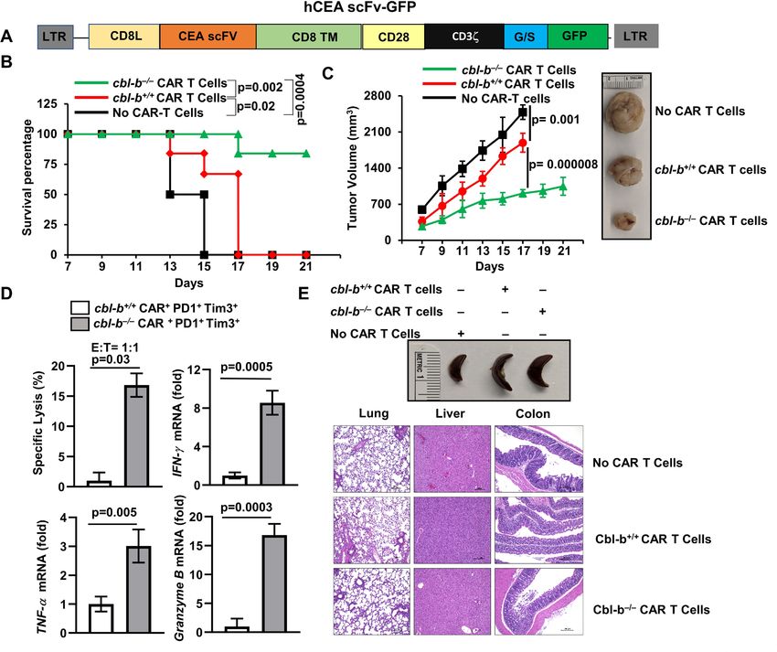
Figure 4 Depletion of Cbl-b inhibits CAR T-cell exhaustion and promotes tumor regression. (A) Schematic representation of
the CEA-reactive CAR construct. (B) 6×10⁶ cbl-b+/+ and cbl-b–/– CAR T cells were adoptively transferred into Rag–/– mice (n=6)
on days 3 and 8 after tumor inoculation. Percentage survival of Rag–/– mice that received cbl-b+/+ and cbl-b–/– CAR T cells.
The mice were considered dead when tumor size became 15 mm. Log-rank test was used for statistical analysis. (C) Tumor
volume is shown; (D) sorted cbl-b+/+ and cbl-b–/– CD8+PD1+Tim3+ TILs were cocultured with MC38-CEA cells (target) labeled
with calcein-AM. Specific lysis and expression of IFN-γ, TNF-α and granzyme B are shown. (E) Representative images of
spleen and H&E-stained sections of the colon, lung and liver. The data are representative of three independent experiments.
Statistics are mean±SD, calculated by Student’s t-test (two-tailed). AM, acetoxymethyl; CAR, chimeric antigen receptor; CEA,
carcinoembryonic antigen; hCEA, human carcinoembryonic antigen; IFN-γ, intergeron gamma; scFv, single-chain variable
fragment; TIL, tumor-infiltrating lymphocyte; TNF-α, tumor necrosis factor alpha.
(figure 4A). We chose CEA as a model antigen because CEA expressed IFN-γ, TNF-α, granzyme B, and IL-2 (online supple-
is overexpressed in many human cancers, most notably in mental figure 2C).
colorectal adenocarcinomas.14 Next, we injected MC38-CEA cells into the right flank of
We retrovirally transduced naïve CD8+ T-cells with either Rag–/– mice, and on days 3 and 8, we inoculated with cbl-b+/+
control (mock) (without hCEAscFv) or hCEAscFv- CD28- or cbl-b–/– CEA-CAR T cells. The use of cbl-b–/– CAR T cells
CD3ζ.GFP. The transduction efficiency varied between 60%
considerably enhanced the survival of Rag–/– mice as shown
and 65% (online supplemental figure 2A). We cocultured
fluorescence-activated cell sorted GFP+ cells (online supple- in figure 4B. In addition, we observed significantly reduced
mental figure 2B) with MC38- CEA cells, a mouse colon tumor growth in Rag–/– mice that received cbl-b–/– CAR T cells
cancer cell line expressing hCEA.15 Real-time PCR analysis compared with the mice that received cbl-b+/+ CAR T cells
showed that CEA-CAR T cells, but not mock transduced cells, (figure 4C).
4 Kumar J, et al. J Immunother Cancer 2021;9:e001688. doi:10.1136/jitc-2020-001688Open access
J Immunother Cancer: first published as 10.1136/jitc-2020-001688 on 18 January 2021. Downloaded from http://jitc.bmj.com/ on February 18, 2021 by guest. Protected by copyright.
To investigate if Cbl-b deficiency effected CAR T-cell exhaus- It was demonstrated that Cbl-b deficiency uncouples the
tion, tumors were collected and CD8+ TILs were enriched. requirement for costimulation for T cells and hence height-
These cells were stained with anti-PD1 and anti-Tim3 anti- ened antitumor activity.3 Our results show that Cbl-b is upreg-
bodies and were analyzed by flow cytometry. The percentage ulated in PD1+Tim3+ exhausted CD8+ T-cells, and depletion of
of PD1+Tim3+ cells were markedly reduced in the tumors Cbl-b restores their effector function, suggesting a novel role
in mice that received cbl-b–/– CAR T cells (between 30% for Cbl-b in CD8+ T-cells. Further, adoptive transfer of CEA-
and 35% in cbl-b+/+ and 4%–6% in cbl-b–/–. Next, we sorted specific CD8+ CAR T cells resulted in reduced tumor growth
the PD1–Tim3– and PD1+Tim3+ cells that were gated on and substantial survival benefit in Rag−/− mice, suggesting the
GFP+ cells and cocultured with MC38-CEA cells to analyze potential of targeting Cbl-b in CAR T cells. However, a signifi-
their tumor killing ability using calcein-acetoxymethyl.16 As cant difference exists between spontaneous tumors that arise
shown in figure 4D, coculture of MC38-CEA cells with cbl-b+/+ in humans and the transplanted tumors. Therefore, further
PD1+Tim3+ CEA-CAR T cells exhibited markedly reduced investigation in spontaneous tumor models and humanized
tumor cell killing ability. However, the cbl-b–/– cells retained mice is essential before the translation into clinical trials.
their tumor killing ability. Further, cbl-b–/– CEA-CAR T cells CAR T cells specific to CEA have shown clinical efficacy
cocultured with MC38-CEA cells expressed IFN-γ, TNF-α and in gastrointestinal adenocarcinomas.23 However, another
granzyme B but not the cbl-b+/+ cells (figure 4D). These data trial failed due to preconditioning- dependent respiratory
collectively suggest that deletion of cbl-b–/– potentiates the toxicity.24 While we did not observe any significant toxicity in
antitumor activity of CAR T cells. Since Cbl-b deficiency in T mice that received cbl-b–/– CAR T cells, further studies using
cells is linked to autoimmunity,3 we looked for signs of toxicity mice transgenically expressing human CEA in gastrointes-
in mice that received cbl-b–/– cells. No significant change in tinal or lung epithelia cells are essential to fully assess the
spleen size was observed between Rag−/− mice that received potential toxicity. Nevertheless, our studies clearly suggest
either cbl-b+/+ or cbl-b–/– cells. Similarly, histological analysis of that Cbl-b could be potentially targeted to overcome CAR
H&E-stained sections of colon, lung and liver tissues of mice T-cell exhaustion.
that received cbl-b–/– CAR T cells did not show any signs of
toxicity (figure 4E).
CONCLUSION
Our studies demonstrate that deficiency of Cbl-b overcomes
DISCUSSION endogenous CD8+ T-cell exhaustion, and deletion of Cbl-b in
Immunotherapy for cancer via checkpoint blockade and CAR T cells render them resistant to exhaustion. Our results
adoptive transfer of tumor antigen- specific CAR T cells could facilitate the development of efficient CAR T- cell
have shown promising results.1 However, only a small subset therapy for solid tumors by targeting Cbl-b.
of patients achieves complete remission with checkpoint
Twitter Marco L Davila @marcoldavila
blockade. Similarly, CAR T cells have been effective against
Contributors J.K, R.K, A.S, E.L.T, M.K performed the experiments, analyzed
hematopoietic malignancies but not against solid tumors. the data and helped to prepare manuscript. M.J.R, A.T, M.D helped to prepare
Several lines of evidence implicate exhaustion in limiting manuscript. K.V conceived the project, designed the experiment and wrote the
the potency of CAR T cells.2 Here, we demonstrate that dele- manuscript.
tion of Cbl-b inhibits CAR T-cell exhaustion, which can be Funding This work was supported by grants from the National Institutes of Health
exploited therapeutically. (R01-DK115668) and Cancer Prevention Research Institute of Texas (RP160577 and
The mechanism by which Cbl-b deficiency leads to such RP190527) to VP and R01- DK117001 to VP and ALT.
a remarkable effect on CD8+ T- cell exhaustion currently Competing interests None declared.
remains unclear. However, recent studies suggest that exhaus- Patient consent for publication Not required.
tion results due to activation of nuclear factor of activated Ethics approval The study protocol has been approved by the institutional review
T-cells (NFAT) in the absence of AP-117 and several studies board of UT Southwestern Medical Center.
have implicated the NFAT-driven transcription factors NR4A7 Provenance and peer review Not commissioned; externally peer reviewed.
and TOX in T-cell exhaustion.18–20 Interestingly, Cbl-b expres- Data availability statement Data are available upon reasonable request. All data
sion was induced by constitutively active NFAT,17 suggesting relevant to the study are included in the article or uploaded as supplementary
the potential role of Cbl-b in NFAT driven mechanisms. information. All the data are available and will be shared upon request.
Previous studies have shown that Cbl-b deficiency leads to Supplemental material This content has been supplied by the author(s). It has
tumor rejection, which is predominantly mediated by CD8+ not been vetted by BMJ Publishing Group Limited (BMJ) and may not have been
peer-reviewed. Any opinions or recommendations discussed are solely those
T-cells.8 11 Further, the antitumor activity of cbl-b–/– CD8+ T-cells of the author(s) and are not endorsed by BMJ. BMJ disclaims all liability and
was shown to be independent of CD4+ T-cell help.8 11 In line responsibility arising from any reliance placed on the content. Where the content
with these studies, we found reduced MC38 tumor growth, includes any translated material, BMJ does not warrant the accuracy and reliability
which was associated with increased infiltration of CD8+ of the translations (including but not limited to local regulations, clinical guidelines,
terminology, drug names and drug dosages), and is not responsible for any error
T-cells in cbl-b–/– mice. However, Cbl-b has also been shown to and/or omissions arising from translation and adaptation or otherwise.
regulate natural killer cells and dendritic cells.21 22 Therefore, Open access This is an open access article distributed in accordance with the
we do not completely exclude the function of these immune Creative Commons Attribution Non Commercial (CC BY-NC 4.0) license, which
cells in Cbl-b-mediated antitumor response. permits others to distribute, remix, adapt, build upon this work non-commercially,
Kumar J, et al. J Immunother Cancer 2021;9:e001688. doi:10.1136/jitc-2020-001688 5Open access
J Immunother Cancer: first published as 10.1136/jitc-2020-001688 on 18 January 2021. Downloaded from http://jitc.bmj.com/ on February 18, 2021 by guest. Protected by copyright.
and license their derivative works on different terms, provided the original work is 12 Chester KA, Begent RH, Robson L, et al. Phage libraries for
properly cited, appropriate credit is given, any changes made indicated, and the use generation of clinically useful antibodies. Lancet 1994;343:455–6.
is non-commercial. See http://c reativecommons.org/licenses/by-nc/4.0 /. 13 Davila ML, Kloss CC, Gunset G, et al. CD19 CAR-targeted
T cells induce long-term remission and B cell aplasia in an
ORCID iDs immunocompetent mouse model of B cell acute lymphoblastic
leukemia. PLoS One 2013;8:e61338.
Marco L Davila http://orcid.org/0000-0002-6270-3065
14 Holzinger A, Abken H. CAR T cells targeting solid tumors:
K Venuprasad http://o rcid.org/0 000-0001-8986-9092 carcinoembryonic antigen (CEA) proves to be a safe target. Cancer
Immunol Immunother 2017;66:1505–7.
15 Robbins PF, Kantor JA, Salgaller M, et al. Transduction and
expression of the human carcinoembryonic antigen gene in a murine
colon carcinoma cell line. Cancer Res 1991;51:3657–62.
REFERENCES 16 Neri S, Mariani E, Meneghetti A, et al. Calcein-acetyoxymethyl
1 June CH, O'Connor RS, Kawalekar OU, et al. CAR T cell cytotoxicity assay: standardization of a method allowing additional
immunotherapy for human cancer. Science 2018;359:1361–5. analyses on recovered effector cells and supernatants. Clin Diagn
2 McLane LM, Abdel-Hakeem MS, Wherry EJ. CD8 T cell exhaustion Lab Immunol 2001;8:1131–5.
during chronic viral infection and cancer. Annu Rev Immunol 17 Martinez GJ, Pereira RM, Äijö T, et al. The transcription factor
2019;37:457–95. NFAT promotes exhaustion of activated CD8⁺ T cells. Immunity
3 Venuprasad K. Cbl-B and itch: key regulators of peripheral T-cell 2015;42:265–78.
tolerance. Cancer Res 2010;70:3009–12. 18 Seo H, Chen J, González-Avalos E, et al. TOX and TOX2 transcription
4 Peng D-J, Zeng M, Muromoto R, et al. Noncanonical K27-linked factors cooperate with NR4A transcription factors to impose CD8+ T
polyubiquitination of TIEG1 regulates FOXP3 expression and tumor cell exhaustion. Proc Natl Acad Sci U S A 2019;116:12410–5.
growth. J Immunol 2011;186:5638–47. 19 Wang X, He Q, Shen H, et al. TOX promotes the exhaustion
5 Klepsch V, Hermann-Kleiter N, Do-Dinh P, et al. Nuclear receptor of antitumor CD8+ T cells by preventing PD1 degradation in
NR2F6 inhibition potentiates responses to PD-L1/PD-1 cancer hepatocellular carcinoma. J Hepatol 2019;71:731–41.
immune checkpoint blockade. Nat Commun 2018;9:1538. 20 Khan O, Giles JR, McDonald S, et al. TOX transcriptionally
6 Li G, Boucher JC, Kotani H, et al. 4-1Bb enhancement of CAR T and epigenetically programs CD8+ T cell exhaustion. Nature
function requires NF-κB and TRAFs. JCI Insight2018;3. 2019;571:211–8.
7 Chen J, López-Moyado IF, Seo H, et al. Nr4A transcription factors 21 Paolino M, Choidas A, Wallner S, et al. The E3 ligase Cbl-b and TAM
limit CAR T cell function in solid tumours. Nature 2019;567:530–4. receptors regulate cancer metastasis via natural killer cells. Nature
8 Loeser S, Loser K, Bijker MS, et al. Spontaneous tumor rejection by 2014;507:508–12.
cbl-b-deficient CD8+ T cells. J Exp Med 2007;204:879–91. 22 Wallner S, Lutz-Nicoladoni C, Tripp CH, et al. The role of the E3
9 Singer M, Wang C, Cong L, et al. A distinct gene module for ligase Cbl-b in murine dendritic cells. PLoS One 2013;8:e65178.
dysfunction uncoupled from activation in tumor-infiltrating T cells. 23 Zhang C, Wang Z, Yang Z, et al. Phase I escalating-dose trial of
Cell 2016;166:e1509:1500–11. CAR-T therapy targeting CEA+ metastatic colorectal cancers. Mol
10 Scott-Browne JP, López-Moyado IF, Trifari S, et al. Dynamic changes Ther 2017;25:1248–58.
in chromatin accessibility occur in CD8 + T cells responding to viral 24 Thistlethwaite FC, Gilham DE, Guest RD, et al. The clinical efficacy of
infection. Immunity 2016;45:1327–40. first-generation carcinoembryonic antigen (CEACAM5)-specific CAR
11 Chiang JY, Jang IK, Hodes R, et al. Ablation of Cbl-b provides T cells is limited by poor persistence and transient pre-conditioning-
protection against transplanted and spontaneous tumors. J Clin dependent respiratory toxicity. Cancer Immunol Immunother
Invest 2007;117:1029–36. 2017;66:1425–36.
6 Kumar J, et al. J Immunother Cancer 2021;9:e001688. doi:10.1136/jitc-2020-001688You can also read