Intraperitoneal collagenase as a novel therapeutic approach in an experimental model of colorectal peritoneal carcinomatosis
←
→
Page content transcription
If your browser does not render page correctly, please read the page content below
www.nature.com/scientificreports
OPEN Intraperitoneal collagenase
as a novel therapeutic
approach in an experimental
model of colorectal peritoneal
carcinomatosis
D. García‑Olmo1,2,3,5, P. Villarejo Campos2,5*, J. Barambio2, S. Garcia Gomez‑Heras4,
L. Vega‑Clemente1, S. Olmedillas‑Lopez1, H. Guadalajara2,3 & M. Garcia‑Arranz1,3
The usefulness of local collagenase in therapeutic approaches to solid tumors has been tested
recently. In this study, we evaluate the safety and efficacy of intraperitoneal collagenase associated
or not to mitomycin for treatment of colorectal peritoneal metastases in an experimental rat model.
Using a fixed-dose procedure, we found that a dose of collagenase of 37 IU/mL administered for
15 min with a hyperthermia pump at 37.5 °C, both in isolation or associated to sequential treatment
with intraperitoneal mitomycin, led to a macroscopic decrease in tumor volume as evaluated by the
modified peritoneal cancer index (mPCI). Concerning the safety of the procedure, the animals showed
no physiological or behavioral disorders during 8 weeks of follow-up. Local treatment for peritoneal
metastases of colorectal origin with intraperitoneal collagenase has proved safe and effective in
an experimental murine model. Therefore, the stroma-first approach by enzymatic breakdown
of collagen from the tumor’s extracellular matrix provides a new therapeutic target for colorectal
peritoneal metastases.
Abbreviations
ANOVA Analysis of variance
ELISA Enzyme-linked immunosorbent assay
mg/kg Milligram/kilogram
mg/m2 Milligram/square meter
mL Millilitre
mPCI Modified peritoneal carcinomatosis index (peritoneal cancer index adapted to this experiment)
p P value
U Units of enzymatic activity
U/mL Units of enzymatic activity/milliliter
The components of the tumor microenvironment are related to drug resistance in several tumors. One such
component, the tumor stroma, protects against the arrival of therapeutic agents to target cancer cells1. However,
this microenvironment may be modified by reshaping the extracellular matrix (composed mainly of collagen
fibers), thereby facilitating the penetration and delivery of drugs into tumors. In this way, therapeutic approaches
could achieve a higher concentration of drugs within tumor cells and improve the tumor response2.
Currently, local administration of collagenase is a standard treatment for benign processes such as Dupuytren’s
disease, Peyronie’s disease, and enzymatic debridement in cutaneous ulcers3–5. Although this treatment is not yet
1
New Therapies Laboratory, Health Research Institute-Fundación Jiménez Díaz University Hospital (IIS-FJD),
Avda. Reyes Católicos, 2, 28040 Madrid, Spain. 2Department of Surgery, Fundación Jiménez Díaz University
Hospital, Avda. Reyes Católicos, 2, 28040 Madrid, Spain. 3Department of Surgery, Universidad Autónoma de
Madrid, C/Arzobispo Morcillo s/n, 28034 Madrid, Spain. 4Department of Human Histology, Universidad Rey Juan
Carlos, Avda de Atenas s/n, 28922 Alcorcón, Spain. 5These authors contributed equally: D. García-Olmo and
P. Villarejo Campos. *email: villarejocampos@yahoo.es
Scientific Reports | (2021) 11:503 | https://doi.org/10.1038/s41598-020-79721-0 1
Vol.:(0123456789)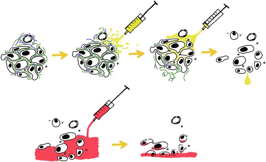
www.nature.com/scientificreports/
Figure 1. Flow-chart. Scheme of the surgical protocol followed for treatment with intraperitoneal collagenase.
First step, evaluation under anesthesia of a modified Peritoneal Carcinomatosis Index (mPCI); second step,
continuous perfusion system (COMBAT-BRS) was placed in the peritoneal cavity, in order to deliver collagenase
or mitomycin in the designed regimen; third step, post-treatment evaluation of the mPCI.
validated for clinical use in cancer, in vivo studies using intratumoral or intravenous routes to deliver collagenase
have been carried out in murine tumor models.
Intravenous dose above 500 μg (0.5%) have been shown to be lethal in murine models due to abdominal and
pulmonary bleeding2. Meanwhile, local administration of intratumoral collagenase at a dose of 37.5 U/mL did
not demonstrate toxicity in any organ and the enzyme did not locate outside the tumor6.
Degradation of the extracellular matrix using intravenous collagenase nanoparticles has been shown to
improve drug penetration in mice bearing cancer7. A chamber-based approach for local collagenase application
in a tumor-bearing rat model has shown that proper control of time, pressure, and enzyme concentration is
essential to limit possible t oxicity8.
To date, no reports of intraperitoneal administration of collagenase for treatment of colorectal peritoneal
metastases have appeared in the literature. The aim of this study is to show the results obtained by using intra-
peritoneal collagenase to treat peritoneal surface carcinomatosis in an experimental rat model.
Methods and results
Legal and ethical considerations. The present study has been approved by the Research Ethics Com-
mittee of the University Hospital Fundación Jiménez Díaz (no. PIC/75-2016) and the Committee on Animal
Research and Ethics of the University General Hospital of Albacete (no. 23-2017). All experiments were per-
formed in adherence of national and international regulations on the protection of experimental animals. The
animals were reared in accordance with Directive 2010/63/UE and maintained with unlimited access to water
and standard rat chow. The environmental conditions (light, ambient temperature, and relative humidity) were
kept constant.
Step 1: Estimation of collagenase solution concentration and acute toxicity. We used a com-
mercially available collagenase for intraperitoneal administration (LYPOSMOL BIOTECH). Irrigation of the
abdominal cavity was carried out with collagenase in a physiological solution (RINGER LACTATE, Braun,
Spain); collagen concentration was expressed in units/milliliters (U/mL). To optimize the enzymatic activity
of collagenase, we used temperatures ranging from 36 to 37.5 °C, depending on the manufacturer’s indications.
Intraperitoneal collagenase and mitomycin (INIBSA, Spain) were administered with the use of a heated pump
perfusion system approved for experimental and clinical use (COMBAT BRS RECIRCULATION SYSTEM,
Hertfordshire, U.K.). When adjusting doses, we calculated the units of enzymatic activity (U), defined as the
catalytic activity responsible for the transformation of 1 µmol of substrate per minute under optimal conditions.
When studying acute intraperitoneal toxicity, we used the fixed-dose method as an alternate means of deter-
mining the lethal dose 50; in this way, we attempted to reduce and refine experimental procedures in animals.
We tested doses of 350, 175, 87.5, 70, 37, 17.5, 8.75, and 3.5 U/mL in Wistar rats with ages ranging from 6 to
8 weeks and an approximate weight of 250 mg. Peripheral blood samples were taken from each animal to quantify
collagenase concentration. Histological studies were performed in all rats.
Results. We found that 350 U/mL was a toxic lethal dose; all animals that received this dose died within 1 h
of administration, and necropsy showed that the cause of death was massive abdominal bleeding. The optimal
dose of intraperitoneal collagenase was 37 U/mL, and the optimal exposure time was 15 min (Fig. 1).
When administered under these conditions, collagenase was not detected in the blood of the animals tested.
Furthermore, histologic examination revealed that concentrations below 70 U/mL did not induce severe tissue
damage.
Step 2: Murine model of peritoneal carcinomatosis. When designing an appropriate experimental
model of colorectal peritoneal carcinomatosis, we chose our previous model consisting of BD-IX syngeneic rats
and the cell line DH/K-12 (also known as DHD/K12-TRb)9.
Animals: We used BD-IX syngeneic rats that were 6 weeks of age, selecting these independently of sex. To
implant the tumor on the peritoneal surface, the animals were weighed and subjected to general anesthesia
(75 mg/kg ketamine plus 25 mg/kg xylocaine). The tumor was implanted by means of an intraperitoneal injec-
tion of 1 × 10⁶ cells in 0.25 ml and phosphate-buffered saline. After the tumor was successfully implanted, we
selected those rats with recognizable peritoneal carcinomatosis by palpating the abdominal wall on a weekly basis
Scientific Reports | (2021) 11:503 | https://doi.org/10.1038/s41598-020-79721-0 2
Vol:.(1234567890)
www.nature.com/scientificreports/
after the first month. Eighty percent of the rats developed palpable peritoneal carcinomatosis within 2 months
of the injection.
Rats with confirmed peritoneal carcinomatosis were randomly distributed among the experimental groups
in the study. Each experimental group consisted of 6 rats (the number of specimens needed was previously
calculated based on the results of preliminary tests).
The control group, which received intraperitoneal collagenase without peritoneal carcinomatosis, was estab-
lished in the previous step (step 1).
Four groups were established: a (Control group), receiving peritoneal carcinomatosis without treatment;
B, the group receiving intraperitoneal collagenase; C, the intraperitoneal mitomycin group, and; D, Sequential
intraperitoneal treatment group (collagenase followed by mitomycin treatment).
The collagenase dose used was 37 U/mL for 15 min at 37.5 °C, in line with our previous toxicity study.
Mitomycin is a chemotherapy drug that has been proven effective for the treatment of peritoneal carcino-
matosis of colorectal origin. The dose of mitomycin used was 35 mg/m2 (usual dose for humans) for 10 min.
Animals were euthanized with an intravenous dose of sodium thiopental at 1 and 8 weeks after the end of
treatment. Euthanasia was also performed when animals showed visible signs of uncontrollable suffering or pain,
according to the endpoint criteria.
Outcomes were evaluated according to three variables:
1. Tumor volume reduction related to each treatment as evidenced by macroscopic examination, based on the
mPCI before and after treatment.
2. The occurrence of adhesions in the abdominal cavity.
3. The Irwin Test, used to evaluate the toxicity of each group.
All animals were evaluated daily after surgery to determine their health and behavior according to a moni-
toring table (data not shown). To detect circulating collagenase in peripheral blood, samples were taken from
each rat. An external laboratory performed ELISA tests against matrix metalloproteinase-1 and -2 (MMT1 and
MMT2). An independent surgeon assessed the extension of the peritoneal metastases based on the mPCI score
and the characteristics of intra-abdominal adhesions.
Variables measurement.
• Modified PCI scale:
• More than 20 nodules equals mPCI of 3
• Between 20 and 5 nodules equals mPCI of 2
• Less to 5 is equivalent to mPCI of 1
• Abdominal adhesion scale:
• no adhesion: 0
• manually separated adhesions: 1
• adhesions that need instruments to separate them: 2
• Irwin test scale (behaviour and autonomic disturbs)
• Absence of disorders: 0
• low disorders: 1
• moderate disorders: 2
• high disorders: 3
Before starting the experimental treatment we performed a laparotomy that allows us to assess the extent of
the tumor disease in the abdominal cavity of each rat and we confirmed extensive peritoneal carcinomatosis,
with a mPCI that reached score 3 in all animals.
Statistical analysis. We applied a mixed-methods design to quantify the observations made with regard to
each variable10. One-way ANOVA followed by Tukey’s post-hoc tests for multiple comparisons were performed.
Results with a value of P < 0.05 were considered significant. Analyses were carried out using the SPSS statistical
package, version 23.0, software for Windows (SPSS, Chicago, IL, USA).
Results. The peritoneal carcinomatosis rate achieved with our model was 80%, according to previous experi-
ences. No unexpected deaths occurred during procedures. We found no traces of collagenase in the ELISA tests
performed on blood samples. All animals were euthanized after 2 months of follow-up, except for rats of the
mitomycin-treated group (C); in this group, 5 rats were euthanized early due to signs of pain and discomfort
that could not be controlled with analgesics (buprenorphine and tramadol according to our follow-up protocol).
The animals were autopsied and macroscopic findings on the necropsy were evaluated (Table 1).
Scientific Reports | (2021) 11:503 | https://doi.org/10.1038/s41598-020-79721-0 3
Vol.:(0123456789)
www.nature.com/scientificreports/
Group Tumor volumen and systemic metastases after treatment Adhesion characteristics Macroscopic tissue damage
Numerous nodules and masses that diffusely involve peritoneal surfaces, omentum
A Film and adhesions No
Lung metastases
Lower tumor volume in peritoneal surfaces without other affected organs
B Fatty tissue lysis of the omentum Adhesion-free Slight tissue damage
No distant metastases
Retracted abdominal organs
Several nodules located on peritoneal surfaces, without other organs involved
C Dense adhesions Hematic omentum
No distant metastases
Inflamed bowel without intestinal perforation
Massive decrease in tumor volume. Small and isolated tumor nodules that were easily
removed from peritoneal surfaces
D Adhesion-free Slight tissue damage
Fatty tissue lysis of the omentum
No distant metastases
Table 1. Macroscopic findings on the necropsy. A or Control Group (untreated peritoneal carcinomatosis), B
or Intraperitoneal Collagenase Group, C or Intraperitoneal Mitomycin Group, D or Sequential Intraperitoneal
Treatment Group (first collagenase treatment followed by mitomycin treatment).
Modified PCI Abdominal adhesions Irwin test
Post hoc Post hoc Post hoc
analysis (P analysis (P analysis (P
Rats groups x ± SD value) x ± SD value ) x ± SD value )
B 0.000 B 0.006 B 0.756
A 3.00 ± 0.00 D 0.000 0.60 ± 0.547 D 0.006 0.60 ± 1.341 D 0.999
C 0.163 C 0.000 C 0.000
A 0.000 A 0.006 A 0.756
B 1.33 ± 0.516 D 0.919 0.00 ± 0.00 D 0.999 0.166 ± 0.408 D 0.634
C 0.004 C 0.000 C 0.000
A 0.163 A 0.000 A 0.000
C 2.40 ± 0.547 B 0.004 2.00 ± 0.00 B 0.000 3.00 ± 0.00 B 0.000
D 0.001 D 0.000 D 0.000
A 0.000 A 0.006 A 0.999
D 1.16 ± 0.408 B 0.919 0.00 ± 0.00 B 0.999 0.666 ± 0.516 B 0.634
C 0.001 C 0.000 C 0.000
Table 2. Statistical analysis of variables. x mean, SD standard deviation. The mean difference is significant at
the 0.05 level, with a 95% confidence interval. Rats groups: A Control group; B, collagenase treatment group;
C, mitomycin treatment group, and; D, collagenase followed by mitomycin treatment group.
The control group (A) had numerous tumors located on peritoneal surfaces and in the lungs of the rats; no
other organs were involved. The intraperitoneal collagenase group (B) showed a low tumor volume on peritoneal
surfaces and no other organ involvement or fatty-tissue lysis of the omentum. In the intraperitoneal mitomycin
group (C), we observed several tumor nodules, retracted abdominal organs, hematic omentum, and inflamed
bowel without perforation. Finally, rats undergoing sequential intraperitoneal treatment (D) showed isolated,
small tumor nodules that were easily removed from peritoneal surfaces.
We observed differences in tumor-volume reduction among groups (F: 22.2, P = 0.000). The collagenase and
sequential groups exhibited greater degrees of reduction (Tukey’s post hoc test, P < 0.05), and between these two
groups there were no differences (P = 0.919).
Regarding the appearance of adhesions after surgery, intergroup differences were found the occurrence of
adhesions in the abdominal cavity (F: 70.6, P = 0.000). Intraabdominal adhesions were less common in the
collagenase and sequential groups (Tukey’s post hoc test, P < 0.05); between these two groups there were no
statistically significant differences (P = 0.999).
Regarding toxicity parameters, there were differences found among groups (F: 16.4, P = 0.000). The intraperi-
toneal mitomycin group had higher toxicity than other experimental groups (Tukey’s post hoc test, P < 0.05).
The analysis of variables among different groups was shown in Table 2.
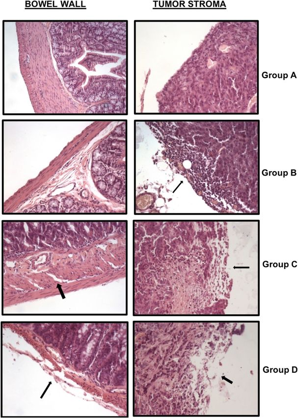
Histopathologic analysis: the control group (A) had several cell implants covered with a hard stroma and
highly conserved structures as previously described. Group B (Intraperitoneal Collagenase) showed less attached
tumor masses and a softer stroma surrounding the tumor. No damage was observed in any other organ except the
slightly thinner intestinal wall. Group C (Intraperitoneal Mitomycin) showed similar tumor masses to Group A,
with hemorrhagic areas in the bowel and involvement of the muscular layers, thinner externally and with edema
internally. Around the tumor masses, mild stroma disorganization and adherences in tissues were described.
Finally, in Group D (Sequential Treatment), the pathologist observed less tumor masses than previous groups
with microscopic hemorrhagic areas in the serosa and muscular layers of the small intestine. Around the tumors,
the stromal was disaggregated and a few tumor cells were free (Fig. 2).
Scientific Reports | (2021) 11:503 | https://doi.org/10.1038/s41598-020-79721-0 4
Vol:.(1234567890)www.nature.com/scientificreports/
Figure 2. Histological intestine and tumor studies. Left: we analyzed the bowel wall in all groups: in
Group B (Intraperitoneal Collagenase Group), a slight thinning of the external muscle layer in the bowel
wall was present; in Group C (Intraperitoneal Mitomycin Group), the serosa layer was broken and
both external and internal muscle layers were affected with this treatment.; and Group D (Sequential
Intraperitoneal Treatment Group: collagenase followed by mitomycin treatment) a thinner external muscle
wall and edema in the internal muscle layer existed, which caused a slight cell disorganization. Right: we
analyzed the effect of treatments on peritoneal tumor implants. In Group B, the stroma and the superficial
tumor area were altered. Group C, mild stroma disorganization was produced. Group D, the stroma was
disaggregated and tumor cells liberated. All photographs are hematoxylin–eosin stained (×200).
Scientific Reports | (2021) 11:503 | https://doi.org/10.1038/s41598-020-79721-0 5
Vol.:(0123456789)www.nature.com/scientificreports/
Figure 3. Stopping collagenase reaction. Hydrolyzed collagen was applied after treatment with collagenase and
we observed a quick cessation of the enzymatic activity.
Ethics approval and consent to participate. The present study was approved by the Research Ethics
Committee of the University Hospital Fundación Jiménez Díaz (nº PIC/75–2016) and the Committee on Animal
Research and Ethics of the University General Hospital of Albacete (Nº 23–2017). All experiments were per-
formed following national and international regulations on the protection of experimental animals.
Discussion
Several tumors characterized by a poor response to drugs have shown an overexpression of the extracellular
matrix with abundant collagen. These fibrous tumors include cholangiocarcinomas, pancreatic cancers, sarcomas,
and even peritoneal metastases11–14.
In addition, these cancers all exhibit poor vascularization in the tumor microenvironment and one that trig-
gers hypoxia, which would play an important role in the tumor-reactive s troma11,15.
The extracellular fibrous matrix of the tumor microenvironment acts as a barrier, hindering access by chemo-
therapy drugs (large-size molecules) to cancer cells and thus weakening tumor r esponse16.
The proteolytic activity of collagenase on the extracellular matrix could facilitate the arrival of chemothera-
peutic agents to the tumor, thereby improving response to chemotherapy.
Collagenase also degrades tumor microvascular-associated collagen and reduces vascular resistance and
microvascular pressure. These two phenomena reduce the transcapillary pressure gradient and facilitate drug
delivery from the capillary to the tumor c ells17.
Therefore, collagenase acts on both the extracellular matrix and tumor microvascularization, increasing
the transcapillary pressure gradient, which facilitates the diffusion and concentration of drugs into the tumor.
We intuited that intraperitoneal administration of collagenase would be suitable for the treatment of perito-
neal carcinomatosis, since collagenase acts directly on the collagen matrix of the tumor implants. Moreover, the
rapid inactivation of collagenase when administered intravenously must be taken into account18.
Direct contact between collagenase and the collagen of tumor implants could be related to a decrease in the
half-life of the enzyme activity and to an absence of toxicity under adjusted time and concentration conditions.
Experimental research has showed that pre-treatment with intravenous collagenase improves the concentra-
tion of drugs in fibrous tumors such as pancreatic cancer7.
Our results reveal that the administration of intraperitoneal collagenase is safe and effective in a murine model
of peritoneal carcinomatosis. Intraperitoneal collagenase solution should be administrated by a recirculating
perfusion system. The extensive distribution of peritoneal metastases makes this route ideal for achieving a
complete distribution of the collagenase solution throughout the abdominal cavity. Preconditioning peritoneal
surfaces with intraperitoneal collagenase may improve the action of chemotherapy drugs administered both
intravenously and intraperitoneally. Furthermore, this enzymatic treatment has been shown to decrease intra-
abdominal adhesions and can assist in releasing tumor masses attached to the omentum and bowel. In addition,
sequential treatment could prevent tissue damage caused by mitomycin (Fig. 4). These histologically confirmed
data suggest that intraperitoneal collagenase is not toxic and may also protect tissues treated with intraperitoneal
chemotherapy.
In our study, we observed that high concentrations of collagenase may produce local toxicity (thinning of
the muscular layer of the bowel), which makes it important to provide homogeneous parameters as concerns
the concentration and length of the enzymatic treatment.
Despite the highest concentration of collagen in the tumor matrix leads to the early stopping of the enzyme
activity, during our study, we decided to test how to neutralize it. Therefore, we added hydrolyzed collagen
(HEMOSNOW; Biom’up, France) after collagenase perfusion and observed an immediate cessation of the reac-
tion (Fig. 3).
In the future, studies should be carried out in large animals before this treatment may be tested in a clinical
trial.
Scientific Reports | (2021) 11:503 | https://doi.org/10.1038/s41598-020-79721-0 6
Vol:.(1234567890)www.nature.com/scientificreports/
Figure 4. First treating the stroma. Preconditioning of peritoneal surfaces by intraperitoneal collagenase
perfusion (yellow) can improve the action of chemotherapy drugs. Treating the stroma first, we released tumor
cells from the stroma, making them vulnerable to chemotherapeutic agents (red).
Study strengths and limitations. The main limitation of our study concerns the toxicity arising from the
group of rats treated with intraperitoneal mitomycin. We chose mitomycin because it is the hyperthermic intra-
peritoneal chemotherapy (HIPEC) drug used to treat patients with peritoneal metastases of colorectal origin.
The usual dose (35 mg/m2) was adjusted to the body surface of each rat. Tissue damage in rats has been reported
with doses of mitomycin higher than 2 mg/kg19.
However, this is also one of the strengths of our research, as we have proven that pre-conditioning treatment
with collagenase could avoid tissue damage.
Data availability
The datasets analysed during the current study available from the corresponding author on reasonable request.
Received: 2 July 2020; Accepted: 7 December 2020
References
1. Werb, Z. & Lu, P. The role of stroma in tumor development. Cancer J. (United States). 21, 250–253 (2015).
2. Dolor, A. & Szoka, F. C. Digesting a path forward: The utility of collagenase tumor treatment for improved drug delivery. Mol.
Pharm. 15, 2069–2083 (2018).
3. Patry, J. & Blanchette, V. Enzymatic debridement with collagenase in wounds and ulcers: A systematic review and meta-analysis.
Int. Wound J. 14, 1055–1065 (2017).
4. Karpinski, M. et al. A systematic review identifying outcomes and outcome measures in Dupuytren’s disease research. J. Hand
Surg. (Eur. Vol.). 45, 513–520 (2020).
5. Natale, C., McLellan, D. M., Yousif, A. & Hellstrom, W. J. G. Review of intralesional collagenase clostridium histolyticum injection
therapy and related combination therapies in the treatment of Peyronie’s disease (an update). Sex. Med. Rev. 20, 30009–30013
(2020).
6. Zheng, X. et al. Ultrasound-guided intratumoral administration of collagenase-2 improved liposome drug accumulation in solid
tumor xenografts. Cancer Chemother. Pharmacol. 67, 173–182 (2011).
7. Zinger, A. et al. Collagenase nanoparticles enhance the penetration of drugs into pancreatic tumors. ACS Nano 13, 11008–11021
(2019).
8. Choi, J. et al. Intraperitoneal immunotherapy for metastatic ovarian carcinoma: Resistance of intratumoral collagen to antibody
penetration. Clin. Cancer Res. 12, 1906–1912 (2006).
9. García-Olmo, D., García-Rivas, M., García-Olmo, D. C. & Atiénzar, M. Orthotopic implantation of colon carcinoma cells provides
an experimental model in the rat that replicates the regional spreading pattern of human colorectal cancer. Cancer Lett. 132,
127–133 (1998).
10. Fetters, M. D., Curry, L. A. & Creswell, J. W. Achieving integration in mixed methods designs—principles and practices. Health
Serv. Res. 48, 2134–2156 (2013).
11. Cadamuro, M. et al. The deleterious interplay between tumor epithelia and stroma in cholangiocarcinoma. Biochim. Biophys. Acta
Mol. Basis Dis. 1864, 1435–1443 (2018).
12. Mahadevan, D. & Von Hoff, D. D. Tumor-stroma interactions in pancreatic ductal adenocarcinoma. Mol. Cancer Ther. 6, 1186–1197
(2007).
Scientific Reports | (2021) 11:503 | https://doi.org/10.1038/s41598-020-79721-0 7
Vol.:(0123456789)www.nature.com/scientificreports/
13. Bai, C. et al. Associations of chemo- and radio-resistant phenotypes with the gap junction, adhesion and extracellular matrix in a
three-dimensional culture model of soft sarcoma. J. Exp. Clin. Cancer Res. 34, 58 (2015).
14. Tan, F. et al. CTHRC1 is associated with peritoneal carcinomatosis in colorectal cancer: A new predictor for prognosis. Med. Oncol.
30, 473 (2013).
15. Natarajan, S. et al. Collagen remodeling in the hypoxic tumor—mesothelial niche promotes ovarian cancer metastasis. Cancer Res.
79, 2271–2284 (2019).
16. Henke, E., Nandigama, R. & Ergün, S. Extracellular matrix in the tumor microenvironment and its impact on cancer therapy.
Front. Mol. Biosci. 6, 160 (2020).
17. Eikenes, L., Bruland, Ø. S., Brekken, C. & de LangeDavies, C. Collagenase increases the transcapillary pressure gradient and
improves the uptake and distribution of monoclonal antibodies in human osteosarcoma xenografts. Cancer Res. 64, 4768–4773
(2004).
18. Xu, F., Huang, X., Wang, Y. & Zhou, S. A size-changeable collagenase-modified nanoscavenger for increasing penetration and
retention of nanomedicine in deep tumor tissue. Mater. Adv. https://doi.org/10.1002/adma.201906745 (2020).
19. Fumagalli, U. et al. Effects of intraperitoneal chemotherapy on anastomotic healing in the rat. J. Surg. Res. 50, 82–87 (1991).
Acknowledgements
The authors acknowledge to Dra. Dolores C Garcia-Olmo for design collaboration on carcinomatosis animal
model, to Mrs. María G Picazo for cell culture and Dra. Mónica Gómez Suarez for animals care; all them from
experimental Research Unit of University Hospital of Albacete, Spain. We also acknowledge to Oliver Shaw
(IIS-FJD, Madrid, Spain) for his assistance in editing this work.
Author contributions
D.G.-O. and P.V.-C. wrote the main manuscript text. D.G.-O., H.G. and M.G.-A. designed the work. M.G.-A.
edited figures. H.G. drew images of Fig. 4. P.V.-C. and J.B. analyzed data. S.G.G.-H. performed the histological
study. L.V.-C. and S.O.-L. collected data. D.G.-O., M.G.-A., H.G., and J.B. performed the experimental assay. All
authors reviewed the manuscript and approved the final version to be published.
Funding
The study was funded by the Institute-Fundación Jiménez Díaz University Hospital (FIIS-FJD). New Therapies
Laboratory. Foundation Health Research.
Competing interests
Authors D. Garcia-Olmo, M. Garcia-Arranz, H. Guadalajara, L. Vega-Clemente, S. Olmedillas-Lopez and J. Bar-
ambio have applied for 1 patent related titled “Chemical Scalpel” (EP 19382118.8). Authors P. Villarejo Campos
and S. Garcia Gomez-Heras declare no competing interests.
Additional information
Correspondence and requests for materials should be addressed to P.V.C.
Reprints and permissions information is available at www.nature.com/reprints.
Publisher’s note Springer Nature remains neutral with regard to jurisdictional claims in published maps and
institutional affiliations.
Open Access This article is licensed under a Creative Commons Attribution 4.0 International
License, which permits use, sharing, adaptation, distribution and reproduction in any medium or
format, as long as you give appropriate credit to the original author(s) and the source, provide a link to the
Creative Commons licence, and indicate if changes were made. The images or other third party material in this
article are included in the article’s Creative Commons licence, unless indicated otherwise in a credit line to the
material. If material is not included in the article’s Creative Commons licence and your intended use is not
permitted by statutory regulation or exceeds the permitted use, you will need to obtain permission directly from
the copyright holder. To view a copy of this licence, visit http://creativecommons.org/licenses/by/4.0/.
© The Author(s) 2021
Scientific Reports | (2021) 11:503 | https://doi.org/10.1038/s41598-020-79721-0 8
Vol:.(1234567890)You can also read