Predictive value of MRI-detected extramural vascular invasion in stage T3 rectal cancer patients before neoadjuvant chemoradiation
←
→
Page content transcription
If your browser does not render page correctly, please read the page content below
Diagn Interv Radiol 2018; 24:128–134 ABDOMINAL IMAGING
© Turkish Society of Radiology 2018 ORIGINAL ARTICLE
Predictive value of MRI-detected extramural vascular invasion in
stage T3 rectal cancer patients before neoadjuvant chemoradiation
Yiqun Sun
PURPOSE
Jianwen Li We set out to explore the probability of MRI-detected extramural vascular invasion (mr-EMVI)
Lijun Shen before chemoradiation to predict responses to chemoradiation and survival in stage T3 rectal
cancer patients.
Xiaolin Wang
Tong Tong METHODS
A total of 100 patients with T3 rectal cancer who underwent MRI examination and received
Yajia Gu neoadjuvant chemoradiation and surgery were enrolled. The correlation between mr-EMVI and
other clinical factors were analyzed by chi-square. Logistic regression model was performed to
select the potential factors influencing tumor responses to neoadjuvant chemoradiation. A Cox
proportional hazards regression model was performed to explore potential predictors of sur-
vival.
RESULTS
The positive mr-EMVI result was more likely to be present in patients with a higher T3 subgroup
(T3a+b = 7.1% vs. T3c+d = 90.1%, P < 0.001) and more likely in patients with mesorectal fascia
involvement than in those without MRF (65% vs. 38.8%, P = 0.034). Compared with mr-EMVI (+)
patients, more mr-EMVI (-) patients showed a good response (staged ≤ ypT2N0) (odds ratio [OR],
3.020; 95% confidence interval [CI], 1.071–8.517; P = 0.037). In univariate analysis, mr-EMVI (+)
(hazard ratio [HR], 5.374; 95% CI, 1.210–23.872; P = 0.027) and lower rectal cancers (HR, 3.326;
95% CI, 1.135–9.743; P = 0.028) were significantly associated with decreased disease-free surviv-
al. A positive mr-EMVI status (HR, 5.727; 95% CI, 1.286–25.594; P = 0.022) and lower rectal cancers
(HR, 3.137; 95% CI, 1.127–8.729; P = 0.029) also served as prognostic factors related to decreased
disease-free survival in multivariate analysis.
CONCLUSION
The mr-EMVI status before chemoradiation is a significant prognostic factor and could be used
for identifying T3 rectal cancer patients who might benefit from neoadjuvant chemoradiation.
T
he definition of histopathologic extramural vascular invasion (EMVI) is tumor cells
From Shanghai Institute of Medical Imaging
(Y.S., X.W., Y.G. cjr.guyajia@vip.163.com) Fudan
invading the veins beyond the muscularis propria, which indicates a poor prognosis
University, Shanghai, China; the Departments of in rectal cancer patients and has drawn great attention in the pathologic reporting of
Radiology (Y.S., T.T. 983352@126.com, Y.G.) and colorectal cancer (1, 2). Magnetic resonance imaging (MRI) has come to play an increasingly
Radiation Oncology (L.S.) Fudan University Shanghai
Cancer Center, Shanghai, China; Department of significant role in evaluating rectal cancer before therapy, because of its multidimensional
Oncology (Y.S., L.S., T.T., Y.G.), Shanghai Medical imaging and excellent soft-tissue contrast. MRI detection of EMVI (mr-EMVI) has also been
College, Fudan University, Shanghai, China;
Department of Urology (J.L.) Shanxi Academy of shown to be accurate and correlate with the pathology of patients who have undergone
Medical Science, Shanxi Dayi Hospital, Shanxi, China. primary surgery (3, 4). Several retrospective studies have confirmed that mr-EMVI may be
*Yiqun Sun, Jianwen Li and Lijun Shen contributed regarded as one of the risk factors for metastasis in rectal cancer (5–7). The newest version
equally to this work and are co-first authors for this of the Society for Medical Oncology (ESMO) guidelines also regards mr-EMVI as a significant
study.
*Yajia Gu and Tong Tong are co-corresponding risk factor (8).
authors for this study. The majority of rectal cancers are identified at the T3 stage (9), and the prognosis for T3
Received 23 August 2017; revision requested 22 rectal cancer is highly variable on whether treated with or without neoadjuvant chemoradi-
October 2017; last revision received 10 February ation (10). The popular and controversial topic is whether all T3 rectal cancer patients need
2018; accepted 17 February 2018.
neoadjuvant chemotherapy (11). It has been reported that the subdivided pT3 could iden-
DOI 10.5152/dir.2018.17286 tify stage II rectal cancer patients (pT3N0) who might not benefit from adjuvant treatment
You may cite this article as: Sun Y, Li J, Shen L, Wang X, Tong T, Gu Y. Predictive value of MRI-detected extramural vascular invasion in stage T3 rectal
cancer patients before neoadjuvant chemoradiation. Diagn Interv Radiol 2018; 24:128-134.
128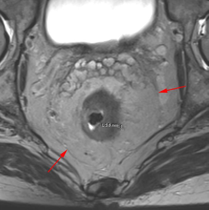
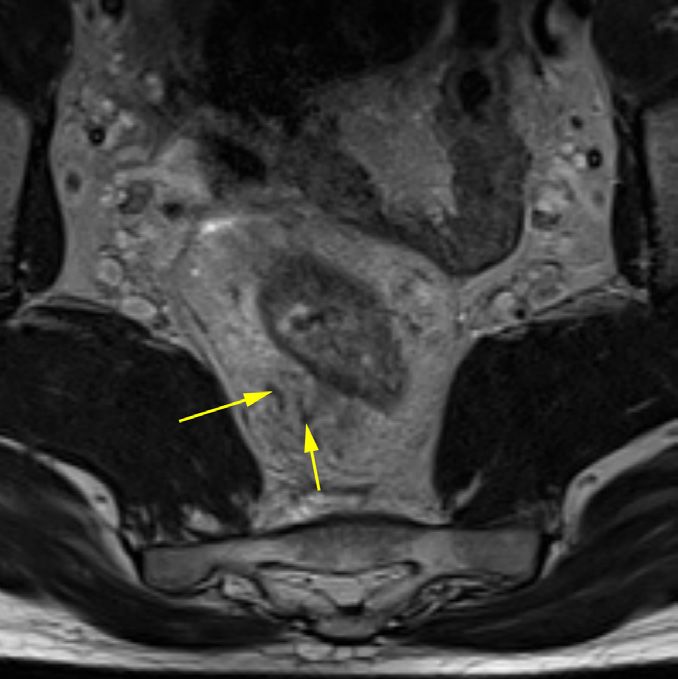
a                                                      b                                                   c

Figure 1. a–c. The performance of mr-EMVI (+) on axial T2-weighted thin-section images (a, b) and coronal T2-weighted image (c). The yellow arrow points
to an extramural vasculature of neighboring image (a). The white arrow points to an invaded extramural vessel (b, c).

(12). While the survival benefit of neoadju-           logic information, independently reviewed           trix, 384×224; section thickness, 3.0 mm;
vant chemoradiation is yet to be quantified            rectal magnetic resonance images. All pa-           bandwidth, 62.50 kHz/pixel) and coronal
in this context, our research may provide              tients had histologically confirmed rectal          T2-weighted imaging scan (TR, 2675 ms;
evidence of the benefits of neoadjuvant                adenocarcinomas, and the TNM stage was              TE, 85 ms; FOV, 240 mm×240 mm; matrix,
chemoradiation in T3 stage rectal cancer               assessed depending on the 7th American              416×224; section thickness, 4.0 mm; band-
patients, particularly when mr-EMVI is de-             Joint Committee on Cancer (AJCC) classifi-          width, 31.25 kHz/pixel).
tected before chemoradiation.                          cation (13). The exclusion criteria were: 1)
  This study aimed to evaluate the prob-               incomplete neoadjuvant chemoradiation               Radiologic evaluation
ability of mr-EMVI, detected prior to neo-             treatment, 2) patients who did not undergo             All images were independently reviewed
adjuvant chemoradiation, as a factor to                surgery within 6–10 weeks after complete            by two radiologists (T.T. is specialized in rec-
predict response to chemoradiation and                 neoadjuvant chemoradiation, and 3) meta-            tal imaging with 9 years of experience and
survival of patients with T3 rectal cancer.            static disease later found, before or at the        Y.Q.S. has 4 years of experience), who were
                                                       time of surgery.                                    blinded to clinical and pathologic informa-
Methods                                                   The neoadjuvant chemoradiation regi-             tion. Two radiologists, in consensus, used a
                                                       men was conventional radiation combined             workstation to review the rectal magnetic
Patients and treatment
                                                       with concurrent 5-fluorouracil (5-FU)-based         resonance images and the T3 sub-classi-
   We obtained ethics committee approval
                                                       chemotherapy. The whole pelvis received a           fication was established depending on
from the local Institutional Review Board
                                                       total dose of 45–55 Gray (Gy) in 1.8–2.0 Gy         radiologic measurement of maximal extra-
and informed consent from patients. Be-
                                                       daily fractions. Surgery was planned to take        mural depth beyond the outer margin of
tween April 2012 and November 2013, 100
                                                       place 6–10 weeks after the completion of            the muscularis propria on axial thin-section
patients with MRI-staged T3 rectal cancer
                                                       chemoradiation. The patient received ad-            T2-weighted images. The definitions used
(N0 or N (+)), who were treated with neo-
                                                       juvant chemotherapy regimens, including             to sub-classify T3a–T3d tumors were tak-
adjuvant chemoradiation followed by sur-               capecitabine or 5-FU.                               en from the ESMO Guidelines (T3a, tumor
gery at our hospital were collected in this                                                                depth 15 mm) (8). If the lymph node
our oncology database. Two radiologists,               treatment on a 3.0 T MRI magnet (Signa Hori-        was of mixed signal intensity and had irreg-
who were blinded to the clinical and patho-            zon, GE Medical Systems) with a phased-ar-          ular, sharp, or obscure borders or the short
                                                       ray body coil. A bowel preparation was not          axis was greater than 5 mm, it was diag-
                                                       needed before MRI examinations. The main            nosed as mr-N positive (+). Rectal cancers
 Main points                                           MRI protocol included sagittal T2-weighted          could be divided into lower and mid-high
 •   mr-EMVI status should be considered as a sig-     imaging scan (repetition time [TR], 2760 ms;        rectal cancers according to a cutoff value of
     nificant prognostic factor to identify stage T3   echo time [TE], 105 ms; field of view [FOV],        a 5 cm distance from the anorectal angle to
     rectal cancer patients who could benefit from     260 mm×260 mm; matrix, 320×224; section             the distal margin of the lower tumor border.
     neoadjuvant chemoradiation.
                                                       thickness, 3.0 mm; bandwidth, 41.67 kHz/            The length of tumor was measured on the
 •   mr-EMVI status may reflect T3 rectal cancer ag-   pixel); axial T2 fast spin-echo (TR, 3960 ms;       sagittal plane along the tumor’s longitudi-
     gressiveness.                                     TE, 105 ms; FOV, 370 mm×370 mm; ma-                 nal axis. The mr-EMVI status was regarded as
 •   mr-EMVI status determinations are likely to       trix, 224×224; section thickness, 5.0 mm;           (+) if typical imaging morphologic appear-
     prove indispensable for individualizing treat-    bandwidth, 62.50 kHz/pixel), T2-weight-             ances could be identified, which included a
     ment and follow-up protocols in T3 rectal can-
                                                       ed thin-section axial images (TR, 3960 ms;          consecutive spread of tumor signal within
     cer patients.
                                                       TE, 105 ms; FOV, 160 mm×160 mm; ma-                 the vascular network, i.e., vessel appeared

                                                                     Predictive value of MRI-detected extramural vascular invasion in T3 rectal cancer •   129
as a tube containing a flow signal void on Table 1. Correlation between mr-EMVI and clinical factors
T2-weighted image (Fig. 1), resulting in ves-
mr-EMVI
sel expansion and irregular vessel borders
on T2-weighted image (Fig. 1b, 1c) (14, 15). n (-) (+) χ2 P
They also evaluated the tumor’s relation- mr-T3 a+b 56 52 4
ship with the mesorectal fascial (MRF) enve- 70.168a b c
d e f
Figure 2. a–f. Panels (a–d) show a more invasive lesion in a 29-year-old man with rectal cancer that staged mr-T3d and MRF (+), with positive mr-EMVI.
The value of maximal extramural depth (EMD) was 20.4 mm and the MRF was invaded (red arrow, c). The yellow arrow points to an extramural vasculature
of neighboring image (a). The white arrow points to an invaded extramural vessel (b–d). Panels (e, f) show a less invasive lesion in a 69-year-old man with
rectal cancer that staged mr-T3b and MRF (-), with negative mr-EMVI. The value of EMD was 3.6 mm and the MRF was negative (red arrows, e). The mr-EMVI
(+) was more likely present in patients with higher T3 sub-classifications and MRF (+) than mr-EMVI (-).
further multivariate analysis showed that
the only factor in our study significantly
EMVI
1.0 influencing the response to neoadjuvant
EMVI (-)
EMVI (+) chemoradiation was mr-EMVI (+) (OR, 3.020;
EMVI (-)-Censored
EMVI (+)-Censored 95% CI, 1.071–8.517; P = 0.037). Fifty-six
0.8 patients were mr-EMVI (-); roughly 50% of
these (27/56) responded well to neoadju-
vant chemoradiation, while roughly 20%
Survival probability
of mr-EMVI (+) (10/44) responded well to
0.6 neoadjuvant chemoradiation (Table 2). The
treatment response to neoadjuvant chemo-
radiation did not appear to be affected by
0.4 other imaging factors.
The follow-up time ranged from 1 to 38
months. During follow-up, 15 recurrences or
metastasis incidences were identified in the
0.2
overall population, including 5 death events.
No patients were lost to clinical follow-up
after treatment. As shown in Fig. 3, the DFS
0.0 was significantly lower in mr-EMVI (+) pa-
.00 10.00 20.00 30.00 40.00
tients than in mr-EMVI (-) patients (P = 0.013).
However, the OS was not significantly lower
Time (Months) in patients with mr-EMVI (+) compared with
Figure 3. Disease-free survival stratified by mr-EMVI in mr-T3 rectal cancer patients. patients with mr-EMVI (-) (P = 0.420).
Predictive value of MRI-detected extramural vascular invasion in T3 rectal cancer • 131Table 2. Univariate and multivariate analysis of influence factors of neoadjuvant treatment of mr-T3 rectal cancer using logistic regression model
Treatment response Univariate analysis Multivariate analysis
Factors Subgroup n Good Poor P OR 95% CI P
mr-T3 a+b 56 22 34
c+d 44 15 29 0.539 1.776 0.749–4.210 0.192
mr-N (-) 6 3 3
(+) 94 34 60 0.667* 1.857 0.306–11.273 0.501
MRF (-) 80 30 50
(+) 20 7 13 0.836 0.955 0.297–3.074 0.955
mr-EMVI (-) 56 27 29
(+) 44 10 34 0.009 3.020 1.071-8.517 0.037
mr-Tumor lengthTable 4. Univariate analysis of prognostic factors for overall survival in mr-T3 patients using Cox quently alter the tumor blood supply. Ra-
proportional hazards regression model* diotherapy of hypoxic tumors can create
free radicals via the application of ionizing
Univariate analysis
radiation (e.g., OH-), and these free radicals
HR (95% Cl) P can irreversibly damage tumor cells, there-
mr-T3 a+b by reducing the sensitivity of radiotherapy.
c+d 2.026 0.514–7.989 0.313 Hypoxia can also restrict the diffusion of
chemotherapeutic agents and promote the
mr-N (-)
expression of multidrug resistance genes,
(+) 0.011 0.042–39.962 0.279 thus rendering the tumor resistant to che-
MRF (-) motherapy and reducing sensitivity to
(+) 3.837 0.292–50.345 0.309 chemoradiotherapy. Previous studies also
have shown that the greater the degree of
mr-EMVI (-)
venous invasion and the larger the diam-
(+) 6.900 0.631–24.678 0.308 eter of the vein that is invaded, the poorer
mr-Tumor lengthuations have been closely associated with Conflict of interest disclosure 11. Glynne-Jones R. Do T3 rectal cancers always
local recurrences and tumor metastasis. The authors declared no conflicts of interest. need radiochemotherapy? Recent Results Can-
cer Res 2014; 203:95–115. [CrossRef]
This study has some limitations. First, 12. Merkel S, Mansmann U, Siassi M, Papadopoulos
we have not evaluated the mr-EMVI status References
T, Hohenberger W, Hermanek P. The prognostic
1. Smith NJ, Barbachano Y, Norman AR, Swift RI,
on restaging MRI or changes in mr-EMVI inhomogeneity in pT3 rectal carcinomas. Int J
Abulafi AM, Brown G. Prognostic significance
status after neoadjuvant chemoradiation. Colorectal Dis 2001;16:298–304. [CrossRef]
of magnetic resonance imaging-detected ex-
Some patient restaging MRI analyses were 13. Edge SB, Compton CC, A.G. F, Byrd DR, Carduc-
tramural vascular invasion in rectal cancer. Br J
ci MA. American Joint Committee on Cancer
not performed using the rectal MRI pro- Surg 2008; 95:229–236. [CrossRef]
(AJCC) Cancer Staging Manual. 7th edition.
tocol, but rather the pelvic MRI protocol, 2. Betge J, Pollheimer MJ, Lindtner RA, et al. In-
2010.
tramural and extramural vascular invasion in
which did not include these high-resolu- 14. Jhaveri K S, Hosseini-Nik H, Thipphavong S, et
colorectal cancer: prognostic significance and
tion thin-section axial T2-weighted images. al. MRI detection of extramural venous inva-
quality of pathology reporting. Cancer 2012;
sion in rectal cancer: correlation with histopa-
It is difficult to evaluate the EMVI status on 118:628–638. [CrossRef]
thology using elastin stain. AJR Am J Roentge-
these images, and we have less experience 3. Brown G, Radcliffe AG, Newcombe RG, Dalli-
nol 2016; 206:747–755. [CrossRef]
in evaluating mr-EMVI after neoadjuvant more NS, Bourne MW, Williams GT. Preopera-
15. Chand M, Evans J, Swift RI, et al. The prognos-
tive assessment of prognostic factors in rectal
chemoradiation. Second, we have not used cancer using high-resolution magnetic reso-
tic significance of postchemoradiotherapy
tumor regression grading (TRG) to describe high-resolution MRI and histopathology detect-
nance imaging. Br J Surg 2003; 90: 355–364.
ed extramural venous invasion in rectal cancer.
tumor response, because TRG scores are [CrossRef]
Ann Surg 2015; 261: 473–479. [CrossRef]
not available in some patients’ histopatho- 4. Smith NJ, Shihab O, Arnaout A, Swift RI, Brown
16. Rubbia-Brandt L, Giostra E, Brezault C, et al.
G. MRI for detection of extramural vascular in-
logic reports. Third, the distribution of pa- Importance of histological tumor response
vasion in rectal cancer. AJR Am J Roentgenol
tients was uneven. T3a is often mistaken for 2008; 191:1517–1522. [CrossRef]
assessment in predicting the outcome in pa-
T2, which may be one of the reasons few tients with colorectal liver metastases treated
5. Sohn B, Lim JS, Kim H, et al. MRI-detected ex-
with neo-adjuvant chemotherapy followed
patients are T3a. Tumor invasion greater tramural vascular invasion is an independent
by liver surgery. Ann Oncol 2007; 18:299–304.
than 15 mm (referring to T3d) is rare, be- prognostic factor for synchronous metastasis
[CrossRef]
in patients with rectal cancer. Eur Radiol 2015;
cause the Chinese mesorectum is generally 17. Tripathi P, Rao SX, Zeng MS. Clinical value of
25:1347–1355. [CrossRef]
thinner than the European and American MRI-detected extramural venous invasion in rec-
6. Seehaus A, Vaccaro C, Quadrelli M, et al. Mag-
mesorectums. Finally, if the follow-up time tal cancer. J Dig Dis 2017; 18:2–12. [CrossRef]
netic resonance and extramural vascular in-
18. Yu J, Huang DY, Xu HX, Li Y, Xu Q. Correlation
were longer, the results would have more vasion in patients with rectal cancer and liver
between magnetic resonance imaging-based
clinical significance. Thus, we have contin- metastases. Acta Gastroenterol Latinoam 2015;
evaluation of extramural vascular invasion and
45:31–36.
ued following enrolled patients for future prognostic parameters of T3 stage rectal can-
7. Bugg WG, Andreou AK, Biswas D, Toms AP,
studies. cer. J Comput Assist Tomogr 2016; 40:537–542.
Williams SM. The prognostic significance of
[CrossRef]
In conclusion, the present analysis of MRI-detected extramural venous invasion in
19. Chand M, Swift RI, Tekkis PP, Chau I, Brown G.
mr-EMVI status not only demonstrates a rectal carcinoma. Clin Radiol 2014; 69:619–623.
Extramural venous invasion is a potential im-
correlation with prognostic factors, but also [CrossRef]
aging predictive biomarker of neoadjuvant
8. Glimelius B, Tiret E, Cervantes A, Arnold D,
shows promising results as far as its value in and ESMO Guidelines Working Group. Rectal
treatment in rectal cancer. Br J Cancer 2014;
predicting tumor response and long-term 110:19–25. [CrossRef]
cancer: ESMO Clinical Practice Guidelines for
20. Chand M, Bhangu A, Wotherspoon A, et al.
outcomes of T3 rectal cancers that receive diagnosis, treatment and follow-up. Ann Oncol
EMVI-positive stage II rectal cancer has similar
neoadjuvant chemoradiation. Multicenter 2013; 24:vi81–vi88. [CrossRef]
clinical outcomes as stage III disease following
9. Tong T, Yao Z, Xu L, et al. Extramural depth of
prospective studies are needed to confirm pre-operative chemoradiotherapy. Ann Oncol
tumor invasion at thin-section MR in rectal
the predictive and prognostic significance cancer: associating with prognostic factors
2014; 25:858–863. [CrossRef]
of mr-EMVI. and ADC value. J Magn Reson Imaging 2014;
40:738–744. [CrossRef]
Funding 10. Shin R, Jeong SY, Yoo HY, et al. Depth of me-
This study was funded by Natural Science Foun- sorectal extension has prognostic significance
dation of China (grant number 81501437) and Imag- in patients with T3 rectal cancer. Dis Colon Rec-
ing Foundation of Fudan University Shanghai Cancer tum 2012; 55:1220–1228. [CrossRef]
Center (Grant No. YX201601 and YX201602).
134 • May–June 2018 • Diagnostic and Interventional Radiology Sun et al.You can also read