Detection of Prostate Cancer Using Deep Learning Framework - IOPscience
←
→
Page content transcription
If your browser does not render page correctly, please read the page content below

IOP Conference Series: Materials Science and Engineering

PAPER • OPEN ACCESS

Detection of Prostate Cancer Using Deep Learning Framework
To cite this article: Abhishek Patel et al 2021 IOP Conf. Ser.: Mater. Sci. Eng. 1022 012073

View the article online for updates and enhancements.

                               This content was downloaded from IP address 46.4.80.155 on 08/03/2021 at 23:33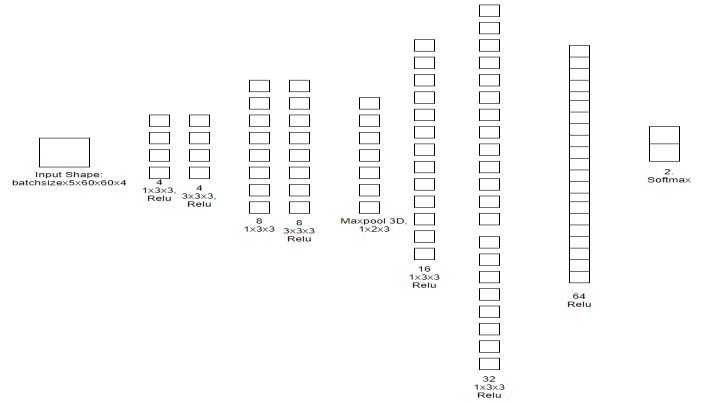
ICCRDA 2020                                                                                                              IOP Publishing
 IOP Conf. Series: Materials Science and Engineering              1022 (2021) 012073           doi:10.1088/1757-899X/1022/1/012073

 Detection of Prostate Cancer Using Deep Learning Framework
                      Abhishek Patel1, Sanjay Kumar Singh2 and Aditya Khamparia3
                      1,2,3
                         School of Computer Science and Engineering, Lovely Professional University,
                      Phagwara, Punjab, India
                      abhishekpatelnp53@gmail.com1,sanjayksingh.012@gmail.com2,aditya.khamparia88
                      @gmail.com3

            Abstract. Recent studies in Prostate Cancer signifies as magnetic resonance imaging
            targets to biopsy shows more enhanced result. The systematic study of Medline,
            Embase, Scopus, Cochrane helps in meta-analysis. Prostate specific antigen is obtained
            from curative radiotherapy. Prostate-specific membrane antigen positron emission
            tomography helps to localize recurrence prostate cancer whether it has increased.
            Prostate specific antigen rising helps to identify and prompted the reason of Prostate-
            specific membrane antigen positron emission tomography imagining. angiogenesis play
            an important role for diagnosis noninvasive cancer with technique contrast-enhanced
            ultrasound. Prostate Cancer accuracy were determined by MRI-targeted biopsy and the
            transrectal ultrasound-guided biopsy.
            Keywords: Prostate Cancer, Magnetic Resonance Imaging (MRI), Prostate specific
            antigen (PSA), Prostate-specific membrane antigen positron emission tomography
            (PSMA-PET), Prostate-specific membrane, Radiotherapy, Prostate Imaging Reporting
            and Data System (PI-RADS).
1. INTRODUCTION

The most common cancers among men is prostate cancer (PCa). Early diagnosis and preparation of care
are critical in decreasing the mortality rate because of PCa. Precise grade prediction is required to ensure
the treatment of cancer. The scaling of prostate cancer can be called as the ordinal classification issue.
Accepted clinical approaches for the diagnosis of clinically relevant prostate cancer (PC) are generally
the combination similar to prostate-specific antigen (PSA) testing, automated rectal analysis, trans-rectal
ultrasound (TRUS) with the magnetic resonance imaging (MRI). Abd the PSA screening does, however,
lead the diagnosis, leading to unnecessarily costly and the painful biopsies needle and possible over-
treatment. The multiparametric MRI that depends mostly on diffusion-weighted imaging (DWI) has
become highly standard for caring in terms of f diagnosis of prostate cancer is setting of the radiology
where the region under the collected operating the characteristic of the curve (ROC) is present. Estimates
suggest that the new cases and deaths with prostate cancer (PCa). In 2017 will be 161, 360 and 26, 730,
respectively. Accurate diagnosis and staging for the prostate cancer are crucial for selecting the most
effective treatment, and eventually to the reduction of PCa morbidity and death.

The field of medical imaging, detection based on computer-aided with a diagnosis which are the
combination of the imaging function engineering and the classification based on ML, has demonstrated
ability to assist radiologists in accurate diagnosis, reducing diagnostic time and diagnostic costs. Deep
learning approaches in various computer vision technique such as the segmentation, the classification and
the object detection have shown promising results. Such methods consist of convolution layers capable
of extracting different features with help of the local low-level features towards the global high-level
features collected from the input photos. A completely linked layer at the edge of the Convolutionary
neural layers transforms convoluted features in terms of the mark probabilities. With different layers
types have been given to enhance the output based on deep learning-based technique with the batch
normalization layer, which normalize the given input layer with zero mean and the unit variant with
dropout layer are seems to be one of the regularization techniques which ignores the nodes which are

               Content from this work may be used under the terms of the Creative Commons Attribution 3.0 licence. Any further distribution
               of this work must maintain attribution to the author(s) and the title of the work, journal citation and DOI.
 Published under licence by IOP Publishing Ltd                          1
ICCRDA 2020 IOP Publishing
IOP Conf. Series: Materials Science and Engineering 1022 (2021) 012073 doi:10.1088/1757-899X/1022/1/012073
selected in randomly order. Nevertheless, optimal combinations and layer structures with an accurate fine-
tuning of the hyper-parameters which are needed to achieve convincing efficiency.
2. LITERATURE REVIEW
A novel and proficient semi supervised system proposed [1] for computerized prostate disease confinement
using the multiparametric attractive reverberation imaging (MRI). The irregular walker (RW) calculation has
end up being exact and quick in division applications. In this mechanized way utilizing discriminative
classifiers, for example: SVM (support vector machine. The proposed strategy creates an
affectability/explicitness pace of 0.76 to 0.86 individually. A Transient Enhanced Ultrasound (TeUS) proposed
[2] , involving the investigation of varieties of signals like backscattered signals which are from the tissue over
an arrangement of ultrasound outlines. propose to utilize profound Recurrent Neural Networks (RNN) which
expressly demonstrate the transient data in TeUS. The exploring a few models of RNN, they showed as the
Long Short-Term Memory (LSTM) systems accomplish the most elevated exactness in isolating malignant
growth from kind tissue present in prostate. If the relative Gleason score[3] has been allocated to the diagnosed
prostate cancer, the accurate therapy followed by the needs to be specified promptly. The assist pathologists
and the radiologists with respect to timely diagnosis, they suggest in this particular paper a system aimed at
inferring the score of Gleason and the therapy for prostate cancer using systematic methods. Contrast-
ultrasonic Imaging (CEUS) technology [4] gave the significant role of angiogenesis in the degree of cancer.
Here, the different deep learning algorithmic models have been trained and the validated against the expert
delineations over with the images of CEUS reported using two different types of the contrast agents. The
presented convolutional neural networks (CNNs) [5] for detection of prostate cancers with the use of AUC.
The CNNs known with the high-level feature that representation the nuclear architecture summarized from the
maps of the nuclear seed and to identify cancers. CNNs obtained the AUC is 0.974 when identifying cancers
(95 per cent CI: 0.961–0.985). Epithelial Network Head with the Grading Network Head, we are presenting a
new regional Convolutionary neural network (R-CNN) [6] system. The suggested model obtained from an
epithelial cell detection precision of 99.07 percent similar to the average of AUC of 0.998 using five-fold cross-
validation. The Magnetic resonance imaging (MRI) [7] . We analyze a fully automatic method of detection
using computer-aided consisting of two phases. The tests show a frequency of 0.42, 0.75 and 0.89 per usual
case at 0.1, 1 and 10 false positives. The RF ultrasound time series [8]. We capture sequential ultrasonic RF
echoes backscattered from tissue in order to form time series of RF while imaging the probe as well as the
tissue which are stationary in place. The growth of Angiogenesis[9] roles in cancer growth that has inspired
work aimed at the non-invasive identification of cancer through imagery of blood perfusion. The time-intensity
curves (TICs) are measured with help of ultrasound imaging. . In this paper [10], they immediately suggest an
adversarial Network for segmentation of prostate cancer. Architecture proposed is a generator network with
the discriminator network. Proposed a constructed computer-aided diagnostic (CAD) [11] tools that help
identify anomalies by the radiologist. Radial Base Function (RBF), polynomial, and the Gaussian and the
Decision Tree for detecting the cancer. The Cross validation was conducted, and output was evaluated with the
terms of receiver operating curve (ROC), the precision, response, the Positive predictive value (PPV), false
positive rate (FPR) and the negative predictive value (NPV). The magnetic Resonance Imaging (MRI) [13].
Here, segmentation is required to automatically or semi-automatically locate the prostate boundary. Fully
Convolutionary Neural Networks (FCNN) have been used to this end recently. Magnetic Resonance Imaging
(MRI) is mostly reliable, the non-invasive and the prostate imaging technique. The multi-parametric MRI
[14] is known to be the safest imaging of the non-invasive tool for the prostate cancer (PCa) diagnosis.
However, in the mp-MRI for the PCa diagnosis are currently limited with some of the critical qualitative or the
interpretation semi-quantitative, resulting in variability between readers and a sub-optimal ability to assess
aggressiveness of lesions. They proposed the novel CNN based on multi-class, FocalNet which will jointly
detect the lesion of PCa and the forecast their aggressiveness with the help of Gleason score (GS). The Scale
2ICCRDA 2020 IOP Publishing
IOP Conf. Series: Materials Science and Engineering 1022 (2021) 012073 doi:10.1088/1757-899X/1022/1/012073
Invariant Trans- Forming function (SIFT) [15] was used to collect local patch details surrounding the boundary.
The size and variation of the SIFT function used for segmentation shall not be defined with the area of the
purpose of concern.
3. FINDINGS FROM LITERATURE
We have systemically studies approx. 18 papers inclusion years from 2005 to 2020, and some meaning-full
findings are highlighted in table 1. We have observed that MRI-TB is an enhanced technique for diagnosing
the prostate cancer with the systematic biopsy. The patients who are suspected, PI-RADS seems having a good
diagnosing with good accuracy. For improving the systematic biopsy, we mainly target the two different
approaches as magnetic resonance imaging (MRI) and the transrectal ultrasonography (TRUS) fusion. The
detection of PC with MRI pathway mainly involves guided systematic pathway for both (biopsy-naive subjects
and those having prior negative biopsy) types of patients. Thus, MRI-TB is enhanced alternative diagnostic
technique for systematic biopsy for the prostate cancer detection.
Table 1: Summary Table of Literature Review
Authors, Year of Database and size Number of Techniques used Finding from
[reference] Publication patients literatures
Y. Artan and I. 2012 Multiparametric Out of 21 Developed the RW technique
S. Yetik [1] MR images of 15 patients,15 graph based with an
confirmed patients confirmed multi-parametric initialization of
were analysed. biopsy patient random automated seed
data were walker(RW) enhanced the
analysed. algorithm for segmentation as
segmenting the increasing the
MRI and later use weights with
of discriminative large
classifier SVM. discriminative
power.
S. Azizi, 2018 TeUS data were 157 subjects Temporal Proposed
S. Bayat, obtained from 157 with prostate Enhanced LSTM- based
P. Yan et al. [2] patients during biopsy and the Ultrasound RNNs technique
prostate biopsy 255 suspicious (TeUS) used for is used for depth
fusion as well as mp-MRI were characterizing the analysis of latent
255 targeted examined. tissue with help features which
biopsy patients of RNN models. helps to
were suspicious Long Short-Term optimize the
from mp-MRI Memory (LSTM) TeUS data.
were used. is used for
gaining the high
accuracy.
L. Brunese, 2019 Dataset of real Totally 36 Infer Gleason Automated real
F. Mercaldo, world analysed patients having score were used time technique is
A.Reginelli et from Cancer 824 slices were with PCa therapy used for
al. [3] Imaging Archive observed. with method improving the
freely available in formal exploiting Gleason score
terms of research in MRI images which supports
purpose. which doesn’t the pathologist
required a biopsy and the
3ICCRDA 2020 IOP Publishing
IOP Conf. Series: Materials Science and Engineering 1022 (2021) 012073 doi:10.1088/1757-899X/1022/1/012073
with a time radiologist while
automation in detecting the
experimental prostate cancer
analysis. in earlier stage.
Y.Feng, F. 2019 CEUS images Available raw Contrast- Deep Learning
Yang, X. Zhou capture from iU22 dataset enhanced technique is
et al. [4] ultrasound system contains 78277 ultrasound used for
have 505x246 raw negative (CEUS) detection of PC
data of AVI sample and technology is from CESU
videos. 9073 positive used for videos which
sample. diagnosing non- overall gives
invasive cancer. 90% of an
It extracts both average
feature temporal accuracy rate.
and the spatial by
performing 3D
CNN operation.
Kwak, Jin Tae 2017 Four tissue TMAs contains CNN approach is Sample tissue
Hewitt, Stephen microarrays 162 tissue used. Microscopy images are of
M. [5] (TMAs) were sample (72 identification of size 5000x5000
taken from begin &89 Epithelial nuclear pixels and the
National Institute cancer in TMA seeds are used for detection of
of Health while A), similarly in constructing nuclear seed
conducting the TMA B it nuclear seed map. takes an average
tissue microarrays contains 149 of 547s due to
research program. sample(76 classification of
begin & 73 epithelium
cancer) and which nearly
TMA C contain give 90% of
157 (73 begin accuracy.
& 86 cancer)
W. Li, 2019 Dataset contains These 513 Gleason grading Proposed
J. Li, 513 images which images are is used for framework helps
K. Sarma et al. were gained from collected from classification. to detect
[6] pathology two different R-CNN epithelial cell
department at sets as 224 framework is and the grading
Cedars-Sinai images from 20 used for of Gleason
Medical Centre. patients and predicting multi- based on the
rest 289 images task model with historical
from 20 used of Epithelial images. R-CNN
patients. Network Head with adding of
and the Grading EHN the
Network Head. proposed model
boosts the
detection of
epithelial cell
and predict the
PC.
4ICCRDA 2020 IOP Publishing
IOP Conf. Series: Materials Science and Engineering 1022 (2021) 012073 doi:10.1088/1757-899X/1022/1/012073
G. Litjens, 2014 Among 165 Totally, 348 Two stage fully Proposed CAD
O. Debats, consecutive studies were automated MRI system detects
J. Barentsz et studies of patients obtained from detection system PC in MRI
al. [7] having PC 18 347 patients were used. images. ROC
lesions and 183 Segmentation curve classifies
case of non-cancer uses multi-atlas the patients.
patients were and the local Evaluation of
analysed for the maxima. CAD system
evaluation of Pharmacokinetic performance is
proposed CAD behaviour the state-of-the-
system. represented. art with the high
Evaluation is specificity than
based upon lesion the radiologist
and operating performance.
characteristic
curve.
M. Moradi, 2009 Comparison of RF 35 patients Analysis of Computer-based
P. time series with dataset were ultrasound RF diagnosing
Abolmaesumi, LF and texture obtained and series of time technique is
D.Siemens et feature were evaluated for with extended proposed with
al. [8] gained for 35 RF time series SVM for an concept of
patients. Hence, it features. obtaining ultrasound RF
enhanced the extended cancer timeseries.
imaging depth, its map which These helps in
frame rate and the enhance the extracting the
acoustic power process of feature value
with an ROI size biopsy. from 1cm2
of the tissue. Sequential proposition of
ultrasound RF the tissue which
echoes were helps the
recorded from radiologist for
agar-gelatin extraction the
mimicking tissue reasonable area
phantoms. of the cancer.
S. Schalk, 2017 Contrast 23 patients’ Blood perfusion CUDI
L. Demi, ultrasound images taken imaging is used implementation
N. Bouhouch et dispersion from 58 for detecting non- is better for
al. [9] imaging(CUDI) datasets which invasive cancer. extracting the
while validating are referred TICs obtained by feature from
the in-vivo of 23 from the ultrasound TICs and helps
patients were radical imaging. Firstly, while investing
parameter are prostatectomy theoretical the tissue
validated with the (RP). connection perfusion.
histopathology between mutual Hence, it can be
after the RP. information and validated other
dispersion. cancer also
where
angiogenesis has
more impact.
G. Zhang, 2019 Dataset contains 120 patient Bi-attention For segmenting
W.Wang, the MR T2- datasets adversarial the PC Bi-
5ICCRDA 2020 IOP Publishing
IOP Conf. Series: Materials Science and Engineering 1022 (2021) 012073 doi:10.1088/1757-899X/1022/1/012073
D. Yang et al. weighted images collected from network used for attention
[10] with a saturation Shanghai Tenth segmenting adversarial
of the fat for People automatically. network were
experimental Hospital. Discriminator compared with
modality having and generator other existing
the vocal size of network technique and
2.
0.9x0.6x3.5mm architecture were observed that it
All the collected used for shows the better
imaging dataset enhancing result than other
are confirmed by adversarial state-of-art
the biopsy learning globally
pathology. performance and proposed.
predicting mask
with true label.
L. Hussain, 2018 Publicly available Totally, 682 CAD is used for Performance of
A. Ahmed, MRI Images MRIs obtained diagnosing prosed model
S. Saeed et al. dataset Harvard from 20 multiresolution were evaluated
[11] university were patients. These MRI. Kernel using PPV,
examined. contain 482 SVM, RBF, NPV, FPR and
images Decision Tree AUC. However,
obtained from and Gaussian it reveals that
prostate technique used proposed feature
subjects and for detecting extraction
remaining 200 PCa. SIFT and methodology is
from EFDs helps in more effective
Brachytherapy scaling the for diagnosing
subjects. feature in ROC the PC.
curve.
L. Gorelick, 2013 All the collected 15 subject Radical Designed the
O. Veksler, images are images are prostatectomy software for
M. Gaed et al. approved by obtained with gives detection of PC
[12] research ethics radical prostatectomy which classifies
board. As all the prostatectomy. information. on haematoxylin
subject are of AdaBoost & eosin-stained
confirmed cases. classification. digital
Here, the prostate Firstly, histopathology
section was portioning of images.
processed by super pixel image Proposed system
embedding and secondly determines high
standard paraffin, labelling tissue level tissue
yielding whole culture is label components
mount. for classifying labelling of the
between cancer super pixel
versus non- without use of
cancer. explicitly
domain
knowledge
which gives the
best result for
detecting the
PC.
6ICCRDA 2020 IOP Publishing
IOP Conf. Series: Materials Science and Engineering 1022 (2021) 012073 doi:10.1088/1757-899X/1022/1/012073
T. 2019 Experimented The MRI imaging is Eight FCNN
Hassanzadeh, dataset was PROMISE12 segmented to based network
L. Hamey, collected from challenge detect boundary. architecture is
K. Ho-Shon four different dataset Improved FCNN proposed for
[13] hospital with based upon 2D segmentation of
employing two network MRI images.
Endo Rectal Coil architecture. Ten- These
(ERC). It includes fold-cross- outperforms is
50 MRI volumes validation shows comparable with
and also 30 MRI competitive and 3D FCNN
volumes without improved result. segmentation
ground truth 3D FCNN with method. Here, it
images which all endorectal coil signifies the
together have and test fold enhanced
1377 images. enhance the performance
performance among the
without further model which
extension of post- transfer the
processing. equal number of
feature map to
generate better
result.
R. Cao, 2019 All the collected 417 patients mp-MRI Multi-class
A. images were were examined considered for CNN framework
Mahammadian performed on four who further diagnosing PCa with focal net is
Bajgiran, different 3T went for a in non-invasive proposed which
S. Afshari scanner (126 Radiotherapy imaging. Multi- consist mutual
Mirak patients on trio, or hormonal class CNNs with loss finding with
et al. [14] 255 on Skyra, 17 therapy. Focal Net and fully utilized
Prisma and the 19 Gleason score are distinctive
patients were used for knowledge
evaluated on predicting obtained from
Verio). Evaluated lesions. 3T mp- multiparametric
on basis of MRI with the MRI images.
standard mp-MRI RALP. Uses of Hence achieve
protocol. FROC used for 89.7% for
analysing and focalNet and the
generating highly 87.9% for
enhancing sensitivity.
performance.
M. Yang, 2013 In the collected Dataset of 52 MRI is mostly Distinctive
X. Li, data each patient patients was used for image descriptor
B. Turkbey, contains one axial obtained from assistance and is proposed to
et al. [15] scan and the TSE 3.0T Philips diagnosis helps in elaborate the PC
MR protocol of Gyroscan. surgical planning position with the
T2-w acquisition. of the prostate help of SVA-
carcinoma. Scale SWIFT
invariant feature framework.
transformation
(SIFT) used for
capturing
7ICCRDA 2020 IOP Publishing
IOP Conf. Series: Materials Science and Engineering 1022 (2021) 012073 doi:10.1088/1757-899X/1022/1/012073
boundary local
patch. Coarse-to-
fine segment
approach is used
obtaining
variation in local
shape.
4. METHODOLOGY
4.1 Dataset & Result
It contains total 158 patients, among which 96 are train case patients and the 62 are of test cases. The
patients MRI sequence contain scan of 2D series which are deployed as 3D form of body. Every patients
MRI consist five different modes as ADC, DWI, Ktrans and T2-weighted transverse sequence as well as
T2-weighted sagittal sequence. The model of 3D CNN was used in these datasets and finally we obtained
the confusion matrix with an accuracy of 0.82, precision of 0.86 as well as recall of 0.78 on the validation
set.
4.2 Proposed architecture of 3D Convolutional Neural Network
Figure 1: Architecture of proposed 3D CNN
5. CONCLUSION & FUTURE WORK
8ICCRDA 2020 IOP Publishing
IOP Conf. Series: Materials Science and Engineering 1022 (2021) 012073 doi:10.1088/1757-899X/1022/1/012073
Detection of prostate cancer in a clinical systematic method. The MRI-TB plays a vital role that systematic
biopsy as it alters and helps in diagnosis in the process of radiography. PI-RADS with the mp-MRI gives
sensitivity and the specificity for detecting prostate cancer. We have compared the different methodology
for detecting the prostate cancer in MRI imaging and the radiology with PI-RADS and the Focal Net were
MRI images were examined with enhanced CNN model for predicting the cancer and non-cancer. The
SVA-SIFT features were taken with a description of the NVFT and SIFT features. In research, it is
observed that robust ML methodology for classification like SVM kernel, Decision Tree and the Bayesian
approach are used for separating cancer cell from the subject like Brachytherapy. Feature extraction is
done with the scale invariant feature transformation (SIFT), elliptic Fourier descriptor (EFDs),
morphology and the texture were used. Cross validation is done by Jack-knife 10-fold for training and
testing the data then the performance was evaluated on the basic of NPV, PPV, FPR and the AUC. Here
accuracy of higher classification is done on basic od single texture and the morphological feature come
from the SVM kernel and while combining them with EFDs and the texture generates good result.
However, the different technique and the result shown in these papers are obtained from current feature
extraction technique which are more efficient for diagnosing and detecting the prostate cancer in the men
with acquiring the high ratio of detection for prostate cancer.
References:
[1] Y. Artan and I. S. Yetik, 2012, “Prostate cancer localization using multiparametric MRI based on
semisupervised techniques with automated seed initialization,” IEEE Trans. Inf. Technol. Biomed., vol.
16, no. 6, pp. 1313–1323,doi: 10.1109/TITB.2012.2201731.
[2] S. Azizi et al., 2018, “Deep recurrent neural networks for prostate cancer detection: Analysis of temporal
enhanced ultrasound,” IEEE Trans. Med. Imaging, vol. 37, no. 12, pp. 2695–2703, doi:
10.1109/TMI.2018.2849959.
[3] L. Brunese, F. Mercaldo, A. Reginelli, and A. Santone, 2019, “Prostate gleason score detection and cancer
treatment through real-time formal verification,” IEEE Access, vol. 7, pp. 186236–186246, doi:
10.1109/ACCESS.2019.2961754.
[4] Y. Feng et al., 2019, “A Deep Learning Approach for Targeted Contrast-Enhanced Ultrasound Based
Prostate Cancer Detection,” IEEE/ACM Trans. Comput. Biol. Bioinforma., vol. 16, no. 6, pp. 1794–1801,
doi: 10.1109/TCBB.2018.2835444.
[5] J. T. Kwak and S. M. Hewitt, 2017, “Lumen-based detection of prostate cancer via convolutional neural
networks,” Med. Imaging 2017 Digit. Pathol., vol. 10140, p. 1014008, doi: 10.1117/12.2253513.
[6] W. Li et al., 2019, “Path R-CNN for Prostate Cancer Diagnosis and Gleason Grading of Histological
Images,” IEEE Trans. Med. Imaging, vol. 38, no. 4, pp. 945–954, doi: 10.1109/TMI.2018.2875868.
[7] G. Litjens, O. Debats, J. Barentsz, N. Karssemeijer, and H. Huisman, 2014, “Computer-aided detection
of prostate cancer in MRI,” IEEE Trans. Med. Imaging, vol. 33, no. 5, pp. 1083–1092, doi:
10.1109/TMI.2014.2303821.
[8] M. Moradi, P. Abolmaesumi, D. R. Siemens, E. E. Sauerbrei, A. H. Boag, and P. Mousavi, 2009,
“Augmenting detection of prostate cancer in transrectal ultrasound images using SVM and RF time
series,” IEEE Trans. Biomed. Eng., vol. 56, no. 9, pp. 2214–2224, doi: 10.1109/TBME.2008.2009766.
9ICCRDA 2020 IOP Publishing
IOP Conf. Series: Materials Science and Engineering 1022 (2021) 012073 doi:10.1088/1757-899X/1022/1/012073
[9] S. G. Schalk et al., 2017, “Contrast-Enhanced Ultrasound Angiogenesis Imaging by Mutual Information
Analysis for Prostate Cancer Localization,” IEEE Trans. Biomed. Eng., vol. 64, no. 3, pp. 661–670, doi:
10.1109/TBME.2016.2571624.
[10] G. Zhang et al., 2019, “A Bi-Attention Adversarial Network for Prostate Cancer Segmentation,” IEEE
Access, vol. 7, pp. 131448–131458, doi: 10.1109/ACCESS.2019.2939389.
[11] L. Hussain et al., 2018, “Prostate cancer detection using machine learning techniques by employing
combination of features extracting strategies,” Cancer Biomark., vol. 21, no. 2, pp. 393–413, doi:
10.3233/CBM-170643.
[12] L. Gorelick et al., 2013, “Prostate histopathology: Learning tissue component histograms for cancer
detection and classification,” IEEE Trans. Med. Imaging, vol. 32, no. 10, pp. 1804–1818, doi:
10.1109/TMI.2013.2265334.
[13] T. Hassanzadeh, L. G. C. Hamey, and K. Ho-Shon, 2019, “Convolutional Neural Networks for Prostate
Magnetic Resonance Image Segmentation,” IEEE Access, vol. 7, no. c, pp. 36748–36760,doi:
10.1109/ACCESS.2019.2903284.
[14] R. Cao et al., 2019, “Joint Prostate Cancer Detection and Gleason Score Prediction in mp-MRI via
FocalNet,” IEEE Trans. Med. Imaging, vol. 38, no. 11, pp. 2496–2506, doi: 10.1109/TMI.2019.2901928.
[15] M. Yang, X. Li, B. Turkbey, P. L. Choyke, and P. Yan, 2013, “Prostate segmentation in MR images using
discriminant boundary features,” IEEE Trans. Biomed. Eng., vol. 60, no. 2, pp. 479–488,doi:
10.1109/TBME.2012.2228644.
10You can also read