FDG-PET/CT images of COVID-19: a comprehensive review - Global Health & Medicine
←
→
Page content transcription
If your browser does not render page correctly, please read the page content below
Global Health & Medicine. 2020; 2(4):221-226.                                                                            Review
DOI: 10.35772/ghm.2020.01056

FDG-PET/CT images of COVID-19: a comprehensive review
Ryogo Minamimoto1,*, Masatoshi Hotta1, Masahiro Ishikane2, Takeshi Inagaki3
1
  Division of Nuclear Medicine, Department of Radiology, National Center for Global Health and Medicine, Tokyo, Japan;
2
  Disease Control and Prevention Center, National Center for Global Health and Medicine, Tokyo, Japan;
3
  Department of General Internal Medicine, National Center for Global Health and Medicine, Tokyo, Japan.

Abstract: Following a lot of reports of coronavirus disease 2019 (COVID-19) CT images, the feature of FDG-PET/
CT imaging of COVID-19 was reported in several articles. Since FDG accumulates in activated inflammatory cells,
FDG-PET/CT has huge potential for diagnosing and monitoring of inflammatory disease. However, FDG-PET/CT
cannot be routinely used in an emergency setting and is not generally recommended as a first choice for diagnosis
of infectious diseases. In this review, we demonstrate FDG-PET/CT imaging features of COVID-19, including our
experience and current knowledge, and discuss the value of FDG-PET/CT in terms of estimating the pathologic
mechanism.

Keywords: COVID-19, FDG, PET/CT, diagnosis

Introduction                                                             metabolism. A PET/CT test can provide metabolic
                                                                         and anatomic information of lesions simultaneously.
The coronavirus disease 2019 (COVID-19) outbreak                         FDG-PET/CT has utility in the staging, restaging, and
that originated in Wuhan, China, spread across the world                 assessment of therapeutic effects in malignancy, and is
within a few months from the first report identified as                  used in the management of patients with malignancy (17).
severe acute respiratory syndrome coronavirus 2 (SARS-                   Because FDG accumulates in activated inflammatory
CoV-2) in January 2020 (1-3). Clinical manifestations                    cells including neutrophils and macrophages, FDG-PET/
and nucleic acid testing (RT-PCR) are essential in the                   CT has huge potential for diagnosing and monitoring
diagnosis of COVID-19 (4). The common features of                        inflammatory disease (18).
chest CT in patients with COVID-19 are multifocal
patchy shadows and ground-glass opacities (5,6) (Figure                  COVID-19 as an incidental finding
1). However, various diseases can mimic these features,
including other viral pneumonias (7), and chest CT                       Patients with cancer and cardiovascular disease have a
is therefore not currently recommended as an initial                     greater risk for worse clinical outcomes of COVID-19
screening tool (8). Following numerous reports regarding                 infections (19). It is particularly noteworthy that the
the CT appearance of COVID-19, several groups have                       incidence of positive CT findings specific to COVID-19
reported [ 18F]-2-fluoro-2-deoxy-D-glucose (FDG) -                       was high among those who were asymptomatic but tested
positron emission tomography/computed tomography                         positive (20). Thus, departments treating patients with
(PET/CT) imaging findings of COVID-19 (9-16). We                         cancer and cardiovascular disease encountered high-risk
experienced two measurements of FDG-PET/CT in                            patients with COVID-19, and imaging departments that
a COVID-19 infected patient, one was 4 weeks from                        possessed a CT scanner have a relatively high incidence
symptoms onset and the other was 4 weeks after the first                 of encountering highly suspicious findings of COVID-19
FDG-PET/CT scan (Figure 2).                                              infection (21). In fact, some reports demonstrate
    Here, we review the features of FDG-PET/CT,                          incidental detection of COVID-19 infection in FDG-
including our experience and the latest knowledge of                     PET/CT examination in patients with malignancy
COVID-19, and discuss the imaging findings to approach                   (11,21,22). With regard to FDG-PET/CT, which is used
the pathological mechanism.                                              in the management of patients with cancer, abnormal
                                                                         findings on chest CT and abnormal FDG uptake related
FDG-PET/CT                                                               to COVID-19 should be surveyed carefully, and we
                                                                         should be alerted immediately if COVID-19 infection is
The glucose analog FDG is a molecular imaging probe                      suspected.
used to evaluate tissue glucose utilization and glucose                      As an advanced preparation, the nuclear medicine

                                                                  (221)
Global Health & Medicine. 2020; 2(4):221-226.                                               www.globalhealthmedicine.com

                                                               FDG-PET/CT imaging findings in COVID-19

                                                               Pneumonia

                                                               The most remarkable features of FDG-PET/CT in
                                                               patients with COVID-19 are increased FDG uptake in
                                                               lung lesions that form segmental ground-glass densities
Figure 1. Chest CT images of COVID-19. Chest CT images         and plaques (10,16,23), which are typical CT findings
at 14 days after symptom onset showed peripheral grand glass   in the early-stage of the disease. In non-small cell lung
opacity with crazy-paving appearance in both lungs, which      cancer (NSCLC), FDG uptake in the lesion correlates
were typical findings of COVID-19 pneumonia.
                                                               with tumor cell density and cell proliferation, thus early
                                                               stage NSCLC featuring ground-glass opacities generally
                                                               shows low FDG uptake (24). Therefore, FDG uptake of
                                                               lung lesions in COVID-19 has an atypical appearance in
                                                               terms of density-based considerations. Similar features
                                                               have been confirmed in active interstitial pneumonia, in
                                                               which FDG uptake reflects activity of the lesion (25).
                                                                    SARS-CoV-2 infects cells expressing the surface
                                                               receptors angiotensin-converting enzyme 2 (ACE2)
                                                               and transmembrane protease serine 2 (TMPRSS2). The
                                                               active replication and release of the virus lead the host
                                                               cell to undergo pyroptosis and release damage-associated
                                                               molecular patterns. These patterns are recognized
                                                               by neighboring epithelial cells, endothelial cells and
                                                               alveolar macrophages, and trigger the generation of pro-
                                                               inflammatory cytokines and chemokines (26). Thus,
                                                               FDG uptake in segmental ground-glass density lesions
                                                               suggests a high level of inflammatory related processes
                                                               occurred in the lesion and looks like an early stage of
                                                               COVID-19 in CT.
                                                                    As a clinical progression of COVID-19, CT
Figure 2. Whole-body FDG-PET images of COVID-19. (A):
                                                               images reveal inflammatory exudation, consolidation,
Whole body FDG-PET image of the patient with COVID-19          and increased density, accompanied by thickening
pneumonia (4 weeks after symptom onset and 3 weeks after       of pulmonary vascular shadowing, bronchus sign,
negative RT-PCRs). (B): Whole-body FDG-PET image of the
patient with COVID-19 pneumonia (4 weeks from previous         paving-stone sign, interlobular septal thickening, and
FDG-PET scan). Intense FDG uptake was confirmed in             pleural effusion (12). In most reports, FDG uptake was
lung lesion and mediastinal lymph node in the first image.     confirmed in progressed lung lesions in patients with
In addition, increased FDG was seen in bone marrow and
spleen. FDG uptake in lung lesion and mediastinal lymph        common COVID-19 manifestation (10,11,13-16). It
node disappeared, and uptake in bone marrow and spleen         is well known that in pneumonia, intense FDG uptake
were decreased as physiological uptake level.
                                                               appears in the active stage and during progression. Das
                                                               et al. observed significant FDG uptake in progressed
department should have established effective procedures        lesions such as lung nodules and cavities in patients with
for patients and staff flow when facing known,                 Middle East respiratory syndrome coronavirus (MERS-
suspected, and incidentally detected COVID-19 patients,        CoV) infection (27).
and should control transmission of the virus while                  At the time of our first FDG-PET/CT scan, the
continuing to provide essential and critical services (23).    respiratory symptoms of the patient were improved, and
    FDG-PET/CT requires a wait of at least 60 min after        chest CT showed a reduction in the size of the segmental
injection and approximately 20 min (depending on the           ground-glass opacities and plaques, decreasing lesion
machine) for scanning one patient. Therefore, patients         density, formulating liner opacity and trabecular
with COVID-19 undergoing FDG-PET/CT would stay                 shadowing were the same as the previous report (28).
longer in the PET/CT department than in the CT scan            It is noteworthy that there was still intense FDG uptake
room. The long-term care of COVID-19 patients in a             in these CT features indicating the recovery stage of
small and closed space with limited equipment will             COVID-19 (Figure 3). In general, metabolic changes
be a burden for any staff in a PET/CT department. For          precede morphological changes; therefore, functional
this reason, FDG-PET/CT is not used routinely in an            imaging using PET is a useful early predictor of the
emergency setting and would not be the first choice for        therapeutic response in inflammation and cancer
diagnosis of infectious diseases.                              lesions. Intense FDG uptake in lung lesions indicates

                                                           (222)
Global Health & Medicine. 2020; 2(4):221-226. www.globalhealthmedicine.com
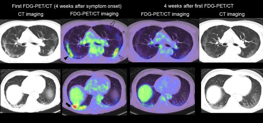
Figure 3. FDG-PET/CT imaging of lung lesion in COVID-19. Left side: CT and fused FDG-PET/CT image of the chest in
the first examination (4 weeks after symptom onset). Right side: CT and fused FDG-PET/CT image of the chest (4 weeks after
the first FDG-PET/CT examination). Intense FDG uptake was seen in liner opacity (black arrowhead), reticular opacity with
consolidation (black arrow), and grand glass opacity with consolidation (open arrowhead) in the lung. Moderate FDG uptake
was confirmed in grand glass opacity (open arrows) at the left upper lobe. All the FDG uptake in the first examination was
significantly decreased in the second PET/CT scan.
Figure 5. Change of CT feature of lymph node over time.
Left side: CT image (14 days from symptom onset), Middle:
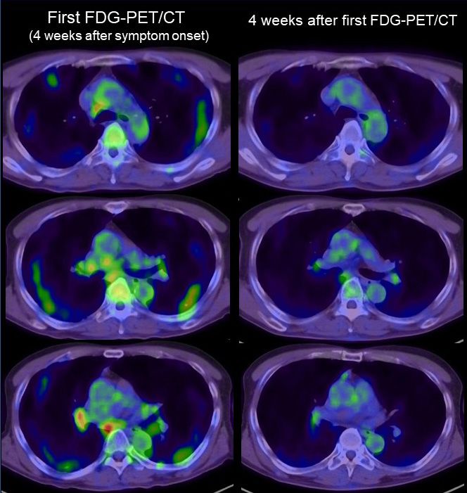
Figure 4. FDG-PET/CT imaging of lymph nodes in CT portion of FDG-PET/CT (4 weeks after symptom onset),
COVID-19. Left side: FDG-PET/CT image of the chest in Right side: CT portion of second FDG-PET/CT (4 weeks after
the first examination (4 weeks after symptom onset). Right first FDG-PET/CT examination). CT image (14 days from
side: FDG-PET/CT image of the chest (4 weeks after the first symptom onset) showed no evidence of mediastinal lymph
FDG-PET/CT examination). Intense FDG uptake was seen in node swelling (arrowhead). Although the size of the lymph
mediastinal and hilar lymph nodes. All the FDG uptake in the node is not significant, it was increased compared to 2 weeks
first examination was significantly decreased in the second from CT imaging and decreased within 4 weeks after the first
FDG-PET/CT scan. FDG-PET/CT examination.
a high level of inflammatory change persists even in with COVID-19 (Figure 4). The FDG uptake in lymph
the recovery stage. However, it is still unclear whether nodes is thought to reflect immunoreactions activated by
the inflammatory change is caused by the remaining inflammatory cells such as neutrophils, monocytes, and
COVID-19 itself, the immunotherapeutic response, or effector T cells by the release of local chemokines. In
angiovascular damage. In our experience, FDG uptake the immune response to viral infections, the number of
in lung lesions was decreased at 4 weeks after the first monocytes in lymphoid tissue increases, thus leading to
FDG-PET/CT scan (Figure 3). Therefore, we should note increased FDG uptake (12,29).
that FDG uptake in the recovery stage will not always A previous study reported that in COVID-19, lymph
forecast disease progression of COVID-19. Considering node enlargement is a rare finding on CT, which present
this finding, it is of interest whether FDG uptake can in < 1% of patients (30). The size and the shape of these
predict potential damage of lung tissue. lymph nodes showing intense FDG uptake was not clear
in some reports, but it is generally small, nonspecific,
Lymph nodes and regular in shape as we identified in our case (Figure
5). In our experience, FDG uptake was confirmed
FDG uptake in lymph nodes is frequently seen in patients in mediastinal lymph nodes without significant
(223)Global Health & Medicine. 2020; 2(4):221-226. www.globalhealthmedicine.com
enlargement, and the uptake decreased during 4 weeks CoV-2 invades host cells via two receptors: angiotensin-
of observation (Figure 4). The CT image showed little converting enzyme 2 (ACE2) and CD147 (42). CD147
change in size of a lymph node during the clinical course is expressed by mesenchymal stem cells of human
(Figure 5), but CT may be less sensitive to host reactions cord blood and bone marrow origin (43), and CD147
compared with FDG-PET/CT, and therefore the actual expression is induced by high glucose concentration
percentage of lymph node involvement may be higher in monocytes (44). Based on this mechanism, FDG
than seen on CT. uptake by bone marrow may be an additional feature of
In contrast, lymph node swelling has been COVID-19. In a COVID-19 patient, more neutrophils
manifested in pneumonia caused by parainfluenza virus and scattered plasma cell infiltration are frequently
and adenovirus (30). FDG uptake in small axillary found in the spleen. The author suggested that
lymph nodes is a common feature just after influenza pathological changes of the spleen might be related to
vaccination (31). In COVID-19, however, several studies the direct attack of virus and the attack of immune cells
have reported negative FDG uptake in these lymph (45). Similar to our case, slight to moderate FDG uptake
nodes, which may occur in the minimally invasive and in the spleen is confirmed in some reports (10,13),
early stages of the disease (32). Therefore, the immune however the significance of this feature is still unknown.
response is weak or almost absent in the early stage and In another report, FDG-PET/CT imaging revealed
becomes more active over time. Moreover, reduction of hypoactivity of the orbitofrontal cortex in COVID-19
FDG uptake in lymph nodes may indicate normalization patients with anosmia (46).
of hyperactive immune response in the body, but further
investigation is necessary to confirm this hypothesis. Conclusions
Possible identification of lesions related to COVID-19 We review the FDG-PET/CT imaging features of
COVID-19, including our experience and current
Patients with COVID-19 show various symptoms that knowledge. FDG-PET/CT may have potential to
can cause damage to the gastrointestinal tract, kidneys, increase our understanding of the mechanism of
heart, bone marrow, and other organs (33). Small vessel COVID-19. Further investigation is required to confirm
vasculitis causing skin disease (34) and symptoms the substantial value of FDG-PET/CT in patients with
similar to those of Kawasaki disease (35) have been COVID-19.
reported as related to COVID-19. However, FDG-PET/
CT is limited in its ability to diagnose small or middle Acknowledgements
vessel aortitis and medium-to-large vessel aortitis. In the
case that vasculitis causes organ damage, the abnormal We thank all the staff in National Center for Global
FDG-PET/CT findings on organs may indirectly Health and Medicine who struggled with COVID-19.
indicate the existence of small-sized or middle-sized We also thank Kaori Saito, Daisuke Horikawa, Tomoya
vessel aortitis (36). Takeuchi, Hisayoshi Mizunuma, Yui Yamada, Satsuki
Damage to endothelial tissue is considered to be the Hironaka, Kazuhiko Nakajima, Hironori Kajiwara,
underlying mechanism of cardiovascular complications and Futoshi Matsunaga for contributing to the PET/CT
in COVID-19 (37). No report has described FDG uptake examination.
by the vascular wall that would suggest endothelial
tissue damage. FDG can visualize metabolically References
active atherosclerosis because FDG is taken up by
macrophages within atherosclerotic plaques (38,39). 1. Zhu N, Zhang D, Wang W, et al. China Novel Coronavirus
However, considering that complications of COVID-19 Investigating and Research Team. A Novel Coronavirus
tend to occur in the elderly, it is questionable whether from Patients with Pneumonia in China, 2019. N Engl J
FDG can distinguish the uptake of FDG caused by Med. 2020; 382:727-733.
2. Centers for Disease Control and Prevention. 2019
atherosclerosis. Further investigation of the relationship
Novel coronavirus, Wuhan, China: 2019-nCoV situation
between endothelial tissue damage and FDG uptake in summary. January 28, 2020. https://stacks.cdc.gov/view/
the arterial wall in COVID-19 is required. cdc/84621 (accessed June 10, 2020).
Because active thrombosis can be depicted as intense 3. Yamayoshi S, Kawaoka Y. Emergence of SARS-CoV-2
FDG uptake, a survey of FDG uptake when thrombosis and its outlook. Global Health & Medicine. 2020; 2:1-2.
is suspected may be of additional value in patients with 4. Youyao Xu, Yizhen Chen, Xiaoyan Tang. Guidelines for
COVID-19 (40). the diagnosis and treatment of coronavirus disease 2019
(COVID-19) in China. Global Health & Medicine. 2020;
Increased FDG uptake in bone marrow may be
2:66-72.
an additional imaging feature in COVID-19 as it was 5. Shi H, Han X, Jiang N, Cao Y, Alwalid O, Gu J, Fan Y,
confirmed in another report (9,13,15,16). Chefer et al. Zheng C. Radiological findings from 81 patients with
reported high uptake by bone marrow over a long period COVID-19 pneumonia in Wuhan, China: a descriptive
of time in a MERS-CoV animal model (41). SARS- study. Lancet Infect Dis. 2020; 20:425-434.
(224)Global Health & Medicine. 2020; 2(4):221-226. www.globalhealthmedicine.com
6. Chung M, Bernheim A, Mei X, Zhang N, Huang M, Zeng in Asymptomatic Patients Undergoing Nuclear Medicine
X, Cui J, Xu W, Yang Y, Fayad ZA, Jacobi A, Li K, Li S, Procedures in a High-Prevalence Region. J Nucl Med.
Shan H. CT Imaging Features of 2019 Novel Coronavirus 2020; 61:632-636.
(2019-nCoV). Radiology. 2020; 295:202-207. 22. Zanoni L, Mosconi C, Cervati V, Diegoli M, Monteduro
7. Hani C, Trieu NH, Saab I, Dangeard S, Bennani S, F, Golfieri R, Fanti S. [18F]-FDG PET/CT for suspected
Chassagnon G, Revel MP. COVID-19 pneumonia: A lymphoma relapse in a patient with concomitant
review of typical CT findings and differential diagnosis. pneumococcal pneumonia during COVID-19 outbreak:
Diagn Interv Imaging. 2020; 101:263-268. unexpected SARS-Cov-2 co-infection despite double RT-
8. ACR Recommendations for the use of Chest Radiography PCR negativity. Eur J Nucl Med Mol Imaging. 2020;
and Computed Tomography (CT) for Suspected 19:1-2.
COVID-19 Infection. https://www.acr.org/Advocacy-and- 23. Paez D, Gnanasegaran G, Fanti S, et al. COVID-19
Economics/ACR-Position-Statements/Recommendations- Pandemic: Guidance for Nuclear Medicine Departments
for-Chest-Radiography-and-CT-for-Suspected-COVID19- Eur J Nucl Med Mol Imaging. 2020; 47:1615-1619.
Infection (accessed June 10, 2020). 24. Dooms C, van Baardwijk A, Verbeken E, van Suylen
9. Zou S, Zhu X. FDG PET/CT of COVID-19. Radiology. RJ, Stroobants S, De Ruysscher D, Vansteenkiste J.
2020: 200770. Association between 18F-fluoro-2-deoxy-D-glucose uptake
10. Qin C, Liu F, Yen TC, Lan X. 18F-FDG PET/CT findings values and tumor vitality: prognostic value of positron
of COVID-19: a series of four highly suspected cases. Eur emission tomography in early-stage non-small cell lung
J Nucl Med Mol Imaging. 2020; 47:1281-1286. cancer. J Thorac Oncol. 2009; 4:822-828.
11. Polverari G, Arena V, Ceci F, Pelosi E, Ianniello A, Poli 25. Win T, Screaton NJ, Porter JC, et al. Pulmonary 18F-FDG
E, Sandri A, Penna D. 18F-Fluorodeoxyglucose uptake uptake helps refine current risk stratification in idiopathic
in patient with asymptomatic severe acute respiratory pulmonary fibrosis (IPF). Eur J Nucl Med Mol Imaging.
syndrome coronavirus 2 (Coronavirus Disease 2019) 2018; 45:806‐815.
Referred to Positron Emission Tomography/Computed 26. Tay MZ, Poh CM, Rénia L, MacAry PA, Ng LFP. The
Tomography for NSCLC Restaging. J Thorac Oncol. trinity of COVID-19: immunity, inflammation and
2020; 15:1078-1080. intervention. Nat Rev Immunol. 2020; 20:363-374.
12. Deng Y, Lei L, Chen Y, Zhang W. The potential added 27. Das KM, Lee EY, Langer RD, Larsson SG. Middle
value of FDG PET/CT for COVID-19 pneumonia. Eur J East Respiratory Syndrome Coronavirus: What Does a
Nucl Med Mol Imaging. 2020; 47:1634-1635. Radiologist Need to Know? AJR Am J Roentgenol. 2016
13. Amini H, Divband G, Montahaei Z, Dehghani T, Kaviani ;206:1193-1201.
H, Adinehpour Z, Akbarian Aghdam R, Rezaee A, Vali 28. Pan F, Ye T, Sun P, Gui S, Liang B, Li L, Zheng D, Wang
R. A case of COVID-19 lung infection first detected by J, Hesketh RL, Yang L, Zheng C. Time Course of Lung
[18F]FDG PET-CT. Eur J Nucl Med Mol Imaging. 2020; Changes at Chest CT during Recovery from Coronavirus
47:1771-1772. Disease 2019 (COVID-19). Radiology. 2020; 295:715-
14. Setti L, Kirienko M, Dalto SC, Bonacina M, Bombardieri 721.
E. FDG-PET/CT findings highly suspicious for 29. Jones HA, Marino PS, Shakur BH, Morrell NW. In vivo
COVID-19 in an Italian case series of asymptomatic assessment of lung inflammatory cell activity in patients
patients. Eur J Nucl Med Mol Imaging. 2020; 47:1649- with COPD and asthma. Eur Respir J. 2003; 21:567-573.
1656. 30. Xu X, Yu C, Qu J, et al. Imaging and clinical features of
15. Liu C, Zhou J, Xia L, Cheng X, Lu D. 18F-FDG PET/CT patients with 2019 novel coronavirus SARS-CoV-2. Eur J
and Serial Chest CT Findings in a COVID-19 Patient With Nucl Med Mol Imaging. 2020; 47:1275-1280.
Dynamic Clinical Characteristics in Different Period. Clin 31. Panagiotidis E, Exarhos D, Housianakou I, Bournazos
Nucl Med. 2020; 45:495-496. A, Datseris I. FDG uptake in axillary lymph nodes after
16. Kamani CH, Jreige M, Pappon M, Fischbacher A, Borens vaccination against pandemic (H1N1). Eur Radiol. 2010;
O, Monney P, Nicod Lalonde M, Schaefer N, Prior JO. 20:1251‐1253.
Added value of 18F-FDG PET/CT in a SARS-CoV-2- 32. Kirienko M, Padovano B, Serafini G, Marchianò A,
infected complex case with persistent fever. Eur J Nucl Gronchi A, Seregni E, Alessi A. [18F]FDG-PET/CT and
Med Mol Imaging. 2020; 16:1-2. clinical findings before and during early Covid-19 onset
17. Wahl RL. Principles and practice of PET and PET/CT. 2nd in a patient affected by vascular tumour. Eur J Nucl Med
ed. Lippincott Williams & Wilkins 2008. Mol Imaging. 2020; 47:1769-1770.
18. Kubota K, Ogawa M. Ji B. Basic science of PET imaging 33. Gao QY, Chen YX, Fang JY. 2019 novel coronavirus
for inflammatory diseases. In Toyama H. et al. ed. "PET/ infection and gastrointestinal tract. J Dig Dis. 2020;
CT for inflammatory diseases" Springer, 2020, pp1-42. 21:125-126.
19. Dai M, Liu D, Liu M, et al. Patients with Cancer Appear 34. Castelnovo L, Capelli F, Tamburello A, Faggioli PM,
More Vulnerable to SARS-CoV-2: A Multicenter Mazzone A. Symmetric cutaneous vasculitis in COVID-19
Study during the COVID-19 Outbreak. Cancer Discov. pneumonia. 2020;10.1111/jdv.16589. doi: 10.1111/
2020;10:783-791. jdv.16589.
20. Inui S, Fujikawa A, Jitsu M, Kunishima N, Watanabe S, 35. Viner RM, Whittaker E. Kawasaki-like disease: emerging
Suzuki Y, Umeda S, Uwabe Y. Chest CT findings in cases complication during the COVID-19 pandemic. Lancet.
from the cruise ship "Diamond Princess" with coronavirus 2020; 395:1741-1743.
disease 2019 (COVID-19). Radiology: Cardiothoracic 36. Farrah TE, Basu N, Dweck M, Calcagno C, Fayad
Imaging. 2020; 2:e200155. ZA, Dhaun N. Advances in Therapies and Imaging for
21. Albano D, Bertagna F, Bertoli M, Bosio G, Lucchini S, Systemic Vasculitis. Arterioscler Thromb Vasc Biol. 2019;
Motta F, Panarotto MB, Peli A, Camoni L, Bengel FM, 39:1520‐1541.
Giubbini R. Incidental Findings Suggestive of COVID-19 37. Varga Z, Flammer AJ, Steiger P, Haberecker M,
(225)Global Health & Medicine. 2020; 2(4):221-226. www.globalhealthmedicine.com
Andermatt R, Zinkernagel AS, Mehra MR, Schuepbach cells reveals common and differentially expressed
RA, Ruschitzka F, Moch H. Endothelial cell infection and markers: identification of angiotensin-converting enzyme
endotheliitis in COVID-19. Lancet. 2020;395:1417‐1418. (CD143) as a marker differentially expressed between
38. Rosenbaum D, Millon A, Fayad ZA. Molecular imaging adult and perinatal tissue sources. Stem Cell Res Ther.
in atherosclerosis: FDG PET. Curr Atheroscler Rep. 2012; 2018; 9:10.
14:429-437. 44. Bao W, Min D, Twigg SM, Shackel NA, Warner FJ,
39. Tawakol A, Migrino RQ, Bashian GG, Bedri S, Vermylen Yue DK, McLennan SV. Monocyte CD147 is induced
D, Cury RC, Yates D, LaMuraglia GM, Furie K, Houser by advanced glycation end products and high glucose
S, Gewirtz H, Muller JE, Brady TJ, Fischman AJ. In vivo concentration: possible role in diabetic complications. Am
18
F-fluorodeoxyglucose positron emission tomography J Physiol Cell Physiol. 2010; 299:C1212-1219.
imaging provides a noninvasive measure of carotid 45. Xu X, Chang XN, Pan HX, et al. Pathological changes
plaque inflammation in patients. J Am Coll Cardiol. 2006; of the spleen in ten patients with coronavirus disease
48:1818-1824. 2019(COVID-19) by postmortem needle autopsy.
40. Rondina MT, Lam UT, Pendleton RC, Kraiss LW, Wanner Zhonghua Bing Li Xue Za Zhi. 2020; 49:576-582. (in
N, Zimmerman GA, Hoffman JM, Hanrahan C, Boucher K, Chinese).
Christian PE, Butterfield RI, Morton KA. 18F-FDG PET 46. Karimi-Galougahi M, Yousefi-Koma A, Bakhshayeshkaram
in the evaluation of acuity of deep vein thrombosis. Clin M, Raad N, Haseli S. 18 FDG PET/CT Scan Reveals
Nucl Med. 2012; 37:1139-1145. Hypoactive Orbitofrontal Cortex in Anosmia of COVID-19.
41. Chefer S, Thomasson D, Seidel J, Reba RC, Bohannon Acad Radiol. 2020; 27:1042-1043.
JK, Lackemeyer MG, Bartos C, Sayre PJ, Bollinger L,
Hensley LE, Jahrling PB, Johnson RF. Modeling [18F]- ----
FDG lymphoid tissue kinetics to characterize nonhuman Received June 15, 2020; Revised July 13, 2020; Accepted
primate immune response to Middle East respiratory July 15, 2020.
syndrome-coronavirus aerosol challenge. EJNMMI Res.
2015; 5:65. Released online in J-STAGE as advance publication July 20,
42. Yan R, Zhang Y, Li Y, Xia L, Guo Y, Zhou Q. Structural 2020.
basis for the recognition of SARS-CoV-2 by full-length
human ACE2. Science. 2020; 367:1444‐1448. *Address correspondence to:
43. Amati E, Perbellini O, Rotta G, Bernardi M, Chieregato Ryogo Minamimoto, Division of Nuclear Medicine, Department
K, Sella S, Rodeghiero F, Ruggeri M, Astori G. High- of Radiology, National Center for Global Health and Medicine.
throughput immunophenotypic characterization of bone 1-21-1, Toyama, Shinjyuku-ku, Tokyo 162-8655, Japan.
marrow- and cord blood-derived mesenchymal stromal E-mail: rminamimoto@hosp.ncgm.go.jp
(226)You can also read